Submitted Successfully!
To reward your contribution, here is a gift for you: A free trial for our video production service.
Thank you for your contribution! You can also upload a video entry or images related to this topic.
Version Summary Created by Modification Content Size Created at Operation
1 + 4595 word(s) 4595 2021-05-24 05:46:11 |
2 format correct + 1744 word(s) 6339 2021-06-28 03:04:57 |

Video Upload Options

Do you have a full video?

Confirm

Are you sure to Delete?
Cite
If you have any further questions, please contact Encyclopedia Editorial Office.
Pallafacchina, G. The Mitochondrial Calcium Channel Discovery. Encyclopedia. Available online: https://encyclopedia.pub/entry/11328 (accessed on 28 March 2024).
Pallafacchina G. The Mitochondrial Calcium Channel Discovery. Encyclopedia. Available at: https://encyclopedia.pub/entry/11328. Accessed March 28, 2024.
Pallafacchina, Giorgia. "The Mitochondrial Calcium Channel Discovery" Encyclopedia, https://encyclopedia.pub/entry/11328 (accessed March 28, 2024).
Pallafacchina, G. (2021, June 25). The Mitochondrial Calcium Channel Discovery. In Encyclopedia. https://encyclopedia.pub/entry/11328
Pallafacchina, Giorgia. "The Mitochondrial Calcium Channel Discovery." Encyclopedia. Web. 25 June, 2021.
The Mitochondrial Calcium Channel Discovery
Edit

The notion of the active role of mitochondria in the decoding and shaping of intracellular Ca2+ signals dates back at the end of the 19th century. However, the identity of the molecule(s) involved in Ca2+ ion transport into mitochondria remained elusive for decades. Only in the last ten years, the factors, and the relative coding genes, mediating Ca2+ entry in mitochondria started to be genetically and biochemically described. The gene for the pore-forming unit of the mitochondrial Ca2+ channel was discovered in 2011, and its product was named mitochondrial Ca2+ uniporter or MCU. The mitochondria Ca2+ uptake regulator 1 gene, MICU1, was cloned one year before, in 2010. The increasing interest of the scientific community towards mitochondrial Ca2+ signaling and metabolism in the subsequent  years led to the identification of many other MCU components and to the description of their 3D structure and physiological role. Here, we will present a brief overview of the land marking discoveries in the history of mitochondrial Ca2+ studies. 

MCU mitochondrial Ca2+ uniporter Ca2+ signaling mitochondrial metabolism

1. Introduction

Every cell type, in every tissue and at any evolutionary level, can communicate with the surrounding environment and with neighboring cells. Both intercellular and extracellular communication play fundamental roles in shaping cell behavior and driving cell fate decisions. Cell-to-cell and environmental signals are normally conveyed by distinct extracellular mediators (hydrophilic or hydrophobic compounds, mechanical, ionic, cell–cell interactions, etc.) that are normally perceived by cells through surface receptors. These receptors convey them into a limited number of intracellular molecules, which are referred to as ‘second messengers’, which, in turn, forward the message to intracellular effectors finally activating the ultimate cellular responses. Despite the plethora of different signals and stimuli that cells may receive, only a few molecules to date have been described as second messengers of intracellular communication. Among them, the most common and, definitively, the most extensively studied is Ca2+.
Ca2+ ions participate in the decoding of a vast range of stimuli and the variety of cellular components involved in the Ca2+ signal transduction is extremely wide, including basically all kinds of components, organelles, and molecules [1][2][3]. The research studies on Ca2+ second messenger started more than one hundred years ago, with the initial recognition of the role of Ca2+ in muscle cell contraction [4]. Since then, the understanding of Ca2+ signaling regulation and dynamics has progressively increased leading to the definition of the concept of Ca2+ compartmentalization and to the demonstration of the existence of microdomains of local high Ca2+ concentration [5], which are crucial for the fine-tuning and correct triggering of the Ca2+-dependent cellular effects [1].
Mitochondria play a fundamental and multifaceted role in the orchestration of cellular Ca2+ signals. Indeed, mitochondria are not a store of rapidly releasable Ca2+ (such as the ER), but rather they efficiently accumulate Ca2+ upon Ca2+ entry from the extracellular space or upon release from ER Ca2+ stores [6]. Upon cytosolic Ca2+ elevation, the entry of Ca2+ into mitochondria exerts a central function in the modulation of cell metabolism. Mitochondria host the enzymes and complexes of the TCA cycle, fatty acid oxidation (FAO), and oxidative phosphorylation (OXPHOS) thus representing the site of the major metabolic pathways and enzymes for cell energy supply, which deserved them the name of ‘cellular powerhouses’. Interestingly, Ca2+ entry and oxidative activity are two strictly intertwined aspects of mitochondrial physiology. The increase of the mitochondrial matrix Ca2+ level stimulates both Ca2+-sensitive dehydrogenases [7][8][9] and respiratory chain complexes [10][11] resident in the organelles, fueling the TCA cycle activity as well as aerobic respiration and thus boosting the overall oxidative metabolism. This makes mitochondria the central hubs for the rapid and effective adaptation of cell metabolism to the changes in energy requirements that are typically decoded as variations of intracellular Ca2+ concentration.
In addition, mitochondria also actively participate in the tuning of global Ca2+ signals thanks to their ability to take up Ca2+ during intracellular Ca2+ elevation with a net result of buffering the cytosolic cation concentration thus modulating the overall cellular Ca2+ response. This buffering capacity is due to two crucial characteristics of the mitochondria: (i) their strategic position in close contacts to the Ca2+ release channels of the ER store [6] and the plasma membrane in immune cells [12] (ii) the presence on their inner membrane of highly selective and efficient machinery for taking up Ca2+, the MCU complex.
Finally, the large buffering capacity of mitochondria can protect cells from Ca2+ overload. Indeed, an excessive accumulation of the cation in the mitochondrial matrix triggers the permeability transition pore (PTP) opening, the release of pro-apoptotic factors, and finally, induction of programmed cell death [13]. Given the strong association between mitochondrial Ca2+ overload and apoptosis induction, the maintenance of mitochondrial Ca2+ homeostasis is thus a crucial aspect for ensuring cell survival [14][15].
Given the extreme relevance of mitochondria Ca2+ signaling for cell physiology, the unveiling of the molecular factors mediating mitochondrial Ca2+ entry and the mechanism(s) of their regulation has been one of the scientific challenges of recent years. In this review, we aim to summarize some of the milestone achievements in the history of mitochondrial Ca2+ research with a particular focus on the recent findings of the mitochondrial Ca2+ uniporter and its role in organ physiology. We will briefly describe the early studies leading to the demonstration of the Ca2+ accumulation capacity of mitochondria, then we will go through the historical chronicle of the discoveries of the mitochondrial Ca2+ uniporter genes and multiple regulators (Figure 1) and we will conclude with an excursus on the physiological relevance of mitochondrial Ca2+ uptake in the context of skeletal muscle tissue.
Figure 1. Timeline of the identification of MCU complex components. The most relevant findings on the structure and composition of the MCU complex discovery are summarized and chronologically presented along a timeline covering the last 10 years. Schematic cartoons show the different components of the MCU complex according to the date of their discovery along the timeline.

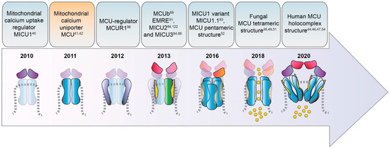
2. Timeline of MCU Identification

It is suggestive to recall that the notion of Ca2+ ions being relevant to organ physiology dates back to more than one century ago when the first report on the physiological action of Ca2+ ions appeared in 1883 [4]. At that time, Ringer described the effects of Ca2+ addition to isolated frog hearts and demonstrated that the supplementation of Ca2+ in the perfusion solution actively induces and sustains the contraction of the organ ex vivo [4]. This seminal observation revealed that Ca2+ is a fundamental messenger within cells, a concept that then extended to virtually every cell type and physiological and pathological process, giving rise to a broad field of study commonly referred to as the field of intracellular Ca2+ signaling. The intrinsic ability of contracting myocytes to operate ex vivo and to rapidly and effectively respond to environmental condition changes made them the ideal experimental system for the investigation of the role of Ca2+ in organ and cell physiology and was extensively exploited by researchers in the following years.
The original concept of the existence of intracellular compartments acting as Ca2+ stores to accumulate the cation required to sustain muscle contraction has been later postulated and demonstrated in 1947 by Heilbrunn [16]. However, although surprising, the identification of the sarco/endoplasmic reticulum (SR/ER) as the principal cellular Ca2+ store came only 20 years later. It was in the 1960′s, with the identification of Ca2+ pumping machinery on intracellular membranes (in particular the calcium pump of the sarcoplasmic reticulum, better known as SERCA) by three independent scientists [17][18][19][20] and the advent of new methodologies for the measurement of intracellular Ca2+ concentration [21] that the ER and its specialized counterpart in muscle cells (the SR) were recognized the main cellular reservoir of Ca2+.
Before that, the pioneering work of Slater & Cleland on cardiac myocyte preparations from rat hearts firstly described some subcellular compartments, called "sarcosomes" at that time, as the entities actively accumulating Ca2+ [22]. Interestingly, these “sarcosomes” did not consist of ER but, instead, they corresponded to isolated mitochondria, to which the addition of Ca2+ caused the block of their oxidative phosphorylation activity. Thus, Ca2+ ions behaved as mitochondria uncouplers. Despite that, Ca2+ appeared to exert a peculiar inhibitory action on mitochondrial OXPHOS activity, which differed from the other irreversible uncouplers known at that period (dinitrophenol, dicoumarol, rotenone, antimycin A, azide, or cyanide), due to the reversibility of its action [23]. This assigned to Ca2+ ions a functional role in mitochondria activity.
After that, a series of land-marking works in the early 60′s experimentally revealed that energized mitochondria can actively take up Ca2+ [24][25][26]. Interestingly, these results anticipated the clear demonstration of a driving force for Ca2+ accumulation in mitochondria, i.e., the chemiosmotic theory, postulated and validated by Peter Mitchell [27]. This theory is based on the following concepts: (i) the activity of the respiratory chain complexes is linked to the extrusion of protons from the matrix to the intermembrane space (IMS) across the inner membrane of mitochondria (IMM). (ii) The accumulation of protons in the IMS generates a difference in the charges across the IMM of around −150 ÷ −180 mV (negative inside) establishing the so-called mitochondrial membrane potential (ΔΨm). (iii) This steep ΔΨm represents the main driving force for the proton gradient-sustained synthesis of ATP and Ca2+ cation entry into the matrix.
Shortly after, at the University of Bristol, Denton and his group made important discoveries on the Ca2+-dependent modulation of three critical oxidative enzymes resident in mitochondria [7][8][9]: pyruvate dehydrogenase phosphatase (the enzyme that dephosphorylates and relieves pyruvate dehydrogenase (PDH) activity allowing the conversion of NAD+, coenzyme A (CoA) and pyruvate into NADH, CO2, and acetyl-CoA, providing substrates to the citric acid (TCA) cycle and cellular respiration), NAD-isocitrate dehydrogenase and oxoglutarate dehydrogenase. The fact that these mitochondrial rate-limiting enzymes are under the control of Ca2+ definitively sets the cation at the center of the cell oxidative metabolism. Moreover, the evidence that mitochondria can take up Ca2+ in response to elevation of cytosolic Ca2+ levels, as shown in insulin-treated epididymal adipose tissue [28], established an active role of mitochondrial Ca2+ entry in shaping cell oxidative metabolism to match the increased cellular energy demands thus tailoring the metabolic outcomes according to the different environmental cues.
Despite all these conceptual advancements, two major issues were destined to puzzle the scientific community for decades. On one end, there is the apparent paradox between the physiological concentration of cytosolic Ca2+, which was estimated in the submicromolar range [29][30][31], and the low affinity of the mitochondrial Ca2+ uptake, whose half-maximal rate (Km) was measured in the order of several µM (reviewed in [32]). On the other end, the fundamental question about the molecular identity of the IMM apparatus responsible for the entry of Ca2+ into mitochondria was still without an answer.
It took around 30 and 50 years, respectively, to find solutions to those two enigmas. The answer to the former came with the advent of innovative and sophisticated technologies allowing the assessment of Ca2+ distribution at the sub-cellular and sub-organellar levels, including mitochondria. Indeed, thanks to the development of Ca2+-sensitive genetic probes and recombinant fluorescent proteins targeted to specific intracellular microdomains [33][34], it was possible to measure variations of Ca2+ concentration in defined and limited areas of the cell (such as the cytosolic face of plasmalemma [35] or the surface of outer mitochondrial membrane [36], or the Golgi cisternae [37]) as well as the relative positioning of the intracellular organelles. These studies were pivotal in the field of cell biology for two main reasons: i) they allowed the observation of the intrinsic heterogeneity of cellular Ca2+ distribution, definitively demonstrating that large variations in Ca2+ concentration are highly regionalized within the cell cytoplasm and allowing the direct measure of Ca2+ levels in the mitochondria matrix as well as in the ER lumen; ii) they pinpointed the fact that organelles, including ER and mitochondria, are in close contact with each other through macromolecular structures involving proteins from both the compartments. In the case of ER-mitochondrial contacts, these structures are biochemically isolated as mitochondria-associated membranes (MAMs) and are formed by membrane channels, as the IP3R and VDAC, respectively, and adaptor proteins of both organelles, such as Grp75, mitofusins, PACS [38]. Upon cell stimulation, the massive release of Ca2+ through the ER membrane clusters of IP3Rs generates microdomains of high Ca2+ concentration right at the mouth of the channel pores, exactly where mitochondria are located. This allows mitochondria to perceive a local cation concentration sufficient to meet the low affinity of the mitochondrial Ca2+ uptake machinery [36][39]. Thus, their strategic position in proximity of ER Ca2+ release channels and their ability to take up Ca2+ with high conductance make mitochondria the ideal operator for cushioning the sudden Ca2+ rise in the cytosol of stimulated cells, thus behaving as an instrumental Ca2+ buffer [6]. The fact that Ca2+ entry into mitochondria stimulates the TCA cycle, respiration, and ATP production then places mitochondrial Ca2+ uptake as a key element for the prompt modulation of cell metabolism to rapidly and efficiently adapt to a variety of environmental cues and energy demands.
Another fundamental advancement in mitochondrial signaling occurred when scientists found the answer to the second big question, i.e., the molecular identity of the mitochondrial Ca2+ uniporter (MCU) machinery. The chronicle of MCU discoveries actually started in 2010, with the identification of the first gene required for the uptake of Ca2+ by mitochondria, CBARA1, coding for the mitochondrial Ca2+ uptake 1 protein (MICU1) [40], then followed by the identification of the mitochondrial channel and the elucidation of its interactors, as described below. The search of the other mitochondrial Ca2+ channel components has been proceeding expansively in the last decade (see next paragraph for a detailed timeline) and it is presently still actively ongoing. The discoveries of many different groups worldwide have been indeed instrumental to provide cell biologists with new knowledge on the functional role of mitochondrial Ca2+ and with new tools for the genetic and molecular intervention on global Ca2+ signaling and cell energetics.

3. Discovery and Characterization of the MCU Complex Components

3.1. MCU

The chronicle of MCU discovery starts with two pioneering studies published in 2011 [41][42] (Figure 1) that finally identified and cloned the long-sought MCU pore-forming unit gene, CCDC109A. MCU is a highly conserved 40 kDa protein ubiquitously expressed in plants, metazoans, protozoans, and fungi but not present in yeast [43]. This pore-forming unit oligomerizes to form the active channel within the inner mitochondrial membrane (IMM) and it directly interacts with the channel regulator MICU1, which was identified one year earlier [40]. These reports clearly demonstrated that the transient downregulation of MCU inhibits the mitochondrial accumulation of Ca2+ that follows the IP3-generating agonist in stimulated cells. Of note, the blunted mitochondrial Ca2+ uptake response occurs without changes in the mitochondria morphology or membrane potential of the MCU-silenced cells [41]. On the contrary, MCU overexpression enhances agonist-induced mitochondrial Ca2+ uptake in mammalian cells. In addition, in vitro experiments, in which recombinant MCU proteins were inserted in a planar lipid bilayer, showed that this pore-subunit alone is sufficient to form the channel. Indeed, in this setting, the MCU electrophysiological activity is completely abolished by the addition of the known inhibitor Ruthenium Red, firmly pointing at MCU as the genuine core component of the mitochondrial Ca2+ machinery.
Sequence and topology analyses revealed that both the N- and C-termini of MCU are located in the mitochondrial matrix and that MCU is endowed with two transmembrane domains, linked by a short highly conserved acidic loop exposed in the intermembrane space (IMS) which contains the so-called acidic “DIME” motif. The acidic residues present in this stretch (in particular E257, D261, E264) are critical for the Ca2+ transport since, if substituted with uncharged residues, MCU mutants failed to rescue the mitochondrial Ca2+ uptake in MCU-silenced cells [41][42]. However, the definitive description of MCU 3D structure had to wait till the very last years, when cryo-EM and X-ray diffraction analyses finally allowed the resolution of full-length MCU structure [44][45][46][47][48][49][50][51]. These studies coherently confirmed that purified MCU from different sources (fungi and metazoan) arranges in a tetramer, confuting previous assumptions on a putative pentameric MCU architecture [52]. Notably, the cryo-EM data also unveiled the exact position of the MCU channel selectivity filter, in which the DIME motif is fundamental for the coordination of Ca2+ ions and which was definitively shown to reside at the beginning of the second transmembrane α-helix [50] and not in the linker region between the two transmembrane helices, as previously suggested [52].
More recently, the structure of the human MCU together with its auxiliary component EMRE was obtained [53]. Each human MCU arranges in tetramers and each subunit complexes with one EMRE peptide. Differently from the three described for fungal MCU, human MCU appears organized in four domains, which are: (i) the N-terminal domain (NTD), (ii) the linker helix domain (LHD) —absent in fungi—, (iii) the coiled-coil domain (CCD), and iv) the transmembrane domain (TMD) (Figure 2).
Figure 2. The MCU holocomplex structure. Schematic representation of the MCU holocomplex (uniplex) components and their relevant domains: the pore-forming subunit MCU (light blue) with the two transmembrane (TM) and coiled-coil (CC) domains and the linker helix domain (LHD); the essential mitochondrial Ca2+ uniporter regulator EMRE (yellow); the mitochondrial Ca2+ uptake proteins MICU1 (violet) and MICU2 (purple), with the EF-hands relevant for the MICU dimer interaction highlighted. The critical residues of the MCU DIME motif forming the Ca2+ selectivity filter are indicated, together with the MICU1 residues of the K-R ring coordinating the MCU acidic region.
Moreover, in the very last year, further insight into MCU channel modulation and function has been gained thanks to the achievement of the human MCU-MICUs holocomplex structure in both the Ca2+-free and Ca2+-bound state by several independent reports. The gating mechanism by which MICU1 regulates the uniporter activity via the conformational change triggered by Ca2+ was finally unveiled [44]. Furthermore, the precise description of the molecular interactions between MCU-EMRE-MICU1-MICU2 in the human MCU supercomplex (MEMMS) has been also obtained [54] (Figure 2). Indeed, MEMMS appears as a 480 kDa integral unit where EMRE coordinates the matrix gate of the MCU channel and MICU proteins interact with the C-terminus of EMRE in the IMS thus enhancing Ca2+ influx through the MCU pore in high [Ca2+] conditions [54] (Figure 3). Finally, the distinct Ca2+-dependent assembly conformations of the beetle and human MCU holocomplexes with human MICUs have also been detailed [46][47]. In the presence of Ca2+, the multiprotein complex shows a two-fold symmetry and consists of two V-shaped MCU-EMRE tetrameric subcomplexes and two MICU1-MICU2 heterodimers that bridge the tops of the subcomplexes (Figure 1). In this setting, the assembly of the MICU1-MICU2 heterodimers to the MCU-EMRE subcomplexes is ensured by the interaction between MICU1 and EMRE [47] (Figure 2). Differently, in the absence of Ca2+, the holocomplex adopts alternative less stable conformations with both monomeric and dimeric forms of the MCU-EMRE tetramers, where the MICU1-MICU2 heterodimer block the channel entrance formed by MCU transmembrane domains [47] (Figure 2 and Figure 3).
Figure 3. The MCU complex activity at low and high intracellular Ca2+ concentration. Schematic representation of the proteins involved in the MCU complex-mediated mitochondrial Ca2+ uptake: the pore-forming subunits MCU (light blue) and MCUb (green), the essential mitochondrial Ca2+ uniporter regulator EMRE (yellow), the mitochondrial Ca2+ uptake proteins MICU1 / MICU1.1 (violet), MICU2/ MICU3 (purple), and the MCU regulator 1 MCUR1 (light violet). The EF-hand Ca2+ binding domains of MICU proteins are indicated as little circles. At low intracellular Ca2+ concentration, the cation does not permeate through the MCU channel since the heterodimers formed by MICU1/MICU1.1–MICU2/MICU3 block the channel pore, thus preventing Ca2+ flux in resting conditions. Differently, at high intracellular Ca2+ concentration, MICU proteins undergo conformational changes relieving the inhibition on MCU and positively regulating channel activity, leading to an efficient mitochondrial Ca2+ uptake. OMM, outer mitochondrial membrane; IMM, inner mitochondrial membrane.
A large body of experimental evidence on the MCU complex functional role has accumulated since the discovery of the MCU. The genetic manipulation of the MCU led to the generation of germline and tissue-specific transgenic models [55][56][57][58][59][60][61][62], which provided pivotal tools for understanding the pathophysiological implications of the mitochondrial Ca2+ signaling in vivo that would have otherwise remained unexplored.
In 2013, three independent groups showed that the MCU silencing/knockout or its overexpression affect survival in different in vivo models [55][63][64]. In Trypanosoma brucei, for instance, the downregulation or the conditional knockout of the uniporter augments the AMP/ATP ratio, thus affecting the parasite growth in vitro. On the contrary, when MCU is overexpressed, mitochondrial Ca2+ accumulation produces a high concentration of ROS and sensitizes trypanosomes to apoptotic stimuli [63]. In zebrafish (Danio rerio), instead, the knockdown of MCU has shown crucial alterations not only during the early step of gastrulation, where the blastomere convergence and extension are altered, but also at later stages of development as the maturation of the notochord and anteroposterior axis formation were strongly impaired in morphant fish [64]. A series of recently published works and our unpublished observation in the zebrafish knockout model obtained with CRISPR/Cas9 gene ablation gave further insight into the role of MCU during development. For example, the ablation of the MCU gene in Danio rerio inhibits mitochondrial Ca2+ influx, reduces oxidative phosphorylation, and induces lipid accumulation, a phenotype that was also observed in the hepatic tissue of liver-specific KO mouse [65]. Notably, the inactivation of MCU has shown to be protective in neurons of Parkinson Disease zebrafish genetic model (namely in pink−/− fish), suggesting the crucial role of the MCU-dependent mitochondrial Ca2+ load on neuronal fitness [66].
As for mammals, the MCU–/– mouse [55] develops normally and displays minor defects without signs of impaired cell survival. Under stress conditions, relatively mild metabolic alterations were observed, such as increased plasma lactate levels in line with impaired exercise performance. However, the same group soon after those findings, showed that embryos from MCU−/− mice in a pure C57BL/6 background were not viable, displaying embryonic lethality at around E 11.5–E 13.5, thus suggesting a major involvement of mitochondrial Ca2+ uptake in organ metabolism and organism development that was compensated in a mixed genetic background [67].
Of note, some years later, it has been reported that the block of MCU-dependent Ca2+ uptake affects Drosophila melanogaster development. In particular, the inhibition of the uniporter activity has been shown to be detrimental for memory establishment during the papulation stage. Indeed, during the development of adult flies, alterations in the structural and functional neuronal substrates, crucial for memory formation, occur in the MCU deficient fly [58].
The genetic manipulation of MCU in C. elegans model also provided additional interesting notions on its role in organism physiology. Indeed, MCU−/− C. elegans is viable and grossly normal, mirroring what was found in the first MCU−/− mouse model described [55], even though it presents some defects in the epidermal wound repair mechanisms. More recently, the characterization of C. elegans deficient of a functional MCU [68] suggested that uniporter activity is essential for mitochondrial Ca2+ transfer during high-intensity stimulation of the worm pharynx muscle. However, a lot still remains to be explored on the role of MCU in this model and most of the knowledge on mitochondrial Ca2+ regulation of muscle physiology has been achieved using the mammalian mouse model where MCU expression was genetically targeted, which we will briefly review in the following paragraphs.

3.2. MCUb

In 2013, Raffaello and co-authors discovered that MCU is not the only pore-forming subunit of the mitochondrial Ca2+ uniporter, since an alternative MCU isoform exists, named MCUb, which crosses the IMM and associates to MCU to form the calcium channel [69] (Figure 1 and Figure 3). The MCUb protein is encoded by the MCU CCDC109a paralog gene CCDC109b. Interestingly, this gene is found in vertebrates, but it is not present in other organisms in which MCU is expressed, such as plants, Nematoda, and Arthropoda. Despite the high structural similarity with MCU, MCUb sequence presents two critical aminoacidic substitutions in the loop region and in the TM1 domain, which explains its inability to transport Ca2+. Indeed, MCUb acts as a negative regulator of MCU activity, drastically reducing mitochondrial Ca2+ currents in vitro in planar lipid bilayer experiments and also when overexpressed in mammalian cells [69]. On the contrary, in other organisms, such as trypanosomatid species, the ortholog of MCUb is capable to conduct the cation and its overexpression facilitates mitochondrial Ca2+ uptake [70].
Interestingly, MCUb displays different expression levels in the different mammalian tissues, and the MCUb:MCU proportion appears also the distinctive feature ensuring the appropriate mitochondrial Ca2+ current to each cell type [69][71]. For instance, a high MCUb/MCU ratio (3:1) is typical of cells with low mitochondrial Ca2+ transients, such as adult cardiomyocytes (Figure 4). In fact, MCUb can be described as a protective gene in cardiac myocytes since i) its expression is transiently induced after ischemia-reperfusion injury and ii) transgenic mice overexpressing MCUb have a reduced mitochondrial Ca2+ uptake ability, thus preventing Ca2+ overload, which is sufficient to protect myocytes from ischemia-reperfusion injury and to decelerate their ongoing necrosis [72][73]. A low MCUb/MCU ratio (1:40) is instead a characteristic of tissues with an extremely high capacity of mitochondrial Ca2+ accumulation, such as skeletal muscle [69][74] (Figure 4).
Figure 4. MCU holocomplex composition in different tissues. Schematic representation of the tissue-specific components of the MCU holo-complex. The presence of a relatively high MCUb:MCU ratio in the heart ensures a re-duced Ca2+ load in cardiomyocyte mitochondria. On the contrary, the expression of the MICU1.1 variant and MICU3 determines an elevated Ca2+ flux in the mitochondria of skeletal muscle fibers and neurons, respectively. IMS, inter-membrane space; IMM, inner mitochondrial membrane.
These lines of evidence highlight the importance of MCU:MCUb proportion in the control of mitochondrial Ca2+ uniporter activity and further investigation will be of fundamental relevance for the understanding of its role in the pathophysiology of different tissues.

3.3. MICU1

MICU1 was actually the first component to be described as the regulator of the long-sought MCU channel and its identification even anticipated that of the pore-forming subunit MCU (Figure 1). Indeed, in 2010, an integrative strategy combining comparative physiology, evolutionary genomics, and organelle proteomics revealed the 54 kDa protein, encoded by the CBARA gene and residing in the IMS, to be the mitochondrial calcium uptake 1 (MICU1), which does not take part to the pore-forming domain of the channel, but it strongly regulates its activity in a Ca2+-dependent way [40][75]. Indeed, after the N-terminal mitochondrial targeting sequence, MICU1 shows two canonical EF-hand Ca2+ binding domains that confers the Ca2+-sensitivity (Figure 2 and Figure 3). In the last decade, several lines of evidence confirmed the initial hypothesis about the role of MICU1 as both MCU gatekeeper at low Ca2+ concentration and MCU positive regulator at high Ca2+ concentration, thus explaining the sigmoid cooperative effect of the MCU activation curve [75].
The downregulation of MICU1 was initially shown to abolish mitochondrial Ca2+ influx in intact and permeabilized HeLa cells [40]. Later studies also demonstrated that the absence of MICU1 also leads to an adaptive Ca2+ accumulation inside the mitochondria matrix, triggering excessive ROS production and the consequent higher sensitivity to apoptotic stress. In addition, the ability of MICU1 to sense cytosolic Ca2+ levels confers to MICU1 the capacity to set the threshold for the activation of mitochondrial Ca2+ uptake. Nevertheless, this occurs without altering the overall kinetics of the channel [75][76].
Two in vivo studies, performed in MICU1-/- mouse models have strengthened this concept and gave additional insight into the physiological role of this MCU regulator. In more detail, the characterization of MICU1-/- transgenic mice reveals that, despite partial postnatal mortality, the viable animals show marked ataxia and muscle weakness [77], a phenotype which is reminiscent of that of human patients bearing MICU1 genomic mutation [78]. Moreover, the MICU1−/− mice display several biochemical defects, including an increase in resting mitochondrial Ca2+ levels, altered mitochondrial morphology, and a decreased ATP production [77]. Interestingly, the loss of MICU1 triggers a sustained pro-inflammatory response after partial hepatectomy and failure of liver regeneration in MICU1-deficient mice. In this scenario, the lack of MICU1 enhances mitochondrial permeability transition pore (PTP) opening in hepatocytes, thus leading to massive necrosis [79].
Interestingly, more recently, evidence of a new role of MICU1 in the regulation of crucial metabolic steps of cell metabolism has emerged. Indeed, MICU1 was shown to inhibit the mitochondrial transport of pyruvate and fatty acids and, interestingly, this MICU1 function appears independent of Ca2+ and MCU core-complex composition [80]. This study reveals a mechanism that controls the MCU-mediated Ca2+ flux machinery and that relies on TCA cycle substrate availability. According to this view, the MICU1 regulatory axis acts as a metabolic homeostatic circuit to protect cells from the risk of bioenergetic crisis and mitochondrial Ca2+ overload during periods of nutrient stress. Altogether these findings [77][79][80] highlight once more the crucial importance of the fine-tuning of mitochondrial Ca2+ uptake by Ca2+ and MICUs, especially in the context of promotion of cell survival under stress conditions.
In addition to that, new important functions of MICU1 in the control of mitochondrial cristae junctions have been revealed by the recent super-resolution structured illumination microscopy (SIM) and electron microscopy studies of Gottschalk and collaborators [81]. According to the authors, MICU1 appears to act as a Ca2+-dependent regulator of cristae junctions’ integrity and cytochrome c localization. Another intriguing feature of MICU1 regulatory function on the determination of uniporter cation selectivity has recently emerged [82]. MICU1 was revealed as the primary responsible for conferring and ensuring the stringent MCU selectivity for Ca2+ over Mn2+ since. Indeed, when present, MICU1 impedes Mn2+ ions to cross the MCU channel pore; on the contrary, in the absence of MICU1, both Ca2+ and Mn2+ cations can enter the mitochondrial matrix. This additional MICU1 checkpoint is of fundamental importance to guarantee cell survival of cells sensitive to Mn2+ such as neurons, thus setting MICU1 as a crucial safeguard against cellular toxicity due to manganese in neurodegenerative diseases [82].

3.4. MICU1.1

A variant of MICU1, named MICU1.1, has been discovered by our group as an alternative splicing product of the MICU1 mRNA [83] (Figure 1). MICU1.1 is well-conserved among species and expressed almost exclusively in skeletal muscle tissue, where it is by far the most abundant MICU moiety (Figure 4), although limited but still appreciable levels are found also in the brain [83]. Similar to MICU1, MICU1.1 behaves as a positive regulator of MCU. Indeed, MICU1.1 expression increases mitochondrial Ca2+ uptake upon stimulation in HeLa cells and skeletal muscle in vivo, and this increase is even higher than that observed after expression of the conventional MICU1 isoform [83]. This behavior is explained by the fact that the muscular MICU1.1-MICU2 heterodimer (see next paragraph for a detailed description of MICU2), binds Ca2+ more efficiently than the canonical MICU1-MICU2 pair, thus activating the uniporter at lower Ca2+ concentrations [83]. This peculiar feature is of critical importance for the physiology of skeletal muscle tissue, and the presence of MICU1.1 is functional to ensure the Ca2+ uptake required for the matching of ATP production to the energy expenditure of muscle contractile activity [83]. These findings shed light on a novel modality by which MCU machinery can be modulated in skeletal muscle mitochondria and widen the scenario of the possible endogenous regulators of mitochondrial Ca2+ uptake, opening the path for future investigations on other tissue-specific isoforms and mechanisms controlling mitochondrial Ca2+ uptake.

3.5. MICU2

The first information about the existence of other genuine MCU regulators, in addition to MICU1, was provided by human genome sequencing studies a few years later [84] (Figure 1). Initially known as EF-hand domain-containing family member A1 (EFHA1), the mitochondrial calcium uptake protein 2 or MICU2, was found to be a paralog of MICU1 [84]. MICU2 resides in the IMS, contains two EF-hand Ca2+-binding domains, and interacts with both MICU1 and MCU [84] (Figure 2). The analysis of MICU2 transcript expression shown that this regulator has a peculiar cell-type distribution: it is present at high levels in the intestine, prostate, and cardiac tissues [84]. Biochemical data evidenced that MICU2 stability is strictly dependent on the presence of MICU1: indeed, silencing of MICU1 leads to loss of also MICU2 protein, while MICU1 (or MCU) overexpression stabilizes both MICUs in mammalian cells [84]. Moreover, the recently reported human MICU1-MICU2 crystal structure [45][85] revealed interesting details on the architecture of the heterodimer and of MICU2 in both the Ca2+-bound and Ca2+-free condition. The MICU1-MICU2 interaction sites have been identified and correspond to Glu242 in MICU1 and Arg352 in MICU2 in the Ca2+-free state, while Phe383 in MICU1 and Glu196 in MICU2 contribute to the interaction of the two proteins in the Ca2+-bound state.
Although the functional role of MICU2 is still controversial, recent findings clarify some key aspects of uniporter modulation by this regulator. In mammalian cells, for example, it has been shown that MICU2 positively regulates MCU activation by controlling the cytosolic [Ca2+] threshold for the relief of MICU1-mediated inhibition of MCU. This function allows MICU2 to restrict the spatial Ca2+ crosstalk between inositol 1,4,5-trisphosphate receptor (InsP3R) and MCU channels [86].
Subsequently, the generation and characterization of the MICU2−/− mouse model highlighted other important properties of this regulator [87]. MICU2 genetic ablation produces a decrease in the threshold for mitochondrial Ca2+ uptake due to loss of the gatekeeping activity and overall loss of MCU-dependent Ca2+ influx due to the destabilization of the entire uniporter complex. These findings lead to the conclusion that the amount of MCU and MICUs proteins is crucial to maintain the stability of the whole complex. Moreover, some phenotypic features of the MICU2−/− mice are in common with models of cardiac pathologies, suggesting that MICU2 may act as a possible cardioprotective factor.

3.6. MICU3

MICU3, previously known as EFHA2, is another MICU1 paralog, originally identified by the same genetic sequence analysis that described MICU2 [84] (Figure 1). This finding added another level of complexity to the regulation of the MCU machinery. MICU3 is an evolutionarily conserved gene since it is present in plants and in vertebrates, with a peculiar tissue-specific distribution: indeed, it is mainly expressed in the brain, much less expressed in skeletal muscle, and virtually absent in other tissues [88][89]. The MICU3 gene encodes a 55 kDa protein that shares 34% and 47% protein sequence similarity with MICU1 and MICU2, respectively [84]. Like the other MICUs, also MICU3 has an N-terminal mitochondrial targeting sequence and binds Ca2+ thanks to the presence of EF-hand domains. The crystal structure of human MICU3 has been recently characterized in both Ca2+-free and Ca2+-bound conditions [90]. This crystallographic analysis revealed a MICU3 3D structure very similar to that of MICU2, in line with the role of these factors as MCU channel gatekeepers at low intracellular Ca2+ levels. Upon cytosolic Ca2+ increase, MICU heterodimers, including those containing MICU3, undergo a conformational change that releases the latch formed upon the uniporter mouth, thus allowing Ca2+ flux through the MCU pore [90] (Figure 3 and Figure 4).
Regarding its functional role, our group showed that MICU3 acts as a positive regulator of mitochondrial Ca2+ uptake through MICU1 [88]. Indeed, MICU3 forms heterodimers exclusively with MICU1, but not with MICU2, and the MICU1-MICU3 interaction leads to a significant increase of mitochondrial Ca2+ uptake, demonstrating the stimulatory action of MICU3 on uniporter activity [88]. Moreover, MICU3 downregulation blocks Ca2+ influx elicited by synaptic activity in primary cortical neurons, suggesting a specific role of this MCU regulator on neuronal function [88]. This line of evidence lets to hypothesize that the primary role of MICU3 is to enhance MCU opening to ensure mitochondrial Ca2+ uptake in response to both small and fast cytosolic Ca2+ rises, typical of synaptic neuronal stimulation (Figure 4).

3.7. EMRE

The Essential MCU REgulator (EMRE) is an additional constituent of the uniporter, discovered by quantitative mass spectrometry analysis of affinity-purified MCU complex components [91] (Figure 1). EMRE is a metazoan-specific protein of 10 kDa, ubiquitously expressed in all mammalian tissue, with one transmembrane domain, a mitochondrial targeting sequence, and a highly conserved C-terminus [91]. Moreover, it is required for the binding of MICU1 to MCU (Figure 2). Initial biochemical and cellular studies revealed that it is required for MCU function [91][92]. Indeed, in yeast cells, reconstituted with human MCU protein, the expression of MCU alone is not sufficient for uniporter activity, because the MCU channel is active only when also EMRE is co-expressed with the MCU pore-forming unit [92]. Interestingly, the knockdown of EMRE led to the loss of mitochondrial Ca2+ uptake to a similar extent to what was observed in MCU-silenced HEK-293T and HeLa cells [91]. The following studies tried to give a more detailed explanation of the EMRE function within the MCU complex [93]. EMRE was shown to control MCU activity by sensing Ca2+ elevation inside the matrix via its C-terminal domain and coordinating the other MCU regulators. Indeed, when EMRE acidic C-terminus was either deleted or substituted with neutral residues, mitochondrial Ca2+ permeation through the uniporter increased and the Ca2+ concentration inside the matrix consistently augmented [93]. After that, a dual-mode of action for the small MCU regulator has been proposed, in contrast with the previously described model [94]. According to this view, EMRE stimulates MCU channel activity via the interaction of transmembrane helices from both proteins, proposing a different EMRE orientation in which the N-terminus is present inside the matrix, while its C-terminal portion faces the IMS. In addition, EMRE was proposed to exert its MCU-regulation activity by binding MICU1 via its conserved C-terminal poly-aspartate tail (Figure 2). In the same year, another interesting aspect of EMRE-dependent MCU regulation has been revealed: the m-AAA protease-mediated degradation of EMRE is an essential event to guarantee the correct MCU-MICU proteins’ assembly [95]. Indeed, the deficiency of m-AAA leads to EMRE accumulation, which prevents the MCU-MICUs association by competing for MCU binding. This generates a constitutively active MCU-EMRE channel that finally induces mitochondrial Ca2+ overload and eventually death of neuronal cells.
Very recently, the cryo-EM structure of the human MCU complex added new insights into the interaction of EMRE within the complex and defined the structural elements by which EMRE exerts its action on the channel [53]. In particular, it confirmed that EMRE crosses the IMM with its N-terminus facing the mitochondrial matrix. Secondly, it proposed that EMRE interaction with MCU occurs along with three major contact points of EMRE N-terminus, thus allowing Ca2+ ions to exit the channel vestibule. Finally, EMRE seems also to be crucial for MCU dimerization since, in its absence, the MCU channel is found as a monomer [53].
These new findings added important notions on the mechanisms of action and regulation of the MCU complex and highlighted the need for a precise orchestration of the holocomplex assembly to ensure its fundamental role in regulating cellular Ca2+ signals, cell metabolism, and cell survival.

References

  1. Raffaello, A.; Mammucari, C.; Gherardi, G.; Rizzuto, R. Calcium at the Center of Cell Signaling: Interplay between Endoplasmic Reticulum, Mitochondria, and Lysosomes. Trends Biochem. Sci. 2016, 41, 1035–1049.
  2. Clapham, D.E. Calcium Signaling. Cell 2007, 131, 1047–1058.
  3. Berridge, M.J.; Lipp, P.; Bootman, M.D. The versatility and universality of calcium signalling. Nat. Rev. Mol. Cell Biol. 2000, 1, 11–21.
  4. Ringer, S. A third contribution regarding the Influence of the Inorganic Constituents of the Blood on the Ventricular Contraction. J. Physiol. 1883, 4, 222–225.
  5. Rizzuto, R.; Pozzan, T. Microdomains of Intracellular Ca2+: Molecular Determinants and Functional Consequences. Physiol. Rev. 2006, 86, 369–408.
  6. Rizzuto, R.; De Stefani, D.; Raffaello, A.; Mammucari, C. Mitochondria as sensors and regulators of calcium signalling. Nat. Rev. Mol. Cell Biol. 2012, 13, 566–578.
  7. Denton, R.M.; Randle, P.J.; Martin, B.R. Stimulation by calcium ions of pyruvate dehydrogenase phosphate phosphatase. Biochem. J. 1972, 128, 161–163.
  8. Denton, R.M.; A Richards, D.; Chin, J.G. Calcium ions and the regulation of NAD+-linked isocitrate dehydrogenase from the mitochondria of rat heart and other tissues. Biochem. J. 1978, 176, 899–906.
  9. McCormack, J.G.; Denton, R.M. The effects of calcium ions and adenine nucleotides on the activity of pig heart 2-oxoglutarate dehydrogenase complex. Biochem. J. 1979, 180, 533–544.
  10. Territo, P.R.; Mootha, V.K.; French, S.A.; Balaban, R.S. Ca2+ activation of heart mitochondrial oxidative phosphorylation: role of the F0/F1-ATPase. Am. J. Physiol. Physiol. 2000, 278, C423–C435.
  11. Glancy, B.; Willis, W.T.; Chess, D.J.; Balaban, R.S. Effect of Calcium on the Oxidative Phosphorylation Cascade in Skeletal Muscle Mitochondria. Biochem. 2013, 52, 2793–2809.
  12. Quintana, A.; Pasche, M.; Junker, C.; Alansary, D.; Rieger, H.; Kummerow, C.; Nuñez, L.; Villalobos, C.; Meraner, P.; Becherer, U.; et al. Calcium microdomains at the immunological synapse: how ORAI channels, mitochondria and calcium pumps generate local calcium signals for efficient T-cell activation. EMBO J. 2011, 30, 3895–3912.
  13. Rasola, A.; Bernardi, P. The mitochondrial permeability transition pore and its involvement in cell death and in disease pathogenesis. Apoptosis 2007, 12, 815–833.
  14. Penna, E.; Espino, J.; De Stefani, D.; Rizzuto, R. The MCU complex in cell death. Cell Calcium 2018, 69, 73–80.
  15. Pinton, P.; Romagnoli, A.; Rizzuto, R.; Giorgi, C. Ca2+ signaling, mitochondria and cell death. Curr. Mol. Med. 2008, 8, 119–130.
  16. Heilbrunn, L.V.; Wiercinski, F.J. The action of various cations on muscle protoplasm. J. Cell. Comp. Physiol. 1947, 29, 15–32.
  17. Hasselbach, W.; Makinose, M. [The calcium pump of the “relaxing granules” of muscle and its dependence on ATP-splitting]. Biochem Z 1961, 333, 518–528.
  18. Hasselbach, W. Structural and enzymatic properties of the calcium transporting membranes of the sarcoplasmic reticulum. Ann. N. Y. Acad. Sci. 1966, 137, 1041–1048.
  19. Ebashi, S.; Lipmann, F. Adenosine triphosphate-linked concentration of calcium ions in a particulate fraction of rabbit muscle. J. Cell Biol. 1962, 14, 389–400.
  20. Weber, A.; Herz, R.; Reiss, I. On the Mechanism of the Relaxing Effect of Fragmented Sarcoplasmic Reticulum. J. Gen. Physiol. 1963, 46, 679–702.
  21. Jöbsis, F.; O’Connor, M. Calcium release and reabsorption in the sartorius muscle of the toad. Biochem. Biophys. Res. Commun. 1966, 25, 246–252.
  22. Slater, E.C.; Cleland, K.W. The effect of calcium on the respiratory and phosphorylative activities of heart-muscle sarcosomes. Biochem. J. 1953, 55, 566–580.
  23. Chance, B. On possible mechanisms for the control of electron transport in the respiratory chain. In Proceedings of the 3rd International Congress of Biochemistry, Brussels, Belgium, 1956.
  24. Stathopulos, P.B.; Zheng, L.; Li, G.-Y.; Plevin, M.J.; Ikura, M. Structural and Mechanistic Insights into STIM1-Mediated Initiation of Store-Operated Calcium Entry. Cell 2008, 135, 110–122.
  25. DeLuca, H.F.; Engstrom, G.W. CALCIUM UPTAKE BY RAT KIDNEY MITOCHONDRIA. Proc. Natl. Acad. Sci. USA 1961, 47, 1744–1750.
  26. Lehninger, A.L.; Rossi, C.S.; Greenawalt, J.W. Respiration-dependent accumulation of inorganic phosphate and Ca++ by rat liver mitochondria. Biochem. Biophys. Res. Commun. 1963, 10, 444–448.
  27. Mitchell, P.J. Coupling of Phosphorylation to Electron and Hydrogen Transfer by a Chemi-Osmotic type of Mechanism. Nat. Cell Biol. 1961, 191, 144–148.
  28. Denton, R.M.; Coore, H.G.; Martin, B.R.; Randle, P.J. Insulin activates Pyruvate Dehydrogenase in Rat Epididymal Adipose Tissue. Nat. New Biol. 1971, 231, 115–116.
  29. Ridgway, E.; Ashley, C. Calcium transients in single muscle fibers. Biochem. Biophys. Res. Commun. 1967, 29, 229–234.
  30. Rudolf, R.; Mongillo, M.; Rizzuto, R.; Pozzan, T. Looking forward to seeing calcium. Nat. Rev. Mol. Cell Biol. 2003, 4, 579–586.
  31. Takahashi, A.; Camacho, P.; Lechleiter, J.D.; Herman, B. Measurement of Intracellular Calcium. Physiol. Rev. 1999, 79, 1089–1125.
  32. Carafoli, E. The interplay of mitochondria with calcium: An historical appraisal. Cell Calcium 2012, 52, 1–8.
  33. Rizzuto, R.; Brini, M.; Pozzan, T. Targeting Recombinant Aequorin to Specific Intracellular Organelles. In Methods in Cell Biology; Elsevier B.V.: Amsterdam, The Netherlands, 1994; Volume 40, pp. 339–358.
  34. De Giorgi, F.; Brini, M.; Bastianutto, C.; Marsault, R.; Montero, M.; Pizzo, P.; Rossi, R.; Rizzuto, R. Targeting aequorin and green fluorescent protein to intracellular organelles. Gene 1996, 173, 113–117.
  35. Marsault, R.; Murgia, M.; Pozzan, T.; Rizzuto, R. Domains of high Ca2+ beneath the plasma membrane of living A7r5 cells. EMBO J. 1997, 16, 1575–1581.
  36. Rizzuto, R.; Pinton, P.; Carrington, W.; Fay, F.S.; Fogarty, K.E.; Lifshitz, L.M.; Tuft, R.A.; Pozzan, T. Close Contacts with the Endoplasmic Reticulum as Determinants of Mitochondrial Ca2+ Responses. Science 1998, 280, 1763–1766.
  37. Pinton, P.; Pozzan, T.; Rizzuto, R. The Golgi apparatus is an inositol 1,4,5-trisphosphate-sensitive Ca2+ store, with functional properties distinct from those of the endoplasmic reticulum. EMBO J. 1998, 17, 5298–5308.
  38. Hayashi, T.; Rizzuto, R.; Hajnoczky, G.; Su, T.-P. MAM: more than just a housekeeper. Trends Cell Biol. 2009, 19, 81–88.
  39. Rizzuto, R.; Brini, M.; Murgia, M.; Pozzan, T. Microdomains with high Ca2+ close to IP3-sensitive channels that are sensed by neighboring mitochondria. Science 1993, 262, 744–747.
  40. Perocchi, F.; Gohil, V.M.; Girgis, H.S.; Bao, X.R.; McCombs, J.E.; Palmer, A.E.; Mootha, V.K. MICU1 encodes a mitochondrial EF hand protein required for Ca2+ uptake. Nat. Cell Biol. 2010, 467, 291–296.
  41. De Stefani, D.; Raffaello, A.; Teardo, E.; Szabò, I.; Rizzuto, R. A forty-kilodalton protein of the inner membrane is the mitochondrial calcium uniporter. Nature 2011, 476, 336–340.
  42. Baughman, J.M.; Perocchi, F.; Girgis, H.S.; Plovanich, M.; Belcher-Timme, C.A.; Sancak, Y.; Bao, X.R.; Strittmatter, L.; Goldberger, O.; Bogorad, R.L.; et al. Integrative genomics identifies MCU as an essential component of the mitochondrial calcium uniporter. Nat. Cell Biol. 2011, 476, 341–345.
  43. Kovács-Bogdán, E.; Sancak, Y.; Kamer, K.J.; Plovanich, M.; Jambhekar, A.; Huber, R.J.; Myre, M.A.; Blower, M.D.; Mootha, V.K. Reconstitution of the mitochondrial calcium uniporter in yeast. Proc. Natl. Acad. Sci. USA 2014, 111, 8985–8990.
  44. Fan, M.; Zhang, J.; Tsai, C.-W.; Orlando, B.J.; Rodriguez, M.; Xu, Y.; Liao, M.; Tsai, M.-F.; Feng, L. Structure and mechanism of the mitochondrial Ca2+ uniporter holocomplex. Nat. Cell Biol. 2020, 582, 129–133.
  45. Wu, W.; Shen, Q.; Zhang, R.; Qiu, Z.; Wang, Y.; Zheng, J.; Jia, Z. The structure of the MICU 1- MICU 2 complex unveils the regulation of the mitochondrial calcium uniporter. EMBO J. 2020, 39.
  46. Wang, Y.; Han, Y.; She, J.; Nguyen, N.X.; Mootha, V.K.; Bai, X.-C.; Jiang, Y. Structural insights into the Ca2+-dependent gating of the human mitochondrial calcium uniporter. eLife 2020, 9, 1–19.
  47. Wang, C.; Jacewicz, A.; Delgado, B.D.; Baradaran, R.; Long, S.B. Structures reveal gatekeeping of the mitochondrial Ca2+ uniporter by MICU1-MICU2. eLife 2020, 9, 1–30.
  48. Nguyen, N.X.; Armache, J.-P.; Lee, C.; Yang, Y.; Zeng, W.; Mootha, V.K.; Cheng, Y.; Bai, X.-C.; Jiang, Y. Cryo-EM structure of a fungal mitochondrial calcium uniporter. Nat. Cell Biol. 2018, 559, 570–574.
  49. Yoo, J.; Wu, M.; Yin, Y.; Herzik, M.A., Jr.; Lander, G.C.; Lee, S.-Y. Cryo-EM structure of a mitochondrial calcium uniporter. Science 2018, 361, 506–511.
  50. Fan, C.; Fan, M.; Orlando, B.J.; Fastman, N.M.; Zhang, J.; Xu, Y.; Chambers, M.G.; Xu, X.; Perry, K.; Liao, M.; et al. X-ray and cryo-EM structures of the mitochondrial calcium uniporter. Nat. Cell Biol. 2018, 559, 575–579.
  51. Baradaran, R.; Wang, C.; Siliciano, A.F.; Long, S.B. Cryo-EM structures of fungal and metazoan mitochondrial calcium uniporters. Nat. Cell Biol. 2018, 559, 580–584.
  52. Oxenoid, K.; Dong, Y.; Cao, C.; Cui, T.; Sancak, Y.; Markhard, A.L.; Grabarek, Z.; Kong, L.; Liu, Z.; Ouyang, B.; et al. Architecture of the mitochondrial calcium uniporter. Nat. Cell Biol. 2016, 533, 269–273.
  53. Wang, Y.; Nguyen, N.X.; She, J.; Zeng, W.; Yang, Y.; Bai, X.-C.; Jiang, Y. Structural Mechanism of EMRE-Dependent Gating of the Human Mitochondrial Calcium Uniporter. Cell 2019, 177, 1252–1261.
  54. Zhuo, W.; Zhou, H.; Guo, R.; Yi, J.; Zhang, L.; Yu, L.; Sui, Y.; Zeng, W.; Wang, P.; Yang, M. Structure of intact human MCU supercomplex with the auxiliary MICU subunits. Protein Cell 2021, 12, 220–229.
  55. Pan, X.; Liu, J.; Nguyen, T.; Liu, C.; Sun, J.; Teng, Y.; Fergusson, M.M.; Rovira, I.I.; Allen, M.; Springer, D.A.; et al. The physiological role of mitochondrial calcium revealed by mice lacking the mitochondrial calcium uniporter. Nat. Cell Biol. 2013, 15, 1464–1472.
  56. Kwong, J.Q.; Lu, X.; Correll, R.N.; Schwanekamp, J.A.; Vagnozzi, R.J.; Sargent, M.A.; York, A.J.; Zhang, J.; Bers, D.M.; Molkentin, J.D. The Mitochondrial Calcium Uniporter Selectively Matches Metabolic Output to Acute Contractile Stress in the Heart. Cell Rep. 2015, 12, 15–22.
  57. Luongo, T.S.; Lambert, J.P.; Yuan, A.; Zhang, X.; Gross, P.; Song, J.; Shanmughapriya, S.; Gao, E.; Jain, M.; Houser, S.R.; et al. The Mitochondrial Calcium Uniporter Matches Energetic Supply with Cardiac Workload during Stress and Modulates Permeability Transition. Cell Rep. 2015, 12, 23–34.
  58. Drago, I.; Davis, R.L. Inhibiting the Mitochondrial Calcium Uniporter during Development Impairs Memory in Adult Drosophila. Cell Rep. 2016, 16, 2763–2776.
  59. Choi, S.; Quan, X.; Bang, S.; Yoo, H.; Kim, J.; Park, J.; Park, K.-S.; Chung, J. Mitochondrial calcium uniporter in Drosophila transfers calcium between the endoplasmic reticulum and mitochondria in oxidative stress-induced cell death. J. Biol. Chem. 2017, 292, 14473–14485.
  60. Hamilton, J.; Brustovetsky, T.; Rysted, J.E.; Lin, Z.; Usachev, Y.M.; Brustovetsky, N. Deletion of mitochondrial calcium uniporter incompletely inhibits calcium uptake and induction of the permeability transition pore in brain mitochondria. J. Biol. Chem. 2018, 293, 15652–15663.
  61. Gherardi, G.; Nogara, L.; Ciciliot, S.; Fadini, G.P.; Blaauw, B.; Braghetta, P.; Bonaldo, P.; De Stefani, D.; Rizzuto, R.; Mammucari, C. Loss of mitochondrial calcium uniporter rewires skeletal muscle metabolism and substrate preference. Cell Death Differ. 2019, 26, 362–381.
  62. Kwong, J.Q.; Huo, J.; Bround, M.J.; Boyer, J.G.; Schwanekamp, J.A.; Ghazal, N.; Maxwell, J.T.; Jang, Y.C.; Khuchua, Z.; Shi, K.; et al. The mitochondrial calcium uniporter underlies metabolic fuel preference in skeletal muscle. JCI Insight 2018, 3.
  63. Huang, G.; Vercesi, A.E.; Docampo, R. Essential regulation of cell bioenergetics in Trypanosoma brucei by the mitochondrial calcium uniporter. Nat. Commun. 2013, 4, 1–14.
  64. Prudent, J.; Popgeorgiev, N.; Bonneau, B.; Thibaut, J.; Gadet, R.; Lopez, J.; Gonzalo, P.; Rimokh, R.; Manon, S.; Houart, C.; et al. Bcl-wav and the mitochondrial calcium uniporter drive gastrula morphogenesis in zebrafish. Nat. Commun. 2013, 4, 2330.
  65. Tomar, D.; Jaña, F.; Dong, Z.; Quinn, W.J.; Jadiya, P.; Breves, S.L.; Daw, C.C.; Srikantan, S.; Shanmughapriya, S.; Nemani, N.; et al. Blockade of MCU-Mediated Ca2+ Uptake Perturbs Lipid Metabolism via PP4-Dependent AMPK Dephosphorylation. Cell Rep. 2019, 26, 3709–3725.
  66. Soman, S.; Keatinge, M.; Moein, M.; Da Costa, M.; Mortiboys, H.; Skupin, A.; Sugunan, S.; Bazala, M.; Kuznicki, J.; Bandmann, O. Inhibition of the mitochondrial calcium uniporter rescues dopaminergic neurons inpink1−/−zebrafish. Eur. J. Neurosci. 2017, 45, 528–535.
  67. Murphy, E.; Pan, X.; Nguyen, T.; Liu, J.; Holmström, K.M.; Finkel, T. Unresolved questions from the analysis of mice lacking MCU expression. Biochem. Biophys. Res. Commun. 2014, 449, 384–385.
  68. Álvarez-Illera, P.; García-Casas, P.; Fonteriz, R.I.; Montero, M.; Alvarez, J. Mitochondrial Ca2+ Dynamics in MCU Knockout C. elegans Worms. Int. J. Mol. Sci. 2020, 21, 8622.
  69. Raffaello, A.; De Stefani, D.; Sabbadin, D.; Teardo, E.; Merli, G.; Picard, A.; Checchetto, V.; Moro, S.; Szabò, I.; Rizzuto, R. The mitochondrial calcium uniporter is a multimer that can include a dominant-negative pore-forming subunit. EMBO J. 2013, 32, 2362–2376.
  70. Chiurillo, M.A.; Lander, N.; Bertolini, M.S.; Storey, M.; Vercesi, A.E.; Docampo, R. Different Roles of Mitochondrial Calcium Uniporter Complex Subunits in Growth and Infectivity of Trypanosoma cruzi. mBio 2017, 8, e00574-17.
  71. Fieni, F.; Lee, S.B.; Jan, Y.N.; Kirichok, Y. Activity of the mitochondrial calcium uniporter varies greatly between tissues. Nat. Commun. 2012, 3, 1–12.
  72. Huo, J.; Lu, S.; Kwong, J.Q.; Bround, M.J.; Grimes, K.M.; Sargent, M.A.; Brown, M.E.; Davis, M.E.; Bers, D.M.; Molkentin, J.D. MCUb Induction Protects the Heart From Postischemic Remodeling. Circ. Res. 2020, 127, 379–390.
  73. Lambert, J.P.; Luongo, T.S.; Tomar, D.; Jadiya, P.; Gao, E.; Zhang, X.; Lucchese, A.M.; Kolmetzky, D.W.; Shah, N.S.; Elrod, J.W. MCUB Regulates the Molecular Composition of the Mitochondrial Calcium Uniporter Channel to Limit Mitochondrial Calcium Overload During Stress. Circulation 2019, 140, 1720–1733.
  74. Mammucari, C.; Raffaello, A.; Reane, D.V.; Gherardi, G.; De Mario, A.; Rizzuto, R. Mitochondrial calcium uptake in organ physiology: from molecular mechanism to animal models. Pflügers Arch. Eur. J. Physiol. 2018, 470, 1165–1179.
  75. Csordás, G.; Golenár, T.; Seifert, E.L.; Kamer, K.J.; Sancak, Y.; Perocchi, F.; Moffat, C.; Weaver, D.; Perez, S.D.L.F.; Bogorad, R.; et al. MICU1 Controls Both the Threshold and Cooperative Activation of the Mitochondrial Ca2+ Uniporter. Cell Metab. 2013, 17, 976–987.
  76. Mallilankaraman, K.; Doonan, P.; Cárdenas, C.; Chandramoorthy, H.C.; Müller, M.; Miller, R.; Hoffman, N.E.; Gandhirajan, R.K.; Molgó, J.; Birnbaum, M.J.; et al. MICU1 Is an Essential Gatekeeper for MCU-Mediated Mitochondrial Ca2+ Uptake that Regulates Cell Survival. Cell 2012, 151, 630–644.
  77. Liu, J.C.; Liu, J.; Holmström, K.M.; Menazza, S.; Parks, R.J.; Fergusson, M.M.; Yu, Z.-X.; Springer, D.A.; Halsey, C.; Liu, C.; et al. MICU1 Serves as a Molecular Gatekeeper to Prevent In Vivo Mitochondrial Calcium Overload. Cell Rep. 2016, 16, 1561–1573.
  78. Logan, C.V.; Szabadkai, G.; A Sharpe, J.; A Parry, D.; Torelli, S.; Childs, A.-M.; Kriek, M.; Phadke, R.; A Johnson, C.; Roberts, N.Y.; et al. Loss-of-function mutations in MICU1 cause a brain and muscle disorder linked to primary alterations in mitochondrial calcium signaling. Nat. Genet. 2013, 46, 188–193.
  79. Antony, A.N.; Paillard, M.; Moffat, C.; Juskeviciute, E.; Correnti, J.; Bolon, B.; Rubin, E.; Csordás, G.; Seifert, E.L.; Hoek, J.B.; et al. MICU1 regulation of mitochondrial Ca2+ uptake dictates survival and tissue regeneration. Nat. Commun. 2016, 7, 10955.
  80. Nemani, N.; Dong, Z.; Daw, C.C.; Madaris, T.R.; Ramachandran, K.; Enslow, B.T.; Rubannelsonkumar, C.S.; Shanmughapriya, S.; Mallireddigari, V.; Maity, S.; et al. Mitochondrial pyruvate and fatty acid flux modulate MICU1-dependent control of MCU activity. Sci. Signal. 2020, 13, eaaz6206.
  81. Gottschalk, B.; Klec, C.; Leitinger, G.; Bernhart, E.; Rost, R.; Bischof, H.; Madreiter-Sokolowski, C.T.; Radulović, S.; Eroglu, E.; Sattler, W.; et al. MICU1 controls cristae junction and spatially anchors mitochondrial Ca2+ uniporter complex. Nat. Commun. 2019, 10, 1–17.
  82. Kamer, K.J.; Sancak, Y.; Fomina, Y.; Meisel, J.D.; Chaudhuri, D.; Grabarek, Z.; Mootha, V.K. MICU1 imparts the mitochondrial uniporter with the ability to discriminate between Ca2+ and Mn2+. Proc. Natl. Acad. Sci. USA 2018, 115, E7960–E7969.
  83. Reane, D.V.; Vallese, F.; Checchetto, V.; Acquasaliente, L.; Butera, G.; De Filippis, V.; Szabò, I.; Zanotti, G.; Rizzuto, R.; Raffaello, A. A MICU1 Splice Variant Confers High Sensitivity to the Mitochondrial Ca 2+ Uptake Machinery of Skeletal Muscle. Mol. Cell 2016, 64, 760–773.
  84. Plovanich, M.; Bogorad, R.L.; Sancak, Y.; Kamer, K.J.; Strittmatter, L.; Li, A.A.; Girgis, H.S.; Kuchimanchi, S.; De Groot, J.; Speciner, L.; et al. MICU2, a Paralog of MICU1, Resides within the Mitochondrial Uniporter Complex to Regulate Calcium Handling. PLoS ONE 2013, 8, e55785.
  85. Wu, W.; Shen, Q.; Lei, Z.; Qiu, Z.; Li, D.; Pei, H.; Zheng, J.; Jia, Z. The crystal structure of MICU 2 provides insight into Ca 2+ binding and MICU 1- MICU 2 heterodimer formation. EMBO Rep. 2019, 20, e47488.
  86. Payne, R.; Hoff, H.; Roskowski, A.; Foskett, J.K. MICU2 Restricts Spatial Crosstalk between InsP 3 R and MCU Channels by Regulating Threshold and Gain of MICU1-Mediated Inhibition and Activation of MCU. Cell Rep. 2017, 21, 3141–3154.
  87. Bick, A.G.; Wakimoto, H.; Kamer, K.J.; Sancak, Y.; Goldberger, O.; Axelsson, A.; DeLaughter, D.M.; Gorham, J.M.; Mootha, V.K.; Seidman, J.G.; et al. Cardiovascular homeostasis dependence on MICU2, a regulatory subunit of the mitochondrial calcium uniporter. Proc. Natl. Acad. Sci. USA 2017, 114, E9096–E9104.
  88. Patron, M.; Granatiero, V.; Espino, J.; Rizzuto, R.; De Stefani, D. MICU3 is a tissue-specific enhancer of mitochondrial calcium uptake. Cell Death Differ. 2018, 26, 179–195.
  89. Márkus, N.M.; Hasel, P.; Qiu, J.; Bell, K.F.S.; Heron, S.; Kind, P.C.; Dando, O.; Simpson, T.I.; Hardingham, G.E. Expression of mRNA Encoding Mcu and Other Mitochondrial Calcium Regulatory Genes Depends on Cell Type, Neuronal Subtype, and Ca2+ Signaling. PLoS ONE 2016, 11, e0148164.
  90. Xing, Y.; Wang, M.; Wang, J.; Nie, Z.; Wu, G.; Yang, X.; Shen, Y. Dimerization of MICU Proteins Controls Ca2+ Influx through the Mitochondrial Ca2+ Uniporter. Cell Rep. 2019, 26, 1203–1212.
  91. Sancak, Y.; Markhard, A.L.; Kitami, T.; Kovács-Bogdán, E.; Kamer, K.J.; Udeshi, N.D.; Carr, S.A.; Chaudhuri, D.; Clapham, D.E.; Li, A.; et al. EMRE Is an Essential Component of the Mitochondrial Calcium Uniporter Complex. Science 2013, 342, 1379–1382.
  92. Yamamoto, T.; Yamagoshi, R.; Harada, K.; Kawano, M.; Minami, N.; Ido, Y.; Kuwahara, K.; Fujita, A.; Ozono, M.; Watanabe, A.; et al. Analysis of the structure and function of EMRE in a yeast expression system. Biochim. Biophys. Acta (BBA) Bioenerg. 2016, 1857, 831–839.
  93. Vais, H.; Mallilankaraman, K.; Mak, D.-O.D.; Hoff, H.; Payne, R.; Tanis, J.E.; Foskett, J.K. EMRE Is a Matrix Ca 2+ Sensor that Governs Gatekeeping of the Mitochondrial Ca 2+ Uniporter. Cell Rep. 2016, 14, 403–410.
  94. Tsai, M.-F.; Phillips, C.B.; Ranaghan, M.; Tsai, C.-W.; Wu, Y.; Willliams, C.; Miller, C. Dual functions of a small regulatory subunit in the mitochondrial calcium uniporter complex. eLife 2016, 5.
  95. König, T.; Tröder, S.E.; Bakka, K.; Korwitz, A.; Richter-Dennerlein, R.; Lampe, P.A.; Patron, M.; Mühlmeister, M.; Guerrero-Castillo, S.; Brandt, U.; et al. The m -AAA Protease Associated with Neurodegeneration Limits MCU Activity in Mitochondria. Mol. Cell 2016, 64, 148–162.
More
Information
Contributor MDPI registered users' name will be linked to their SciProfiles pages. To register with us, please refer to https://encyclopedia.pub/register :
View Times: 521
Revisions: 2 times (View History)
Update Date: 28 Jun 2021
1000/1000