Submitted Successfully!
To reward your contribution, here is a gift for you: A free trial for our video production service.
Thank you for your contribution! You can also upload a video entry or images related to this topic.
Version Summary Created by Modification Content Size Created at Operation
1 + 4707 word(s) 4707 2021-06-22 04:48:39 |
2 format correct -21 word(s) 4686 2021-06-24 03:15:38 |

Video Upload Options

Do you have a full video?

Confirm

Are you sure to Delete?
Cite
If you have any further questions, please contact Encyclopedia Editorial Office.
Matos, P. Inflammatory Tumor Microenvironment. Encyclopedia. Available online: https://encyclopedia.pub/entry/11190 (accessed on 19 April 2024).
Matos P. Inflammatory Tumor Microenvironment. Encyclopedia. Available at: https://encyclopedia.pub/entry/11190. Accessed April 19, 2024.
Matos, Paulo. "Inflammatory Tumor Microenvironment" Encyclopedia, https://encyclopedia.pub/entry/11190 (accessed April 19, 2024).
Matos, P. (2021, June 23). Inflammatory Tumor Microenvironment. In Encyclopedia. https://encyclopedia.pub/entry/11190
Matos, Paulo. "Inflammatory Tumor Microenvironment." Encyclopedia. Web. 23 June, 2021.
Inflammatory Tumor Microenvironment
Edit

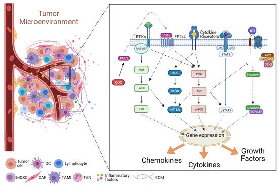
The development of tumors requires an initiator event, usually exposure to DNA damaging agents that cause genetic alterations such as gene mutations or chromosomal abnormalities, leading to deregulated cell proliferation. Although the mere stochastic accumulation of further mutations may cause tumor progression, it is now clear that an inflammatory microenvironment has a major tumor-promoting influence on initiated cells, in particular when a chronic inflammatory reaction already existed before the initiated tumor cell was formed. Moreover, inflammatory cells become mobilized in response to signals emanating from tumor cells. In both cases, the microenvironment provides signals that initiated tumor cells perceive by membrane receptors and transduce via downstream kinase cascades to modulate multiple cellular processes and respond with changes in cell gene expression, metabolism, and morphology. Cytokines, chemokines, and growth factors are examples of major signals secreted by immune cells, fibroblast, and endothelial cells and mediate an intricate cell-cell crosstalk in an inflammatory microenvironment, which contributes to increased cancer cell survival, phenotypic plasticity and adaptation to surrounding tissue conditions. Eventually, consequent changes in extracellular matrix stiffness and architecture, coupled with additional genetic alterations, further fortify the malignant progression of tumor cells, priming them for invasion and metastasis.

tumor microenvironment inflammation signal transduction cancer

1. Introduction

In the human body, complex physiological processes need to be coordinated at a cellular level. Circulating cytokines, hormones, and growth factors control aspects such as cell proliferation, differentiation, metabolism, angiogenesis, apoptosis, and senescence. Cells respond to such signals from their environment through sensors at the cell surface, namely receptor proteins that propagate their activation to intracellular proteins via sequential protein kinase signaling, often translocating into the nucleus, where transcription factors become activated, resulting in changes in gene expression that subsequently alter the cell’s biological responses.
Tumor cells develop several well-defined features that cause dysregulation of cellular signal transduction pathways, leading to increased cell proliferation, resistance to apoptosis, metabolic changes, genetic instability, induction of angiogenesis, and increased migratory capacity. This dysregulation involves genetic mutations and epigenetic changes in the tumor cells but also a complex interplay and exchange of signals with surrounding non-neoplastic cells and the extracellular matrix (ECM), designated as the tumor microenvironment (TME) [1][2].
 

2. Cancer-Associated Inflammation (CAI) and the TME

2.1. Mediators of Cancer-Associated Inflammation (CAI)

Soluble components in the TME are derived both from stroma and tumor cells. The main inflammatory mediators are cytokines, which include chemokines, interferons, interleukins, and tumor necrosis factors. Some are signaling molecules that trigger a specific biological function in the receptor-expressing target cells, whereas chemokines function by attracting immune cells to sites of inflammation. Cytokines modify the behavior of both tumor and inflammatory cells, influencing the type, abundance, and activity of the latter in the TME.

2.1.1. Pro-Inflammatory Factors

The main primary inflammatory cytokines are IL-1β, IL-6, and TNF-α that use type I transmembrane receptors with extracellular immunoglobulin domains for signaling [3]. These pro-inflammatory cytokines are not expressed in healthy tissue but become upregulated by an inflammatory insult to protect the host. Paradoxically, during carcinogenesis, they do not protect the host but promote cancer cell survival. In many cases, the reason for this paradox of cytokine function is the presence of a smoldering subclinical-level of chronic inflammation, continuously drawing in inflammatory cells [4][5].
IL-1β is crucial to initiate acute inflammation and is mainly synthesized as a precursor protein in tissue-resident macrophages, monocytes, or neutrophils. Upon their activation, IL-1β is cleaved by intracellular caspase-1 within the inflammasome complex and can then be secreted. Inflammasome activation can be initiated by cytosolic pattern recognition receptors (PRRs) in response to microbial pathogen-associated molecular patterns (PAMPs) or danger-associated molecular patterns (DAMPs) generated by the host cell. Apparently, intestinal epithelial cells also contain inflammasome components and are able to release IL-1β as a first line of defense against environmental pathogens that then promotes differentiation of monocytes to macrophages [6][7][8][9]. When IL-1β binds to a cell with IL1R, signal transduction leads to activation of the transcription factor NF-κB, which stimulates expression of pro-inflammatory cytokine genes, including IL-6 and TNF-α (see 3.2 for details). Tumor cells can also express IL1R, which in breast cancer cells was shown to stimulate IL-6 production [10], or in colon cells to drive sustained NF-κB activation involved in cell proliferation [11]. IL-1β further promotes tumor development by stimulating secretion of VEGF by malignant cells and influencing angiogenesis to increase blood vessel density in tumors [9][12][13].
Altogether, IL-1β in the TME is generally pro-tumorigenic, sustaining chronic non-resolved inflammation, endothelial cell activation, tumor angiogenesis, and the induction of immune-suppressive cells, although divergent data ascribing tumor-inhibiting effects to IL-1β exist [14]. Clinically, high serum levels of IL-1β correlate with bad prognosis in cancer patients and detection of IL-1β within the TME generally predicts tumor growth and metastasis [15].
IL-6 is critical in the final maturation of B-cells, T cell differentiation into Th1, Th2 and Treg phenotypes, and also inhibits functional maturation of DC to activate effector T-lymphocytes, blocking the anticancer immunity [16]. IL-6 is produced mainly by fibroblasts, monocytes, and macrophages, but also tumor cells, which can enhance IL-6 production by activated stromal fibroblasts.
IL-6R with its associated gp130 co-receptor activates JAK-STAT signaling leading to phosphorylation and nuclear translocation of STAT3 transcription factor and expression of IL-6-responsive genes. IL-6-activated STAT3 has been shown to be a survival and/or proliferation factor in certain cancers [17] and high serum IL-6 levels were reported in various cancer types and associated with poor prognosis [18], with anti-IL-6 receptor antibodies showing anti-tumor and anti-angiogenic activity in vivo [19].
TNF-α is a potent inflammatory mediator in any inflammatory reaction of the innate immune system. It induces the expression of chemokines and endothelial and cellular adhesion molecules in order to facilitate the recruitment of effector immune cells to the site of infection [3]. TNF-α is first synthesized as a transmembrane precursor (mTNF-α) requiring release by proteolytic cleavage through TACE (or ADAM17) [3][20]. A soluble trimer (sTNF-α) is the ligand for TNF receptors with TNFR1 being widely expressed on different cell types. TNFR1 is a single transmembrane glycoprotein containing an intracellular death domain, to which upon activation the adaptor molecule TRADD and TRAF2 are recruited, leading to the addition of linear ubiquitin chains to RIPK1 via the ubiquitin-E3 ligase activity of TRAF2. If the formation of this so-called Complex I is successful, the ubiquitin chains act as scaffolds for additional factors that lead to the activation of NF-κB, JNK, and p38 pathways and induction of cytokine signaling and cell survival [20]. Alternatively, failure of RKIP1 ubiquitination leads to Complex II formation by recruitment of Fas-associated death domain (FADD) and activation of pro-caspase-8 to mediate cell death via apoptosis or necroptosis, hence the initial designation as tumor-necrosis factor [21].
The therapeutic administration of neutralizing anti-TNF-α antibodies effectively reduces local and systemic inflammation in patients with rheumatoid arthritis or Crohn’s Disease [22] but may also increase the risk for malignant diseases [23].

2.1.2. Pro-Inflammatory Chemokines in the TME

More than 50 different chemokines exist, mainly from the CC and CXC subfamilies, that attract immune cells to sites of inflammation, including the TME [24]. One example is IL-8/CXCL8 that is recognized by the G protein-coupled receptors CXCR1 and CXCR2, and signals via phosphatidylinositol-3 kinase (PI3K) and MAPK [25] (see Section 3.2 for details). The main role of IL-8 in inflammation is the recruitment of neutrophils but increased expression of IL-8 and/or its receptors has been characterized in cancer cells, where IL-8 acts as an autocrine growth factor inducing proliferation and preventing apoptosis. Chemokines also modulate stromal cells in the TME to release growth and angiogenic factors [25][26].
Other soluble factors in the TME are reactive oxygen species (ROS) that are secreted either by activated immune cells such as neutrophils or MDSCs, or as intercellular signaling molecule such as nitric oxide secreted by M1-type macrophages or cancer cells. Their presence can increase DNA damage and the mutation rate in cancer cells. On the other hand, excess ROS production by altered metabolism of cancer cells contributes to immunosuppression, as immune effector cells such as NK and Treg cells are inhibited by ROS [27].

2.1.3. Anti-Inflammatory Mediators—IL-10

Anti-inflammatory cytokines are functionally defined as those inhibiting the synthesis of pro-inflammatory cytokines such as IL-1 or TNF-α. Their function is mainly to limit the magnitude of the immune response and prevent damage to the inflamed host tissue by allowing tissue repair and regeneration. Major anti-inflammatory cytokines include IL-1 receptor antagonist (ra), IL-4, IL-10, IL-11, and IL-13, but also soluble and decoy variants of TNF, IL-1, or IL-18 receptors.
A key immunosuppressive cytokine is IL-10, which is produced by a variety of immune but also non-hematopoietic and cancer cells. Upon binding of IL-10, the receptor IL-10R inhibits the activation of NF-κB and STAT3 that are required for transcription of pro-inflammatory cytokine genes [28].
IL-10 is an essential regulator of intestinal homeostasis, and mice and humans deficient in either IL-10 or its receptor (IL-10R) develop spontaneous intestinal inflammation. In addition, polymorphisms or mutations in the IL-10 locus confer increased genetic risk for developing ulcerative colitis or Crohn’s disease [29][30][31]. Another phenomenon of insufficient anti-inflammatory response is known as the cytokine storm, characterized by an aggressive pro-inflammatory response with loss of homeostasis of the immune response to either pathogens, like SARS-CoV-2 [32] or in response to cancer immunotherapy [33][34].
The presence of IL-10 in the TME may thus suggest that it undermines the immune response of macrophages to cancer; however, IL-10 is also required for the expansion of tumor-specific cytotoxic CD8+ T cells to control tumor cells [35].

2.1.4. The Transforming Growth Factor-β (TGF-β)

Virtually all human cell types are responsive to TGF-β and a dual role has been described in relation to cancer.
In normal or initiated cancer cells, TGF-β suppresses growth. The active TGF-β dimer activates a pair of receptor serine/threonine kinases known as the type I and type II receptors, which propagate the signal by phosphorylating Smad transcription factors. After trimerization, phospho-SMAD2, SMAD3, and SMAD4 translocate to the nucleus and bind to their specific SMAD binding element in the promoter of target genes. For its growth-inhibitory effect, Smads mobilize cyclin-dependent kinase (CDK) inhibitors and suppress expression of c-Myc. Later in tumorigenesis, mutations in the tumor-suppressive arm of the pathway (e.g., the TGFBR2 or SMAD4 genes) allows TGF-β to promote expression of the transcription factors SNAIL and SLUG, which stimulate EMT and invasiveness [36].
In the TME, TGF-β is mostly secreted by TAMs and Tregs as an immune-suppressive molecule [37]. Similar to IL-10, TGF-β inactivates NK cells and promotes differentiation of TAN into the N2 phenotype or of T cells into the Treg class [38]. TGFβ further stimulates the activation of mesenchymal stem cells, tissue fibroblasts, or endothelial cells into CAFs that produce matrix metalloproteases and degrade the ECM, facilitating tumor cell invasion, and also produce cytokines, promoting cancer cell proliferation and angiogenesis. Thus, TGFβ acts in the TME as an immune suppressor, an inducer of tumor-cell mitogens and a promoter of carcinoma invasion [36][39].

2.2. Signaling in the Inflammatory TME

Many signaling events and pathways have been shown to mediate the contribution of inflammatory cues in the TME to the tumorigenic process. However, a set of molecules and pathways are considered to be prime drivers of cancer-associated inflammation (CAI) (summarized in Figure 2).
Figure 2. Schematic representation of the signaling pathways that are activated in tumor cells or tumor-associated stromal cells and modulate the interplay within the inflammatory tumor microenvironment, promoting tumor development and progression (see text for details). TAM—tumor-associated macrophage; TAN—tumor-associated neutrophil; DC—dendritic cells; CAF—cancer-associated fibroblast; MDSC—Myeloid-derived suppressor cells; ECM—Extracellular matrix. Figure created with BioRender.com.

2.2.1. The JAK/STAT Pathway

The Janus kinase (JAK) family of nonreceptor tyrosine kinases includes JAK1, JAK2, JAK3, and TYK2. JAK family proteins associate with the intracellular domain of cytokine receptors, transducing cytokine-induced signals to the signal transducer and activator of transcription (STAT) family of transcription factors [40]. The JAK/STAT pathway contributes to the regulation of many cellular processes, including immunity, cell growth, cell death, and differentiation. Dysregulation of JAK/STAT signaling underlies several pathogenic conditions related to chronic inflammation, autoimmune diseases, and cancer [40]. Among the seven mammalian STAT family members, STAT3 has been reported as having a vital role in modulating both endogenous and exogenous inflammatory signaling in tumors, by mediating the expression of inflammatory molecules triggered by oncogenic stimuli [41].
Most of these inflammatory mediators are produced by stromal immune cells such as TAMs and MDSCs, although some are produced by the tumor cells [1]. Underlying this inflammatory interplay is the continuous upregulation of STAT3 activity, which has been detected in as many as 50% of all human tumors [41]. In tumor cells, STAT3 promotes gene expression of many pro-inflammatory products such as IL-6, IL-10, and TNF-α. These, in turn, stimulate immune cell receptors that activate their STAT3 signaling pathways, further potentiating the expression of pro-tumorigenic cytokines, chemokines, and growth factors, thus establishing a STAT3 positive feedback loop between tumors and the inflammatory TME [1][42].
Notably, STAT3 signaling is also known to play an important role in tumor-associated angiogenesis. Upregulated STAT3 signaling in both tumor and stromal cells leads to the production of several angiogenic stimuli [43]. For example, the pro-inflammatory cytokine IL-6 activates STAT3 via the classical IL-6R/gp130-JAK-STAT3 pathway, leading to VEGFA expression by binding of activated STAT3 to the promoter of the VEGF gene [44]. Moreover, pro-inflammatory cytokines act via STAT3 not only on stromal and tumor cells but also on endothelial cells. For instance, stimulation of endothelial cells by IL-17 induced STAT3-mediated expression and secretion of additional inflammatory factors such as growth-related oncogene-α (GRO-α), GM-CSF, and IL-8 [45]. These factors also mediate the recruitment of additional inflammatory cells, including neutrophils, to the TME, namely to the perivascular stroma, thus promoting another tumorigenic feedforward cycle [43]. Of note, it was reported that TNF-α-induced IL-10 also participates in angiogenesis by promoting endothelial progenitor cell migration, adhesion, and tubule formation, through activation of the STAT3 pathway and induction of VEGF and MMP-9 expression [46]. Additionally, VEGF secretion also promotes breast and lung cancer stem cell self-renewal via VEGF receptor-2 (VEGFR-2)/JAK2/STAT3 pathway-mediated upregulation of MYC and SOX2 expression [47]. Finally, STAT3 activity has also been implicated in the evasion from immune surveillance, by promoting the expression of PD-L1 and PD-L2 in cancer cells, suppressing immune cell activity [48].

2.2.2. The NF-κB Pathway

Nuclear factor kappa-light-chain-enhancer of activated B cells (NF-κB) is a transcription factor assembled by the dimerization of two of five members of the Rel family of proteins that share a Rel homology domain in their N-terminus. However, whereas subunits RelA (p65), RelB, and c-Rel, have a transactivation domain in their C-termini, the p50 and p52 subunits do not. The classical NF-κB heterodimer is a combination of the p65 (RelA) and p50 subunits. While in an inactivated state, NF-κB is located in the cytosol complexed with the inhibitory protein nuclear factor of kappa light polypeptide gene enhancer in B-cells inhibitor alpha (IκBα). A variety of extracellular signals, stimulating various receptor types, result in the activation of the IκB kinase (IKK). IKK, in turn, phosphorylates the IκBα protein, which results in its dissociation from NF-κB dimers and eventual proteasomal degradation, allowing activated NF-κB to translocate into the nucleus and bind to the promoters of multiple target genes [49].
Although initially identified as a central mediator of immune cell stimulation, the NF-κB pathway is also activated in tumor cells and has been reported to promote tumor cell proliferation, survival, and invasion [1][50][51][52]. Nevertheless, the upregulation of NF-κB activity does not seem to be associated with oncogenic mutations of Rel proteins or their direct regulators [53]. On the contrary, the activation of NF-κB is mainly driven by inflammatory cytokines present in the TME, such as TNF-α, IL-6, IL-1, and IL-8 [53][54]. Indeed, persistent chronic inflammation can trigger abnormal NF-κB activity in precancerous lesions, promoting tumor development, which is then sustained by the inflammatory microenvironment, induced and maintained after malignant transformation [53][55]. The magnitude of NF-κB upregulation in tumors has been further highlighted by recent data showing that it can be detected in high levels in the plasma of breast and colon cancer patients, correlating with increased systemic inflammation markers such as, TNF-α, IL-6, and C-reactive protein (CRP) [56].
Notably, studies have shown that there is an important interplay between STAT3 and NF-κB signaling pathways in the development of inflammation-induced tumors. The concomitant activation of these two factors in the tumor and stromal immune cells results in secretion of a large number of tumor-promoting cytokines, including TNF-α, IL-1β, IL-6, IL-8, and VEGF [1][53][54]. This again triggers positive feedback loops where the inflammatory cytokines mediate the initiation signals to non-tumor stromal cells, including endothelial cells. NF-κB binds to the promoter of VEGFR2 and stimulates its expression in endothelial cells. The activation of VEGFR2 by the VEGF in the TME further promotes the activation of downstream angiogenesis signals in endothelial cells [57]. NF-κB also binds the IL-8 gene promoter, and stimulates its transcription by stromal and tumor cells, an inflammatory chemokine that also functions as a pro-angiogenic agent [53]. By binding to the C-X-C motif chemokine receptor 2 (CXCR2), IL-8 upregulates VEGF levels in endothelial cells, also via the NF-κB pathway, leading to the autocrine activation of VEGFR2 [58].
Thus, the crosstalk between tumor and tumor-associated stromal cells feeds back to the TME, further stimulating angiogenesis but also tumor cell proliferation, epithelial-mesenchymal transition (EMT), invasion, metastasis, and chemoresistance [1][42]. Indeed, NF-κB activation is a well-known mechanism by which chemoresistance to anticancer agents arises, namely through the upregulation of a multitude of mediators, including anti-apoptotic genes [59]. Moreover, activated NF-κB also induces the expression of MMP-2 and MMP-9, again synergizing with STAT3 activity to facilitate EMT, invasion and metastasis [60][61]. NF-κB also regulates the expression of another important mediator of the inflammatory TME, often found overexpressed in malignant tumors—cyclooxygenase-2 (COX-2) [54][62].

2.2.3. The COX2/PGE2 Pathway

As we discussed above, inflammatory mediators can be produced by the different stromal cell types or directly by the cancer cells themselves. Besides the crucial cytokines, chemokines, growth factors, and matrix remodeling factors already mentioned, several other tumor-sustaining mediators are known to contribute to the inflammatory microenvironment. Among this, prostaglandin E2 (PGE2) clearly stands out [63]. PGE2 is a prostanoid lipid associated with the promotion of cancer cell survival, growth, migration, and invasion, also participating in tumor-associated angiogenesis, and immunosuppression [54][63]. Cyclooxygenase (COX)-1 and -2 are the rate-limiting enzymes for prostaglandin synthesis from arachidonic acid [63]. COX-1 is constitutively expressed in a wide range of normal tissues and works as a housekeeping enzyme responsible for maintaining tissue homeostasis. COX-2 is nearly absent in most normal cells, but is often overexpressed in multiple cancers, including colorectal, breast, stomach, lung, and pancreatic cancers, and is associated with poor prognosis [62][64]. Of note, COX-2-derived PGE2 has been shown to mediate the crosstalk between colonic tumor cells and TAMs [65], induce the accumulation and activation of MDSCs [66][67], and promote the tumor growth of mutant BRAFV600E melanomas by suppressing immunity and enhancing tumor-promoting inflammation [65][68][69].The depletion of COX-2 or downstream PGE2 synthases modifies the TME inflammatory signaling profile from pro-tumorigenic to anticancer pathways [54][62]. Moreover, the inhibition of COX-2 synergizes with PD-1 blockade to improve tumor cell eradication and augment the numbers of functional tumor-specific CTLs in patients with advanced epithelial ovarian cancer [70]. These data indicate the critical role of COX-2 and PGE2 in modulating the TME to an immunosuppressive status. Notably, several clinical studies have shown that the long-term use of non-steroidal anti-inflammatory drugs, which act by inhibiting COX activity, may reduce the risk of developing several types of cancer, although the optimal preventive drug dosages and treatment durations remain to be fully clarified [71].
Several stimuli from the inflammatory TME can elevate the expression of COX-2 or upregulate COX-2/PGE2 signaling axis. For example, TME cues were shown to activate the receptor tyrosine kinase ephrin-A receptor 2 (EPHA2), which signals through the TGF-β pathway to stimulate COX-2 expression in pancreatic cancer [72]. In addition, reduction of Receptor-interacting protein kinase 3 (RIPK3), a key element in colonic mucosal repair, elicited NF-κB-mediated upregulation of COX-2 and consequent increased PGE2 production in colorectal cancer cells and associated MDSCs [73]. Moreover, PGE2 exacerbated the immunosuppressive activity of MDSCs, accelerated tumor growth, and further suppressed RIPK3 expression, in another example of a TME -associated feedforward cycle [73]. Notably, inhibition of COX-2 or PGE2 receptors reversed the immunosuppressive activity of MDSCs and dampened tumorigenesis [73].
In another example, histone deacetylase 6 (HDAC6), one of class II histone deacetylases, was frequently found associated with poor survival outcomes when upregulated in the CAFs from breast cancer patients [74]. HDAC6 upregulation in CAFs was a crucial epigenetic mediator to promote an immunosuppressive TME by regulating STAT3 activation and promoting STAT3-dependent expression of COX-2 and PGE2 synthesis [74]. Finally, the frequent cancer-associated aberrant activity of classical mitogen-activated protein kinase (MAPK) cascades, such as the MAPK/ERK and p38 MAPK pathways, also promoting the upregulation of COX-2/PGE2, thus favoring immunosuppressive TME and promoting the progression of various cancer types [62][75][76][77].

2.2.4. The PI3K/Akt Pathway

The phosphatidylinositol 3-kinase (PI3K)/AKT (Protein kinase B, PKB) pathway is a crucial coordinator of intracellular signaling in response to the extracellular stimuli. Consequently, its dysregulation has been reported to have a broad role in mediating the inflammatory signaling between tumors and the TME [78]. On one hand, upstream receptor tyrosine kinases, the p110α catalytic subunit of PI3K, the downstream effector kinase AKT, and the negative pathway regulator PTEN (a lipid phosphatase that dephosphorylates phosphatidylinositol (3,4,5)-trisphosphate (PIP3) to phosphatidylinositol (4,5)-bisphosphate (PIP2)), are all often mutated in a wide range of human tumors, contributing to the malignant transformation of cells by promoting tumor growth, but also invasion and metastasis [79]. For example, activation of mechanistic target of rapamycin (mTOR) serine/threonine kinase downstream of PI3K/AKT promotes tumor cell growth, proliferation, and survival, and also facilitates tumor cell motility through cytoskeletal reorganization [80]. Moreover, activated AKT signaling also facilitates EMT through differential phosphorylation on Twist, a key transcriptional regulator controlling cell plasticity, in response to growth factors and inflammatory cytokines in the TME [81]. On the other hand, various inflammatory factors, such as IL-1, IL-6, IL-17A, and TFN-α, stimulate AKT activation in tumor cells and, conversely, leading to the synthesis and secretion of more inflammatory mediators, including IL-8, IL-6 and CCL2 [79][82].
As described above, the transcriptional regulation of most of these cytokines is primarily controlled by NF-κB and STAT3 pathways. However, NF-κB and STAT3 activities are also tightly controlled via cross-talks with other key intracellular pathways, including the PI3K/AKT/mTOR pathway [78]. For example, AKT can activate the IKK complex, namely through a direct mTOR-IKK interaction [83], and part of TNF-α-induced AKT pro-oncogenic signaling is relayed through the NF-κB pathway [84]. In addition, in hepatocellular carcinoma, TME IL-17 robustly induced IL-6 expression and STAT3 activation in an AKT-dependent manner [85]. In turn, IL-6 activated JAK2/STAT3 signaling leading to IL-8, MMP-2, and VEGF upregulation, which promoted neutrophil infiltration and increased tumor vascularity [85].
Indeed, several reports have shown that multiple pathways downstream of activated AKT participate in the recruitment, proliferation, and differentiation of various TME-associated leukocytes, such as TAMs, TANs, and lymphocytes, thereby further promoting inflammation [78][86]. Thus, the dysregulation of PI3K/AKT signaling favors the aggregation of reactive immune cells in the TME, resulting in the release and accumulation of ROS in the tumor site by means of the oxidative stress response [1][81]. Notably, increased ROS levels in the TME can feedback and further stimulate AKT signaling in tumor cells. For instance, in ovarian cancer cells, epidermal growth factor (EGF)-induced increase in ROS levels promoted the activation of the AKT/mTOR/ p70 S6 Kinase 1 (p70S6K1) axis, which mediate the expression of (HIF-1 and VEGF, promoting tumor angiogenesis [87].
Interestingly, TAM-secreted IL-1 and TNF-α also promotes the phosphorylation of pro-apoptotic Bcl-2 family member BAD by activating the PI3K/AKT pathway, preventing BAD from inhibiting the activity of survival protein Bcl-2 and Bcl-xL, favoring neovascularization by promoting endothelial cell survival through a mechanism that also involves NF-κB activity [88][89]. Notably, despite limited by resistance and adverse effects, it has been suggested that the anti-angiogenic effects of PI3K inhibitors might account, at least in part, for their reported therapeutic effectiveness within the advanced stages of multiple cancers, including lymphoma, glioblastoma multiforme, melanoma, colorectal, lung, breast, and hepatocellular carcinomas [90][91][92].

2.2.5. The Wnt Pathway

The Wnt pathway is one of the central mechanisms regulating tissue morphogenesis during embryogenesis and repair [93]. It is thus not surprising that anomalous Wnt signaling has been associated with several cancers, namely colorectal, breast, lung, oral, cervical, and hematopoietic malignancies [94].
To date, 19 distinct Wnt proteins (ligands), 10 frizzled receptors (FZD), and several co-receptors have been identified in mammals [95]. In the canonical Wnt pathway, Wnt proteins bind to their complex receptor FZD/LRP5/6 (low-density lipoprotein receptor related protein), preventing proteasomal degradation of β-catenin. This allows β-catenin to be translocated to the nucleus, where it binds to transcription factors of the T cell factor (TCF)/lymphoid enhancer factor (LEF) family, activating the expression of multiple target genes [93][96]. Conversely, in the absence of Wnt ligands, the phosphorylation of β-catenin by the so-called destruction complex (composed of axin, Adenomatous Polyposis Coli (APC), and kinases CK1, and GSK3β) leads to its ubiquitination by β-TrCP ubiquitin ligase, targeting it for proteasomal degradation. As a result, the pool of β-catenin in the cytosol is depleted, and its nuclear translocation is blocked, restraining the transcription of Wnt target genes [96]. The APC protein serves as the building platform for the assembly of the β-catenin destruction complex. Loss-of-function mutations in APC, which occur early on in cancers such as colon cancer, lead to loss of β-catenin regulation and aberrant activation of Wnt signaling [96]. Indeed, familial adenomatous polyposis (FAP), an inherited condition characterized by numerous adenomatous polyps in the large intestine and highly increased risk of colon cancer, results from germline mutation in the APC gene [97].
Several growth factors secreted by stromal cells of TME have also been reported to induce the activation of Wnt signaling in tumor cells. For example, augmented hepatocyte growth factor (HGF) levels in colorectal cancer (CRC) upregulate β-catenin expression via the PI3K pathway and promote β-catenin dissociation from c-Met (HGF receptor) at the plasma membrane enhancing the activity of the β-catenin-regulated TCF family of transcription factors [98].
Also in CRC, TAMs express increasing levels of Wnt ligands Wnt2 and Wnt5a during progression from normal colorectal adenoma to carcinoma, suggesting that paracrine Wnt activation by macrophages may result in cancer progression [99]. A study, using mouse models of breast cancer, showed that Wnt7b, another Wnt ligand produced by TAMs, initiated the canonical Wnt pathway in TECs expressing LRP5 and Frizzled, leading to β-catenin-mediated transcription of cell cycle genes, thus linking Wnt signaling to tumor angiogenesis [100].
Notably, PGE2 secreted by TECs is also known to activate β-catenin signaling and help in the proliferation of CRC cells. PGE2-stimulated EP2 receptors promote the dissociation of GSK3β from the destruction complex while simultaneously inducing its inactivation via PI3K/AKT-mediated phosphorylation. This leads to translocation of β-catenin to the nucleus of cancer cells, promoting tumor proliferation and progression [101].
Finally, recent evidence indicates that the aberrant expression of Wnt5a in some tumors, including melanoma, non-small-cell lung cancer, and ovarian cancer, can drive a Wnt5a/NF-κB/IL-6/STAT3 positive feedback loop that contributes to an immunosuppressive tumor microenvironment [102][103]. Wnt5a expression is upregulated by several cytokines including TNF-α, IFN-γ, IL-1β, and IL-6, in both immune and non-immune cells [104][105][106][107][108].
This upregulation can be prevented by pre-incubation with either NF-κB or STAT3 inhibitors, suggesting that these pathways are implicated in Wnt5a transcription [105][109][110]. Conversely, Wnt5a can also induces NF-κB and STAT3 activity, thus generating a continuous feedforward loop in the TME [111][112][113]. At some point, particular subpopulations of stromal immune cells arise that respond to Wnt5a signals by promoting the synthesis of IL-10, thus generating tolerogenic microenvironments [102]. For instance, a recent study determined that a Wnt5a+CD68+/CD68+ TAMs ratio was significantly associated with poor prognosis in CRC patients and that the Wnt5a+ TAMs were of an M2-like subtype [110]. Wnt5a induced TAMs to secrete IL-10 by stimulating a CaKMII-ERK1/2-STAT3-dependent pathway, and IL-10 then acted autocrinally to induce M2 polarization of these TAMs. Furthermore, Wnt5a-induced M2 TAMs promoted CRC cells proliferation, migration and invasion, and the knockdown of Wnt5a significantly impaired the pro-tumor functions of TAMs [110].

References

  1. Li, L.; Yu, R.; Cai, T.; Chen, Z.; Lan, M.; Zou, T.; Wang, B.; Wang, Q.; Zhao, Y.; Cai, Y. Effects of Immune Cells and Cytokines on Inflammation and Immunosuppression in the Tumor Microenvironment. Int. Immunopharmacol. 2020, 88, 106939.
  2. Balkwill, F.R.; Capasso, M.; Hagemann, T. The Tumor Microenvironment at a Glance. J. Cell Sci. 2012, 125, 5591–5596.
  3. Turner, M.D.; Nedjai, B.; Hurst, T.; Pennington, D.J. Cytokines and Chemokines: At the Crossroads of Cell Signalling and Inflammatory Disease. Biochim. Biophys. Acta 2014, 1843, 2563–2582.
  4. Dinarello, C.A. The Paradox of Pro-Inflammatory Cytokines in Cancer. Cancer Metastasis Rev. 2006, 25, 307–313.
  5. Colotta, F.; Allavena, P.; Sica, A.; Garlanda, C.; Mantovani, A. Cancer-Related Inflammation, the Seventh Hallmark of Cancer: Links to Genetic Instability. Carcinogenesis 2009, 30, 1073–1081.
  6. Schenk, M.; Fabri, M.; Krutzik, S.R.; Lee, D.J.; Vu, D.M.; Sieling, P.A.; Montoya, D.; Liu, P.T.; Modlin, R.L. Interleukin-1β Triggers the Differentiation of Macrophages with Enhanced Capacity to Present Mycobacterial Antigen to T Cells. Immunology 2014, 141, 174–180.
  7. Apte, R.N.; Krelin, Y.; Song, X.; Dotan, S.; Recih, E.; Elkabets, M.; Carmi, Y.; Dvorkin, T.; White, R.M.; Gayvoronsky, L.; et al. Effects of Micro-Environment- and Malignant Cell-Derived Interleukin-1 in Carcinogenesis, Tumour Invasiveness and Tumour-Host Interactions. Eur. J. Cancer Oxf. Engl. 1990 2006, 42, 751–759.
  8. Gelfo, V.; Romaniello, D.; Mazzeschi, M.; Sgarzi, M.; Grilli, G.; Morselli, A.; Manzan, B.; Rihawi, K.; Lauriola, M. Roles of IL-1 in Cancer: From Tumor Progression to Resistance to Targeted Therapies. Int. J. Mol. Sci. 2020, 21, 9.
  9. Voronov, E.; Apte, R.N. IL-1 in Colon Inflammation, Colon Carcinogenesis and Invasiveness of Colon Cancer. Cancer Microenviron. Off. J. Int. Cancer Microenviron. Soc. 2015, 8, 187–200.
  10. Oh, K.; Lee, O.-Y.; Park, Y.; Seo, M.W.; Lee, D.-S. IL-1β Induces IL-6 Production and Increases Invasiveness and Estrogen-Independent Growth in a TG2-Dependent Manner in Human Breast Cancer Cells. BMC Cancer 2016, 16, 724.
  11. Terzić, J.; Grivennikov, S.; Karin, E.; Karin, M. Inflammation and Colon Cancer. Gastroenterology 2010, 138, 2101–2114.e5.
  12. Akagi, Y.; Liu, W.; Xie, K.; Zebrowski, B.; Shaheen, R.M.; Ellis, L.M. Regulation of Vascular Endothelial Growth Factor Expression in Human Colon Cancer by Interleukin-1beta. Br. J. Cancer 1999, 80, 1506–1511.
  13. Carmi, Y.; Dotan, S.; Rider, P.; Kaplanov, I.; White, M.R.; Baron, R.; Abutbul, S.; Huszar, M.; Dinarello, C.A.; Apte, R.N.; et al. The Role of IL-1β in the Early Tumor Cell-Induced Angiogenic Response. J. Immunol. 2013, 190, 3500–3509.
  14. Baker, K.J.; Houston, A.; Brint, E. IL-1 Family Members in Cancer; Two Sides to Every Story. Front. Immunol. 2019, 10, 1197.
  15. Lewis, A.M.; Varghese, S.; Xu, H.; Alexander, H.R. Interleukin-1 and Cancer Progression: The Emerging Role of Interleukin-1 Receptor Antagonist as a Novel Therapeutic Agent in Cancer Treatment. J. Transl. Med. 2006, 4, 48.
  16. Kitamura, H.; Ohno, Y.; Toyoshima, Y.; Ohtake, J.; Homma, S.; Kawamura, H.; Takahashi, N.; Taketomi, A. Interleukin-6/STAT3 Signaling as a Promising Target to Improve the Efficacy of Cancer Immunotherapy. Cancer Sci. 2017, 108, 1947–1952.
  17. Kumari, N.; Dwarakanath, B.S.; Das, A.; Bhatt, A.N. Role of Interleukin-6 in Cancer Progression and Therapeutic Resistance. Tumour Biol. J. Int. Soc. Oncodev. Biol. Med. 2016, 37, 11553–11572.
  18. Rossi, J.-F.; Lu, Z.-Y.; Jourdan, M.; Klein, B. Interleukin-6 as a Therapeutic Target. Clin. Cancer Res. Off. J. Am. Assoc. Cancer Res. 2015, 21, 1248–1257.
  19. Nagasaki, T.; Hara, M.; Nakanishi, H.; Takahashi, H.; Sato, M.; Takeyama, H. Interleukin-6 Released by Colon Cancer-Associated Fibroblasts Is Critical for Tumour Angiogenesis: Anti-Interleukin-6 Receptor Antibody Suppressed Angiogenesis and Inhibited Tumour-Stroma Interaction. Br. J. Cancer 2014, 110, 469–478.
  20. Gough, P.; Myles, I.A. Tumor Necrosis Factor Receptors: Pleiotropic Signaling Complexes and Their Differential Effects. Front. Immunol. 2020, 11, 585880.
  21. Micheau, O.; Tschopp, J. Induction of TNF Receptor I-Mediated Apoptosis via Two Sequential Signaling Complexes. Cell 2003, 114, 181–190.
  22. Fakhoury, M.; Negrulj, R.; Mooranian, A.; Al-Salami, H. Inflammatory Bowel Disease: Clinical Aspects and Treatments. J. Inflamm. Res. 2014, 7, 113–120.
  23. Pereira, R.; Faria, R.; Lago, P.; Torres, T. Infection and Malignancy Risk in Patients Treated with TNF Inhibitors for Immune-Mediated Inflammatory Diseases. Curr. Drug Saf. 2017, 12, 162–170.
  24. Nagarsheth, N.; Wicha, M.S.; Zou, W. Chemokines in the Cancer Microenvironment and Their Relevance in Cancer Immunotherapy. Nat. Rev. Immunol. 2017, 17, 559–572.
  25. Waugh, D.J.J.; Wilson, C. The Interleukin-8 Pathway in Cancer. Clin. Cancer Res. Off. J. Am. Assoc. Cancer Res. 2008, 14, 6735–6741.
  26. Bikfalvi, A.; Billottet, C. The CC and CXC Chemokines: Major Regulators of Tumor Progression and the Tumor Microenvironment. Am. J. Physiol. Cell Physiol. 2020, 318, C542–C554.
  27. Wang, L.; Kuang, Z.; Zhang, D.; Gao, Y.; Ying, M.; Wang, T. Reactive Oxygen Species in Immune Cells: A New Antitumor Target. Biomed. Pharmacother. 2021, 133, 110978.
  28. Moore, K.W.; de Waal Malefyt, R.; Coffman, R.L.; O’Garra, A. Interleukin-10 and the Interleukin-10 Receptor. Annu. Rev. Immunol. 2001, 19, 683–765.
  29. Zhu, L.; Shi, T.; Zhong, C.; Wang, Y.; Chang, M.; Liu, X. IL-10 and IL-10 Receptor Mutations in Very Early Onset Inflammatory Bowel Disease. Gastroenterol. Res. 2017, 10, 65–69.
  30. Franke, A.; McGovern, D.P.B.; Barrett, J.C.; Wang, K.; Radford-Smith, G.L.; Ahmad, T.; Lees, C.W.; Balschun, T.; Lee, J.; Roberts, R.; et al. Genome-Wide Meta-Analysis Increases to 71 the Number of Confirmed Crohn’s Disease Susceptibility Loci. Nat. Genet. 2010, 42, 1118–1125.
  31. Shouval, D.S.; Biswas, A.; Goettel, J.A.; McCann, K.; Conaway, E.; Redhu, N.S.; Mascanfroni, I.D.; Al Adham, Z.; Lavoie, S.; Ibourk, M.; et al. Interleukin-10 Receptor Signaling in Innate Immune Cells Regulates Mucosal Immune Tolerance and Anti-Inflammatory Macrophage Function. Immunity 2014, 40, 706–719.
  32. Coperchini, F.; Chiovato, L.; Ricci, G.; Croce, L.; Magri, F.; Rotondi, M. The Cytokine Storm in COVID-19: Further Advances in Our Understanding the Role of Specific Chemokines Involved. Cytokine Growth Factor Rev. 2021, 58, 82–91.
  33. Rossi, J.-F.; Lu, Z.Y.; Massart, C.; Levon, K. Dynamic Immune/Inflammation Precision Medicine: The Good and the Bad Inflammation in Infection and Cancer. Front. Immunol. 2021, 12, 595722.
  34. Cobb, D.A.; Lee, D.W. Cytokine Release Syndrome Biology and Management. Cancer J. Sudbury Mass 2021, 27, 119–125.
  35. Oft, M. IL-10: Master Switch from Tumor-Promoting Inflammation to Antitumor Immunity. Cancer Immunol. Res. 2014, 2, 194–199.
  36. Massagué, J. TGFbeta in Cancer. Cell 2008, 134, 215–230.
  37. Gonzalez, H.; Hagerling, C.; Werb, Z. Roles of the Immune System in Cancer: From Tumor Initiation to Metastatic Progression. Genes Dev. 2018, 32, 1267–1284.
  38. Itatani, Y.; Kawada, K.; Sakai, Y. Transforming Growth Factor-β Signaling Pathway in Colorectal Cancer and Its Tumor Microenvironment. Int. J. Mol. Sci. 2019, 20, 5822.
  39. Derynck, R.; Turley, S.J.; Akhurst, R.J. TGFβ Biology in Cancer Progression and Immunotherapy. Nat. Rev. Clin. Oncol. 2021, 18, 9–34.
  40. Coricello, A.; Mesiti, F.; Lupia, A.; Maruca, A.; Alcaro, S. Inside Perspective of the Synthetic and Computational Toolbox of JAK Inhibitors: Recent Updates. Mol. Basel Switz. 2020, 25, 3321.
  41. Guanizo, A.C.; Fernando, C.D.; Garama, D.J.; Gough, D.J. STAT3: A Multifaceted Oncoprotein. Growth Factors Chur Switz. 2018, 36, 1–14.
  42. Ji, Z.; He, L.; Regev, A.; Struhl, K. Inflammatory Regulatory Network Mediated by the Joint Action of NF-KB, STAT3, and AP-1 Factors Is Involved in Many Human Cancers. Proc. Natl. Acad. Sci. USA 2019, 116, 9453–9462.
  43. Fu, L.-Q.; Du, W.-L.; Cai, M.-H.; Yao, J.-Y.; Zhao, Y.-Y.; Mou, X.-Z. The Roles of Tumor-Associated Macrophages in Tumor Angiogenesis and Metastasis. Cell. Immunol. 2020, 353, 104119.
  44. Leng, K.; Xu, Y.; Kang, P.; Qin, W.; Cai, H.; Wang, H.; Ji, D.; Jiang, X.; Li, J.; Li, Z.; et al. Akirin2 Is Modulated by MiR-490-3p and Facilitates Angiogenesis in Cholangiocarcinoma through the IL-6/STAT3/VEGFA Signaling Pathway. Cell Death Dis. 2019, 10, 262.
  45. Yuan, S.; Zhang, S.; Zhuang, Y.; Zhang, H.; Bai, J.; Hou, Q. Interleukin-17 Stimulates STAT3-Mediated Endothelial Cell Activation for Neutrophil Recruitment. Cell. Physiol. Biochem. Int. J. Exp. Cell. Physiol. Biochem. Pharmacol. 2015, 36, 2340–2356.
  46. Wang, Y.; Chen, Q.; Zhang, Z.; Jiang, F.; Meng, X.; Yan, H. Interleukin-10 Overexpression Improves the Function of Endothelial Progenitor Cells Stimulated with TNF-α through the Activation of the STAT3 Signaling Pathway. Int. J. Mol. Med. 2015, 35, 471–477.
  47. Zhao, D.; Pan, C.; Sun, J.; Gilbert, C.; Drews-Elger, K.; Azzam, D.J.; Picon-Ruiz, M.; Kim, M.; Ullmer, W.; El-Ashry, D.; et al. VEGF Drives Cancer-Initiating Stem Cells through VEGFR-2/Stat3 Signaling to Upregulate Myc and Sox2. Oncogene 2015, 34, 3107–3119.
  48. Chen, L.; Han, X. Anti-PD-1/PD-L1 Therapy of Human Cancer: Past, Present, and Future. J. Clin. Investig. 2015, 125, 3384–3391.
  49. Bloom, M.J.; Saksena, S.D.; Swain, G.P.; Behar, M.S.; Yankeelov, T.E.; Sorace, A.G. The Effects of IKK-Beta Inhibition on Early NF-Kappa-B Activation and Transcription of Downstream Genes. Cell. Signal. 2019, 55, 17–25.
  50. Greten, F.R.; Eckmann, L.; Greten, T.F.; Park, J.M.; Li, Z.-W.; Egan, L.J.; Kagnoff, M.F.; Karin, M. IKKbeta Links Inflammation and Tumorigenesis in a Mouse Model of Colitis-Associated Cancer. Cell 2004, 118, 285–296.
  51. Karin, M.; Cao, Y.; Greten, F.R.; Li, Z.-W. NF-KappaB in Cancer: From Innocent Bystander to Major Culprit. Nat. Rev. Cancer 2002, 2, 301–310.
  52. Aggarwal, B.B. Nuclear Factor-KappaB: The Enemy Within. Cancer Cell 2004, 6, 203–208.
  53. Lalle, G.; Twardowski, J.; Grinberg-Bleyer, Y. NF-ΚB in Cancer Immunity: Friend or Foe? Cells 2021, 10, 355.
  54. Fan, Y.; Mao, R.; Yang, J. NF-ΚB and STAT3 Signaling Pathways Collaboratively Link Inflammation to Cancer. Protein Cell 2013, 4, 176–185.
  55. Taniguchi, K.; Karin, M. NF-ΚB, Inflammation, Immunity and Cancer: Coming of Age. Nat. Rev. Immunol. 2018, 18, 309–324.
  56. Papila, K.B.; Sozer, V.; Cigdem, K.P.; Durmus, S.; Kurtulus, D.; Papila, C.; Gelisgen, R.; Uzun, H. Circulating Nuclear Factor-Kappa B Mediates Cancer-Associated Inflammation in Human Breast and Colon Cancer. J. Med. Biochem. 2021, 40, 150–159.
  57. Dong, F.; Zhou, X.; Li, C.; Yan, S.; Deng, X.; Cao, Z.; Li, L.; Tang, B.; Allen, T.D.; Liu, J. Dihydroartemisinin Targets VEGFR2 via the NF-ΚB Pathway in Endothelial Cells to Inhibit Angiogenesis. Cancer Biol. Ther. 2014, 15, 1479–1488.
  58. Martin, D.; Galisteo, R.; Gutkind, J.S. CXCL8/IL8 Stimulates Vascular Endothelial Growth Factor (VEGF) Expression and the Autocrine Activation of VEGFR2 in Endothelial Cells by Activating NFkappaB through the CBM (Carma3/Bcl10/Malt1) Complex. J. Biol. Chem. 2009, 284, 6038–6042.
  59. Godwin, P.; Baird, A.M.; Heavey, S.; Barr, M.P.; O’Byrne, K.J.; Gately, K. Targeting Nuclear Factor-Kappa B to Overcome Resistance to Chemotherapy. Front. Oncol. 2013, 3, 120.
  60. De, D.; Chowdhury, P.; Panda, S.K.; Ghosh, U. Ethanolic Extract of Leaf of Dillenia Pentagyna Reduces In-Vitro Cell Migration and Induces Intrinsic Pathway of Apoptosis via Downregulation of NF-Κβ in Human NSCLC A549 Cells. J. Cell. Biochem. 2019, 120, 19841–19857.
  61. Huang, L.; Jian, Z.; Gao, Y.; Zhou, P.; Zhang, G.; Jiang, B.; Lv, Y. RPN2 Promotes Metastasis of Hepatocellular Carcinoma Cell and Inhibits Autophagy via STAT3 and NF-ΚB Pathways. Aging 2019, 11, 6674–6690.
  62. Pu, D.; Yin, L.; Huang, L.; Qin, C.; Zhou, Y.; Wu, Q.; Li, Y.; Zhou, Q.; Li, L. Cyclooxygenase-2 Inhibitor: A Potential Combination Strategy With Immunotherapy in Cancer. Front. Oncol. 2021, 11, 637504.
  63. Wang, D.; Dubois, R.N. Eicosanoids and Cancer. Nat. Rev. Cancer 2010, 10, 181–193.
  64. Ferrer, M.D.; Busquets-Cortés, C.; Capó, X.; Tejada, S.; Tur, J.A.; Pons, A.; Sureda, A. Cyclooxygenase-2 Inhibitors as a Therapeutic Target in Inflammatory Diseases. Curr. Med. Chem. 2019, 26, 3225–3241.
  65. Zelenay, S.; van der Veen, A.G.; Böttcher, J.P.; Snelgrove, K.J.; Rogers, N.; Acton, S.E.; Chakravarty, P.; Girotti, M.R.; Marais, R.; Quezada, S.A.; et al. Cyclooxygenase-Dependent Tumor Growth through Evasion of Immunity. Cell 2015, 162, 1257–1270.
  66. Pan, Q.; Bao, L.W.; Merajver, S.D. Tetrathiomolybdate Inhibits Angiogenesis and Metastasis through Suppression of the NFkappaB Signaling Cascade. Mol. Cancer Res. MCR 2003, 1, 701–706.
  67. Wang, D.; DuBois, R.N. Immunosuppression Associated with Chronic Inflammation in the Tumor Microenvironment. Carcinogenesis 2015, 36, 1085–1093.
  68. Escuin-Ordinas, H.; Atefi, M.; Fu, Y.; Cass, A.; Ng, C.; Huang, R.R.; Yashar, S.; Comin-Anduix, B.; Avramis, E.; Cochran, A.J.; et al. COX-2 Inhibition Prevents the Appearance of Cutaneous Squamous Cell Carcinomas Accelerated by BRAF Inhibitors. Mol. Oncol. 2014, 8, 250–260.
  69. Panza, E.; De Cicco, P.; Ercolano, G.; Armogida, C.; Scognamiglio, G.; Anniciello, A.M.; Botti, G.; Cirino, G.; Ianaro, A. Differential Expression of Cyclooxygenase-2 in Metastatic Melanoma Affects Progression Free Survival. Oncotarget 2016, 7, 57077–57085.
  70. Miao, J.; Lu, X.; Hu, Y.; Piao, C.; Wu, X.; Liu, X.; Huang, C.; Wang, Y.; Li, D.; Liu, J. Prostaglandin E2 and PD-1 Mediated Inhibition of Antitumor CTL Responses in the Human Tumor Microenvironment. Oncotarget 2017, 8, 89802–89810.
  71. Gu, K.J.; Li, G. An Overview of Cancer Prevention: Chemoprevention and Immunoprevention. J. Cancer Prev. 2020, 25, 127–135.
  72. Markosyan, N.; Li, J.; Sun, Y.H.; Richman, L.P.; Lin, J.H.; Yan, F.; Quinones, L.; Sela, Y.; Yamazoe, T.; Gordon, N.; et al. Tumor Cell-Intrinsic EPHA2 Suppresses Anti-Tumor Immunity by Regulating PTGS2 (COX-2). J. Clin. Investig. 2019, 129, 3594–3609.
  73. Yan, G.; Zhao, H.; Zhang, Q.; Zhou, Y.; Wu, L.; Lei, J.; Wang, X.; Zhang, J.; Zhang, X.; Zheng, L.; et al. A RIPK3-PGE2 Circuit Mediates Myeloid-Derived Suppressor Cell-Potentiated Colorectal Carcinogenesis. Cancer Res. 2018, 78, 5586–5599.
  74. Li, A.; Chen, P.; Leng, Y.; Kang, J. Histone Deacetylase 6 Regulates the Immunosuppressive Properties of Cancer-Associated Fibroblasts in Breast Cancer through the STAT3-COX2-Dependent Pathway. Oncogene 2018, 37, 5952–5966.
  75. Chiang, K.-H.; Shieh, J.-M.; Shen, C.-J.; Chang, T.-W.; Wu, P.-T.; Hsu, J.-Y.; Tsai, J.-P.; Chang, W.-C.; Chen, B.-K. Epidermal Growth Factor-Induced COX-2 Regulates Metastasis of Head and Neck Squamous Cell Carcinoma through Upregulation of Angiopoietin-like 4. Cancer Sci. 2020, 111, 2004–2015.
  76. Smakman, N.; Kranenburg, O.; Vogten, J.M.; Bloemendaal, A.L.A.; van Diest, P.; Borel Rinkes, I.H.M. Cyclooxygenase-2 Is a Target of KRASD12, Which Facilitates the Outgrowth of Murine C26 Colorectal Liver Metastases. Clin. Cancer Res. Off. J. Am. Assoc. Cancer Res. 2005, 11, 41–48.
  77. Lim, H.J.; Park, J.H.; Jo, C.; Yoon, K.; Koh, Y.H. Cigarette Smoke Extracts and Cadmium Induce COX-2 Expression through γ-Secretase-Mediated P38 MAPK Activation in C6 Astroglia Cells. PLoS ONE 2019, 14, e0212749.
  78. Tang, F.; Wang, Y.; Hemmings, B.A.; Rüegg, C.; Xue, G. PKB/Akt-Dependent Regulation of Inflammation in Cancer. Semin. Cancer Biol. 2018, 48, 62–69.
  79. Noorolyai, S.; Shajari, N.; Baghbani, E.; Sadreddini, S.; Baradaran, B. The Relation between PI3K/AKT Signalling Pathway and Cancer. Gene 2019, 698, 120–128.
  80. Xu, K.; Liu, P.; Wei, W. MTOR Signaling in Tumorigenesis. Biochim. Biophys. Acta 2014, 1846, 638–654.
  81. Tang, H.; Massi, D.; Hemmings, B.A.; Mandalà, M.; Hu, Z.; Wicki, A.; Xue, G. AKT-Ions with a TWIST between EMT and MET. Oncotarget 2016, 7, 62767–62777.
  82. Chen, Y.; Kijlstra, A.; Chen, Y.; Yang, P. IL-17A Stimulates the Production of Inflammatory Mediators via Erk1/2, P38 MAPK, PI3K/Akt, and NF-ΚB Pathways in ARPE-19 Cells. Mol. Vis. 2011, 17, 3072–3077.
  83. Dan, H.C.; Cooper, M.J.; Cogswell, P.C.; Duncan, J.A.; Ting, J.P.-Y.; Baldwin, A.S. Akt-Dependent Regulation of NF-B Is Controlled by MTOR and Raptor in Association with IKK. Genes Dev. 2008, 22, 1490–1500.
  84. Factor, V.; Oliver, A.L.; Panta, G.R.; Thorgeirsson, S.S.; Sonenshein, G.E.; Arsura, M. Roles of Akt/PKB and IKK Complex in Constitutive Induction of NF-KappaB in Hepatocellular Carcinomas of Transforming Growth Factor Alpha/c-Myc Transgenic Mice. Hepatol. Baltim. Md 2001, 34, 32–41.
  85. Gu, F.-M.; Li, Q.-L.; Gao, Q.; Jiang, J.-H.; Zhu, K.; Huang, X.-Y.; Pan, J.-F.; Yan, J.; Hu, J.-H.; Wang, Z.; et al. IL-17 Induces AKT-Dependent IL-6/JAK2/STAT3 Activation and Tumor Progression in Hepatocellular Carcinoma. Mol. Cancer 2011, 10, 150.
  86. Xue, G.; Zippelius, A.; Wicki, A.; Mandalà, M.; Tang, F.; Massi, D.; Hemmings, B.A. Integrated Akt/PKB Signaling in Immunomodulation and Its Potential Role in Cancer Immunotherapy. J. Natl. Cancer Inst. 2015, 107.
  87. Liu, L.-Z.; Hu, X.-W.; Xia, C.; He, J.; Zhou, Q.; Shi, X.; Fang, J.; Jiang, B.-H. Reactive Oxygen Species Regulate Epidermal Growth Factor-Induced Vascular Endothelial Growth Factor and Hypoxia-Inducible Factor-1alpha Expression through Activation of AKT and P70S6K1 in Human Ovarian Cancer Cells. Free Radic. Biol. Med. 2006, 41, 1521–1533.
  88. Madge, L.A.; Pober, J.S. A Phosphatidylinositol 3-Kinase/Akt Pathway, Activated by Tumor Necrosis Factor or Interleukin-1, Inhibits Apoptosis but Does Not Activate NFkappaB in Human Endothelial Cells. J. Biol. Chem. 2000, 275, 15458–15465.
  89. Liu, S.; Shen, H.; Xu, M.; Liu, O.; Zhao, L.; Liu, S.; Guo, Z.; Du, J. FRP Inhibits Ox-LDL-Induced Endothelial Cell Apoptosis through an Akt-NF-B-Bcl-2 Pathway and Inhibits Endothelial Cell Apoptosis in an ApoE-Knockout Mouse Model. Am. J. Physiol. Endocrinol. Metab. 2010, 299, E351–E363.
  90. Massacesi, C.; Di Tomaso, E.; Urban, P.; Germa, C.; Quadt, C.; Trandafir, L.; Aimone, P.; Fretault, N.; Dharan, B.; Tavorath, R.; et al. PI3K Inhibitors as New Cancer Therapeutics: Implications for Clinical Trial Design. OncoTargets Ther. 2016, 9, 203–210.
  91. Suvarna, V.; Murahari, M.; Khan, T.; Chaubey, P.; Sangave, P. Phytochemicals and PI3K Inhibitors in Cancer-An Insight. Front. Pharmacol. 2017, 8, 916.
  92. Mishra, R.; Patel, H.; Alanazi, S.; Kilroy, M.K.; Garrett, J.T. PI3K Inhibitors in Cancer: Clinical Implications and Adverse Effects. Int. J. Mol. Sci. 2021, 22, 3464.
  93. Niehrs, C. The Complex World of WNT Receptor Signalling. Nat. Rev. Mol. Cell Biol. 2012, 13, 767–779.
  94. Azbazdar, Y.; Karabicici, M.; Erdal, E.; Ozhan, G. Regulation of Wnt Signaling Pathways at the Plasma Membrane and Their Misregulation in Cancer. Front. Cell Dev. Biol. 2021, 9, 631623.
  95. Caspi, M.; Wittenstein, A.; Kazelnik, M.; Shor-Nareznoy, Y.; Rosin-Arbesfeld, R. Therapeutic Targeting of the Oncogenic Wnt Signaling Pathway for Treating Colorectal Cancer and Other Colonic Disorders. Adv. Drug Deliv. Rev. 2021, 169, 118–136.
  96. Li, X.; Ortiz, M.A.; Kotula, L. The Physiological Role of Wnt Pathway in Normal Development and Cancer. Exp. Biol. Med. Maywood NJ 2020, 245, 411–426.
  97. Bugter, J.M.; Fenderico, N.; Maurice, M.M. Mutations and Mechanisms of WNT Pathway Tumour Suppressors in Cancer. Nat. Rev. Cancer 2021, 21, 5–21.
  98. Rasola, A.; Fassetta, M.; De Bacco, F.; D’Alessandro, L.; Gramaglia, D.; Di Renzo, M.F.; Comoglio, P.M. A Positive Feedback Loop between Hepatocyte Growth Factor Receptor and Beta-Catenin Sustains Colorectal Cancer Cell Invasive Growth. Oncogene 2007, 26, 1078–1087.
  99. Smith, K.; Bui, T.D.; Poulsom, R.; Kaklamanis, L.; Williams, G.; Harris, A.L. Up-Regulation of Macrophage Wnt Gene Expression in Adenoma-Carcinoma Progression of Human Colorectal Cancer. Br. J. Cancer 1999, 81, 496–502.
  100. Ojalvo, L.S.; Whittaker, C.A.; Condeelis, J.S.; Pollard, J.W. Gene Expression Analysis of Macrophages That Facilitate Tumor Invasion Supports a Role for Wnt-Signaling in Mediating Their Activity in Primary Mammary Tumors. J. Immunol. 2010, 184, 702–712.
  101. Castellone, M.D.; Teramoto, H.; Williams, B.O.; Druey, K.M.; Gutkind, J.S. Prostaglandin E2 Promotes Colon Cancer Cell Growth through a Gs-Axin-Beta-Catenin Signaling Axis. Science 2005, 310, 1504–1510.
  102. Lopez-Bergami, P.; Barbero, G. The Emerging Role of Wnt5a in the Promotion of a Pro-Inflammatory and Immunosuppressive Tumor Microenvironment. Cancer Metastasis Rev. 2020, 39, 933–952.
  103. Asem, M.S.; Buechler, S.; Wates, R.B.; Miller, D.L.; Stack, M.S. Wnt5a Signaling in Cancer. Cancers 2016, 8, 79.
  104. Pereira, C.; Schaer, D.J.; Bachli, E.B.; Kurrer, M.O.; Schoedon, G. Wnt5A/CaMKII Signaling Contributes to the Inflammatory Response of Macrophages and Is a Target for the Antiinflammatory Action of Activated Protein C and Interleukin-10. Arterioscler. Thromb. Vasc. Biol. 2008, 28, 504–510.
  105. Ge, X.-P.; Gan, Y.-H.; Zhang, C.-G.; Zhou, C.-Y.; Ma, K.-T.; Meng, J.-H.; Ma, X.-C. Requirement of the NF-ΚB Pathway for Induction of Wnt-5A by Interleukin-1β in Condylar Chondrocytes of the Temporomandibular Joint: Functional Crosstalk between the Wnt-5A and NF-ΚB Signaling Pathways. Osteoarthr. Cartil. 2011, 19, 111–117.
  106. Catalán, V.; Gómez-Ambrosi, J.; Rodríguez, A.; Pérez-Hernández, A.I.; Gurbindo, J.; Ramírez, B.; Méndez-Giménez, L.; Rotellar, F.; Valentí, V.; Moncada, R.; et al. Activation of Noncanonical Wnt Signaling through WNT5A in Visceral Adipose Tissue of Obese Subjects Is Related to Inflammation. J. Clin. Endocrinol. Metab. 2014, 99, E1407–E1417.
  107. Park, S.-Y.; Kang, M.-J.; Han, J.-S. Interleukin-1 Beta Promotes Neuronal Differentiation through the Wnt5a/RhoA/JNK Pathway in Cortical Neural Precursor Cells. Mol. Brain 2018, 11, 39.
  108. Linnskog, R.; Mohapatra, P.; Moradi, F.; Prasad, C.P.; Andersson, T. Demonstration of a WNT5A-IL-6 Positive Feedback Loop in Melanoma Cells: Dual Interference of This Loop More Effectively Impairs Melanoma Cell Invasion. Oncotarget 2016, 7, 37790–37802.
  109. Rozovski, U.; Harris, D.M.; Li, P.; Liu, Z.; Jain, P.; Ferrajoli, A.; Burger, J.A.; Bose, P.; Thompson, P.A.; Jain, N.; et al. STAT3-Induced Wnt5a Provides Chronic Lymphocytic Leukemia Cells with Survival Advantage. J. Immunol. 2019, 203, 3078–3085.
  110. Liu, Q.; Yang, C.; Wang, S.; Shi, D.; Wei, C.; Song, J.; Lin, X.; Dou, R.; Bai, J.; Xiang, Z.; et al. Wnt5a-Induced M2 Polarization of Tumor-Associated Macrophages via IL-10 Promotes Colorectal Cancer Progression. Cell Commun. Signal. CCS 2020, 18, 51.
  111. Chen, Y.; Chen, L.; Yu, J.; Ghia, E.M.; Choi, M.Y.; Zhang, L.; Zhang, S.; Sanchez-Lopez, E.; Widhopf, G.F.; Messer, K.; et al. Cirmtuzumab Blocks Wnt5a/ROR1 Stimulation of NF-ΚB to Repress Autocrine STAT3 Activation in Chronic Lymphocytic Leukemia. Blood 2019, 134, 1084–1094.
  112. Barbero, G.; Castro, M.V.; Villanueva, M.B.; Quezada, M.J.; Fernández, N.B.; DeMorrow, S.; Lopez-Bergami, P. An Autocrine Wnt5a Loop Promotes NF-ΚB Pathway Activation and Cytokine/Chemokine Secretion in Melanoma. Cells 2019, 8, 1060.
  113. Ikeda, T.; Nishita, M.; Hoshi, K.; Honda, T.; Kakeji, Y.; Minami, Y. Mesenchymal Stem Cell-Derived CXCL16 Promotes Progression of Gastric Cancer Cells by STAT3-Mediated Expression of Ror1. Cancer Sci. 2020, 111, 1254–1265.
More
Information
Subjects: Immunology
Contributor MDPI registered users' name will be linked to their SciProfiles pages. To register with us, please refer to https://encyclopedia.pub/register :
View Times: 799
Revisions: 2 times (View History)
Update Date: 29 Mar 2022
1000/1000