Submitted Successfully!
To reward your contribution, here is a gift for you: A free trial for our video production service.
Thank you for your contribution! You can also upload a video entry or images related to this topic.
Version Summary Created by Modification Content Size Created at Operation
1 + 3017 word(s) 3017 2021-06-02 08:13:37 |
2 format correct Meta information modification 3017 2021-06-08 11:54:16 |

Video Upload Options

Do you have a full video?

Confirm

Are you sure to Delete?
Cite
If you have any further questions, please contact Encyclopedia Editorial Office.
Chung, M. Oral Mucosal Pain. Encyclopedia. Available online: https://encyclopedia.pub/entry/10621 (accessed on 29 March 2024).
Chung M. Oral Mucosal Pain. Encyclopedia. Available at: https://encyclopedia.pub/entry/10621. Accessed March 29, 2024.
Chung, Man-Kyo. "Oral Mucosal Pain" Encyclopedia, https://encyclopedia.pub/entry/10621 (accessed March 29, 2024).
Chung, M. (2021, June 08). Oral Mucosal Pain. In Encyclopedia. https://encyclopedia.pub/entry/10621
Chung, Man-Kyo. "Oral Mucosal Pain." Encyclopedia. Web. 08 June, 2021.
Oral Mucosal Pain
Edit

The oral cavity is a portal into the digestive system, which exhibits unique sensory properties. Like facial skin, the oral mucosa needs to be exquisitely sensitive and selective, in order to detect harmful toxins versus edible food. Chemosensation and somatosensation by multiple receptors, including transient receptor potential channels, are well-developed to meet these needs. In contrast to facial skin, however, the oral mucosa rarely exhibits itch responses. Like the gut, the oral cavity performs mechanical and chemical digestion. Therefore, the oral mucosa needs to be insensitive, to some degree, in order to endure noxious irritation. Persistent pain from the oral mucosa is often due to ulcers, involving both tissue injury and infection. Trigeminal nerve injury and trigeminal neuralgia produce intractable pain in the orofacial skin and the oral mucosa, through mechanisms distinct from those seen in the spinal area, which is particularly difficult to predict or treat. The diagnosis and treatment of idiopathic chronic pain, such as atypical odontalgia (idiopathic painful trigeminal neuropathy or post-traumatic trigeminal neuropathy) and burning mouth syndrome, remain especially challenging. The central integration of gustatory inputs might modulate chronic oral and facial pain. A lack of pain in chronic inflammation inside the oral cavity, such as chronic periodontitis, involves the specialized functioning of oral bacteria. A more detailed understanding of the unique neurobiology of pain from the orofacial skin and the oral mucosa should help us develop novel methods for better treating persistent orofacial pain.

chronic pain mucosa pain orofacial pain

1. Introduction

Orofacial skin and the oral mucosa protect the body from physical and chemical damage, infection, dehydration, and heat loss. Even though both oral mucosa and facial skin are covered by highly specialized stratified epithelia, the two tissues are structurally different in many ways: hair follicles and sweat glands exist in the skin, while the oral mucosa surrounds the teeth and contains taste buds and minor salivary glands. The oral mucosa is more permeable than skin. Nonkeratinized mucosa, such as the floor of the mouth and the buccal mucosa, is more permeable than other regions of the oral mucosa, and transmucosal drug delivery is under active development [1]. As the oral mucosa heals after injury faster than skin and without scar tissue, the cellular, molecular, and immunologic differences between oral mucosa and skin have been widely studied, and the oral mucosa has been used as a model for developing methods for scarless cutaneous healing [2]. Therefore, distinct sensations arising from the oral mucosa and facial skin have drawn much attention [3][4].
As a portal into the digestive system, the oral cavity is exposed to a dynamic environment featuring mechanical, thermal, and chemical stimuli due to the ingestion and mastication of various foods. The oral mucosa exhibits sensory properties, similar to both facial skin and the gut. Like facial skin, the oral mucosa requires an exquisite level of sensitivity to mechanical, thermal, and chemical stimuli, in order to detect the properties of foods and to prevent the ingestion of harmful materials. Pain from the oral mucosa also modulates jaw movements and masticatory activities [5]. At the same time, similar to the gut, the oral mucosa needs to be somewhat insensitive to stimuli, in order to resist the mechanical mastication of hard food or to endure the swallowing of hot drinks. When ingested food is perceived to be unpleasant, the food is spit out of the oral cavity for protection. In addition to toxic food materials, multiple etiologies cause acute or chronic pathological pain in the oral cavity. Oral pain critically affects the quality of life, as it degrades vital functions, such as eating and swallowing, especially when the pain is chronic [6]. Here, we review the characteristics of the chemosensory and somatosensory functions of the oral mucosa, as well as its neurobiological mechanisms, in comparison to those in facial skin. We also review the pathological conditions inducing acute or chronic oral and facial pain and discuss their underlying mechanisms. A better understanding of the neurobiological mechanisms of oral and facial pain should help in the development of more effective methods for managing the associated conditions, eventually improving the quality of care for patients. Although orofacial pain is derived from different tissues due to a diverse etiology, including an autonomic function (Table 1), in this study, we focused on several subtypes of pain from the oral cavity and face that are more difficult to diagnose or treat.
Table 1. Classification overview of the International Classification of Orofacial Pain (ICOP) *.
Table Subtype
1. Orofacial pain attributed to disorders of dentoalveolar and anatomically related structures 1.1 Dental pain
1.2 Oral mucosal, salivary gland, and jawbone pains
2. Myofascial orofacial pain 2.1 Primary myofascial orofacial pain
2.2 Secondary myofascial orofacial pain
3. Temporomandibular joint (TMJ) pain 3.1 Primary temporomandibular joint pain
3.2 Secondary temporomandibular joint pain
4. Orofacial pain attributed to lesion or disease of the cranial nerves 4.1Pain attributed to lesion or diseaseofthetrigeminalnerve
4.2 Pain attributed to lesion or disease of the glossopharyngeal nerve
5. Orofacial pains resembling presentations of primary headaches 5.1 Orofacial migraine
5.2 Tension-type orofacial pain
5.3 Trigeminal autonomic orofacial pain
5.4 Neurovascular orofacial pain
6. Idiopathic orofacial pain 6.1 Burning mouth syndrome (BMS)
6.2 Persistent idiopathic facial pain (PIFP)
6.3 Persistent idiopathic dentoalveolar pain
6.4 Constant unilateral facial pain with additional attacks (CUFPA)
* Cephalalgia, 40:129–221 (2020). Bold, subtype of pain focused on in this review.

2. Physiological Somatosensation and Pain from Oral Mucosa and Facial Skin

Peripheral nociception in orofacial tissues occurs at the peripheral branches of the trigeminal nerves—the fifth cranial nerves (Figure 1). Trigeminal ganglia harbor neuronal cell bodies of sensory neurons, projecting to both the peripheral and central sides. Nociceptive nerves synapse with second-order neurons within the trigeminal nucleus complex, especially the caudal area [7], which relays the nociceptive signals to the brain regions involved in sensory discriminative and affective pain.
Figure 1. Innervation of the craniofacial sensory nerves, and the sources of persistent orofacial pain from tissue or nerve injuries. The trigeminal nerve is the fifth cranial nerve. The trigeminal ganglia (TG) contain neuronal cell bodies of sensory neurons projecting to the orofacial structures. The first branch (V1) projects to the forehead skin and dura through the ophthalmic nerve. The second branch (V2) projects to the skin, mucosa, and teeth of the upper jaw through the maxillary nerve. The third branch (V3) projects to the skin, mucosa, and teeth of the lower jaw, including the tongue mucosa. The central branches of the trigeminal sensory neurons project to the trigeminal nucleus complex in the brainstem and make synaptic connections with second-order neurons. The pain-sensing nociceptors from the orofacial area are highly connected with the caudal region of the trigeminal nucleus complex (trigeminal subnucleus caudalis; Vc). The Vc neurons relay signals to various ascending pain pathways within the brain. The tongue mucosa is also innervated by gustatory nerves, such as the chorda tympani nerve (Ch tym n), which is a part of the facial nerve (the seventh cranial nerve). Taste signals are transmitted through the chorda tympani nerve, relayed in the nucleus tract solitarius in the brainstem, and transmitted to the central taste pathways. Persistent pain from the orofacial area can be derived from multiple etiologies of injuries to the tissue or nerves. Irradiation or chemotherapy can cause oral mucositis. Oral cancer often causes pain from the oral mucosa. The direct injury of peripheral nerves due to facial trauma or tooth extraction can lead to the development of painful post-traumatic trigeminal neuropathy (PTTN). Trigeminal neuralgia [8] is a distinct entity of chronic pain, derived from compression of the central root of the trigeminal nerve. Some idiopathic chronic orofacial pain, such as burning mouth syndrome (BMS) or persistent idiopathic dentoalveolar pain, are regarded to be of neuropathic origin.

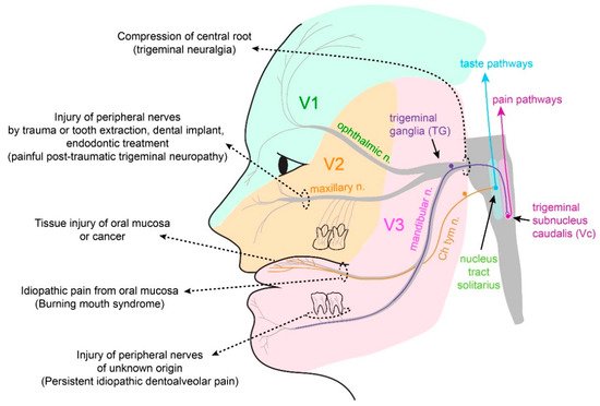
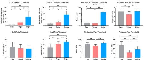
2.1. Somatosensation of Oral Mucosa and Facial Skin

The thermal and mechanical sensitivity of the oral mucosa differ from that of facial skin. Different intraoral sites also show different sensitivities. Extraoral skin and the tongue tip are generally more sensitive than the gingival mucosa, as has been shown by quantitative sensory testing (QST) [9] (Figure 2). The face and tongue are more sensitive to cold, warmth, and mechanical stimuli than the gingiva. The heat pain threshold is also higher in the gingiva, while the mechanical pain and vibration detection thresholds are lower in the tongue than other tissues. The pressure pain threshold is lowest in the tongue and highest in the face. There is no difference among the oral mucosa and facial skin in terms of the cold pain threshold. Interestingly, paradoxical heat sensations upon cooling do not occur in the skin or tongue but occurred in the gingiva of 71% of subjects, suggesting a poor thermal discrimination ability of the gingiva [9]. Other reports have also shown similar tendencies, with minor differences. Tactile sensitivities of the lower lip, anterior tongue, and buccal mucosa are greater than that of the soft palate, posterior tongue, and posterior pharyngeal wall [10]. The tip of the tongue is the most sensitive, in terms of tactile sensitivity, followed by the hard palate, lateral tongue, buccal mucosa, and gingiva [11]. The pressure pain threshold is greater in the maxillary gingiva than the mandible but showed no differences at sites along the tooth rows [12]. Cold sensitivity at the tongue tip is higher when compared to the chin, whereas warmth and heat pain sensitivity are lower [13]. Responses to cold and warm stimuli are poorer in the oral mucosa than in the supraorbital skin and nasal mucosa [14].
Figure 2. Different sensations of intraoral mucosa and extraoral skin. Results of quantitative sensory testing (QST) in 21 healthy subjects (13 women and 8 men; mean 40.4 years) on the cheek, tip of the tongue, and gingival mucosa of the upper premolar region. Mean ± SD; * p < 0.05, ** p < 0.01, and *** p < 0.001; paired t-test following Bonferroni correction for multiple comparisons. Plots were redrawn using previously published data from Pigg et al. [9], with kind permission.
The oral mucosa is well-developed to detect various chemical stimuli with considerable sensitivity. The topical application of capsaicin to the oral mucosa produces a burning sensation, where capsaicin sensitivity is similar between the supraorbital skin and the oral mucosa but less than in the nasal mucosa [14]. The topical application of capsaicin on the oral mucosa led to strong pain from the subsequent heat stimulus both at the site of application (primary hyperalgesia) but also outside the site of application (secondary hyperalgesia) [15]. In addition to capsaicin, mustard oil and cinnamaldehyde also enhance the pain caused by the application of heat (49 °C) to the tongue [16]. In contrast, subjects who frequently eat foods containing capsaicin show a slightly higher warmth detection threshold than subjects who rarely eat foods containing capsaicin, suggesting desensitization by the chronic ingestion of capsaicin [17]. Mustard oil and cinnamaldehyde also modestly enhance the pain caused by the application of cold to the tongue. Menthol ingestion increases both the warm and cold detection thresholds [17] but does not affect the pain intensity in response to subsequent heat or cold stimuli [15].

2.2. Properties and Projections of Primary Afferents in Oral Mucosa and Facial Skin

Microneurographic studies in humans have shown that oral mucosal mechanoreceptors exhibit both fast and slow adapting properties [18]. The stimulation of fast-adapting fibers produces sensations of vibration, whereas the stimulation of slow-adapting fibers produces sensations of constant pressure. Unlike facial skin, however, there are no fast-adapting mechanoreceptors with the properties of Pacinian corpuscle afferents [18]. In the oral mucosa of experimental animals, three types of mechanoreceptors exist: Meissner corpuscles, Ruffini endings, and Merkel cells. Free nerve endings serve as thermoreceptors or nociceptors [19]. In mice, Merkel cells, Meissner corpuscles, and glomerular corpuscles have been found in the tongue, hard palate, and gingival mucosa, but no Pacinian corpuscles or Ruffini endings were found [20]. The density of Merkel cells in the hard palate decreases in aged mice [20]. The functional properties of oral mucosal nociceptive afferents are not well-known. In Wistar rats, using a single unit recording of a lingual nerve innervated to the mandibular gingival mucosa [21], it has been found that 46% of fibers are non-nociceptive low-threshold mechanoreceptors, and 54% of fibers are classified as nociceptive. Based on their conduction velocity in the oral mucosa, the nociceptive fibers can be classified as Aδ (58%) or C fibers (42%). Based on their responses to thermal, mechanical, and chemical stimuli, oral mucosal nociceptors are classified into four types: Aδ high-threshold mechanonociceptors, Aδ mechano–heat nociceptors, Aδ polymodal nociceptors, and C polymodal nociceptors. The mechanical threshold of each of these receptors is higher than that in skin, whereas the heat threshold is similar. Unlike skin afferents, in the oral mucosa, the size of the receptive field is greater in C polymodal nociceptors than in Aδ nociceptors. Aδ polymodal nociceptors are rare in the skin but enriched in visceral afferents [21]. A recent study has also shown the characteristics of afferents innervated to the tongue in mice [22]. Approximately 50% of lingual afferents are c fibers, while approximately 30% are Aδ fibers. In this study, the implantation of oral squamous carcinoma into the tongue increased spontaneous firing, decreased the mechanical thresholds of the c and Aδ mechanoreceptors, and reduced the proportion of mechanically insensitive fibers [22].
In rodents, approximately 40–45% of buccal mucosal afferents express transient receptor potential cation channel subfamily V member 1 (TRPV1) and transient receptor potential cation channel subfamily A member 1 (TRPA1) [23]), which may transduce the burning pain caused by capsaicin and mustard oil. These afferents include peptidergic afferents, and, in the buccal mucosa, capsaicin and mustard oil evoke the release of the calcitonin gene-related peptide (CGRP) [24]. The retrograde labeling of gingival mucosa afferents in the trigeminal ganglia (TG) of rats and mice has shown that gingival mucosal afferents are small- to medium-sized, and their average size is smaller than tooth pulp afferents [25][26]. In rats, approximately 50% of gingival afferents are CGRP-positive, while 25% of gingival afferents are substance P-positive [27]; in contrast, in mice, only 23% of gingival afferents are CGRP-positive [26]. In mouse gingiva, CGRP-positive afferents are highly colocalized with TRPV1 [26]. In rats, 76% of gingival afferents are tropomyosin receptor kinase A-positive neurons, and 50% are isolectin B4-binding neurons [21]. Transient receptor potential cation channel subfamily M member 8 (TRPM8)-expressing fibers in the oral mucosa [28] likely mediate the sensation due to menthol. Menthol itself does not evoke the release of CGRP from the buccal mucosa but enhances the cold-evoked release of CGRP, which depends on TRPM8 [24]. The overexpression of neurturin—a member of the glial cell line-derived neurotrophic factor (GDNF)—in keratinocytes increases the expression of TRPM8 in TG and increases the oral sensation generated by menthol [29]. A recent study showed that the tongue mucosa and muscle are innervated by neurons expressing CGRP (25% of tongue afferents), TRPV1 (17%), 5HT3A (21%), TrkC (31%), parvalbumin (14%), NPY2R (12%), and Mrgprd (7%), which suggests the projection of peptidergic and non-peptidergic nociceptors, proprioceptors, and low-threshold mechanoreceptors [30]; however, the functional implications of these differences have not been studied well.
The ascending pathways of orofacial pain have been thoroughly reviewed elsewhere [31]. Trigeminal subnucleus caudalis (Vc) neurons are critical hubs for transmitting oral and facial pain [7], which are activated by noxious or non-noxious stimuli applied to the oral cavity, as well as the face. The effects of the lingual application of a variety of chemical stimuli, such as capsaicin, ethanol, histamine, mustard oil, nicotine, acid, and piperine, have been assessed [32]. Other regions, such as cranial nerves (C1/C2), subnuclei interpolaris (Vi), Vi/Vc, and subnuclei oralis (Vo), are also known to be involved in orofacial pain. Mapping phosphorylated extracellular signal-regulated kinase (pERK) following the injection of capsaicin into various orofacial areas showed the somatotopic arrangement of neurons in Vc and the upper cervical spinal cord and revealed distinct chemical transmission pathways between the intraoral and extraoral facial sites [33]. Capsaicin injection into the extraoral ophthalmic, maxillary, or mandibular regions induces pERK+ neurons in the ventral, middle, and dorsal portions of Vc, respectively. In contrast, the localization of pERK+ neurons by capsaicin injection into the intraoral mucosa does not show distinct segregation. Capsaicin injection into the tongue or lower gum induces pERK+ neurons localized to the dorsal half of the Vc, whereas capsaicin injection into the anterior hard palate, upper gum, or buccal mucosa induces pERK+ neurons in both dorsal and ventral Vc. Unlike extraoral injection, capsaicin injection into intraoral sites produces a large number of pERK+ neurons in contralateral Vc [33]. Oral and facial regions also show a rostrocaudal somatotopic pattern: oral regions are represented in rostral Vc, whereas the lateral face regions are represented more in caudal Vc [7].

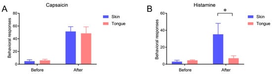
2.3. Itch Sensation of Oral Mucosa Is Weaker Than That of Facial Skin

The itch sensation in the oral cavity has not been well-studied. Although an itching sensation associated with allergic reactions occurs in the oral mucosa, histamine-induced flares are limited to the skin, as histamine is not associated with the itch sensation in human oral mucosa [34]. Indeed, the injection of histamine into the mucosa at the dorsal surface of the tongue rarely produces itch-like responses in mice, whereas the same dose of histamine evokes robust scratching upon injection into the cheek skin (Figure 3). Histamine-induced itches largely depend on TRPV1 and TRPV1-expressing afferents [35]. As a capsaicin injection into the tongue mucosa produces robust nocifensive behaviors, a lack of itch response is not due to the lack of TRPV1-expressing afferent projection into the tongue mucosa. It is possible that oral mucosa afferents are devoid of itch-specific factors, such as phospholipase β3 [35]. Indeed, the tongue mucosa is rarely projected by afferents expressing MrgprA3 or the gastrin-releasing peptide, two well-established markers for itch-sensing afferents [30][36]. It is clear that further studies on the neurobiology of pruriceptors in the oral mucosa are needed.
Figure 3. Lack of histamine-induced itch response from the tongue mucosa. The number of behavioral responses assessed 5 min before and after the injection of capsaicin (10 µg/10 µl (A)) or histamine (20 µg/10 µl (B)) into the facial skin or tongue of C57BL/6 mice. Under isoflurane anesthesia, capsaicin or histamine was injected into the facial skin subcutaneously or into the submucosa of the dorsum of the tongue. Before and after the injection of capsaicin into the facial skin, the number of wipings of the injected skin using the ipsilateral hind paw was counted. Before and after histamine injection into the facial skin, the number of scratchings of the injected site using the ipsilateral forepaw was counted. Upon the injection of capsaicin or histamine into the tongue, the number of instances of wiping, scratching, and grooming of the face using the bilateral forepaws was counted. n = 4 in each group. * p < 0.05 in post-test following two-way repeated measures ANOVA.

References

  1. Targhotra, M.; Chauhan, M.K. An Overview on Various Approaches and Recent Patents on Buccal Drug Delivery Systems. Curr. Pharm. Des. 2020, 26, 39.
  2. Glim, J.E.; Van Egmond, M.; Niessen, F.B.; Everts, V.; Beelen, R.H.J. Detrimental dermal wound healing: What can we learn from the oral mucosa? Wound Repair Regen. 2013, 21, 648–660.
  3. Bradley, R.M. Essentials of Oral Physiology; Mosby: St. Louis, MO, USA, 1995.
  4. Dubner, R. The Neural Basis of Oral and Facial Function; Springer: Boston, MA, USA, 1978.
  5. Zhao, N.N.; Whittle, T.; Murray, G.M.; Peck, C.C. The effects of capsaicin-induced intraoral mucosal pain on jaw movements in humans. J. Orofac. Pain 2012, 26, 277–287.
  6. Mirabile, A.; Airoldi, M.; Ripamonti, C.; Bolner, A.; Murphy, B.; Russi, E.; Numico, G.; Licitra, L.; Bossi, P. Pain management in head and neck cancer patients undergoing chemo-radiotherapy: Clinical practical recommendations. Crit. Rev. Oncol. 2016, 99, 100–106.
  7. Sessle, B.J. Acute and Chronic Craniofacial Pain: Brainstem Mechanisms of Nociceptive Transmission and Neuroplasticity, and Their Clinical Correlates. Crit. Rev. Oral Biol. Med. 2000, 11, 57–91.
  8. Campillo, E.R.D.R.; López-López, J.; Chimenos-Küstner, E. Response to topical clonazepam in patients with burning mouth syndrome: A clinical study. Bulletin GIRSO 2010, 49, 19–29.
  9. Pigg, M.; Baad-Hansen, L.; Svensson, P.; Drangsholt, M.; List, T. Reliability of intraoral quantitative sensory testing (QST). Pain 2010, 148, 220–226.
  10. Bearelly, S.; Cheung, S.W. Sensory Topography of Oral Structures. JAMA Otolaryngol. Neck Surg. 2017, 143, 73–80.
  11. Cordeiro, P.G.; Schwartz, M.; Neves, R.; Tuma, R. A Comparison of Donor and Recipient Site Sensation in Free Tissue Reconstruction of the Oral Cavity. Ann. Plast. Surg. 1997, 39, 461–468.
  12. McMillan, A.S. Pain-pressure threshold in human gingivae. J. Orofac. Pain 1995, 9, 44–50.
  13. Wang, Y.; Mo, X.; Zhang, J.; Fan, Y.; Wang, K.; Peter, S. Quantitative sensory testing (QST) in the orofacial region of healthy Chinese: Influence of site, gender and age. Acta Odontol. Scand. 2017, 76, 58–63.
  14. Poulsen, C.E.; Bendixen, K.H.; Terkelsen, A.J.; May, A.; Hansen, J.; Svensson, P. Region-Specific Effects of Trigeminal Capsaicin Stimulation. J. Oral Facial Pain Headache 2019, 33, 318–330.
  15. Naganawa, T.; Baad-Hansen, L.; Ando, T.; Svensson, P. Influence of topical application of capsaicin, menthol and local anesthetics on intraoral somatosensory sensitivity in healthy subjects: Temporal and spatial aspects. Exp. Brain Res. 2015, 233, 1189–1199.
  16. Albin, K.C.; Carstens, M.I.; Carstens, E. Modulation of Oral Heat and Cold Pain by Irritant Chemicals. Chem. Sens. 2008, 33, 3–15.
  17. Kalantzis, A.; Robinson, P.; Loescher, A. Effects of capsaicin and menthol on oral thermal sensory thresholds. Arch. Oral Biol. 2007, 52, 149–153.
  18. Trulsson, M.; Essick, G.K. Sensations Evoked by Microstimulation of Single Mechanoreceptive Afferents Innervating the Human Face and Mouth. J. Neurophysiol. 2010, 103, 1741–1747.
  19. Jacobs, R.; Wu, C.-H.; Goossens, K.; Van Loven, K.; Van Hees, J.; Van Steenberghe, D. Oral mucosal versus cutaneous sensory testing: A review of the literature. J. Oral Rehabil. 2002, 29, 923–950.
  20. Moayedi, Y.; Duenas-Bianchi, L.F.; Lumpkin, E.A. Somatosensory innervation of the oral mucosa of adult and aging mice. Sci. Rep. 2018, 8, 9975.
  21. Toda, K.; Ishii, N.; Nakamura, Y. Characteristics of mucosal nociceptors in the rat oral cavity: An in vitro study. Neurosci. Lett. 1997, 228, 95–98.
  22. Grayson, M.; Furr, A.; Ruparel, S. Depiction of Oral Tumor-Induced Trigeminal Afferent Responses Using Single-Fiber Electrophysiology. Sci. Rep. 2019, 9, 4574.
  23. Urata, K.; Shinoda, M.; Honda, K.; Lee, J.; Maruno, M.; Ito, R.; Gionhaku, N.; Iwata, K. Involvement of TRPV1 and TRPA1 in Incisional Intraoral and Extraoral Pain. J. Dent. Res. 2015, 94, 446–454.
  24. Kichko, T.I.; Neuhuber, W.; Kobal, G.; Reeh, P.W. The roles of TRPV1, TRPA1 and TRPM8 channels in chemical and thermal sensitivity of the mouse oral mucosa. Eur. J. Neurosci. 2018, 47, 201–210.
  25. Kovačič, U.; Tesovnik, B.; Molnar, N.; Cör, A.; Skalerič, U.; Gašperšič, R. Dental pulp and gingivomucosa in rats are innervated by two morphologically and neurochemically different populations of nociceptors. Arch. Oral Biol. 2013, 58, 788–795.
  26. Wang, S.; Kim, M.; Ali, Z.; Ong, K.; Pae, E.K.; Chung, M.K. TRPV1 and TRPV1-Expressing Nociceptors Mediate Orofacial Pain Behaviors in a Mouse Model of Orthodontic Tooth Movement. Front. Physiol. 2019, 10, 1207.
  27. Gašperšič, R.; Kovačič, U.; Cör, A.; Skaleric, U. Unilateral ligature-induced periodontitis influences the expression of neuropeptides in the ipsilateral and contralateral trigeminal ganglion in rats. Arch. Oral Biol. 2008, 53, 659–665.
  28. Yajima, T.; Sato, T.; Hosokawa, H.; Kondo, T.; Saito, M.; Shimauchi, H.; Ichikawa, H. Distribution of transient receptor potential melastatin-8-containing nerve fibers in rat oral and craniofacial structures. Ann. Anat. Anat. Anz. 2015, 201, 1–5.
  29. Wang, T.; Jing, X.; DeBerry, J.J.; Schwartz, E.S.; Molliver, D.C.; Albers, K.M.; Davis, B.M. Neurturin Overexpression in Skin Enhances Expression of TRPM8 in Cutaneous Sensory Neurons and Leads to Behavioral Sensitivity to Cool and Menthol. J. Neurosci. 2013, 33, 2060–2070.
  30. Wu, P.; Arris, D.; Grayson, M.; Hung, C.-N.; Ruparel, S. Characterization of sensory neuronal subtypes innervating mouse tongue. PLOS ONE 2018, 13, e0207069.
  31. Chichorro, J.G.; Porreca, F.; Sessle, B. Mechanisms of craniofacial pain. Cephalalgia 2017, 37, 613–626.
  32. Carstens, E.; Kuenzler, N.; Handwerker, H.O. Activation of neurons in rat trigeminal subnucleus caudalis by different irritant chemicals applied to oral or ocular mucosa. J. Neurophysiol. 1998, 80, 465–492.
  33. Noma, N.; Tsuboi, Y.; Kondo, M.; Matsumoto, M.; Sessle, B.J.; Kitagawa, J.; Saito, K.; Iwata, K. Organization of pERK-immunoreactive cells in trigeminal spinal nucleus caudalis and upper cervical cord following capsaicin injection into oral and craniofacial regions in rats. J. Comp. Neurol. 2008, 507, 1428–1440.
  34. Bay, B.; Hilliges, M.; Weidner, C.; Sandborgh-Englund, G. Response of human oral mucosa and skin to histamine provocation: Laser Doppler perfusion imaging discloses differences in the nociceptive nervous system. Acta Odontol. Scand. 2009, 67, 99–105.
  35. Imamachi, N.; Park, G.H.; Lee, H.; Anderson, D.J.; Simon, M.I.; Basbaum, A.I.; Han, S.-K. TRPV1-expressing primary afferents generate behavioral responses to pruritogens via multiple mechanisms. Proc. Natl. Acad. Sci. USA 2009, 106, 11330–11335.
  36. Barry, D.M.; Liu, X.-T.; Liu, B.; Liu, X.-Y.; Gao, F.; Zeng, X.; Liu, J.; Yang, Q.; Wilhelm, S.; Yin, J.; et al. Exploration of sensory and spinal neurons expressing gastrin-releasing peptide in itch and pain related behaviors. Nat. Commun. 2020, 11, 1–14.
More
Information
Contributor MDPI registered users' name will be linked to their SciProfiles pages. To register with us, please refer to https://encyclopedia.pub/register :
View Times: 2.0K
Revisions: 2 times (View History)
Update Date: 08 Jun 2021
1000/1000