Submitted Successfully!
To reward your contribution, here is a gift for you: A free trial for our video production service.
Thank you for your contribution! You can also upload a video entry or images related to this topic.
Version Summary Created by Modification Content Size Created at Operation
1 + 4676 word(s) 4676 2021-08-20 07:58:56 |
2 format correct -1409 word(s) 3267 2021-09-17 10:45:27 | |
4 format done -2362 word(s) 905 2022-04-13 12:17:56 |

Video Upload Options

Do you have a full video?

Confirm

Are you sure to Delete?
Cite
If you have any further questions, please contact Encyclopedia Editorial Office.
Tavares, A.P.; F. Nunes, J.C.; Cristóvão, R.; Santos-Ebinuma, V.; Faria, J.L.; Silva, C.; Neves, M.C.; Freire, M. L-Asparaginase-Based Biosensors. Encyclopedia. Available online: https://encyclopedia.pub/entry/13391 (accessed on 16 April 2024).
Tavares AP, F. Nunes JC, Cristóvão R, Santos-Ebinuma V, Faria JL, Silva C, et al. L-Asparaginase-Based Biosensors. Encyclopedia. Available at: https://encyclopedia.pub/entry/13391. Accessed April 16, 2024.
Tavares, Ana Paula, João C. F. Nunes, Raquel Cristóvão, Valeria Santos-Ebinuma, Joaquim Luís Faria, Cláudia Silva, Márcia C. Neves, Mara Freire. "L-Asparaginase-Based Biosensors" Encyclopedia, https://encyclopedia.pub/entry/13391 (accessed April 16, 2024).
Tavares, A.P., F. Nunes, J.C., Cristóvão, R., Santos-Ebinuma, V., Faria, J.L., Silva, C., Neves, M.C., & Freire, M. (2021, August 20). L-Asparaginase-Based Biosensors. In Encyclopedia. https://encyclopedia.pub/entry/13391
Tavares, Ana Paula, et al. "L-Asparaginase-Based Biosensors." Encyclopedia. Web. 20 August, 2021.
Peer Reviewed
L-Asparaginase-Based Biosensors

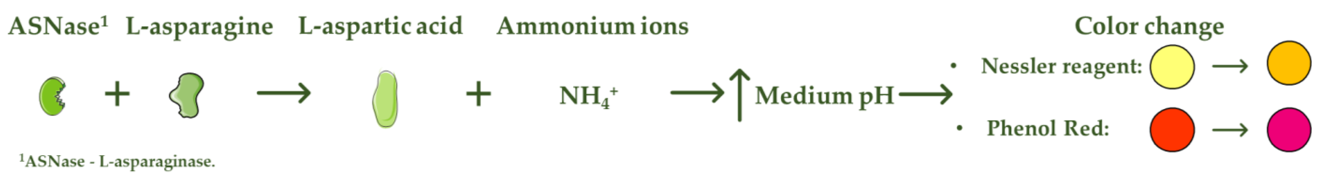
L-asparaginase (ASNase) is an aminohydrolase enzyme widely used in the pharmaceutical and food industries. Although currently its main applications are focused on the treatment of lymphoproliferative disorders such as acute lymphoblastic leukemia (ALL) and acrylamide reduction in starch-rich foods cooked at temperatures above 100 °C, its use as a biosensor in the detection and monitoring of L-asparagine levels is of high relevance. ASNase-based biosensors are a promising and innovative technology, mostly based on colorimetric detection since the mechanism of action of ASNase is the catalysis of the L-asparagine hydrolysis, which releases L-aspartic acid and ammonium ions, promoting a medium pH value change followed by color variation. ASNase biosensing systems prove their potential for L-asparagine monitoring in ALL patients, along with L-asparagine concentration analysis in foods, due to their simplicity and fast response. 

L-asparaginase biosensor L-asparagine monitoring ammonia pharmaceutical food industry
Biosensors are analytical systems consisting of an immobilized biological element combined with a suitable transducer to quantify an analyte [1]. Depending on the biosensor purpose, the most common transducers are electrochemical, piezoelectric, optical, thermometric or magnetic [2]. Biosensors usually generate an electronic or optical signal proportional to the particular interaction between the analyte and the immobilized recognition compound. The low equipment costs, high sensitivity and precision and easy operation have led to increased demand for this type of systems compared to traditional analytical methods [3]. The biological component may be constituted by different bio-recognition elements, such as enzymes, bacteria, tissues, antibodies or nucleic acids, etc. Among them, enzyme-based biosensors have been increasingly used due to their recognized high specificity and exceptional biorecognition capabilities [4]. The enzyme immobilization procedure can influence the biosensor stability, specificity, sensitivity and reproducibility. Therefore, the method used to attach the enzyme onto the electrode must guarantee the stability of the active site and the biomolecule activity. The selection of a suitable technique depends on the biological component nature, the transducer type, the analyte characteristics and the operating conditions. The most used enzyme immobilization methods for the design and development of biosensors are classified into physical adsorption, covalent binding and entrapment [5]. Physical adsorption comprises low associated costs and improved enzymatic performance, whereas entrapment (within the framework of a support) allows enzyme preservation and high enzymatic activity levels. However, physical adsorption can lead to nonspecific adsorption and enzyme desorption, while entrapment can lead to mass transfer limitations and enzyme leakage. On the other hand, by applying a covalent bond between the enzyme and the immobilization support, the enzyme leaching is avoided, allowing for the recovery and reuse of the support/enzyme. Nevertheless, in this last option, the enzyme’s active center amino acids should not be involved in covalent bonding to guarantee high levels of enzyme activity, which is defined by the enzyme binding orientation [6][7].
The enzyme L-asparaginase (E.C.3.5.1.1, ASNase) is a chemotherapeutic agent for the treatment of lymphoproliferative disorders, specifically acute lymphoblastic leukemia (ALL), lymphomas and natural killer cell tumors. The tumor-inhibitory characteristics of ASNase were first reported in animal trials in the 1950s [8][9]. ASNase catalyzes the hydrolysis of L-asparagine into L-aspartic acid and ammonia. Since healthy cells can produce L-asparagine internally, whereas leukemic cells depend on this crucial extracellular amino acid for their growth, prolonged deprivation of L-asparagine in blood leads malignant lymphoid cells into apoptosis [10]. L-asparagine concentration varies from 10−6 to 10−4 M in healthy blood serum samples and from 10−3 to 10−2 M in leukemia blood serum samples [11]. Thus, monitoring L-asparagine depletion in ALL patients is essential to assess the efficacy of ASNase therapy [12]. Full L-asparagine depletion and high ASNase activity are both associated with improved outcomes in ALL patients [13].
ASNase also has an important application in the food industry for acrylamide mitigation from heat-processed foods. Acrylamide is referred to as a Group 2A carcinogen (“probably carcinogenic to humans”) by the International Agency for Research on Cancer (IARC) and by the World Health Organization (WHO) [14][15]. According to the Food and Drug Administration (FDA), the food products with the highest acrylamide concentrations (up to 8440 µg.kg−1) are cereals, french fries, potato chips and cookies, whose average daily acrylamide intake varies from 0.03 to 0.05 µg.kg−1 body weight [6][16]. Nevertheless, the tolerable daily acrylamide intake to avoid carcinogenic risk is 2.6 µg kg−1 body weight [17]. The pre-treatment of starchy foods with ASNase before cooking transforms L-asparagine into L-aspartic acid, and the Maillard reaction takes place without the contribution of L-asparagine, avoiding the acrylamide formation in the final food product [18]. Since the amino acid L-asparagine is not mainly responsible for the taste and appearance of the processed foods, the desired organoleptic characteristics are preserved [19]. Thus, ASNase is already used to lower the acrylamide dosage in several food products, such as potatoes, bread, french fries, coffee and biscuits [6].
ASNase-based biosensors are a promising and innovative technology that can be used both to detect and monitor the level of L-asparagine in blood serum samples of leukemic patients and in different food samples [6]. The methods currently used to detect L-asparagine are based on spectroscopy and chromatography techniques [20], whereas the biosensor operation mode is mostly based on colorimetric detection. The L-asparagine hydrolysis releases ammonium ions, promoting, consequently, a change in the pH value of the medium followed by a variation in the color [21], being the mode of action of colorimetric ASNase-based biosensors (Figure 1). Although there are few reports on the development of ASNase-based biosensors, biosensing systems prove to be imperative for pharmaceutical/food industrial applications due to their simplicity and fast response, while allowing online L-asparagine monitoring.
Encyclopedia 01 00065 g001
Figure 1. Mode of action of colorimetric ASNase based biosensors.

References

  1. Thévenot, D.R.; Toth, K.; Durst, R.A.; Wilson, G.S. Electrochemical biosensors: Recommended definitions and classification 1 International Union of Pure and Applied Chemistry: Physical Chemistry Division, Commission I.7 (Biophysical Chemistry); Analytical Chemistry Division, Commission V.5 (Electroanalytical). Biosens. Bioelectron. 2001, 16, 121–131.
  2. Bahadır, E.B.; Sezgintürk, M.K. Applications of commercial biosensors in clinical, food, environmental, and biothreat/biowarfare analyses. Anal. Biochem. 2015, 478, 107–120.
  3. Turner, A.; Karube, I.; Wilson, G.S. Biosensors: Fundamentals and Applications, 1st ed.; Oxford University Press: Oxford, UK, 1987; ISBN 0198547242.
  4. Songa, E.A.; Okonkwo, J.O. Recent approaches to improving selectivity and sensitivity of enzyme-based biosensors for organophosphorus pesticides: A review. Talanta 2016, 155, 289–304.
  5. Zhilei, W.; Zaijun, L.; Xiulan, S.; Yinjun, F.; Junkang, L. Synergistic contributions of fullerene, ferrocene, chitosan and ionic liquid towards improved performance for a glucose sensor. Biosens. Bioelectron. 2010, 25, 1434–1438.
  6. Nunes, J.C.F.; Cristóvão, R.O.; Freire, M.G.; Santos-Ebinuma, V.C.; Faria, J.L.; Silva, C.G.; Tavares, A.P.M. Recent strategies and applications for L-asparaginase confinement. Molecules 2020, 25, 5827.
  7. Brena, B.; González-Pombo, P.; Batista-Viera, F. Immobilization of enzymes: A literature survey. In Ebolaviruses; Springer Science and Business Media LLC: Berlin/Heidelberg, Germany, 2013; Volume 1051, pp. 15–31. ISBN 9781627035491.
  8. Kidd, J.G. Regression of transplanted lymphomas induced in vivo by means of normal guinea pig serum. I. Course of transplanted cancers of various kinds in mice and rats given guinea pig serum, horse serum, or rabbit serum. J. Exp. Med. 1953, 98, 565–582.
  9. Kidd, J.G. Regression of transplanted lymphomas induced in vivo by means of normal guinea pig serum. II. Studies on the nature of the active serum constituent: Histological mechanism of the regression: Tests for effects of guinea pig serum on lymphoma cells in vitro. J. Exp. Med. 1953, 98, 583–606.
  10. Ando, M.; Sugimoto, K.; Kitoh, T.; Sasaki, M.; Mukai, K.; Ando, J.; Egashira, M.; Schuster, S.M.; Oshimi, K. Selective apoptosis of natural killer-cell tumours by L-asparaginase. Br. J. Haematol. 2005, 130, 860–868.
  11. Sandeep, P.; Raman, K.; Kuldeep, K. Enzyme based asparagine biosensor for the detection of asparagine levels in leukemic samples. Int. J. Appl. Biol. Pharm. Technol. 2015, 6, 40–43.
  12. Avramis, V.I.; Panosyan, E.H. Pharmacokinetic/pharmacodynamic relationships of asparaginase formulations: The past, the present and recommendations for the future. Clin. Pharmacokinet. 2005, 44, 367–393.
  13. Paillassa, J.; Leguay, T.; Thomas, X.; Huguet, F.; Audrain, M.; Lheritier, V.; Vianey-Saban, C.; Acquaviva-Bourdain, C.; Pagan, C.; Dombret, H.; et al. Monitoring of asparagine depletion and anti-L-asparaginase antibodies in adult acute lymphoblastic leukemia treated in the pediatric-inspired GRAALL-2005 trial. Blood Cancer J. 2018, 8, 45.
  14. World Health Organization. IARC monographs on the evaluation of carcinogenic risks to humans—Volume 63. Dry cleaning, some chlorinated solvents and other industrial chemicals. Anal. Chim. Acta 1996, 336, 229–230.
  15. Joint Food and Agriculture Organization; World Health Organization. Health Implications of Acrylamide in Food: Report of a Joint FAO/WHO Consultation, WHO Headquarters, Geneva, Switzerland, 25–27 June 2002; World Health Organization: Geneva, Switzerland, 2002; ISBN 9241562188.
  16. Abt, E.; Robin, L.P.; McGrath, S.; Srinivasan, J.; DiNovi, M.; Adachi, Y.; Chirtel, S. Acrylamide levels and dietary exposure from foods in the United States, an update based on 2011–2015 data. Food Addit. Contam. Part A Chem. Anal. Control. Expo. Risk Assess. 2019, 36, 1475–1490.
  17. Tardiff, R.G.; Gargas, M.L.; Kirman, C.R.; Leigh Carson, M.; Sweeney, L.M. Estimation of safe dietary intake levels of acrylamide for humans. Food Chem. Toxicol. 2010, 48, 658–667.
  18. Xu, F.; Oruna-Concha, M.-J.; Elmore, J.S. The use of asparaginase to reduce acrylamide levels in cooked food. Food Chem. 2016, 210, 163–171.
  19. Parker, J.K.; Balagiannis, D.P.; Higley, J.; Smith, G.; Wedzicha, B.L.; Mottram, D.S. Kinetic model for the formation of acrylamide during the finish-frying of commercial french fries. J. Agric. Food Chem. 2012, 60, 9321–9331.
  20. Zubavichus, Y.; Fuchs, O.; Weinhardt, L.; Heske, C.; Umbach, E.; Denlinger, J.D.; Grunze, M. Soft X-ray-induced decomposition of amino acids: An XPS, mass spectrometry, and NEXAFS study. Radiat. Res. 2004, 161, 346–358.
  21. Kumar, K.; Kataria, M.; Verma, N. Plant asparaginase-based asparagine biosensor for leukemia. Artif. Cells Nanomed. Biotechnol. 2013, 41, 184–188.
More
Information
Contributors MDPI registered users' name will be linked to their SciProfiles pages. To register with us, please refer to https://encyclopedia.pub/register : , , , , , , ,
View Times: 856
Online Date: 20 Aug 2021
1000/1000