Submitted Successfully!
To reward your contribution, here is a gift for you: A free trial for our video production service.
Thank you for your contribution! You can also upload a video entry or images related to this topic.
Version Summary Created by Modification Content Size Created at Operation
1 + 3945 word(s) 3945 2021-05-31 10:27:38 |
2 format correct Meta information modification 3945 2021-06-23 04:39:30 |

Video Upload Options

Do you have a full video?

Confirm

Are you sure to Delete?
Cite
If you have any further questions, please contact Encyclopedia Editorial Office.
Jiang, I. Microbiota and Endometriosis. Encyclopedia. Available online: https://encyclopedia.pub/entry/11157 (accessed on 23 April 2024).
Jiang I. Microbiota and Endometriosis. Encyclopedia. Available at: https://encyclopedia.pub/entry/11157. Accessed April 23, 2024.
Jiang, Irene. "Microbiota and Endometriosis" Encyclopedia, https://encyclopedia.pub/entry/11157 (accessed April 23, 2024).
Jiang, I. (2021, June 22). Microbiota and Endometriosis. In Encyclopedia. https://encyclopedia.pub/entry/11157
Jiang, Irene. "Microbiota and Endometriosis." Encyclopedia. Web. 22 June, 2021.
Microbiota and Endometriosis
Edit

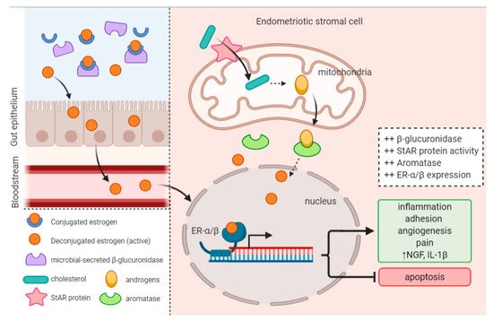
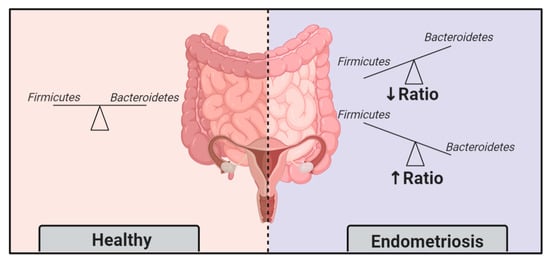
Imbalances in gut and reproductive tract microbiota composition, known as dysbiosis, disrupt normal immune function, leading to the elevation of proinflammatory cytokines, compromised immunosurveillance and altered immune cell profiles, all of which may contribute to the pathogenesis of endometriosis. Over time, this immune dysregulation can progress into a chronic state of inflammation, creating an environment conducive to increased adhesion and angiogenesis, which may drive the vicious cycle of endometriosis onset and progression. Recent studies have demonstrated both the ability of endometriosis to induce microbiota changes, and the ability of antibiotics to treat endometriosis. Endometriotic microbiotas have been consistently associated with diminished Lactobacillus dominance, as well as the elevated abundance of bacterial vaginosis-related bacteria and other opportunistic pathogens. Possible explanations for the implications of dysbiosis in endometriosis include the Bacterial Contamination Theory and immune activation, cytokine-impaired gut function, altered estrogen metabolism and signaling, and aberrant progenitor and stem-cell homeostasis. 

endometriosis microbiota dysbiosis estrogen estrobolome metabolome Lactobacillus vaginal microbiota uterine microbiota gut microbiota inflammation immune dysregulation antibiotics probiotics

1. Endometriosis

1.1. Introduction of Endometriosis

Endometriosis is a complex gynaecological disease characterised by the presence of endometrial glands and stroma outside the uterus [1]. This tissue is often found at sites in the pelvis, including ovaries, fallopian tubes, peritoneal surfaces, the bowel and bladder, but can also engraft in distant organs [1][2]. Much like the eutopic endometrium, these histologic lesions respond to estrogen and are driven to proliferate and bleed alongside the menstrual cycle [1]. Thus, the disease primarily manifests between menarche and menopause, affecting approximately 10% of reproductive-aged women [1][3][4][5]. However, the true prevalence of endometriosis remains enigmatic because the condition presents differently across patients, ranging from symptomatic to asymptomatic independent of its severity, and reliable non-invasive tests are not yet available [2][6].
Like the uterine lining, endometriotic implants bleed during menstruation, activating local inflammation and inducing pelvic pain [1][2]. Often chronic, the disease can have significant impact on a woman’s physical, mental, sexual and social wellbeing [7][8][9][10]. Prolonged inflammation at the lesions can lead to formation of adhesions and scarring (fibrosis), as well as debilitating symptoms including severe pelvic pain, dysmenorrhea, dyspareunia, dyschezia and subfertility [1][11]. Symptoms can be relieved by surgically excising the peritoneal implants, or by supressing lesion growth and bleeding through hormonal modulation [12][13]. Although many management approaches enhance fertility and relieve pain, the benefit is moderate and associated with high recurrence rates and side-effects of hormonal therapy and risks of surgery [1][14]. The current standard of clinical diagnosis involves surgical visualisation [2], making it not only costly and invasive to diagnose, but also limits our ability to study it in the asymptomatic general population.

1.2. Aetiology and Pathogenesis

Endometriosis is a multifactorial disease, and its aetiology and pathogenesis are still ill-established. One of the most widely accepted theories on the origin of ectopic endometrial tissues is “Retrograde Menstruation”, which refers to the reflux of menstrual debris with viable endometrial cells via the fallopian tubes into the pelvic cavity [1][15][16]. Once there, cells in the endometrial deposits must adhere to peritoneal surfaces and proliferate in order to develop into invasive lesions (Figure 1) [17][18]. Endometrial stromal cells from women with endometriosis display increased adhesive properties as a result of altered integrin profiles, allowing them to adhere to the peritoneal lining [18][19]. Cellular adhesion is further enhanced by the inflammatory peritoneal environment, which is a hallmark of endometriosis. For example, abundantly present pro-inflammatory cytokine interleukin-8 (IL-8) stimulates cells to adhere to extracellular proteins [20], thus regulating the initial establishment of the disease.
Figure 1. Aetiology and pathogenesis of endometriosis.
To survive and expand, endometriotic implants require a blood supply. The process of angiogenesis is regulated by various angiogenic factors, such as vascular endothelial growth factor (VEGF), which has upregulated expression in the peritoneal fluid of patients with endometriosis [18][21][22]. VEGF in the peritoneal fluid (PF) is primarily produced by macrophages, and its expression is directly regulated by estradiol and progesterone [23]. Tumor necrosis factor-α (TNF-α) and IL-8, also secreted by peritoneal macrophages, are other potent inducers of angiogenesis and lesion proliferation [24][25]. TNF-α is a predominant product of activated macrophages, which stimulates other leukocytes to produce IL-6 and more TNF-α. Its role in stimulating endometrial cell adhesion and inducing angiogenesis is necessary in the initial stages of endometriosis establishment [26]. In addition, excessive pelvic blood leads to the generation of reactive oxygen species (ROS), which cause tissue damage and exposes tissues, favoring angiogenesis [11].
The persistence of endometrial debris in the peritoneum may overload the immune system, causing low-grade inflammation, and over time possibly lead to chronic immune dysregulation [27]. This results in poor immunosurveillance, allowing the foreign tissue to escape immune defenses, and has immense consequences for endometriosis [21], as we review below.

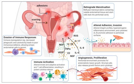
1.3. A Disease of the Immune System

In endometriosis, the peritoneal environment is in a chronic state of local inflammation, and contains immune cells with altered functions. This immune dysregulation in endometriosis creates an ideal environment for disease progression [21]. At present, it is unclear whether immune dysfunction is a pathophysiological hallmark or cause of endometriosis. In either case, there is a strong association demonstrated by the following findings. Table 1 summarizes how immune dysregulation is embodied in the major types of immune cells involved.
Table 1. Dysregulation of peritoneal innate and adaptive immunity creates environment conducive to endometriosis onset and progression.
  Macrophages Neutrophils NK Cells T Cells B Cells
  Ijms 22 05644 i001 Ijms 22 05644 i002 Ijms 22 05644 i003 Ijms 22 05644 i004 Ijms 22 05644 i005
Immune Dysregulation
  • High numbers in PF and endometriotic lesions
  • ↓ phagocytosis
  • ↑ cytokine secretion (TNF-α, IL-6, IL-1β, NF-κB, VEGF)
  • Altered phenotype: more proinflammatory
  • Elevated numbers in PF
  • Preconditioned by bacterial presence
  • Recruited to lesions by IL-8
  • Impaired by aberrant immune environment (IL-6, TGF-β)
  • Altered activating/ inhibitory receptor pattern
  • Altered subset proportions
  • ↑ TH2 ↑ TH17
  • Increased IL-17 secretion
  • Produce anti-endometrial autoantibodies
  • ↑ IL-6 ↑ IL-17
Implications on Endometriosis
  • ↓ ability to eliminate refluxed endometrial tissue
  • Promote further immune activation and inflammation
  • Lesion growth & vascularization
  • Involved in early lesion formation
  • Promote angiogenesis
  • Reduced cytotoxic capacity =
  • ↓ immunosurveillance
  • Lesion survival
  • Suppressed cell-mediated immunity = ↓ immunosurveillance
  • Stimulate production of proinflammatory cytokines and pro-angiogenic factors
  • Endometriosis progression
  • Contribute to inflammation, disease progression
  • Role is unclear

1.3.1. Elevated Inflammatory Mediators

Cytokines and prostaglandins are key players in the initiation, propagation and regulation of immune responses, including inflammation processes. Surges of cytokines lead to signaling cascades and activation of immune cell activity, recruiting more immune cells, and leading to further cytokine production. In the peritoneum, these molecules are produced by various leukocytes, mainly macrophages and stromal cells of the ectopic endometrial tissues, which elicit a localised immune and inflammatory response [2][21][28]. The stromal cells produce IL-6 at similar rates as macrophages, and have further increased production when stimulated by TNF-α [29]. It was found that in women with endometriosis, even eutopic endometrial cells produce higher quantities of IL-6 under basal conditions when compared to women with endometriosis [30]. Furthermore, events that occur in women with endometriosis such as overexpression of NF-κB by peritoneal macrophages and endometriotic cells, activation of MAPK pathways and production of ROS all contribute to cytokine production [31]. Additionally, women with endometriosis have been reported to have increased numbers of immune cells in the PF, which secrete various growth factors and cytokines, enhancing survival and proliferation of the ectopic endometrial cells [18][32]. Another study investigating plasma inflammatory markers found that elevated plasma levels of IL-1β and TNF-α were associated with increased risk for endometriosis [32]. As a result of all this, the PF of women with endometriosis is a potent mixture of cytokines whose positive feedback mechanism maintains a chronic state of inflammation.

1.3.2. Macrophages: Principal Contributors to the Pathogenesis of Endometriosis

The peritoneal fluid of women with endometriosis contains a higher number of activated macrophages in comparison to healthy controls, and these immune cells are postulated to be the primary contributors to the pathogenesis of endometriosis, in part due to their high level of cytokine secretion [18][33]. They are recruited to the peritoneal cavity by various chemo-attractants, including IL-8, and are the main source of IL-6 [18]. Their activity produces the perfect environment for the adhesion, survival and progression of ectopic endometrial implants [18][33][34][35].
These important immune cells phagocytose pathogens, present antigen, and play a critical role in tissue regeneration, angiogenesis and wound healing [36]. In the healthy endometrium, their numbers fluctuate throughout the menstrual cycle, heightening in the secretory phase [37]. This allows them to phagocytose cell debris and apoptotic cells, effectively cleaning up after endometrial shedding. However, this normal fluctuation is not observed in women with endometriosis, which could contribute to the survival ability of refluxed endometrial cells in the peritoneum [33][38].
Furthermore, the peritoneal macrophage population in endometriotic women are phenotypically distinct; they exhibit decreased phagocytic capacity and increased activation of NF-κB pathways, leading to the downstream upregulation of proinflammatory cytokines (TNF-α, IL-1β, and IL-6), proangiogenic factors (VEGF), growth factors and adhesion molecules [2][21][33][39][40]. Macrophages can be phenotypically categorised as “classically activated” (M1) or “alternatively activated” (M2), and their polarisation state depends on their microenvironment [33]. M1 are involved in proinflammatory responses, while M2 are involved in anti-inflammatory responses, tissue repair and angiogenesis [38]. A recent study revealed that in women with endometriosis, peritoneal M1 exhibited exaggerated proinflammatory qualities and M2 tended to switch toward the proinflammatory phenotype of M1 [33]. This supports important previous findings in mice that macrophages infiltrating endometriotic lesions express markers of activation and are necessary for lesion growth and vascularisation [35]. However, the mechanisms of macrophage plasticity are still under debate. Nonetheless, these findings suggest that peritoneal macrophages of women with endometriosis have reduced ability to clear out invasive endometriotic cells, and instead contribute to their growth.

1.3.3. Preconditioned Neutrophils

The PF of women with endometriosis also contains higher numbers of neutrophils, recruited by potent chemoattractant IL-8 and preconditioned by bacterial presence [21][41][42]. A study found that neutrophil infiltration in ectopic endometrial tissues peaked in the early stages of lesion formation and subsequently declined, indicating an important role for neutrophils in early lesion formation [34].

1.3.4. Impaired Natural Killer Cells

The peritoneal immune environment in endometriosis patients is known to impair natural killer (NK) cell activity, and is an example of immune dysregulation in endometriosis [21]. NK cells in diseased women express altered patterns of activating and inhibitory receptors, and display reduced cytotoxicity when exposed to IL-6 and transforming growth factor beta (TGF-β) [21][43]. This immunosuppressive activity partially explains how ectopic endometrial cells are able to evade immunosurveillance and persist in the peritoneal cavity [21].

1.3.5. Altered T cell Differentiation

T cell subset profiles are altered in women with endometriosis [21]. Cytokine secretion by T helper (TH) cells is shifted toward TH2, which is involved in the suppression of cell-mediated immunity, potentially lending to poor immunosurveillance [44][45]. There are also higher numbers of TH17 cells in the PF of endometriosis patients, and consequently higher concentrations of IL-17 [46]. The presence of elevated TH17 cells and IL-17 plays an established role in promoting chronic inflammation [32][47]. IL-17 stimulates production of cytokines that induce angiogenesis and inflammation, contributing to the progression of endometriosis [48].

1.3.6. Activated B Cells

B cells are also implicated in endometriosis, although their role is mostly speculative [21]. They are known to produce anti-endometrial autoantibodies, IL-6 and IL-17, which contribute to inflammation [21][49][50].
It is evident that peritoneal immune dysfunction is deeply involved in endometriosis, and accumulating evidence suggests that presence of pathogenic, non-commensal bacteria in the gut and uterine microbiome may be a contributing factor.

1.4. Estrogen Levels and Signaling Is Altered in Endometriosis

Estrogen is heavily involved in many aspects of endometriosis, and the disease is also considered a hormone-dependent disease, as it bears symptoms restricted to the reproductive period and is responsive to hormonal treatment [47][51]. In fact, a 2017 study found that estrogen is necessary to induce endometriosis [52]. In women, estrogen stimulates the growth of ectopic endometrial tissues and inflammatory activity, and endometriosis has been associated with alterations in estrogen signaling [47]. For instance, endometriotic women have a heightened proinflammatory and anti-apoptotic response to estradiol [53]. This may be attributed to the changes in nuclear estrogen receptor expression.
Endometriotic lesions express higher levels of estrogen receptor-β (ER-β), whose signaling promotes lesion growth by inhibiting TNF-α-induced apoptosis, activating an inflammasome which increases IL-1β, and enhancing cellular adhesion and proliferation [54]. In this study, they found that TNF-α, detected abundantly in the peritoneum of women with endometriosis, cooperates indirectly with ER-β to incite these events [54]. In murine models, expression of nuclear estrogen receptors (ER-α and ER-β) is altered in lesions, and this ER signaling is necessary for lesion establishment [55]. They found that ER-α signaling drove proliferation, adhesion and angiogenesis of ectopic lesions [55].
Another consequence of estrogen in endometriosis is its ability to affect peripheral nerve fibres directly or indirectly through the upregulation of various growth factors, including nerve growth factors (NGF), contributing to nociceptive pain [56].
Three key factors dysregulating estrogen availability in endometriotic women include expression of estrogen-synthesis enzymes, the estrobolome and the metabolome.
In endometriosis, estradiol is made available through systemic hormones and locally in the peritoneal environment through aromatase and steroidogenic acute regulatory protein (StAR) activity [57]. Aromatase is an enzyme that converts androgens into estrogens, and StAR is a transport protein that regulates the transfer of cholesterol in the mitochondria required for steroidogenesis. The upregulated expression of these in endometriotic lesions contributes to the increased availability of estrogen, and drives the disease (Figure 2) [47][57]. In contrast, normal endometrial tissue lacks these enzymes, and is unable to synthesize estrogen [51].
Figure 2. Altered estrobolome activity and upregulated enzyme expression produces a hyperestrogenic environment that promotes endometriosis onset and progression.
Furthermore, estrogen metabolism is known to be regulated by the estrobolome, a collection of genes in the gut microbiome involved in estrogen metabolism [58][59]. Estrobolome activity modulates the amount of excess estrogen that is expelled from or reabsorbed into the body [59]. When this activity is impaired, typically as a result of imbalances in the gut microbiome, excess estrogen can be retained in the body and travel from the gut to the endometrial and peritoneal environment via circulation [59][60]. This contributes to the hyperestrogenic state that drives endometriosis, and provides a possible mechanism as to how dysbiosis in the gut microbiota may be involved in the disease.
Finally, the metabolome also plays a role in regulating circulating estrogen [61]. The metabolome refers to the total metabolites in a given environment, in this case the gut. It is heavily influenced by gut microbial activity, and includes consequential neuro-active metabolites that affect the brain and its signalling [62][63][64]. This bidirectional link is called the gut-brain-axis, and these compounds bind to host gonadotropin-releasing hormone (GnRH) receptors to stimulate production of luteinising hormone (LH) and follicle-stimulating hormone (FSH), which subsequently stimulate the production of estrogen [61][65].

2. Evidence of an Intricate Connection

Recently, research on the involvement of the microbiota in endometriosis has begun to accrue. It is postulated that dysbiosis may be involved in dysregulating the immune system and altering estrogen metabolism. Having discussed the extensive role of the immune system and estrogen signaling in endometriosis, it would seem inevitable that the microbiota plays a critical role in the disease.
Studies have shown that patients with pelvic inflammatory disease (PID), which results from the ascension of vaginal bacteria up into the uterus, fallopian tubes and ovaries, are associated with a threefold increase in risk of developing endometriosis [66], possibly suggesting that the disease may have an infectious etiology, at least in part [39].
Researchers have found evidence suggesting the gut and female reproductive tract microbiota may be inextricably linked to the onset and progression of endometriosis. This novel perspective on endometriosis opens the door to many preventative, diagnostic and therapeutic possibilities, and is an emerging area of research.

2.1. Endometriotic Women Exhibit Altered Microbiotas

A recent study by Ata et al. sought to compare the vaginal, cervical and gut microbiota composition of women with Stage III/IV endometriosis to healthy controls [67]. Remarkably, they did indeed detect a difference at the genus level. In the cervical microbiota of endometriotic women, they found increased abundance of potentially pathogenic species including GardnerellaStreptococcusEscherichiaShigella and Ureaplasma. Stool microbiota of the endometriotic group were Shigella and Escherichia dominant. Interestingly, they found a complete absence of Atopobium, a gynaecologic pathogen, in the vagina and cervix of the endometriotic group. Another study reported high incidence of Atopobium vaginae in women with endometrial cancer, and suggested that Atopobium can facilitate intracellular Porphyromonas infection, leading to disrupted cell regulatory functions and carcinogenic trigger [68]. Conversely, they found A. vaginae to have lower incidence in women with benign gynaecologic pathologies, suggesting a possible connection through a different mechanism of action, since endometriosis is also a benign gynaecologic pathology [68]. Several other studies have also found that uterine microbiota composition is altered in women with uterine diseases, including endometriosis, demonstrating its clinical relevance [69][70][71]. For example, researchers found an elevated abundance of Streptococcaceae, Moraxellaceae, Staphylococcaceae and Enterobacteriacea, and lowered Lactobacillacae in endometriotic women [69]. Recently, Hernandes et al. found that, compared with eutopic endometrium, ectopic lesions have higher microbial diversity [70]. In Wei et al.’s attempt to characterize microbiota composition and distribution along the FRT in endometriotic women, they found in conformance that the lower FRT was Lactobacillus dominant, and significant differences in community diversity appeared and increased from the cervix up into the endometrium and PF [71].
In general, studies to date have consistently found increases in BV-associated bacteria and opportunistic pathogens, and a decrease in Lactobacillus in the reproductive tract of endometriotic women [72][73][67].

2.2. Endometriosis Induces Gut Microbiota Alterations

In a study where mice were injected with intraperitoneal endometrial tissue to induce endometriosis, it was demonstrated that after 42 days of endometriotic lesion persistence, a distinct gut microbiota develops [74]. In other words, endometriosis progression was able to change the gut microbiota. Among the observed differences, the nearly doubled Firmicutes/Bacteroidetes ratio in endometriotic mice was discriminative and concrete [74]. A previous study in 2002 also found similar differences in microbiota profiles in rhesus monkeys [75]. Compared to healthy controls, monkeys with endometriosis had lower Lactobacilli and higher gram-negative bacteria [75]. The ratio Firmicutes/Bacteroidetes is widely accepted as a feature of dysbiosis (Figure 3); hence, these momentous findings support that endometriosis induces gut microbiota alterations.
Figure 3. The Firmicutes/Bacteroidetes ratio, an indicator of dysbiosis, is altered in endometriosis patients.

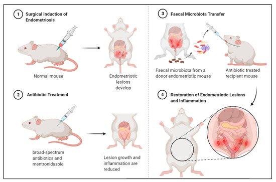
2.3. Faecal Microbiota Transfer Induces Endometriosis

Findings from a compelling mouse model study (Figure 4) support that a distinct gut microbiota promotes endometriosis [76]. In this study, mice were subjected to surgical induction of endometriosis, and then treated with antibiotics which reduced lesion size. Subsequently, they received faecal microbiota transfers from endometriotic mice, which restored lesion growth and associated inflammation [76].
Figure 4. Antibiotic treatment can reduce endometriotic lesion growth and peritoneal inflammation, and subsequent faecal microbiota transfer from diseased mouse can restore lesion growth and inflammation. Mouse experiment demonstrates bidirectional relationship between endometriosis and gut microbiota.

2.4. Diet-Induced Gut Microbiota Changes Reduce Endometriosis Risk

Another interesting finding is that women with a high omega-3 polyunsaturated fatty acids (PUFAs) intake have lower risk for endometriosis [77][78]. A similar diet showed anti-inflammatory effects and suppressed endometriotic lesion formation in murine models [79][80]. It is reasonable to speculate that this can be at least partially attributed to diet-induced modification of the gut flora. Research has shown that diets high in PUFAs and probiotic supplements may alter the gut flora, and may contribute to the prevention and treatment of various diseases, including osteoporosis and obesity [81][82].

3. What Can This Mean for Endometriosis Care

It is evident that bacterial presence in both the gut and uterus plays a major role in endometriosis. But what does this mean for patients? Could the modulation of the microbiota be a therapeutic or preventative approach? Could certain microbial compositions or the presence of microbiota-based biomarkers be used as screening or diagnostic tools?

3.1. Gynaecologic and Obstetric Applications of Microbiota Modulation

Microbiota modulation through antibiotics is already broadly applied in the field of gynaecology and obstetrics [83]. In accordance with ample research showing uterine dysbiosis threatens fertility and pregnancy outcomes, it is suggested that intervention options such as uterine lavage or antibiotics to eradicate microbes or pro/prebiotics and improve the microbiota could be valuable [83]. In a recent clinical setting, broad-spectrum antibiotics and pre/probiotics were employed to achieve Lactobacillus-dominant uterine microbiota (from previously non-Lactobacillus-dominant), which led to higher pregnancy rates [84] — A hopeful finding that encourages further investigation of this approach. Moreover, many studies show that treatment of chronic endometritis with antibiotics leads to improved reproductive outcomes [85][86][87][88].

3.2. Treating Endometriosis with Antibiotics

Antibiotics may be a promising approach for treating endometriosis. In fact, broad-spectrum antibiotic treatments have demonstrated efficacy for treating endometriosis in animal models [76]. A recent study found that use of broad-spectrum antibiotics inhibited ectopic lesions, while treatment with metronidazole significantly decreased inflammation and reduced lesion size, possibly by lessening Bacteroidetes presence [76]. Peculiarly, treatment with neomycin did not produce the same results, indicating that lesion-growth-promoting bacteria are metronidazole-sensitive and neomycin-resistant [76].

3.3. Treating Endometriosis with Probiotics

Alternatively, probiotic intervention, the administration of live microorganisms, could be another effective approach [83]. For instance, in randomised, placebo-controlled trials, oral administration of Lactobacillus has been shown to ameliorate endometriosis-associated pain in women [89][90], and reduce endometriotic lesions in mice by increasing IL-12 concentration and NK cell activity [83][91][92]. Dysbiosis and endometriotic-inflammation leads to impaired NK cell activity, and the probiotic treatment reversed this immune dysregulation. Lactobacillus probiotic treatment not only improved endometriosis, but is also capable of preventing its growth in rats [91]. These impressive frontiers warrant research and testing.

3.4. A Mechanism for Known Treatments

New research is showing that some known endometriosis treatments may have actually been working through gut microbiota modulation [93]. For example, letrozole, an aromatase inhibitor that reduces estrogen levels and Shaofu Zhuyu decoction (SFZYD), a traditional Chinese medicine that inhibits cellular proliferation, promotes apoptosis, and reduces angiogenesis in ectopic endometrial tissues, have been shown to inhibit the progression of endometriosis and reduce inflammation in mice [93]. In a 2020 study, it was found that both letrozole and SFZYD exert their therapeutic effect in part through restoration of the gut microbiota; they both attenuated the Firmicutes/Bacteroidetes ratio, which was elevated in the untreated endometriotic group, and restored α-diversity and Ruminococcaceae abundance in the gut microbiota [93]. Loss of Ruminococcaceae may exacerbate peritoneal inflammation, as it may be negatively correlated to apoptosis of intestinal epithelial cells and murine IL-6 levels [94]. Therefore, these treatments at least partially function through restoring gut microbiota health.

3.5. Side Effects and Challenges

Unfortunately, the use of antibiotics is known to introduce off-target effects and new interventions such as pre/probiotics have poorly understood pharmacological mechanisms with difficult-to-prove clinical efficacy. For example, it is well known that routine or overuse of antibiotics increases the risk of antimicrobial resistance, which is currently one of the largest threats to global human health [95]. Furthermore, the core FRT microbiota should be well characterised before applying any such treatments [83]. Antibiotics are widely effective in treating infection by reducing abundance or eliminating pathogenic species, however its use alters microbial community profiles, and can create lasting disruptions to healthy microbiotas [96][97]. A study examining the urinary microbiotas of women during BV and after oral administration of metronidazole found that while it was an effective treatment, it also significantly decreased the Shannon diversity, an effect which persisted for up to 28 days [96]. Furthermore, the antibiotic treatment created complex changes to the microbiota composition, and healthy microbial community could not be restored [96]. Compared to BV, dysbiosis related to endometriosis is far less studied, and therefore the convoluted effects of antibiotic treatments are largely undetermined. The use of antibiotics to treat endometriosis still requires extensive research, but is an area of potential. There are many unanswered questions regarding its practical use, and the diagnosis of an “abnormal uterine microbiota” to indicate such treatment remains a major hurdle to overcome.

3.6. Opportunities for Diagnostics

Another appealing application of the microbiota-endometriosis relationship lies in diagnostics. So much is yet to be learned about the mechanisms involved, but based on the current state of knowledge that there is an appreciable difference in the microbiotas of women with endometriosis, we can imagine its potential value as a diagnostic or screening tool. Significant differences in the community diversity of cervical microbiota in endometriotic women indicate that cervical samples may be used as an endometriosis risk indicator [71]. A recent study found for the first time that vaginal microbiome profiles could be successfully used to predict the rASRM (revised American Society for Reproductive Medicine) stage of endometriosis [98]. These exciting findings hopefully incite further research into non-invasive diagnostics and screening tools, as traditional techniques are limited and remain a challenge today.

References

  1. Zondervan, K.T.; Becker, C.M.; Koga, K.; Missmer, S.A.; Taylor, R.N.; Viganò, P. Endometriosis. Nat. Rev. Dis. Primers 2018, 4, 9.
  2. Zondervan, K.T.; Becker, C.M.; Missmer, S.A. Endometriosis. N. Engl. J. Med. 2020, 382, 1244–1256.
  3. Busacca, M.; Vignali, M. Ovarian endometriosis: From pathogenesis to surgical treatment. Curr. Opin. Obstet. Gynecol. 2003, 15, 321–326.
  4. Dunselman, G.A.J.; Vermeulen, N.; Becker, C.; Calhaz-Jorge, C.; D’Hooghe, T.; De Bie, B.; Heikinheimo, O.; Horne, A.W.; Kiesel, L.; Nap, A.; et al. ESHRE Guideline: Management of Women with Endometriosis. Hum. Reprod. 2014, 29, 400–412.
  5. Eskenazi, B.; Warner, M.L. Epidemiology of endometriosis. Obstet. Gynecol. Clin. N. Am. 1997, 24, 235–258.
  6. Vercellini, P.; Fedele, L.; Aimi, G.; Pietropaolo, G.; Consonni, D.; Crosignani, P.G. Association between Endometriosis Stage, Lesion Type, Patient Characteristics and Severity of Pelvic Pain Symptoms: A Multivariate Analysis of over 1000 Patients. Hum. Reprod. 2007, 22, 266–271.
  7. Arion, K.; Orr, N.L.; Noga, H.; Allaire, C.; Williams, C.; Bedaiwy, M.A.; Yong, P.J. A Quantitative Analysis of Sleep Quality in Women with Endometriosis. J. Women’s Health 2020, 29, 1209–1215.
  8. Shum, L.K.; Bedaiwy, M.A.; Allaire, C.; Williams, C.; Noga, H.; Albert, A.; Lisonkova, S.; Yong, P.J. Deep Dyspareunia and Sexual Quality of Life in Women with Endometriosis. Sex. Med. 2018, 6, 224–233.
  9. Sepulcri, R.d.P.; do Amaral, V.F. Depressive Symptoms, Anxiety, and Quality of Life in Women with Pelvic Endometriosis. Eur. J. Obstet. Gynecol. Reprod. Biol. 2009, 142, 53–56.
  10. Simoens, S.; Dunselman, G.; Dirksen, C.; Hummelshoj, L.; Bokor, A.; Brandes, I.; Brodszky, V.; Canis, M.; Colombo, G.L.; DeLeire, T.; et al. The Burden of Endometriosis: Costs and Quality of Life of Women with Endometriosis and Treated in Referral Centres. Hum. Reprod. 2014, 29, 2073.
  11. Vercellini, P.; Vigano, P.; Somigliana, E.; Fedele, L. Endometriosis: Pathogenesis and Treatment. Nat. Rev. Endocrinol. 2014, 10, 261–276.
  12. Bedaiwy, M.A.; Barker, N.M. Evidence Based Surgical Management of Endometriosis. Middle East. Fertil. Soc. J. 2012, 17, 57–60.
  13. Bedaiwy, M.A.; Alfaraj, S.; Yong, P.; Casper, R. New Developments in the Medical Treatment of Endometriosis. Fertil. Steril. 2017, 107, 555–565.
  14. Vercellini, P.; Somigliana, E.; Viganò, P.; Abbiati, A.; Daguati, R.; Crosignani, P.G. Endometriosis: Current and Future Medical Therapies. Best Pract. Res. Clin. Obstet. Gynaecol. 2008, 22, 275–306.
  15. Yovich, J.L.; Rowlands, P.K.; Lingham, S.; Sillender, M.; Srinivasan, S. Pathogenesis of Endometriosis: Look No Further than John Sampson. Reprod. Biomed. Online 2020, 40, 7–11.
  16. Halme, J.; Hammond, M.G.; Hulka, J.F.; Raj, S.G.; Talbert, L.M. Retrograde Menstruation in Healthy Women and in Patients with Endometriosis. Obstet. Gynecol. 1984, 64, 151–154.
  17. Laschke, M.W.; Menger, M.D. The Gut Microbiota: A Puppet Master in the Pathogenesis of Endometriosis? Am. J. Obstet. Gynecol. 2016, 215, 68.e1–68.e4.
  18. Harada, T.; Iwabe, T.; Terakawa, N. Role of Cytokines in Endometriosis. Fertil. Steril. 2001, 76, 1–10.
  19. Klemmt, P.A.B.; Carver, J.G.; Koninckx, P.; McVeigh, E.J.; Mardon, H.J. Endometrial Cells from Women with Endometriosis Have Increased Adhesion and Proliferative Capacity in Response to Extracellular Matrix Components: Towards a Mechanistic Model for Endometriosis Progression. Hum. Reprod. 2007, 22, 3139–3147.
  20. Garcia-Velasco, J.A.; Arici, A. P-255. Interleukin-8 Stimulates Endometrial Stromal Cell Adhesion to Fibronectin. Hum. Reprod. 1999, 14, 268.
  21. Symons, L.K.; Miller, J.E.; Kay, V.R.; Marks, R.M.; Liblik, K.; Koti, M.; Tayade, C. The Immunopathophysiology of Endometriosis. Trends Mol. Med. 2018, 24, 748–762.
  22. Cosín, R.; Gilabert-Estellés, J.; Ramón, L.A.; Gómez-Lechón, M.J.; Gilabert, J.; Chirivella, M.; Braza-Boïls, A.; España, F.; Estellés, A. Influence of Peritoneal Fluid on the Expression of Angiogenic and Proteolytic Factors in Cultures of Endometrial Cells from Women with Endometriosis. Hum. Reprod. 2010, 25, 398–405.
  23. McLaren, J.; Prentice, A.; Charnock-Jones, D.S.; Millican, S.A.; Müller, K.H.; Sharkey, A.M.; Smith, S.K. Vascular Endothelial Growth Factor Is Produced by Peritoneal Fluid Macrophages in Endometriosis and Is Regulated by Ovarian Steroids. J. Clin. Investig. 1996, 98, 482–489.
  24. Iwabe, T.; Harada, T.; Tsudo, T.; Nagano, Y.; Yoshida, S.; Tanikawa, M.; Terakawa, N. Tumor Necrosis Factor-α Promotes Proliferation of Endometriotic Stromal Cells by Inducing Interleukin-8 Gene and Protein Expression. J. Clin. Endocrinol. Metab. 2000, 85, 824–829.
  25. Leibovich, S.J.; Polverini, P.J.; Shepard, H.M.; Wiseman, D.M.; Shively, V.; Nuseir, N. Macrophage-Induced Angiogenesis Is Mediated by Tumour Necrosis Factor- α. Nature 1987, 329, 630–632.
  26. Nothnick, W.B. Treating Endometriosis as an Autoimmune Disease. Fertil. Steril. 2001, 76, 223–231.
  27. García-Peñarrubia, P.; Ruiz-Alcaraz, A.J.; Martínez-Esparza, M.; Marín, P.; Machado-Linde, F. Hypothetical Roadmap towards Endometriosis: Prenatal Endocrine-Disrupting Chemical Pollutant Exposure, Anogenital Distance, Gut-Genital Microbiota and Subclinical Infections. Hum. Reprod. Update 2020, 26, 214–246.
  28. Shigesi, N.; Kvaskoff, M.; Kirtley, S.; Feng, Q.; Fang, H.; Knight, J.C.; Missmer, S.A.; Rahmioglu, N.; Zondervan, K.T.; Becker, C.M. The Association between Endometriosis and Autoimmune Diseases: A Systematic Review and Meta-Analysis. Hum. Reprod. Update 2019, 25, 486–503.
  29. Tsudo, T.; Harada, T.; Iwabe, T.; Tanikawa, M.; Nagano, Y.; Ito, M.; Taniguchi, F.; Terakawa, N. Altered Gene Expression and Secretion of Interleukin-6 in Stromal Cells Derived from Endometriotic Tissues. Fertil. Steril. 2000, 73, 205–211.
  30. Tseng, J.F.; Ryan, I.P.; Milam, T.D.; Murai, J.T.; Schriock, E.D.; Landers, D.V.; Taylor, R.N. Interleukin-6 Secretion in Vitro Is up-Regulated in Ectopic and Eutopic Endometrial Stromal Cells from Women with Endometriosis. J. Clin. Endocrinol. Metab. 1996, 81, 1118–1122.
  31. Beste, M.T.; Pfäffle-Doyle, N.; Prentice, E.A.; Morris, S.N.; Lauffenburger, D.A.; Isaacson, K.B.; Griffith, L.G. Molecular Network Analysis of Endometriosis Reveals a Role for C-Jun–Regulated Macrophage Activation. Sci. Transl. Med. 2014, 6, 222ra16.
  32. Mu, F.; Harris, H.R.; Rich-Edwards, J.W.; Hankinson, S.E.; Rimm, E.B.; Spiegelman, D.; Missmer, S.A. A Prospective Study of Inflammatory Markers and Risk of Endometriosis. Am. J. Epidemiol. 2018, 187, 515–522.
  33. Vallvé-Juanico, J.; Santamaria, X.; Vo, K.C.; Houshdaran, S.; Giudice, L.C. Macrophages Display Proinflammatory Phenotypes in the Eutopic Endometrium of Women with Endometriosis with Relevance to an Infectious Etiology of the Disease. Fertil. Steril. 2019, 112, 1118–1128.
  34. Lin, Y.-J.; Lai, M.-D.; Lei, H.-Y.; Wing, L.-Y.C. Neutrophils and Macrophages Promote Angiogenesis in the Early Stage of Endometriosis in a Mouse Model. Endocrinology 2006, 147, 1278–1286.
  35. Bacci, M.; Capobianco, A.; Monno, A.; Cottone, L.; Di Puppo, F.; Camisa, B.; Mariani, M.; Brignole, C.; Ponzoni, M.; Ferrari, S.; et al. Macrophages Are Alternatively Activated in Patients with Endometriosis and Required for Growth and Vascularization of Lesions in a Mouse Model of Disease. Am. J. Pathol. 2009, 175, 547–556.
  36. Hirayama, D.; Iida, T.; Nakase, H. The Phagocytic Function of Macrophage-Enforcing Innate Immunity and Tissue Homeostasis. Int. J. Mol. Sci. 2017, 19, 92.
  37. Salamonsen, L.A.; Zhang, J.; Brasted, M. Leukocyte Networks and Human Endometrial Remodelling. J. Reprod. Immunol. 2002, 57, 95–108.
  38. Berbic, M.; Schulke, L.; Markham, R.; Tokushige, N.; Russell, P.; Fraser, I.S. Macrophage Expression in Endometrium of Women with and without Endometriosis. Hum. Reprod. 2009, 24, 325–332.
  39. Bedaiwy, M.A. Endometrial Macrophages, Endometriosis, and Microbiota: Time to Unravel the Complexity of the Relationship. Fertil. Steril. 2019, 112, 1049–1050.
  40. Lousse, J.-C.; Van Langendonckt, A.; González-Ramos, R.; Defrère, S.; Renkin, E.; Donnez, J. Increased Activation of Nuclear Factor-Kappa B (NF-ΚB) in Isolated Peritoneal Macrophages of Patients with Endometriosis. Fertil. Steril. 2008, 90, 217–220.
  41. Milewski, Ł.; Dziunycz, P.; Barcz, E.; Radomski, D.; Roszkowski, P.I.; Korczak-Kowalska, G.; Kamiński, P.; Malejczyk, J. Increased Levels of Human Neutrophil Peptides 1, 2, and 3 in Peritoneal Fluid of Patients with Endometriosis: Association with Neutrophils, T Cells and IL-8. J. Reprod. Immunol. 2011, 91, 64–70.
  42. Karmarkar, D.; Rock, K.L. Microbiota Signalling through MyD88 Is Necessary for a Systemic Neutrophilic Inflammatory Response. Immunology 2013, 140, 483–492.
  43. Wu, M.-Y.; Yang, J.-H.; Chao, K.-H.; Hwang, J.-L.; Yang, Y.-S.; Ho, H.-N. Increase in the Expression of Killer Cell Inhibitory Receptors on Peritoneal Natural Killer Cells in Women with Endometriosis. Fertil. Steril. 2000, 74, 1187–1191.
  44. Podgaec, S.; Abrao, M.S.; Dias, J.A., Jr.; Rizzo, L.V.; de Oliveira, R.M.; Baracat, E.C. Endometriosis: An Inflammatory Disease with a Th2 Immune Response Component. Hum. Reprod. 2007, 22, 1373–1379.
  45. Antsiferova, Y.S.; Sotnikova, N.Y.; Posiseeva, L.V.; Shor, A.L. Changes in the T-Helper Cytokine Profile and in Lymphocyte Activation at the Systemic and Local Levels in Women with Endometriosis. Fertil. Steril. 2005, 84, 1705–1711.
  46. Gogacz, M.; Winkler, I.; Bojarska-Junak, A.; Tabarkiewicz, J.; Semczuk, A.; Rechberger, T.; Adamiak, A. Increased Percentage of Th17 Cells in Peritoneal Fluid Is Associated with Severity of Endometriosis. J. Reprod. Immunol. 2016, 117, 39–44.
  47. Laux-Biehlmann, A.; d’Hooghe, T.; Zollner, T.M. Menstruation Pulls the Trigger for Inflammation and Pain in Endometriosis. Trends Pharmacol. Sci. 2015, 36, 270–276.
  48. Ahn, S.H.; Edwards, A.K.; Singh, S.S.; Young, S.L.; Lessey, B.A.; Tayade, C. IL-17A Contributes to the Pathogenesis of Endometriosis by Triggering Proinflammatory Cytokines and Angiogenic Growth Factors. J. Immunol. 2015, 195, 2591–2600.
  49. Shen, P.; Fillatreau, S. Antibody-Independent Functions of B Cells: A Focus on Cytokines. Nat. Rev. Immunol. 2015, 15, 441–452.
  50. Randall, G.W.; Gantt, P.A.; Poe-Zeigler, R.L.; Bergmann, C.A.; Noel, M.E.; Strawbridge, W.R.; Richardson-Cox, B.; Hereford, J.R.; Reiff, R.H. Serum Antiendometrial Antibodies and Diagnosis of Endometriosis. Am. J. Reprod. Immunol. 2007, 58, 374–382.
  51. Chantalat, E.; Valera, M.-C.; Vaysse, C.; Noirrit, E.; Rusidze, M.; Weyl, A.; Vergriete, K.; Buscail, E.; Lluel, P.; Fontaine, C.; et al. Estrogen Receptors and Endometriosis. Available online: (accessed on 26 January 2021).
  52. Galvankar, M.; Singh, N.; Modi, D. Estrogen Is Essential but Not Sufficient to Induce Endometriosis. J. Biosci. 2017, 42, 251–263.
  53. Reis, F.M.; Petraglia, F.; Taylor, R.N. Endometriosis: Hormone Regulation and Clinical Consequences of Chemotaxis and Apoptosis. Hum. Reprod. Update 2013, 19, 406–418.
  54. Han, S.J.; Jung, S.Y.; Wu, S.-P.; Hawkins, S.M.; Park, M.J.; Kyo, S.; Qin, J.; Lydon, J.P.; Tsai, S.Y.; Tsai, M.-J.; et al. Estrogen Receptor β Modulates Apoptosis Complexes and the Inflammasome to Drive the Pathogenesis of Endometriosis. Cell 2015, 163, 960–974.
  55. Burns, K.A.; Rodriguez, K.F.; Hewitt, S.C.; Janardhan, K.S.; Young, S.L.; Korach, K.S. Role of Estrogen Receptor Signaling Required for Endometriosis-Like Lesion Establishment in a Mouse Model. Endocrinology 2012, 153, 3960–3971.
  56. Morotti, M.; Vincent, K.; Becker, C.M. Mechanisms of Pain in Endometriosis. Eur. J. Obstet. Gynecol. Reprod. Biol. 2017, 209, 8–13.
  57. Bulun, S.E. Endometriosis. N. Engl. J. Med. 2009, 360, 268–279.
  58. Ervin, S.M.; Li, H.; Lim, L.; Roberts, L.R.; Liang, X.; Mani, S.; Redinbo, M.R. Gut Microbial β-Glucuronidases Reactivate Estrogens as Components of the Estrobolome That Reactivate Estrogens. J. Biol. Chem. 2019, 294, 18586–18599.
  59. Baker, J.M.; Al-Nakkash, L.; Herbst-Kralovetz, M.M. Estrogen–Gut Microbiome Axis: Physiological and Clinical Implications. Maturitas 2017, 103, 45–53.
  60. Goedert, J.J.; Jones, G.; Hua, X.; Xu, X.; Yu, G.; Flores, R.; Falk, R.T.; Gail, M.H.; Shi, J.; Ravel, J.; et al. Investigation of the Association between the Fecal Microbiota and Breast Cancer in Postmenopausal Women: A Population-Based Case-Control Pilot Study. J. Natl. Cancer Inst. 2015, 107.
  61. Mikhael, S.; Punjala-Patel, A.; Gavrilova-Jordan, L. Hypothalamic-Pituitary-Ovarian Axis Disorders Impacting Female Fertility. Available online: (accessed on 18 February 2021).
  62. Chen, J.; Li, Y.; Tian, Y.; Huang, C. Interaction between Microbes and Host Intestinal Health: Modulation by Dietary Nutrients and Gut-Brain-Endocrine-Immune Axis. Curr. Protein Pept. Sci. 2015, 16, 592.
  63. Baj, A.; Moro, E.; Bistoletti, M.; Orlandi, V.; Crema, F.; Giaroni, C. Glutamatergic Signaling Along the Microbiota-Gut-Brain Axis. Available online: (accessed on 18 February 2021).
  64. Jones, L.A.; Sun, E.W.; Martin, A.M.; Keating, D.J. The Ever-Changing Roles of Serotonin. Int. J. Biochem. Cell Biol. 2020, 125, 105776.
  65. Hiller-Sturmhöfel, S.; Bartke, A. The Endocrine System. Alcohol Health Res. World 1998, 22, 153–164.
  66. Tai, F.-W.; Chang, C.Y.-Y.; Chiang, J.-H.; Lin, W.-C.; Wan, L. Association of Pelvic Inflammatory Disease with Risk of Endometriosis: A Nationwide Cohort Study Involving 141,460 Individuals. J. Clin. Med. 2018, 7, 379.
  67. Ata, B.; Yildiz, S.; Turkgeldi, E.; Brocal, V.P.; Dinleyici, E.C.; Moya, A.; Urman, B. The Endobiota Study: Comparison of Vaginal, Cervical and Gut Microbiota Between Women with Stage 3/4 Endometriosis and Healthy Controls. Sci. Rep. 2019, 9, 2204.
  68. Walther-António, M.R.S.; Chen, J.; Multinu, F.; Hokenstad, A.; Distad, T.J.; Cheek, E.H.; Keeney, G.L.; Creedon, D.J.; Nelson, H.; Mariani, A.; et al. Potential Contribution of the Uterine Microbiome in the Development of Endometrial Cancer. Available online: (accessed on 14 January 2021).
  69. Khan, K.N.; Fujishita, A.; Masumoto, H.; Muto, H.; Kitajima, M.; Masuzaki, H.; Kitawaki, J. Molecular Detection of Intrauterine Microbial Colonization in Women with Endometriosis. Eur. J. Obstet. Gynecol. Reprod. Biol. 2016, 199, 69–75.
  70. Hernandes, C.; Silveira, P.; Sereia, A.F.R.; Christoff, A.P.; Mendes, H.; de Oliveira, L.F.V.; Podgaec, S. Microbiome Profile of Deep Endometriosis Patients: Comparison of Vaginal Fluid, Endometrium and Lesion. Available online: (accessed on 19 January 2021).
  71. Wei, W.; Zhang, X.; Tang, H.; Zeng, L.; Wu, R. Microbiota Composition and Distribution along the Female Reproductive Tract of Women with Endometriosis. Ann. Clin. Microbiol. Antimicrob. 2020, 19, 15.
  72. Dols, J.A.M.; Molenaar, D.; van der Helm, J.J.; Caspers, M.P.M.; de Kat Angelino-Bart, A.; Schuren, F.H.J.; Speksnijder, A.G.C.L.; Westerhoff, H.V.; Richardus, J.H.; Boon, M.E.; et al. Molecular Assessment of Bacterial Vaginosis by Lactobacillus Abundance and Species Diversity. BMC Infect. Dis. 2016, 16, 180.
  73. Dols, J.A.M.; Smit, P.W.; Kort, R.; Reid, G.; Schuren, F.H.J.; Tempelman, H.; Bontekoe, T.R.; Korporaal, H.; Boon, M.E. Microarray-Based Identification of Clinically Relevant Vaginal Bacteria in Relation to Bacterial Vaginosis. Am. J. Obstet. Gynecol. 2011, 204, 305.e1–305.e7.
  74. Yuan, M.; Li, D.; Zhang, Z.; Sun, H.; An, M.; Wang, G. Endometriosis Induces Gut Microbiota Alterations in Mice. Hum. Reprod. 2018, 33, 607–616.
  75. Bailey, M.T.; Coe, C.L. Endometriosis Is Associated with an Altered Profile of Intestinal Microflora in Female Rhesus Monkeys. Hum. Reprod. 2002, 17, 1704–1708.
  76. Chadchan, S.B.; Cheng, M.; Parnell, L.A.; Yin, Y.; Schriefer, A.; Mysorekar, I.U.; Kommagani, R. Antibiotic Therapy with Metronidazole Reduces Endometriosis Disease Progression in Mice: A Potential Role for Gut Microbiota. Hum. Reprod. 2019, 34, 1106–1116.
  77. Missmer, S.A.; Chavarro, J.E.; Malspeis, S.; Bertone-Johnson, E.R.; Hornstein, M.D.; Spiegelman, D.; Barbieri, R.L.; Willett, W.C.; Hankinson, S.E. A Prospective Study of Dietary Fat Consumption and Endometriosis Risk. Hum. Reprod. 2010, 25, 1528–1535.
  78. Hopeman, M.M.; Riley, J.K.; Frolova, A.I.; Jiang, H.; Jungheim, E.S. Serum Polyunsaturated Fatty Acids and Endometriosis. Reprod. Sci. 2015, 22, 1083–1087.
  79. Tomio, K.; Kawana, K.; Taguchi, A.; Isobe, Y.; Iwamoto, R.; Yamashita, A.; Kojima, S.; Mori, M.; Nagamatsu, T.; Arimoto, T.; et al. Omega-3 Polyunsaturated Fatty Acids Suppress the Cystic Lesion Formation of Peritoneal Endometriosis in Transgenic Mouse Models. PLoS ONE 2013, 8, e73085.
  80. Attaman, J.A.; Stanic, A.K.; Kim, M.; Lynch, M.P.; Rueda, B.R.; Styer, A.K. The Anti-Inflammatory Impact of Omega-3 Polyunsaturated Fatty Acids During the Establishment of Endometriosis-Like Lesions. Am. J. Reprod. Immunol. 2014, 72, 392–402.
  81. Ilich, J.Z.; Kelly, O.J.; Kim, Y.; Spicer, M.T. Low-Grade Chronic Inflammation Perpetuated by Modern Diet as a Promoter of Obesity and Osteoporosis. Arch. Ind. Hyg. Toxicol. 2014, 65, 139–148.
  82. Kelly, O.J.; Gilman, J.C.; Kim, Y.; Ilich, J.Z. Long-Chain Polyunsaturated Fatty Acids May Mutually Benefit Both Obesity and Osteoporosis. Nutr. Res. 2013, 33, 521–533.
  83. Molina, N.M.; Sola-Leyva, A.; Saez-Lara, M.J.; Plaza-Diaz, J.; Tubić-Pavlović, A.; Romero, B.; Clavero, A.; Mozas-Moreno, J.; Fontes, J.; Altmäe, S. New Opportunities for Endometrial Health by Modifying Uterine Microbial Composition: Present or Future? Available online: (accessed on 18 January 2021).
  84. Kyono, K.; Hashimoto, T.; Kikuchi, S.; Nagai, Y.; Sakuraba, Y. A Pilot Study and Case Reports on Endometrial Microbiota and Pregnancy Outcome: An Analysis Using 16S RRNA Gene Sequencing among IVF Patients, and Trial Therapeutic Intervention for Dysbiotic Endometrium. Reprod. Med. Biol. 2019, 18, 72–82.
  85. Cicinelli, E.; Matteo, M.; Tinelli, R.; Pinto, V.; Marinaccio, M.; Indraccolo, U.; De Ziegler, D.; Resta, L. Chronic Endometritis due to Common Bacteria Is Prevalent in Women with Recurrent Miscarriage as Confirmed by Improved Pregnancy Outcome after Antibiotic Treatment. Reprod. Sci. 2014, 21, 640–647.
  86. Cicinelli, E.; Matteo, M.; Trojano, G.; Mitola, P.C.; Tinelli, R.; Vitagliano, A.; Crupano, F.M.; Lepera, A.; Miragliotta, G.; Resta, L. Chronic Endometritis in Patients with Unexplained Infertility: Prevalence and Effects of Antibiotic Treatment on Spontaneous Conception. Am. J. Reprod. Immunol. 2018, 79, e12782.
  87. McQueen, D.B.; Bernardi, L.A.; Stephenson, M.D. Chronic Endometritis in Women with Recurrent Early Pregnancy Loss and/or Fetal Demise. Fertil. Steril. 2014, 101, 1026–1030.
  88. Zhang, Y.; Xu, H.; Liu, Y.; Zheng, S.; Zhao, W.; Wu, D.; Lei, L.; Chen, G. Confirmation of Chronic Endometritis in Repeated Implantation Failure and Success Outcome in IVF-ET after Intrauterine Delivery of the Combined Administration of Antibiotic and Dexamethasone. Am. J. Reprod. Immunol. 2019, 82, e13177.
  89. Khodaverdi, S.; Mohammadbeigi, R.; Khaledi, M.; Mesdaghinia, L.; Sharifzadeh, F.; Nasiripour, S.; Gorginzadeh, M. Beneficial Effects of Oral Lactobacillus on Pain Severity in Women Suffering from Endometriosis: A Pilot Placebo-Controlled Randomized Clinical Trial. Int. J. Fertil. Steril. 2019, 13, 178–183.
  90. Itoh, H.; Uchida, M.; Sashihara, T.; Ji, Z.-S.; Li, J.; Tang, Q.; Ni, S.; Song, L.; Kaminogawa, S. Lactobacillus Gasseri OLL2809 Is Effective Especially on the Menstrual Pain and Dysmenorrhea in Endometriosis Patients: Randomized, Double-Blind, Placebo-Controlled Study. Cytotechnology 2011, 63, 153–161.
  91. Uchida, M.; Kobayashi, O. Effects of Lactobacillus Gasseri OLL2809 on the Induced Endometriosis in Rats. Biosci. Biotechnol. Biochem. 2013, 77, 1879–1881.
  92. Itoh, H.; Sashihara, T.; Hosono, A.; Kaminogawa, S.; Uchida, M. Lactobacillus Gasseri OLL2809 Inhibits Development of Ectopic Endometrial Cell in Peritoneal Cavity via Activation of NK Cells in a Murine Endometriosis Model. Cytotechnology 2011, 63, 205–210.
  93. Cao, Y.; Jiang, C.; Jia, Y.; Xu, D.; Yu, Y. Letrozole and the Traditional Chinese Medicine, Shaofu Zhuyu Decoction, Reduce Endometriotic Disease Progression in Rats: A Potential Role for Gut Microbiota. Evid. Based Complement. Altern. Med. 2020, 2020, 3687498.
  94. Meng, J.; Meng, J.; Banerjee, S.; Banerjee, S.; Zhang, L.; Sindberg, G.; Moidunny, S.; Li, B.; Robbins, D.J.; Girotra, M.; et al. Opioids Impair Intestinal Epithelial Repair in HIV-Infected Humanized Mice. Available online: (accessed on 20 January 2021).
  95. Llor, C.; Bjerrum, L. Antimicrobial resistance: Risk associated with antibiotic overuse and initiatives to reduce the problem. Ther Adv. Drug Saf. 2014, 5, 229–241.
  96. Gottschick, C.; Deng, Z.L.; Vital, M.; Masur, C.; Abels, C.; Pieper, D.H.; Wagner-Döbler, I. The urinary microbiota of men and women and its changes in women during bacterial vaginosis and antibiotic treatment. Microbiome. 2017, 5, 99.
  97. Zhang, S.; Chen, D.C. Facing a new challenge: The adverse effects of antibiotics on gut microbiota and host immunity. Chin. Med. J. 2019, 132, 1135–1138.
  98. Perrotta, A.R.; Borrelli, G.M.; Martins, C.O.; Kallas, E.G.; Sanabani, S.S.; Griffith, L.G.; Alm, E.J.; Abrao, M.S. The Vaginal Microbiome as a Tool to Predict RASRM Stage of Disease in Endometriosis: A Pilot Study. Reprod. Sci. 2020, 27, 1064–1073.
More
Information
Contributor MDPI registered users' name will be linked to their SciProfiles pages. To register with us, please refer to https://encyclopedia.pub/register :
View Times: 969
Revisions: 2 times (View History)
Update Date: 23 Jun 2021
1000/1000