Submitted Successfully!
To reward your contribution, here is a gift for you: A free trial for our video production service.
Thank you for your contribution! You can also upload a video entry or images related to this topic.
Version Summary Created by Modification Content Size Created at Operation
1 + 1980 word(s) 1980 2021-07-06 11:30:49 |
2 Format correct Meta information modification 1980 2021-07-27 03:31:24 |

Video Upload Options

Do you have a full video?

Confirm

Are you sure to Delete?
Cite
If you have any further questions, please contact Encyclopedia Editorial Office.
Burgos, M. Sex Maintenance in Mammals. Encyclopedia. Available online: https://encyclopedia.pub/entry/12468 (accessed on 20 April 2024).
Burgos M. Sex Maintenance in Mammals. Encyclopedia. Available at: https://encyclopedia.pub/entry/12468. Accessed April 20, 2024.
Burgos, Miguel. "Sex Maintenance in Mammals" Encyclopedia, https://encyclopedia.pub/entry/12468 (accessed April 20, 2024).
Burgos, M. (2021, July 26). Sex Maintenance in Mammals. In Encyclopedia. https://encyclopedia.pub/entry/12468
Burgos, Miguel. "Sex Maintenance in Mammals." Encyclopedia. Web. 26 July, 2021.
Sex Maintenance in Mammals
Edit

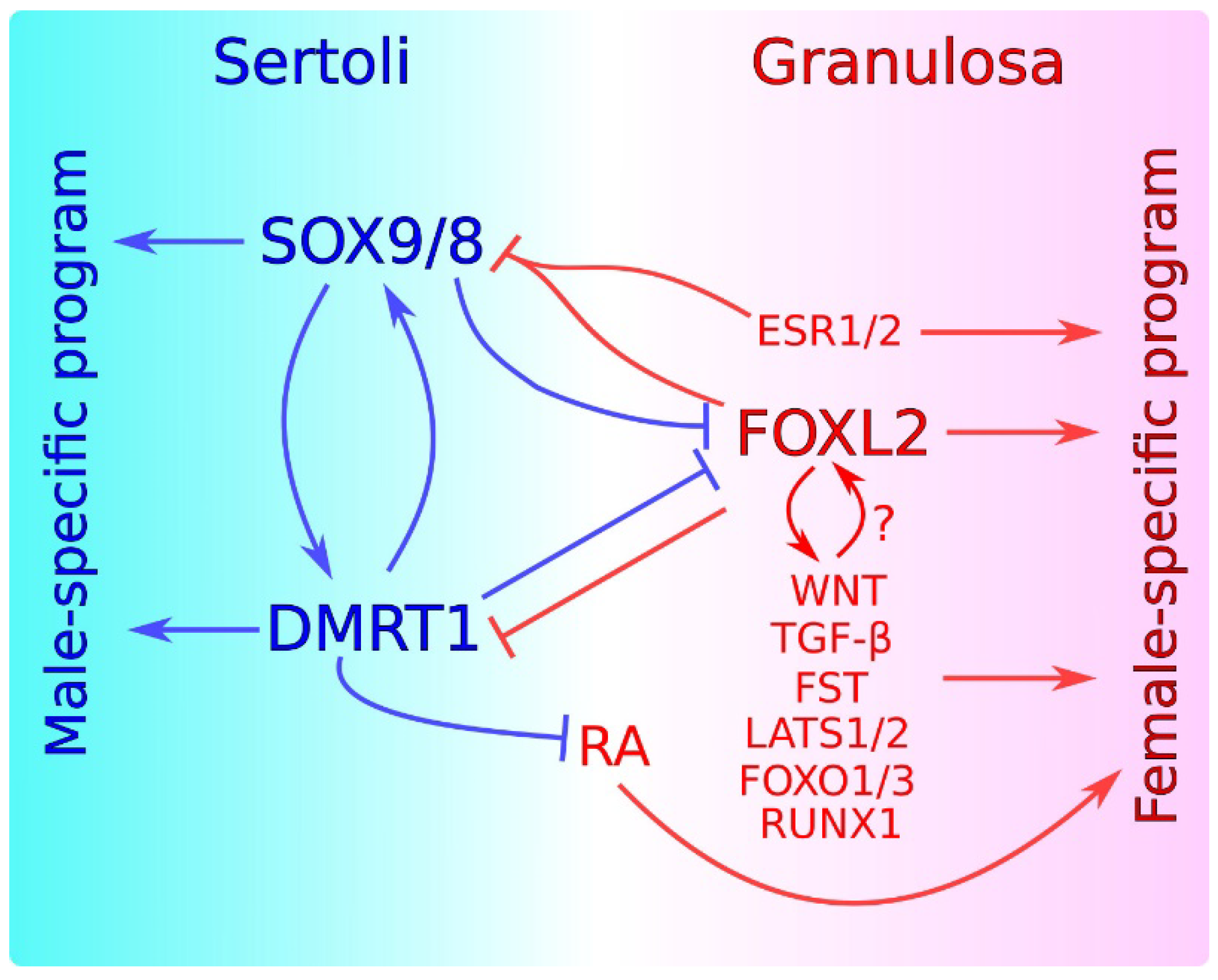
Sex maintenance in mammals is important in sexual differentiation. The crucial event in mammalian sexual differentiation occurs at the embryonic stage of sex determination, when the bipotential gonads differentiate as either testes or ovaries, according to the sex chromosome constitution of the embryo, XY or XX, respectively. Once differentiated, testes produce sexual hormones that induce the subsequent differentiation of the male reproductive tract. On the other hand, the lack of masculinizing hormones in XX embryos permits the formation of the female reproductive tract. It was long assumed that once the gonad is differentiated, this developmental decision is irreversible. However, several findings in the last decade have shown that this is not the case and that a continuous sex maintenance is needed. Deletion of Foxl2 in the adult ovary lead to ovary-to-testis transdifferentiation and deletion of either Dmrt1 or Sox9/Sox8 in the adult testis induces the opposite process. In both cases, mutant gonads were genetically reprogrammed, showing that both the male program in ovaries and the female program in testes must be actively repressed throughout the individual’s life. In addition to these transcription factors, other genes and molecular pathways have also been shown to be involved in this antagonism. 

mammalian sex maintenance sex determination testis differentiation ovary differentiation gonadal cells transdifferentiation gonadal genetic reprograming

1. Introduction

The assumption that gonadal differentiation is irreversible was motivated by the fact that most cases of XX or XY sex reversal could be explained by functional failure of sex-specific factors at the sex determination stage, suggesting that alterations were never produced at later stages. However, cases of transdifferentiation between gonadal sex-specific cell lines, mainly the supporting cell line, after sex determination have long been described. For instance, the ovary of old rats contained structures resembling testis cords that were not present in young rats [1]. Additionally, several cases were reported in which testis-like structures developed in the ovary after the loss of the meiotic germ cells. E12.0 mouse ovaries transplanted beneath the kidney capsules of adult male mice initially developed as ovaries, but seminiferous cords with Sertoli-like cells and testosterone-producing Leydig cells started to develop from day 12 after transplantation in regions depleted of oocytes [2]. Similarly, E14.5 rat ovaries cultured in a medium conditioned by either fetal or young testes, lost germ cells, and developed testis-like cords after 12 days of culture [3], and undifferentiated tammar wallaby ovaries transplanted under the skin of young male pouch also became depleted of germ cells and contained seminiferous-like cords 25 days after transplantation [4]. In addition, granulosa cells survive, proliferate, and subsequently acquire morphological characteristics of Sertoli cells in rat ovarian follicles depleted of oocytes by irradiation [5]. We also showed that after depletion of germ cells in the medullary region of the developing XX gonad of the Iberian mole, Talpa occidentalis, testis cord-like structures are formed and Leydig cells subsequently appear in the interstitial spaces [6].
Another case of granulosa-to-Sertoli cell transdifferentiation was observed in freemartinism, a syndrome in which XX female cattle fetuses with male twins exhibit female-to-male sex reversal with varying degrees of female reproductive tract misdevelopment [7]. In about 50% of the cases, the XX gonads present seminiferous-like tubules [7], and in the most severe cases, Leydig-like cells were also described [8]. Freemartinism occurs when chorionic vascular anastomosis allows the transfer of male gonad derived hormones to the female twin embryo, affecting its sexual development. Because the male reproductive ducts (Müllerian ducts) regress at the same time in the freemartin female as in her male twin, it was proposed that the Anti-Müllerian hormone (AMH, also known as Müllerian inhibiting substance, MIS), a member of the transforming growth factor β family produced by Sertoli cells that induces the regression of Müllerian ducts in male fetuses [9], could be the factor responsible for the syndrome. Indeed, in vitro culture of E14.5 rat ovaries in the presence of purified bovine, AMH showed a reduction of the gonadal volume, oocyte depletion, and differentiation of Sertoli-like cells [10]. Likewise, transgenic mice chronically expressing Amh presented ovaries with few germ cells at birth that were lost afterward, coinciding with the formation of seminiferous-like tubules [9]. The molecular mechanism underlying this process remains unknown. It has been suggested that it is the loss of oocytes derived from the presence of AMH (this hormone is cytotoxic for oocytes), rather than the presence of AMH itself, which causes the transdifferentiation [11][12]. However, this is controversial as the effect of oocyte depletion on granulosa cell transdifferentiation is not well understood. It seems to depend on the stage of germ cell development, and does not occur if the germ cells are depleted before the pre-meiotic stages [13]. However, since postnatal oocyte depletion (like in ovaries exposed to AMH) leads to transdifferentiation in some cases [5], but not in others [14], it cannot be ruled out that AMH may have a direct role in the process of granulosa cell transdifferentiation.
Cell transdifferentiation has also been described in cases of human gonadal cancers. The Sertoli-Leydig cell tumor (SLCT) of the ovary is a rare type of tumor normally affecting middle-aged women, which is characterized by the presence of testicular structures including Sertoli-like cells and Leydig cells that produce androgens [15]. On the other hand, cases of granulosa-cell tumors have also been reported in which neoplastic proliferation of intratubular sex cord cells progresses to an invasive tumor, simultaneously experiencing granulosa cell differentiation and losing Sertoli cell features [16][17]. However, in both cases, supporting cell transdifferentiation may be just a secondary consequence of the dramatic alterations taking place in the genetic program of tumor cells.
Additional evidence of the plasticity of the gonadal cell fate came from transgenic mice. Sry ectopic expression in XX embryonic gonads using a heat-shock-inducible system revealed that Sox9 expression was upregulated and maintained in pre-granulosa cells when Sry expression was induced during the E11.0–11.25 critical time window, but not afterward [18]. However, fine monitoring of granulosa cells in these mice revealed that the SRY-dependent Sox9 inducibility was not as transient in a subpopulation of pre-granulosa cells near the mesonephric tissue, which maintained that capability throughout fetal and early postnatal stages. Furthermore, when E13.5 ovaries were grafted into adult male nude mice, the heat-shock Sry transgene was also able to induce Sox9 expression in differentiated granulosa cells [19].
Finally, chromatin accessibility landscape analyses using purified Sertoli cells have shown that this cell type maintains open chromatin regions near female-promoting genes that are enriched in transcription factor binding motifs for male-promoting genes such as Sox9 and Dmrt1, indicating that these genes are continuously acting as silencers by binding repressors of the granulosa cell fate [20][21]. ChIP-seq for H3K27me3 and H3K4me3 provided results consistent with this notion. H3K27me3 indicates promoter repression, whereas H3K4me3 evidences promoter activation. ChIP-seq experiments performed using purified adult Sertoli cells showed that male-promoting genes harbored H3K4me3 and were depleted in H3K27me3, whereas female-promoting genes were enriched for both marks, indicating that female-determining genes persist in a poised state even long after Sertoli cell differentiation [22]. These results indicate that cells of the supporting lineage require the opposite sex to be permanently repressed, and explain why the loss of such repressors can lead to transdifferentiation into the opposite cell lineage even long after the sex determination stage.
Cases of XX and XY sex reversal induced after sex determination have been associated with a number of sex-specific genes and pathways. We will review the most outstanding cases as follows.

2. Antagonism between Male and Female Factors in Sexual Cell Fate Maintenance

Sex determination involves not only the activation of genes necessary for the differentiation of a sexual fate, but also the active repression of the genetic program of the opposite sex [23][24][25][26][27][28][29]. Some of these molecular interactions operate throughout life, and are summarized in Figure 1. The SOX9/SOX8-DMRT1 axis plays an central role in the maintenance of the Sertoli cell fate. Several studies have indicated that SOX9/SOX8 may control Dmrt1 expression. Sertoli cell-specific ablation of Sox9/8 at E13.5, shortly after the sex determination stage, leads to a rapid downregulation of Dmrt1 which is observable just four days later, at E17.5 [30]. In contrast, in mice with a Sertoli cell-specific deletion of Dmrt1Sox9 is downregulated much later, at P14, coinciding with Foxl2 upregulation [31]. This suggests that, whereas Dmrt1 expression seems to be dependent of SOX9, Sox9 expression is independent of DMRT1 and that the loss of Sox9 after Dmrt1 ablation is a secondary consequence of Foxl2 upregulation. In contrast, other results suggest that DMRT1 might regulate Sox9 expression as DMRT1 binds near the Sox9 locus in P28 mouse testes [31]. In any case, we currently know that the three transcription factors cooperate in the maintenance of the Sertoli cell fate, as Sertoli-to-granulosa transdifferentiation in the postnatal testis is faster and more efficient when Dmrt1Sox9, and Sox8 are deleted in Sertoli cells, compared to single deletion of either Dmrt1 or Sox8/9 alone [32][33].
A recent study identified genes with Sertoli- and granulosa-biased postnatal expression and showed that many of them were associated with sex-biased differentially-accessible chromatin regions (DARs). In postnatal Sertoli cells, many of the Sertoli-biased DARs were bound by both DMRT1 and SOX9, confirming again postnatal cooperation between the two transcription factors in maintaining the Sertoli cell fate. Furthermore, ChIP-seq analysis of granulosa cells ectopically expressing Dmrt1 or Sox9 indicated that DMRT1 and SOX9 jointly bind many sites, although SOX9 was unable to bind most of these sites in the absence of DMRT1, suggesting that during the transdifferentiation process, DMRT1 acts as a pioneer factor promoting chromatin accessibility at regions where SOX9 binds subsequently [33]. Nevertheless, the fact that ectopic SOX9 expression can reprogram sex-biased gene expression in vitro without activating Dmrt1 indicates that DMRT1-independent actions of SOX9 also exist during the Sertoli-to-granulosa transdifferentiation process [33]. Activation of the SOX9/SOX8-DMRT1 axis is essential to maintain Foxl2 repressed, a process that is mediated by the antagonism of DMRT1 on the feminizing action of retinoic acid (RA) [31][30]. Two independent genetic cascades are necessary for embryonic and early postnatal maintenance of the female fate, one controlled by WNT signaling and the other by FOXL2 [34][35]. In the adult ovary, FOXL2 alone is essential to maintain the granulosa cell fate, whereas the involvement of WNT genes in female sex maintenance remains to be elucidated [14]. FOXL2 cooperates with ERα and ERβ in maintaining the granulosa cell fate by directly repressing the Sox9 promoter [14][36]. In addition, the action of TGF-β, FST and LATS1/2, RUNX1, FOXO1/3 is necessary for maintaining the granulosa cell fate. Although it is known (1) that FOXL2 can cooperate with members of the TGF-β pathway in maintaining Fst expression [37][38]; (2) that FOXL2 is phosphorylated by LATS1, a process that seems to be important for granulosa cell differentiation and follicle maturation [39]; and (3) that FOXL2 and RUNX1 exhibit overlaps in chromatin binding in fetal ovaries [40], most of the molecular mechanisms governing the functional relationship among these female promoting genes remain unknown.
Figure 1. Current model for the maintenance of sex-specific supporting cell fates in adult gonads. In Sertoli cells, SOX9/8 establish a feed-forward regulatory loop with DMRT1, necessary for the maintenance of the male-specific program and for preventing the expression of ovary-promoting genes including FOXL2. DMRT1 inhibits RA signaling, which induces the expression of ovarian genes. In granulosa cells, FOXL2 interacts with ESR1/2 and probably with other genes and molecular pathways including WNT, TGFβ, FST, LATS1/2, FOXO1/3, and RUNX1 to maintain the female-specific program. FOXL2 together with ESR1/2 negatively regulates SOX9/8 and/or DMRT1. Male- and female-promoting genes are in blue and red, respectively. Blue and red lines represent an action exerted by male- and female-promoting genes, respectively. Positive regulation is indicated by arrows. Negative regulation is indicated by perpendicular lines.
Finally, we would like to mention that alterations of the balance between male- and female-promoting factors are associated with gonadal diseases, particularly sex cord tumors. For example, a mutation of FOXL2 (C134W) is found in more than 97% of adult-type granulosa cell tumors [41]. In addition, other molecules and pathways such as inhibins, TGF-β, WNT, and SOX9 are associated with the pathogenesis or prognosis of gonadal tumors [42][43][44]. Thus, the exact knowledge of how the sex is maintained is important to understand gonadal function under normal and pathological conditions.

References

  1. Engle, E.T. Tubular Adenomas and Testis-Like Tubules of the Ovaries of Aged Rats. Cancer Res. 1946, 6, 578–582.
  2. Taketo-Hosotani, T.; Merchant-Larios, H.; Thau, R.B.; Koide, S.S. Testicular Cell Differentiation in Fetal Mouse Ovaries Following Transplantation into Adult Male Mice. J. Exp. Zool. 1985, 236, 229–237.
  3. Prépin, J.; Hida, N. Influence of Age and Medium on Formation of Epithelial Cords in the Rat Fetal Ovary In Vitro. Reproduction 1989, 87, 375–382.
  4. Whitworth, D.J.; Shaw, G.; Renfree, M.B. Gonadal Sex Reversal of the Developing Marsupial Ovary In Vivo and In Vitro. Development 1996, 122, 4057–4063.
  5. Guigon, C.J.; Coudouel, N.; Mazaud-Guittot, S.; Forest, M.G.; Magre, S. Follicular Cells Acquire Sertoli Cell Characteristics after Oocyte Loss. Endocrinology 2005, 146, 2992–3004.
  6. Barrionuevo, F.J.; Zurita, F.; Burgos, M.; Jiménez, R. Testis-like Development of Gonads in Female Moles. New Insights on Mammalian Gonad Organogenesis. Dev. Biol. 2004, 268, 39–52.
  7. Jost, A.; Perchellet, J.P.; Prepin, J.; Vigier, B. The Prenatal Development of Bovine Freemartins. In Intersexuality in the Animal Kingdom; Reinboth, R., Ed.; Springer: Berlin/Heidelberg, Germany, 1975; pp. 392–406. ISBN 978-3-642-66069-6.
  8. Dominguez, M.M.; Liptrap, R.M.; Croy, B.A.; Basrur, P.K. Hormonal Correlates of Ovarian Alterations in Bovine Freemartin Fetuses. Anim. Reprod. Sci. 1990, 22, 181–201.
  9. Behringer, R.R.; Finegold, M.J.; Cate, R.L. Müllerian-Inhibiting Substance Function during Mammalian Sexual Development. Cell 1994, 79, 415–425.
  10. Vigier, B.; Watrin, F.; Magre, S.; Tran, D.; Josso, N. Purified Bovine AMH Induces a Characteristic Freemartin Effect in Fetal Rat Prospective Ovaries Exposed to It In Vitro. Development 1987, 100, 43–55.
  11. Whitworth, D.J. XX Germ Cells: The Difference between an Ovary and a Testis. Trends Endocrinol. Metab. 1998, 9, 2–6.
  12. Rios-Rojas, C.; Bowles, J.; Koopman, P. On the Role of Germ Cells in Mammalian Gonad Development: Quiet Passengers or Back-Seat Drivers? Reproduction 2015, 149, R181–R191.
  13. Maatouk, D.M.; Mork, L.; Hinson, A.; Kobayashi, A.; McMahon, A.P.; Capel, B. Germ Cells Are Not Required to Establish the Female Pathway in Mouse Fetal Gonads. PLoS ONE 2012, 7, e47238.
  14. Uhlenhaut, N.H.; Jakob, S.; Anlag, K.; Eisenberger, T.; Sekido, R.; Kress, J.; Treier, A.-C.; Klugmann, C.; Klasen, C.; Holter, N.I.; et al. Somatic Sex Reprogramming of Adult Ovaries to Testes by FOXL2 Ablation. Cell 2009, 139, 1130–1142.
  15. Durmuş, Y.; Kılıç, Ç.; Çakır, C.; Yüksel, D.; Boran, N.; Karalök, A.; Boyraz, G.; Turan, A.T. Sertoli–Leydig Cell Tumor of the Ovary: Analysis of a Single Institution Database and Review of the Literature. J. Obstet. Gynaecol. Res. 2019, 45, 1311–1318.
  16. Kao, C.-S.; Cornejo, K.M.; Ulbright, T.M.; Young, R.H. Juvenile Granulosa Cell Tumors of the Testis. Am. J. Surg. Pathol. 2015, 39, 1159–1169.
  17. Cornejo, K.M.; Young, R.H. Adult Granulosa Cell Tumors of the Testis: A Report of 32 Cases. Am. J. Surg. Pathol. 2014, 38, 1242–1250.
  18. Hiramatsu, R.; Matoba, S.; Kanai-Azuma, M.; Tsunekawa, N.; Katoh-Fukui, Y.; Kurohmaru, M.; Morohashi, K.; Wilhelm, D.; Koopman, P.; Kanai, Y. A Critical Time Window of Sry Action in Gonadal Sex Determination in Mice. Development 2009, 136, 129–138.
  19. Harikae, K.; Miura, K.; Shinomura, M.; Matoba, S.; Hiramatsu, R.; Tsunekawa, N.; Kanai-Azuma, M.; Kurohmaru, M.; Morohashi, K.; Kanai, Y. Heterogeneity in Sexual Bipotentiality and Plasticity of Granulosa Cells in Developing Mouse Ovaries. J. Cell Sci. 2013, 126, 2834–2844.
  20. Maatouk, D.M.; Natarajan, A.; Shibata, Y.; Song, L.; Crawford, G.E.; Ohler, U.; Capel, B. Genome-Wide Identification of Regulatory Elements in Sertoli Cells. Development 2017, 144, 720–730.
  21. Garcia-Moreno, S.A.; Futtner, C.R.; Salamone, I.M.; Gonen, N.; Lovell-Badge, R.; Maatouk, D.M. Gonadal Supporting Cells Acquire Sex-Specific Chromatin Landscapes during Mammalian Sex Determination. Dev. Biol. 2019, 446, 168–179.
  22. Garcia-Moreno, S.A.; Lin, Y.-T.; Futtner, C.R.; Salamone, I.M.; Capel, B.; Maatouk, D.M. CBX2 Is Required during Male Sex Determination to Repress Female Fate at Bivalent Loci. bioRxiv 2018.
  23. Barrionuevo, F.J.; Burgos, M.; Scherer, G.; Jiménez, R. Genes Promoting and Disturbing Testis Development. Histol. Histopatol. 2012, 11, 1361–1383.
  24. Sekido, R.; Lovell-Badge, R. Genetic Control of Testis Development. Sex. Dev. 2013, 7, 21–32.
  25. Svingen, T.; Koopman, P. Building the Mammalian Testis: Origins, Differentiation, and Assembly of the Component Cell Populations. Genes Dev. 2013, 27, 2409–2426.
  26. Ewen, K.A.; Koopman, P. Mouse Germ Cell Development: From Specification to Sex Determination. Mol. Cell. Endocrinol. 2010, 323, 76–93.
  27. Lin, Y.-T.; Capel, B. Cell Fate Commitment during Mammalian Sex Determination. Curr. Opin. Genet. Dev. 2015, 32, 144–152.
  28. Nef, S.; Stévant, I.; Greenfield, A. Chapter Six—Characterizing the bipotential mammalian gonad. In Current Topics in Developmental Biology; Capel, B., Ed.; Sex Determination in Vertebrates; Academic Press: Cambridge, MA, USA, 2019; Volume 134, pp. 167–194.
  29. Vining, B.; Ming, Z.; Bagheri-Fam, S.; Harley, V. Diverse Regulation but Conserved Function: SOX9 in Vertebrate Sex Determination. Genes 2021, 12, 486.
  30. Georg, I.; Barrionuevo, F.; Wiech, T.; Scherer, G. Sox9 and Sox8 Are Required for Basal Lamina Integrity of Testis Cords and for Suppression of FOXL2 during Embryonic Testis Development in Mice1. Biol. Reprod. 2012, 87, 1–11.
  31. Matson, C.K.; Murphy, M.W.; Sarver, A.L.; Griswold, M.D.; Bardwell, V.J.; Zarkower, D. DMRT1 Prevents Female Reprogramming in the Postnatal Mammalian Testis. Nature 2011, 476, 101–104.
  32. Minkina, A.; Matson, C.K.; Lindeman, R.E.; Ghyselinck, N.B.; Bardwell, V.J.; Zarkower, D. DMRT1 Protects Male Gonadal Cells from Retinoid-Dependent Sexual Transdifferentiation. Dev. Cell 2014, 29, 511–520.
  33. Lindeman, R.E.; Murphy, M.W.; Agrimson, K.S.; Gewiss, R.L.; Bardwell, V.J.; Gearhart, M.D.; Zarkower, D. The Conserved Sex Regulator DMRT1 Recruits SOX9 in Sexual Cell Fate Reprogramming. Nucleic Acids Res. 2021, 49, 6144–6164.
  34. Ottolenghi, C.; Pelosi, E.; Tran, J.; Colombino, M.; Douglass, E.; Nedorezov, T.; Cao, A.; Forabosco, A.; Schlessinger, D. Loss of Wnt4 and Foxl2 Leads to Female-to-Male Sex Reversal Extending to Germ Cells. Hum. Mol. Genet. 2007, 16, 2795–2804.
  35. Pannetier, M.; Chassot, A.-A.; Chaboissier, M.-C.; Pailhoux, E. Involvement of FOXL2 and RSPO1 in Ovarian Determination, Development, and Maintenance in Mammals. Sex. Dev. 2016, 10, 167–184.
  36. Georges, A.; L’Hôte, D.; Todeschini, A.L.; Auguste, A.; Legois, B.; Zider, A.; Veitia, R.A. The Transcription Factor FOXL2 Mobilizes Estrogen Signaling to Maintain the Identity of Ovarian Granulosa Cells. eLife 2014, 3, e04207.
  37. Blount, A.L.; Schmidt, K.; Justice, N.J.; Vale, W.W.; Fischer, W.H.; Bilezikjian, L.M. FoxL2 and Smad3 Coordinately Regulate Follistatin Gene Transcription*. J. Biol. Chem. 2009, 284, 7631–7645.
  38. Kashimada, K.; Pelosi, E.; Chen, H.; Schlessinger, D.; Wilhelm, D.; Koopman, P. FOXL2 and BMP2 Act Cooperatively to Regulate Follistatin Gene Expression during Ovarian Development. Endocrinology 2011, 152, 272–280.
  39. Pisarska, M.D.; Kuo, F.-T.; Bentsi-Barnes, I.K.; Khan, S.; Barlow, G.M. LATS1 Phosphorylates Forkhead L2 and Regulates Its Transcriptional Activity. Am. J. Physiol.-Endocrinol. Metab. 2010, 299, E101–E109.
  40. Nicol, B.; Grimm, S.A.; Chalmel, F.; Lecluze, E.; Pannetier, M.; Pailhoux, E.; Dupin-De-Beyssat, E.; Guiguen, Y.; Capel, B.; Yao, H.H.-C. RUNX1 Maintains the Identity of the Fetal Ovary through an Interplay with FOXL2. Nat. Commun. 2019, 10, 5116.
  41. Shah, S.P.; Köbel, M.; Senz, J.; Morin, R.D.; Clarke, B.A.; Wiegand, K.C.; Leung, G.; Zayed, A.; Mehl, E.; Kalloger, S.E.; et al. Mutation of FOXL2 in Granulosa-Cell Tumors of the Ovary. N. Engl. J. Med. 2009, 360, 2719–2729.
  42. Fang, X.; Ni, N.; Gao, Y.; Vincent, D.F.; Bartholin, L.; Li, Q. A Novel Mouse Model of Testicular Granulosa Cell Tumors. Mol. Hum. Reprod. 2018, 24, 343–356.
  43. Färkkilä, A.; Haltia, U.-M.; Tapper, J.; McConechy, M.K.; Huntsman, D.G.; Heikinheimo, M. Pathogenesis and Treatment of Adult-Type Granulosa Cell Tumor of the Ovary. Ann. Med. 2017, 49, 435–447.
  44. Onder, S.; Hurdogan, O.; Bayram, A.; Yilmaz, I.; Sozen, H.; Yavuz, E. The Role of FOXL2, SOX9, and β-Catenin Expression and DICER1 Mutation in Differentiating Sex Cord Tumor with Annular Tubules from Other Sex Cord Tumors of the Ovary. Virchows Arch. 2021.
More
Information
Contributor MDPI registered users' name will be linked to their SciProfiles pages. To register with us, please refer to https://encyclopedia.pub/register :
View Times: 718
Revisions: 2 times (View History)
Update Date: 27 Jul 2021
1000/1000