Submitted Successfully!
To reward your contribution, here is a gift for you: A free trial for our video production service.
Thank you for your contribution! You can also upload a video entry or images related to this topic.
Version Summary Created by Modification Content Size Created at Operation
1 + 2359 word(s) 2359 2021-06-04 07:55:01 |
2 format correct Meta information modification 2359 2021-06-25 09:36:47 | |
3 move out from EC Meta information modification 2359 2021-09-22 04:32:14 |

Video Upload Options

Do you have a full video?

Confirm

Are you sure to Delete?
Cite
If you have any further questions, please contact Encyclopedia Editorial Office.
Grange, P. Cutibacterium acnes. Encyclopedia. Available online: https://encyclopedia.pub/entry/11300 (accessed on 16 April 2024).
Grange P. Cutibacterium acnes. Encyclopedia. Available at: https://encyclopedia.pub/entry/11300. Accessed April 16, 2024.
Grange, Philippe. "Cutibacterium acnes" Encyclopedia, https://encyclopedia.pub/entry/11300 (accessed April 16, 2024).
Grange, P. (2021, June 25). Cutibacterium acnes. In Encyclopedia. https://encyclopedia.pub/entry/11300
Grange, Philippe. "Cutibacterium acnes." Encyclopedia. Web. 25 June, 2021.
Cutibacterium acnes
Edit

Cutibacterium acnes is a member of the skin microbiota found predominantly in regions rich in sebaceous glands. It is involved in maintaining healthy skin and has long been considered a commensal bacterium. Its involvement in various infections has led to its emergence as an opportunist pathogen. Interactions between C. acnes and the human host, including the human skin microbiota, promote the selection of C. acnes strains capable of producing several virulence factors that increase inflammatory capability. This pathogenic property may be related to many infectious mechanisms, such as an ability to form biofilms and the expression of putative virulence factors capable of triggering host immune responses or enabling C. acnes to adapt to its environment. During the past decade, many studies have identified and characterized several putative virulence factors potentially involved in the pathogenicity of this bacterium. These virulence factors are involved in bacterial attachment to target cells, polysaccharide-based biofilm synthesis, molecular structures mediating inflammation, and the enzymatic degradation of host tissues. C. acnes, like other skin-associated bacteria, can colonize various ecological niches other than skin. It produces several proteins or glycoproteins that could be considered to be active virulence factors, enabling the bacterium to adapt to the lipophilic environment of the pilosebaceous unit of the skin, but also to the various organs it colonizes. 

C. acnes inflammation innate immunity virulence factors characterization

1. C. acnes Characteristics

The genus Cutibacterium acnes (C. acnes, formerly known as Propionibacterium acnes or P. acnes, see below) is a commensal lipophilic Gram-positive bacterium. C. acnes is described as diphtheroid or coryneform because it is rod-shaped and slightly curved with a width of 0.4 to 0.7 µm and length of 3 to 5 µm. Anaerobic bacteria are characterized by their inability to grow on solid media in the presence of atmospheric oxygen. However, C. acnes is considered an aerotolerant anaerobe because it possesses enzymatic systems able to detoxify oxygen, allowing it to be sustained on the surface of the skin [1].
Following its isolation [2]C. acnes was first included in the genus Bacillus as Bacillus acnes, and then in the genus Corynebacterium as Corynebacterium acnes or “anaerobic corynebacteria” because of its morphology. Based on its ability to produce propionic acid via its anaerobic catabolism, it was then assigned to the genus Propionibacterium, subsequently renamed Cutibacterium. Genus Cutibacterium belongs to a branch of Actinobacter and can be split into two groups, one containing the so-called “classic or dairy” species, bringing together saprophytic species isolated from non-human-pathogenic dairy products, and the other containing commensal “skin” species, most found on the surface of human skin. Classic species, such as Propionibacterium freundenreichii, which is essential for the ripening of Swiss cheeses, or Propionibacterium acidipropionici, known for its beneficial effects in the bovine rumen, have been studied in considerable detail due to their importance to the agri-food industry. By contrast, the pathophysiology of cutaneous species is less well understood.
Phylogenetic and genome analyses of classic and cutaneous species have highlighted differences in the 16S RNA gene sequences and the core genome between species. This finding led to proposals to reclassify many species into four different genera:
(1)
The genus Propionibacterium, comprising the species P. freundenreichiiP. cyclohexanicumP. acidifaciens, and P. australianse.
(2)
The new genus Acidipropionibacterium, comprising the species A. jenseniiA. thoeniiA. acidipropioniciA. microaerophilumA. damnosum, and A. olivae.
(3)
The new genus Pseudopropionibacterium, containing a single species: P. propionicum.
(4)
The new genus Cutibacterium, comprising cutaneous Propionibacterium bacteria belonging to the species C. acnesC. avidumC. granulosum, and C. humerusiiP. acnes has thus been renamed C. acnes [3]. Moreover, the genus C. acnes has been further subdivided into subspecies, such as C. acnes subsp. defendens [4][5], and C. acnes subsp. elongatum [6] (Figure 1).
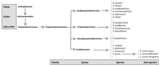
Figure 1. New classification of C. acnes strains.
Unlike other Gram-positive bacteria, C. acnes has a unique cell wall and envelope, containing phosphatidylinositol, triacylglycerol, and many other common lipids [7]. The cell wall of C. acnes consists of peptidoglycan (PNG), but of a type different from that of other Gram-positive bacteria, in that the peptide chain contains the L-acid L-diaminopelic acid and D-alanine. Analyses of C. acnes lipoglycans have also revealed the presence of a lipid anchor based on fatty acids and shown that the polysaccharide moiety contains significant amounts of mannose, glucose, and galactose, together with an amino sugar thought to be a diaminohexuronic acid [8]C. acnes has been shown to survive for long periods of time in human tissues with a low oxidative potential. Indeed, C. acnes has all of the proteins required for oxidative phosphorylation (NAPDH dehydrogenase/complex I, cytochrome c reductase, cytochrome c oxidase, and FoF1-type ATP synthase). It also possesses the cytochrome d oxidase genes, enabling it to grow in conditions in which limited amounts of oxygen are present and to tolerate oxygen for a few hours. Despite its tolerance of the presence of air, C. acnes cannot be reliably detected by culture in aerobic conditions due to its very slow growth (5 to 7 days), associated with a division time of about five hours [9]. A limited number of C. acnes strains possess a linear plasmid carrying several genes, including a locus for tight adherence (tad) encoding adhesive Flp (fimbrial low-molecular weight protein) pili [10]. Antibiotics were found to be effective against inflammatory acne, but the selection pressure exerted by antibiotic treatment for this condition has led to the induction of antibiotic resistance in up to 40% of C. acnes strains (resistance to erythromycin, clindamycin, and tetracycline), increasing the likelihood of treatment failure [11]. Interestingly, it should be noted that the anaerobic species may have intrinsic resistance due to a lack of molecular mechanisms involved in the uptake of the antibiotic [1]. Therefore, antimicrobial susceptibility testing on C. acnes strains should be implemented largely in order to adapt antibiotic treatments and to obtain epidemiological data on C. acnes antibiotic resistance. In this review, we summarize current knowledge concerning characterized C. acnes virulence factors and their possible implication in the pathogenicity of C. acnes, which may interest researchers and clinicians investigating the physiopathology of C. acnes.

2. C. acnes Classification

C. acnes strains were previously classified into two main types, I and II, on the basis of their cell wall carbohydrate content and serum lectin responses [12][13]. It was then suggested that C. acnes strains could be distinguished on the basis of random amplification of polymorphic DNA (RAPD) analysis [14]. Analyses of the sequences of the non-ribosomal housekeeping genes RecA and tly, and the use of the QUBPa1 and QUBPa2 mAbs, specific for type I and II strains, respectively, subsequently showed that the two types corresponded to evolutionary lineages of C. acnes displaying both genetic and phenotypic differences. An additional phylotype, type III, corresponding to strains with filamentous appendages, was then added to the classification [15][16]. Based on 16S rRNA gene analysis, C. acnes strains were divided into ribotypes (RTs), which could be used to differentiate between types I, II, and III, associated with healthy skin or acne [17]. RT4, RT5 and RT8 appeared to be found in acne lesions, whereas RT2 and RT6 were mostly found on healthy skin [18]. Multi-locus sequence typing (MLST) and single-locus sequence typing (SLST) methods have been developed for this species, to increase typing resolution. Depending on the number and nature of the housekeeping genes selected, two schemes for discriminating between C. acnes strains have been described. The Belfast scheme, using seven target genes, can differentiate type I into clades IA1, IA2, IB, and IC [19][20]. A multiplex touchdown PCR typing method has also been developed that can discriminate between the IA1, IA2, IB, IC, type II, and type III phylotypes, providing a rapid overview of the C. acnes types present in a population [21]. The other scheme, the Aarhus scheme, uses nine target genes to split type I into clades I-1a, I-1b, and I-2 [22]. This scheme appears to be the most discriminant of these two schemes [23]. SLST and whole-genome sequencing (WGS) methods were subsequently used to develop a new scheme capable of differentiating C. acnes strains into clades IA-1, IA-2, IB-1, IB-2, IB-3, IC, II, and III [24], but some C. acnes strains were not identified with this scheme [25] (Table 1). MS-based typing for routine analysis has been evaluated with the MALDI-TOF MS method, to characterize C. acnes strains in diverse samples [26]. This method is very useful for rapidly discriminating C. acnes phylotypes, although missing some of them [26]. However, this approach was improved using profiling of identified biomarkers, such as ribosomal subunit proteins, and named MALDI-MS prototyping, which allowed discrimination of all of the C. acnes phylotypes. Although this method needs to ultrafiltrate bacteria whole pellets in order to analyze the concentrated protein fraction, it is nonetheless compatible with the analysis of a large number of C. acnes strains [27]. Another attempt to facilitate C. acnes typing involved the use of the MLVA method to analyze the polymorphism of 13 VNTRs; the results of this method were well-correlated with those for the MLST and SLST methods [28]. Nomenclatures highlight the need for standardization. WGS is of potential interest for this purpose because it should provide a higher-resolution phylogeny more rapidly and at lower cost [29][30].
Table 1. C. acnes phylotypes.
Phylotypes
C. acnes subspecies   WGS/Ribotyping a SLST b MLST8 c CC MLST9 d CC MALDI-TOF e MLVA13 f
C. acnes acnes IA-1 RT1, RT5 A1-A34 IA1 CC1 I-1a CC18 IA IAI
  RT532 B1            
IA-2 RT1, RT4 C1-C5 IA1 CC3 I-1a CC3 IA IA2
  RT5              
IB-1 RT8 D1-D5 IA1 CC4 I-1a CC28 IB IB
    E1-E9       CC31    
IB-2 RT3, RT16 F1-F14 IA2 CC2 I-1b CC28 IB  
IB-3 RT1 H1-H8 IB CC5 I-2 CC36 IB  
IC RT5 G1 IC CC107 / / IB(IC) /
C. acnes defendens   RT2, RT6 K1-K25   CC6   CC53    
II RT6   II CC30 II CC60 II II
        CC71        
        CC72        
C. acnes elongatum III RT9 L1-L10 III CC77 III CC43 III III
WGS: whole genome sequencing; CC: clonal complex. a Ribotyping [17]b SLST [3] c Belfast scheme [19][20]d Aarhus scheme [22]e MALDI-TOF analysis [31]f MLVA13 analysis [28].

3. Skin Microbiota and Acne

Human skin consists of a stratified, cellular epidermis and an underlying dermis of connective tissue. The epidermis is characterized by a stratified squamous epithelium composed of about 80–90% keratinocytes, which progressively move from the epidermal basement membrane towards the surface of the skin, forming several well-defined layers during this transit. Other cells resident within the epidermis include melanocytes, Langerhans’ cells, and Merkel cells. The epidermis has several very important functions in the protection of the body against environmental hazards, and it also acts as an immunological barrier, modulating the microbial population of the skin [32][33][34]. The dermis is characterized by a layer of fibroblast cells that also contains immune cells, such as macrophages, neutrophils, and resident T cells.
Acne vulgaris is a chronic inflammatory skin disease mostly found in the teenage population but also in young adults [35]. In a large majority of cases, acne is not a serious disease but if left untreated, can have serious consequences both physically and psychologically. However, there are severe forms of acne that cause permanent scarring, which may have severe consequences for personality development in young people, leading to social and economic problems. Adolescents suffering from acne are more anxious, socially inhibited, and aggressive than adolescents without acne. Furthermore, acne is the only skin disease for which outcome has been implicated as a risk factor for suicide, particularly in men [36][37].
Acne vulgaris is a disorder affecting the pilosebaceous unit (PSU), resulting in both inflammatory and non-inflammatory clinical lesions. Most patients have a mixture of non-inflammatory comedones and inflammatory papules, pustules, and nodules. Acne ranges in severity from mild symptoms to rare cases of severe rare fulminant infection, with a small subset of patients displaying a highly destructive inflammatory response that is often associated with scarring [38]. The hair follicle or pilosebaceous unit (PSU) groups together sebaceous glands, a hair, and a follicle duct made up of epithelial cells. The sebaceous glands produce sebum which flows into the infundibulum to be released to the surface of the skin. Sebum contains a wide variety of lipids, such as squalene, wax esters, triglycerides, and free fatty acids. The production of sebum is linked to the interaction between the PSU and the thyroid and androgenic hormones, and is dependent on age [39]. Acne occurs mostly in teenagers, in whom hormonal secretion is often imbalanced, leading to an increase in sebum secretion by the sebaceous glands in the PSU [40]. The accumulation of sebum in the infundibulum induces keratinocyte proliferation in the follicular wall, promoting PSU obstruction and the formation of comedones. PSU obstruction leads to hypoxia, favoring the development of C. acnes. By interacting with the cells of the PSU, this bacterium then triggers the expression of pro-inflammatory molecules, resulting in strong inflammation. Acne vulgaris thus appears to be a multifactorial disorder, involving sebaceous hyperplasia, follicular hyperkeratinization, and colonization by C. acnes, acting as an opportunistic pathogen. C. acnes has also been shown to influence the formation of comedones by secreting propionic acid, which modulates keratinocyte differentiation [41].
However, the long-standing hypothesis that acne results from C. acnes proliferation has evolved, because this bacterium is also involved in the maintenance of healthy skin and can act as an opportunistic pathogen in various inflammatory conditions, including acne. Amplification- and sequencing-based methods have shed considerable light on the huge diversity of the skin microbial community. Despite the possible interference of sampling methods with the microbiome profile identified [42][43], it has been demonstrated that healthy skin harbors microorganisms from multiple kingdoms: bacteria, fungi, and viruses. Most skin bacteria belong to four phyla: Actinobacteria (Corynebacterineae, Propionibacterineae), Proteobacteria, Firmicutes (Staphylococcaceae), and Bacteroidetes. The composition of the skin microbiota varies between individuals, but C. acnes seems to occur mostly at sebaceous sites (on the face, back, and pre-thoracic region), reflecting its ability to survive in anaerobic and lipid-rich conditions. By contrast, Staphylococcus and Corynebacterium species are mostly found at humid sites, with a more diverse bacterial population occupying dry sites [17][44][45]C. acnes is, by far, the most abundant bacterium in the skin microbiota, and its load does not seem to differ between healthy skin and skin affected by acne. It has been suggested that inflammatory acne is triggered by an imbalance in the skin microbiota associated with the selection of specific types of C. acnes [25][45][46]. It has been shown that phylotypes IA-1 are strongly associated with inflammatory acne and acne fulminans. These phylotypes produce high levels of porphyrins, which can promote inflammation [17][18][20][22][38][47]. The population of cutaneous microorganisms should be considered a dynamic feature. Bacteria can interact with each other (S. epidermidis—C. acnes) and may also be influenced by changes in host characteristics (hormonal disorders, stress, environmental changes), promoting the selection of pathogenic strains of C. acnes capable of producing several virulence factors (biofilm, surface proteins) increasing inflammatory capacity [48][49][50]. The skin also reacts to the commensal microbiota, with effects on the immune response and epidermal development, contributing to skin health and disease [51]. Modulation of the population of C. acnes strains on the skin, without inducing a negative reaction, could therefore potentially be used in the treatment of microbiome-related diseases [52].

References

  1. Gajdács, M.; Spengler, G.; Urbán, E. Identification and Antimicrobial Susceptibility Testing of Anaerobic Bacteria: Rubik’s Cube of Clinical Microbiology? Antibiotics 2017, 6, 25.
  2. Wickham, L. The Microbial Origin of Baldness: Sabouraud’s Researches into the Relations between Seborrhoea, Alopecia Areata, and Baldness. Br. Med. J. 1897, 1, 1028–1030.
  3. Scholz, C.F.P.; Kilian, M. The Natural History of Cutaneous Propionibacteria, and Reclassification of Selected Species within the Genus Propionibacterium to the Proposed Novel Genera Acidipropionibacterium gen. nov., Cutibacterium gen. nov. and Pseudopropionibacterium gen. nov. Int. J. Syst. Evol. Microbiol. 2016, 66, 4422–4432.
  4. McDowell, A.; Barnard, E.; Liu, J.; Li, H.; Patrick, S. Proposal to Reclassify Propionibacterium acnes Type I as Propionibacterium acnes subsp. acnes subsp. nov. and Propionibacterium acnes Type II as Propionibacterium acnes subsp. defendens subsp. nov. Int. J. Syst. Evol. Microbiol. 2016, 66, 5358–5365.
  5. McDowell, A.; Barnard, E.; Liu, J.; Li, H.; Patrick, S. Corrigendum: Proposal to Reclassify Propionibacterium acnes Type I as Propionibacterium acnes subsp. acnes subsp. nov. and Propionibacterium acnes Type II as Propionibacterium acnes subsp. defendens subsp. nov. Int. J. Syst. Evol. Microbiol. 2017, 67, 4880–4880.
  6. Dekio, I.; Culak, R.; Misra, R.; Gaulton, T.; Fang, M.; Sakamoto, M.; Ohkuma, M.; Oshima, K.; Hattori, M.; Klenk, H.-P.; et al. Dissecting the Taxonomic Heterogeneity within Propionibacterium acnes: Proposal for Propionibacterium acnes subsp. acnes subsp. nov. and Propionibacterium acnes subsp. elongatum subsp. nov. Int. J. Syst. Evol. Microbiol. 2015, 65, 4776–4787.
  7. Jeon, J.; Park, S.C.; Her, J.; Lee, J.W.; Han, J.-K.; Kim, Y.-K.; Kim, K.P.; Ban, C. Comparative Lipidomic Profiling of the Human Commensal Bacterium Propionibacterium acnes and Its Extracellular Vesicles. RSC Adv. 2018, 8, 15241–15247.
  8. Whale, G.A.; Sutcliffe, I.C.; Morrisson, A.R.; Pretswell, E.L.; Emmison, N. Purification and Characterisation of Lipoglycan Macroamphiphiles from Propionibacterium acnes. Antonie Van Leeuwenhoek 2004, 86, 77–85.
  9. Hall, G.S.; Pratt-Rippin, K.; Meisler, D.M.; Washington, J.A.; Roussel, T.J.; Miller, D. Growth Curve for Propionibacterium acnes. Curr. Eye Res. 1994, 13, 465–466.
  10. Davidsson, S.; Carlsson, J.; Mölling, P.; Gashi, N.; Andrén, O.; Andersson, S.-O.; Brzuszkiewicz, E.; Poehlein, A.; Al-Zeer, M.A.; Brinkmann, V.; et al. Prevalence of Flp Pili-Encoding Plasmids in Cutibacterium acnes Isolates Obtained from Prostatic Tissue. Front. Microbiol. 2017, 8, 2241.
  11. Dreno, B.; Thiboutot, D.; Gollnick, H.; Bettoli, V.; Kang, S.; Leyden, J.J.; Shalita, A.; Torres, V. Antibiotic Stewardship in Dermatology: Limiting Antibiotic Use in Acne. Eur. J. Dermatol. 2014, 24, 330–334.
  12. Johnson, J.L.; Cummins, C.S. Cell Wall Composition and Deoxyribonucleic Acid Similarities among the Anaerobic Coryneforms, Classical Propionibacteria, and Strains of Arachnia propionica. J. Bacteriol. 1972, 109, 1047–1066.
  13. Webster’, G.F.; Cummins, C.S. Use of Bacteriophage Typing to Distinguish Propionibacterium acnes Types I and II. J. Clin. Microbiol. 1978, 7, 84–90.
  14. Perry, A.L.; Worthington, T.; Hilton, A.C.; Lambert, P.A.; Stirling, A.J.; Elliott, T.S.J. Analysis of Clinical Isolates of Propionibacterium acnes by Optimised RAPD. FEMS Microbiol. Lett. 2003, 228, 51–55.
  15. McDowell, A.; Valanne, S.; Ramage, G.; Tunney, M.M.; Glenn, J.V.; McLorinan, G.C.; Bhatia, A.; Maisonneuve, J.-F.; Lodes, M.; Persing, D.H.; et al. Propionibacterium acnes Types I and II Represent Phylogenetically Distinct Groups. J. Clin. Microbiol. 2005, 43, 9.
  16. McDowell, A.; Perry, A.L.; Lambert, P.A.; Patrick, S. A New Phylogenetic Group of Propionibacterium acnes. J. Med Microbiol. 2008, 57, 218–224.
  17. Fitz-Gibbon, S.; Tomida, S.; Chiu, B.-H.; Nguyen, L.; Du, C.; Liu, M.; Elashoff, D.; Erfe, M.C.; Loncaric, A.; Kim, J.; et al. Propionibacterium acnes Strain Populations in the Human Skin Microbiome Associated with Acne. J. Investig. Dermatol. 2013, 133, 2152–2160.
  18. McDowell, A.; Nagy, I.; Magyari, M.; Barnard, E.; Patrick, S. The Opportunistic Pathogen Propionibacterium acnes: Insights into Typing, Human Disease, Clonal Diversification and CAMP Factor Evolution. PLoS ONE 2013, 8, e70897.
  19. McDowell, A.; Gao, A.; Barnard, E.; Fink, C.; Murray, P.I.; Dowson, C.G.; Nagy, I.; Lambert, P.A.; Patrick, S. A Novel Multilocus Sequence Typing Scheme for the Opportunistic Pathogen Propionibacterium acnes and Characterization of Type I Cell Surface-Associated Antigens. Microbiology 2011, 157, 1990–2003.
  20. McDowell, A.; Barnard, E.; Nagy, I.; Gao, A.; Tomida, S.; Li, H.; Eady, A.; Cove, J.; Nord, C.E.; Patrick, S. An Expanded Multilocus Sequence Typing Scheme for Propionibacterium acnes: Investigation of ‘Pathogenic’, ‘Commensal’ and Antibiotic Resistant Strains. PLoS ONE 2012, 7, e41480.
  21. Barnard, E.; Nagy, I.; Hunyadkürti, J.; Patrick, S.; McDowell, A. Multiplex Touchdown PCR for Rapid Typing of the Opportunistic Pathogen Propionibacterium acnes. J. Clin. Microbiol. 2015, 53, 1149–1155.
  22. Lomholt, H.B.; Kilian, M. Population Genetic Analysis of Propionibacterium acnes Identifies a Subpopulation and Epidemic Clones Associated with Acne. PLoS ONE 2010, 5, e12277.
  23. Kilian, M.; Scholz, C.F.P.; Lomholt, H.B. Multilocus Sequence Typing and Phylogenetic Analysis of Propionibacterium acnes. J. Clin. Microbiol. 2012, 50, 1158–1165.
  24. Scholz, C.F.P.; Jensen, A.; Lomholt, H.B.; Brüggemann, H.; Kilian, M. A Novel High-Resolution Single Locus Sequence Typing Scheme for Mixed Populations of Propionibacterium acnes in Vivo. PLoS ONE 2014, 9, e104199.
  25. Paugam, C.; Corvec, S.; Saint-Jean, M.; Le Moigne, M.; Khammari, A.; Boisrobert, A.; Nguyen, J.M.; Gaultier, A.; Dréno, B. Propionibacterium acnes Phylotypes and Acne Severity: An Observational Prospective Study. J. Eur. Acad. Dermatol. Venereol. 2017, 31, e398–e399.
  26. Broly, M.; Ruffier d’Epenoux, L.; Guillouzouic, A.; Le Gargasson, G.; Juvin, M.-E.; Leroy, A.G.; Bémer, P.; Corvec, S. Propionibacterium/Cutibacterium Species–Related Positive Samples, Identification, Clinical and Resistance Features: A 10-Year Survey in a French Hospital. Eur J. Clin. Microbiol. Infect. Dis 2020, 39, 1357–1364.
  27. Teramoto, K.; Okubo, T.; Yamada, Y.; Sekiya, S.; Iwamoto, S.; Tanaka, K. Classification of Cutibacterium acnes at Phylotype Level by MALDI-MS Proteotyping. Proc. Jpn. Acad. Ser. B 2019, 95, 612–623.
  28. Hauck, Y.; Soler, C.; Gérôme, P.; Vong, R.; Macnab, C.; Appere, G.; Vergnaud, G.; Pourcel, C. A Novel Multiple Locus Variable Number of Tandem Repeat (VNTR) Analysis (MLVA) Method for Propionibacterium acnes. Infect. Genet. Evol. 2015, 33, 233–241.
  29. Tomida, S.; Nguyen, L.; Chiu, B.-H.; Liu, J.; Sodergren, E.; Weinstock, G.M.; Li, H. Pan-Genome and Comparative Genome Analyses of Propionibacterium acnes Reveal Its Genomic Diversity in the Healthy and Diseased Human Skin Microbiome. mBio 2013, 4, e00003-13.
  30. Dagnelie, M.-A.; Khammari, A.; Dréno, B.; Corvec, S. Cutibacterium acnes Molecular Typing: Time to Standardize the Method. Clin. Microbiol. Infect. 2018, 24, 1149–1155.
  31. Nagy, E.; Urbán, E.; Becker, S.; Kostrzewa, M.; Vörös, A.; Hunyadkürti, J.; Nagy, I. MALDI-TOF MS Fingerprinting Facilitates Rapid Discrimination of Phylotypes I, II and III of Propionibacterium acnes. Anaerobe 2013, 20, 20–26.
  32. Belkaid, Y.; Segre, J.A. Dialogue between Skin Microbiota and Immunity. Science 2014, 346, 954–959.
  33. Grice, E.A. The Intersection of Microbiome and Host at the Skin Interface: Genomic- and Metagenomic-Based Insights. Genome Res. 2015, 25, 1514–1520.
  34. Borkowski, A.W.; Gallo, R.L. The Coordinated Response of the Physical and Antimicrobial Peptide Barriers of the Skin. J. Investig. Dermatol. 2011, 131, 285–287.
  35. Svensson, A.; Ofenloch, R.F.; Bruze, M.; Naldi, L.; Cazzaniga, S.; Elsner, P.; Goncalo, M.; Schuttelaar, M.-L.A.; Diepgen, T.L. Prevalence of Skin Disease in a Population-Based Sample of Adults from Five European Countries. Br. J. Dermatol. 2018, 178, 1111–1118.
  36. Cotterill, J.A.; Cunliffe, W.J. Suicide in Dermatological Patients. Br. J. Dermatol. 1997, 137, 246–250.
  37. Zaenglein, A.L. Acne Vulgaris. N. Engl. J. Med. 2018, 379, 1343–1352.
  38. Bocquet-Trémoureux, S.; Corvec, S.; Khammari, A.; Dagnelie, M.-A.; Boisrobert, A.; Dreno, B. Acne Fulminans and Cutibacterium acnes Phylotypes. J. Eur. Acad. Dermatol. Venereol. 2020, 34, 827–833.
  39. Stewart, M.E.; Steele, W.A.; Downing, D.T. Changes in the Relative Amounts of Endogenous and Exogenous Fatty Acids in Sebaceous Lipids during Early Adolescence. J. Invest. Dematol. 1989, 92, 371–378.
  40. Deplewski, D.; Rosenfield, R.L. Role of Hormones in Pilosebaceous Unit Development. Endocr. Rev. 2000, 21, 30.
  41. Tax, G.; Urbán, E.; Palotás, Z.; Puskás, R.; Kónya, Z.; Bíró, T.; Kemény, L.; Szabó, K. Propionic Acid Produced by Propionibacterium acnes Strains Contributes to Their Pathogenicity. Acta Dermatol. Venerol. 2016, 96, 43–49.
  42. Hall, J.B.; Cong, Z.; Imamura-Kawasawa, Y.; Kidd, B.A.; Dudley, J.T.; Thiboutot, D.M.; Nelson, A.M. Isolation and Identification of the Follicular Microbiome:IImplications for Acne Research. J. Investig. Dermatol. 2018, 138, 2033–2040.
  43. Prast-Nielsen, S.; Tobin, A.-M.; Adamzik, K.; Powles, A.; Hugerth, L.W.; Sweeney, C.; Kirby, B.; Engstrand, L.; Fry, L. Investigation of the Skin Microbiome: Swabs vs. Biopsies. Br. J. Dermatol. 2019, 181, 572–579.
  44. Dreno, B.; Martin, R.; Moyal, D.; Henley, J.B.; Khammari, A.; Seité, S. Skin Microbiome and Acne Vulgaris: Staphylococcus, a New Actor in Acne. Exp. Dermatol. 2017, 26, 798–803.
  45. Grice, E.A.; Segre, J.A. The Skin Microbiome. Nat. Rev Microbiol. 2011, 9, 244–253.
  46. Nakase, K.; Hayashi, N.; Akiyama, Y.; Aoki, S.; Noguchi, N. Antimicrobial Susceptibility and Phylogenetic Analysis of Propionibacterium acnes Isolated from Acne Patients in Japan between 2013 and 2015. J. Dermatol. 2017, 44, 1248–1254.
  47. Johnson, T.; Kang, D.; Barnard, E.; Li, H. Strain-Level Differences in Porphyrin Production and Regulation in Propionibacterium acnes Elucidate Disease Associations. mSphere 2016, 1, e00023-15.
  48. Dréno, B.; Pécastaings, S.; Corvec, S.; Veraldi, S.; Khammari, A.; Roques, C. Cutibacterium acnes (Propionibacterium acnes) and Acne Vulgaris: A Brief Look at the Latest Updates. J. Eur. Acad. Dermatol. Venereol. 2018, 32, 5–14.
  49. Xu, H.; Li, H. Acne, the Skin Microbiome, and Antibiotic Treatment. Am. J. Clin. Dermatol. 2019, 20, 335–344.
  50. Barnard, E.; Shi, B.; Kang, D.; Craft, N.; Li, H. The Balance of Metagenomic Elements Shapes the Skin Microbiome in Acne and Health. Sci. Rep. 2016, 6, 39491.
  51. Meisel, J.S.; Sfyroera, G.; Bartow-McKenney, C.; Gimblet, C.; Bugayev, J.; Horwinski, J.; Kim, B.; Brestoff, J.R.; Tyldsley, A.S.; Zheng, Q.; et al. Commensal Microbiota Modulate Gene Expression in the Skin. Microbiome 2018, 6, 20.
  52. Paetzold, B.; Willis, J.R.; Pereira de Lima, J.; Knödlseder, N.; Brüggemann, H.; Quist, S.R.; Gabaldón, T.; Güell, M. Skin Microbiome Modulation Induced by Probiotic Solutions. Microbiome 2019, 7, 95.
More
Information
Contributor MDPI registered users' name will be linked to their SciProfiles pages. To register with us, please refer to https://encyclopedia.pub/register :
View Times: 1.9K
Revisions: 3 times (View History)
Update Date: 22 Sep 2021
1000/1000