 +1 credit
| Version | Summary | Created by | Modification | Content Size | Created at | Operation |
|---|---|---|---|---|---|---|
| 1 | Udoka Okaro | + 1747 word(s) | 1747 | 2021-04-21 04:47:28 | | | |
| 2 | Rita Xu | Meta information modification | 1747 | 2021-04-28 11:06:16 | | |
Video Upload Options
Bartonella henselae (B. henselae) is a gram-negative bacterium that causes cat scratch disease, bacteremia, and endocarditis, as well as other clinical presentations. B. henselae has been shown to form a biofilm in vitro that likely plays a role in the establishment and persistence of the bacterium in the host.
1. Introduction
Since the first report of microbial biofilms nearly 40 years ago, two decades passed before interest began to grow in studies that examined the clinical significance of biofilm formation [1]. Studies that elucidate the complexity and dynamics of bacterial biofilms have continued to grow in recent years. As a result, increased data has become available establishing the intricate relationship between gene regulation, biofilm formation, and disease progression.
The genus Bartonella consists of numerous species, some of which are known to cause Trench fever, Carrion’s disease, and cat scratch disease (CSD) [2]. Trench fever, originally described more than 100 years ago as infecting nearly one million troops during World War I, is caused by B. quintana [3]. Evidence of Carrion’s disease can be traced back to pre-Inca cultures, but the illness was not attributed to infection with B. bacilliformis until the early 1900s [4]. CSD caused by B. henselae remains one of the most common infections caused by bacteria in the genus Bartonella. The Centers for Disease Control and Prevention (CDC) estimates more than 12,500 diagnosed cases of CSD annually in the US, although the disease is prevalent worldwide [5][6][7][8]. Recently, Bartonella species have been isolated from a wide array of species ranging from terrestrial animals to sea inhabitants, demonstrating the ability of Bartonella to adapt and survive in a diverse range of hosts [9][10][11].
Amongst the almost 50 defined or proposed Bartonella species, B. henselae is a gram-negative, intracellular zoonotic bacterium that infects both cats and humans [12]. Cats, the natural reservoir of B. henselae, generally do not show symptoms of infection. In several case reports, however, B. henselae has been isolated in cats with a variety of clinical symptoms including endocarditis, seizure disorders, ocular disease, and hyperglobulinaemia [13]. B. henselae infection occurs in humans when the bacterium is accidently transmitted through the scratch or bite of a cat, but other modes of transmission such as red ant bites have been reported [14]. Flea infestation on domestic animal reservoirs such as cats and dogs are of rising concern, as resistance to flea control insecticides is increasing [15].
B. henselae is able to adhere to host cells and form a biofilm (Figure 1) [16][17][18]. The ability of B. henselae or any microbe to form a biofilm has been linked to chronic diseases [19][20]. Upon diagnosis with a systemic B. henselae infection such as endocarditis, patients may be treated with ciprofloxacin or azithromycin depending on the severity of symptoms [21]. Treatment failure in patients diagnosed with B. henselae endocarditis has been largely attributed to the ability of B. henselae to form a biofilm and resist antibiotics. Recent in vitro data suggests drug combination treatments are more effective in eliminating B. henselae chronic infection, compared to current single drug treatments [22]. Despite the continued use of antibiotics, systemic cases of B. henselae infection remain difficult to treat and often require more invasive treatment courses [23]. Biofilm-associated antibiotic resistance or tolerance is a major mechanism used by pathogenic bacteria located within the extracellular matrix or extracellular polymeric substance (EPS). This EPS matrix has been shown to increase antibiotic resistance or tolerance by several mechanisms including inhibiting antibiotic penetration, secreting enzymes to degrade antibiotics, and activating signaling pathways for adaptation and survival [24]. Biofilm regulation in B. henselae consists of a complex gene regulatory system that allows the bacteria to survive in different phases of the lifecycle, resist antibiotic treatment, and persist in the human host [19][25]. The ineffectiveness of current antibiotic treatment for biofilm associated infections warrants further investigation of the mechanism by which B. henselae regulates biofilm-associated genes.
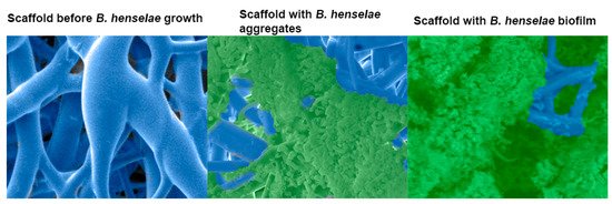
Figure 1. Scanning electron micrograph of a 48 h Bartonella henselae (B. henselae) biofilm growing on a 3-dimensional nanofibrous scaffold. Left, scaffold before bacterial growth. Middle, bacterial growth, adhesion, and aggregation around the scaffold branches. Right, B. henselae biofilm covering the scaffold and eclipsing the bacterial cells. Biofilm was preserved by the addition of the cationic dye, Alcian blue.
Given the history of the Bartonella genus, the increased prevalence of Bartonella in a variety of species, and the poor treatment efficacy of systemic B. henselae infection, it is necessary to determine how B. henselae survives and persists, specifically through the formation of biofilms. Understanding such mechanism(s) will aid in the development of more effective treatments for B. henselae infection.
2. Clinical Importance
Three different Bartonella species—B. bacilliformis, B. quintana, and B. henselae—are the species most commonly associated with acute or chronic infections in humans [26]. In general, the severity of clinical outcomes correlates with the patient’s immune status; therefore, the more severe cases typically occur in immunocompromised individuals [27]. B. henselae is known to show several clinical manifestations, such as cat scratch disease (CSD), a condition characterized by lymphadenopathy and mostly reported in children [28][29], chronic lymphadenopathy [28][30], fever with persistent bacteremia [31], bacillary angiomatosis [32], neurological conditions [33], peliosis hepatitis [34], and life-threatening infective endocarditis, which is usually reported as blood-culture-negative endocarditis [31][35][36].
Fleas are competent vectors for numerous microbial pathogens. Ctenocephalides felis (C. felis), known as cat fleas, are an opportunistic blood feeder and the arthropod vector for B. henselae; however, other vectors such as ticks have been proposed [37][38][39][40][41][42]. Cats are reported to be the predominant host for B. henselae, but B. henselae has been isolated from a variety of hosts [43][44][45][46]. Most cats are asymptomatic when infected, but the exceptions usually develop a fever and local inflammation at the site of inoculum [46][47][48].
Bartonella infections begin with the inoculation of the bacteria, which is usually associated with the feeding of the arthropod vector on an infected cat. Bacterial load from the flea gut is excreted in the flea fecal matter and subsequently onto the cat [49]. Human infections occur through a cat scratch inoculating the host with bacteria from the flea fecal matter.
In the life cycle of B. henselae, biofilms are implicated in both the flea and the mammalian hosts. A previous report from our laboratory showed scanning electron micrographs with bacteria in the gut and bacterial biofilm in the fecal matter of laboratory i-fected cat fleas [19]. The ability of Bartonella to form biofilms in vertebrate hosts has been reported in the literature [50][51][52]. A mouse model of B. tayolrii infection was shown to demonstrate persistent bacteremia, liver lesions, and eventually death [51]. The masses observed in the liver and kidney appeared embedded in an amorphous matrix, which can be defined as a biofilm, providing an experimental model to study human disease progression in an immunocompromised host. In Edouard et al. (2015) [50], a study with 106 patients provided evidence of endocarditis by B. quintana and B. henselae. The above studies provide evidence that Bartonella biofilm communities are indeed an integral part of the vegetative mass associated with infective endocarditis [50][51][52]. B. henselae is a fastidious bacterium with particular nutritional requirements; hence, it is challenging to isolate and culture the bacterium from clinical samples [53]. Laboratory diagnosis is usually accomplished by one or more of the following diagnostic techniques: PCR, serology, isolation with extended incubation periods, or histopathology [54][55][56][57].
Bartonella species have a noteworthy ability to evade the host immune system and resist antimicrobial agents. While some isolates are susceptible to minocycline and macrolide antibiotics such as erythromycin, clarithromycin, azithromycin, and fluoroquinolone compounds with relatively low minimal inhibitory concentrations (MICs) [58][59][60], clinical experience has shown that Bartonella infection treatment failures are a major concern, as most of these antimicrobial classes exhibit only bacteriostatic properties [60][61][62]. It is expected that growth in biofilms allows Bartonella species to persist in the face of stress including antimicrobial treatment and the host immune response. Multiple studies and guidelines support 2 to 6 weeks of treatment for endocarditis using at least two antibiotics, one of which is an aminoglycoside [58][63][64][65]. Considering the clinical manifestations and antibiotic resistance of B. henselae, understanding the mechanisms by which B. henselae initiates biofilm formation is critical to comprehend how it causes chronic disease in humans.
3. Biofilm Formation, Composition, and Life Cycle
Bacteria grow as free-floating planktonic cells or as coordinated aggregates embedded in a matrix, referred to as a biofilm [66]. Biofilms can be single or multispecies communities, can thrive on most surfaces, and may be surface associated (agar, contact lens) or submerged under a static or shear flow condition (such as those formed in an artificial cardiac valve or indwelling catheter) [67][68]. Because of the diversity of biofilm surfaces, the well-organized structures of the colonies, and a characteristic anti-microbial resistance or tolerance, biofilms have received significant attention and are currently investigated for their role in infectious diseases [20][69]. Adhesion or aggregation is the first step of biofilm formation, virulence, and host cell interactions in most bacteria [70]. Outer membrane adhesins facilitate bacterial adhesion to a biotic or abiotic substrate and self-aggregation. Bacteria first adhere, then aggregate to allow the chemical signaling and quorum sensing communication required for the aggregates to secrete the proteins, polysaccharides, and extracellular DNA (eDNA) required for the assembly of the biofilm [71]. The B. henselae biofilm has been shown to contain proteins, polysaccharides, and eDNA, and both DNase and proteinase K have been shown to result in biofilm destruction [16].
The growth of B. henselae as a biofilm was first reported by Kyme et al. [72] as an auto-aggregative phase variation later linked to the expression of a surface adhesin called BadA. In the Bartonella genus, the roles of BadA and Vomps proteins are well documented for the adhesion steps in B. henselae and B. quintana, respectively. BadA is a trimeric auto transporter (TAA), and the role of TAAs have been significantly documented in other gram-negative bacteria [73][74][75]. A genetic deletion of badA in different strains of B. henselae led to failure to adhere efficiently and form a biofilm [12][16][76].
In humans, it has been speculated that B. henselae infects erythrocytes and may persist in these cells [77]. Evidence also supports persistence in endothelial progenitor cells, which presents the possibility for host immune system evasion [78][79][80]. B. henselae is speculated to form biofilms in the gut of the cat flea, which helps the bacteria persist and replicate in the gut [81]. The bacterial load is excreted in the fecal matter, where it forms a biofilm that protects the bacteria, which persists about 10–12 days in the flea fecal matter before human inoculation through the cat claw [19][49]. We propose that the biofilm represents an additional niche that provides the platform for seeding planktonic cells into the bloodstream, causing host immune reactions, disease conditions, and persistence in the face of antimicrobial treatments.
References
- Costerton, J.W.; Geesey, G.G.; Cheng, K.-J. How Bacteria Stick. Sci. Am. 1978, 238, 86–95.
- Lins, K.D.A.; Drummond, M.R.; Velho, P.E.N.F. Cutaneous manifestations of bartonellosis. An. Bras. de Dermatol. 2019, 94, 594–602.
- Ruiz, J. Bartonella quintana, past, present, and future of the scourge of World War, I. Acta Pathol. Microbiol. Immunol. Scand. 2018, 126, 831–837.
- Gomes, C.; Ruiz, J. Carrion’s disease: The sound of silence. Clin. Microbiol. Rev. 2017, 31, e00056-17.
- Nelson, C.A.; Saha, S.; Mead, P.S. Cat-Scratch Disease in the United States, 2005–2013. Emerg. Infect. Dis. 2016, 22, 1741–1746.
- Fournier, P.-E.; Robson, J.; Zeaiter, Z.; McDougall, R. Improved culture from lymph nodes of patients with cat scratch disease and genotypic characterization of Bartonella henselae isolates in Australia. J. Clin. Microbiol. 2002, 40, 3620–3624.
- Kelly, P.J.; Meads, N.; Theobald, A.; Fournier, P.-E.; Raoult, D. Rickettsia felis, Bartonella henselae, and B. clarridgeiae, New Zealand. Emerg. Infect. Dis. 2004, 10, 967–968.
- Maruyama, S.; Izumikawa, K.; Miyashita, M.; Kabeya, H.; Mikami, T.; Yamanouchi, H.; Sasaki, E.; Yoshida, H.; Izumikawa, K. First isolation of Bartonella henselae type I from a cat-scratch disease patient in Japan and its molecular analysis. Microbiol. Immunol. 2004, 48, 103–109.
- Valentine, K.H.; Harms, C.A.; Cadenas, M.B.; Birkenheuer, A.J.; Marr, H.S.; Braun-McNeill, J.; Maggi, R.G.; Breitschwerdt, E.B. Bartonella DNA in loggerhead sea turtles. Emerg. Infect. Dis. 2007, 13, 949–950.
- Mascarelli, P.E.; Elmore, S.A.; Jenkins, E.J.; Alisauskas, R.T.; Walsh, M.; Breitschwerdt, E.B.; Maggi, R.G. Vector-borne pathogens in arctic foxes, Vulpes lagopus, from Canada. Res. Vet. Sci. 2015, 99, 58–59.
- Mascarelli, P.E.; McQuillan, M.; Harms, C.A.; Harms, R.V.; Breitschwerdt, E.B. Bartonella henselae and B. koehlerae DNA in birds. Emerg. Infect. Dis. 2014, 20, 490–492.
- Okaro, U.; Addisu, A.; Casanas, B.; Anderson, B. Bartonella species, an emerging cause of blood-culture-negative endocarditis. Clin. Microbiol. Rev. 2017, 30, 709–746.
- Stützer, B.; Hartmann, K. Chronic Bartonellosis in cats: What are the potential implications? J. Feline Med. Surg. 2012, 14, 612–621.
- Guru, P.K.; Agarwal, A.; Fritz, A. A miraculous recovery: Bartonella henselae infection following a red ant bite. BMJ Case Rep. 2018, 2018, bcr2017222326.
- Rust, M.K. The Biology and ecology of cat fleas and advancements in their pest management: A review. Insects 2017, 8, 118.
- Okaro, U.; Green, R.; Mohapatra, S.; Anderson, B. The trimeric autotransporter adhesin BadA is required for in vitro biofilm formation by Bartonella henselae. npj Biofilms Microbiomes 2019, 5, 1–9.
- Riess, T.; Andersson, S.G.; Lupas, A.; Schaller, M.; Schäfer, A.; Kyme, P.; Martin, J.; Wälzlein, J.-H.; Ehehalt, U.; Lindroos, H.; et al. Bartonella Adhesin A mediates a proangiogenic host cell response. J. Exp. Med. 2004, 200, 1267–1278.
- Szczesny, P.; Linke, D.; Ursinus, A.; Bär, K.; Schwarz, H.; Riess, T.M.; Kempf, V.A.J.; Lupas, A.N.; Martin, J.; Zeth, K. Structure of the head of the Bartonella Adhesin BadA. PLoS Pathog. 2008, 4, e1000119.
- Okaro, U.; George, S.; Valdes, S.; Macaluso, K.; Anderson, B. A non-coding RNA controls transcription of a gene encoding a DNA binding protein that modulates biofilm development in Bartonella henselae. Microb. Pathog. 2020, 147, 104272.
- Bjarnsholt, T. The role of bacterial biofilms in chronic infections. Acta Pathol. Microbiol. Immunol. Scand. 2013, 121, 1–58.
- Florin, T.A.; Zaoutis, T.E.; Zaoutis, L.B. Beyond cat scratch disease: Widening spectrum of Bartonella henselae infection. Pediatrics 2008, 121, e1413–e1425.
- Zheng, X.; Ma, X.; Li, T.; Shi, W.; Zhang, Y. Effect of different drugs and drug combinations on killing stationary phase and biofilms recovered cells of Bartonella henselae in vitro. BMC Microbiol. 2020, 20, 87–89.
- Angelakis, E.; Raoult, D. Pathogenicity and treatment of Bartonella infections. Int. J. Antimicrob. Agents 2014, 44, 16–25.
- Hall, C.W.; Mah, T.F. Molecular mechanisms of biofilm-based antibiotic resistance and tolerance in pathogenic bacteria. FEMS Microbiol. Rev. 2017, 41, 276–301.
- Vestby, L.K.; Grønseth, T.; Simm, R.; Nesse, L.L. Bacterial biofilm and its role in the pathogenesis of disease. Antibiotics 2020, 9, 59.
- Karem, K.L.; Paddock, C.D.; Regnery, R.L. Bartonella henselae, B. quintana, and B. bacilliformis: Historical pathogens of emerging significance. Microbes Infect. 2000, 2, 1193–1205.
- Resto-Ruiz, S.; Burgess, A.; Anderson, B.E. The role of the host immune response in pathogenesis of Bartonella henselae. DNA Cell Biol. 2003, 22, 431–440.
- Jackson, L.A.; Perkins, B.A.; Wenger, J.D. Cat scratch disease in the United States: An analysis of three national databases. Am. J. Public Health 1993, 83, 1707–1711.
- Debre, R. Cat scratch disease. Mars. Med. 1950, 87, 375–378.
- Klotz, S.A.; Ianas, V.; Elliott, S.P. Cat-scratch disease. Am. Fam. Physician 2011, 83, 152–155.
- Jacomo, V.; Kelly, P.J.; Raoult, D. Natural history of Bartonella infections (an exception to Koch’s postulate). Clin. Diagn. Lab. Immunol. 2002, 9, 8.
- Stoler, M.H.; Bonfiglio, T.A.; Steigbigel, R.T.; Pereira, M. An atypical subcutaneous infection associated with acquired immune deficiency syndrome. Am. J. Clin. Pathol. 1983, 80, 714–718.
- Chomel, B.B.; Kasten, R.W.; Sykes, J.E.; Boulouis, H.-J.; Breitschwerdt, E.B. Clinical impact of persistent Bartonella Bacteremia in humans and animals. Ann. N. Y. Acad. Sci. 2003, 990, 267–278.
- Perkocha, L.A.; Geaghan, S.M.; Yen, T.B.; Nishimura, S.L.; Chan, S.P.; Garcia-Kennedy, R.; Honda, G.M.D.; Stoloff, A.C.M.D.; Klein, M.D.H.Z.; Goldman, M.D.R.L.; et al. Clinical and pathological features of bacillary peliosis hepatis in association with human immunodeficiency virus infection. N. Engl. J. Med. 1990, 323, 1581–1586.
- Hadfield, T.; Warren, R.; Kass, M.; Brun, E.; Levy, C. Endocarditis caused by Rochalimaea henselae. Hum. Pathol. 1993, 24, 1140–1141.
- Holmes, A.H.; Greenough, T.C.; Balady, G.J.; Regnery, R.L.; Anderson, B.E.; O’Keane, J.C.; Fonger, J.D.; McCrone, E.L. Bartonella henselae Endocarditis in an Immunocompetent Adult. Clin. Infect. Dis. 1995, 21, 1004–1007.
- Chung, C.Y.; Kasten, R.W.; Paff, S.M.; Van Horn, B.A.; Vayssier-Taussat, M.; Boulouis, H.-J.; Chomel, B.B. Bartonella spp. DNA associated with biting flies from California. Emerg. Infect. Dis. 2004, 10, 1311–1313.
- Sanogo, Y.O.; Zeaiter, Z.; Caruso, G.; Merola, F.; Shpynov, S.; Brouqui, P.; Raoult, D. Bartonella henselae in Ixodes ricinus ticks (Acari: Ixodida) removed from humans, Belluno province, Italy. Emerg. Infect. Dis. 2003, 9, 329–332.
- Chomel, B.B.; Kasten, R.W.; Floyd-Hawkins, K.; Chi, B.; Yamamoto, K.; Roberts-Wilson, J.; Koehler, J.E.; Pedersen, N.C.; Abbott, R.C. Experimental transmission of Bartonella henselae by the cat flea. J. Clin. Microbiol. 1996, 34, 1952–1956.
- Zając, V.; Wójcik-Fatla, A.; Dutkiewicz, J.; Szymańska, J. Bartonella henselae in eastern Poland: The relationship between tick infection rates and the serological response of individuals occupationally exposed to tick bites. J. Vector Ecol. 2015, 40, 75–82.
- Cotté, V.; Bonnet, S.; Le Rhun, D.; Le Naour, E.; Chauvin, A.; Boulouis, H.J.; Lecuelle, B.; Lilin, T.; Vayssier-Taussat, M. Transmission of Bartonella henselae by Ixodes ricinus. Emerg. Infect. Dis. 2008, 14, 1074.
- Angelakis, E.; Billeter, S.A.; Breitschwerdt, E.B.; Chomel, B.B.; Raoult, D. Potential for Tick-borne Bartonelloses. Emerg. Infect. Dis. 2010, 16, 385–391.
- McElroy, K.M.; Blagburn, B.L.; Breitschwerdt, E.B.; Mead, P.S.; McQuiston, J.H. Flea-associated zoonotic diseases of cats in the USA: Bartonellosis, flea-borne rickettsioses, and plague. Trends Parasitol. 2010, 26, 197–204.
- Hutchinson, M.J.; Jacobs, D.E.; Mencke, N. Establishment of the cat flea (Ctenocephalides felis felis) on the ferret (Mustela putorius furo) and its control with imidacloprid. Med. Vet. Èntomol. 2001, 15, 212–214.
- Araújo, F.R.; Silva, M.P.A.; Lopes, A.; Ribeiro, O.C.; Pires, P.P.; Carvalho, C.; Balbuena, C.B.A.; Villas, A.; Ramos, J. Severe cat flea infestation of dairy calves in Brazil. Vet. Parasitol. 1998, 80, 83–86.
- Koehler, J.E.; Glaser, C.A.; Tappero, J.W. Rochalimaea henselae infection. A new zoonosis with the domestic cat as reservoir. J. Am. Med. Assoc. 1994, 271, 531–535.
- Guptill, L.; Slater, L.; Wu, C.; Lin, T.; Glickman, L.T.; Welch, D.F.; HogenEsch, H. Experimental infection of young specific pathogen-free cats with Bartonella henselae. J. Infect. Dis. 1997, 176, 206–216.
- O’Reilly, K.L.; Bauer, R.W.; Freeland, R.L.; Foil, L.D.; Hughes, K.J.; Rohde, K.R.; Roy, A.F.; Stout, R.W.; Triche, P.C. Acute clinical disease in cats following infection with a pathogenic strain of Bartonella henselae (LSU16). Infect. Immun. 1999, 67, 3066–3072.
- Bouhsira, E.; Franc, M.; Boulouis, H.-J.; Jacquiet, P.; Raymond-Letron, I.; Liénard, E. Assessment of persistence of Bartonella henselae in Ctenocephalides felis. Appl. Environ. Microbiol. 2013, 79, 7439–7444.
- Edouard, S.; Nabet, C.; Lepidi, H.; Fournier, P.E.; Raoult, D. Bartonella, a common cause of endocarditis: A report on 106 cases and review. J. Clin. Microbiol. 2015, 53, 824–829.
- Chiaraviglio, L.; Duong, S.; Brown, D.A.; Birtles, R.J.; Kirby, J.E. An immunocompromised murine model of chronic Bartonella infection. Am. J. Pathol. 2010, 176, 2753–2763.
- Spach, D.H.; Callis, K.P.; Paauw, D.S.; Houze, Y.B.; Schoenknecht, F.D.; Welch, D.F.; Rosen, H.; Brenner, D.J. Endocarditis caused by Rochalimaea quintana in a patient infected with human immunodeficiency virus. J. Clin. Microbiol. 1993, 31, 692–694.
- Minnick, M.F.; Anderson, B.E. Bartonella. In Molecular Medical Microbiology, 2nd ed.; Tang, Y.-W., Sails, A., Eds.; Academic Press: Boston, MA, USA, 2015; pp. 1911–1939.
- Koehler, J.E.; Quinn, F.D.; Berger, T.G.; LeBoit, P.E.; Tappero, J.W. Isolation of rochalimaea species from cutaneous and osseous lesions of bacillary angiomatosis. N. Engl. J. Med. 1992, 327, 1625–1631.
- Houpikian, P.; Raoult, D. Blood culture-negative endocarditis in a reference center: Etiologic diagnosis of 348 cases. Medicine 2005, 84, 162–173.
- Caponetti, G.C.; Pantanowitz, L.; Marconi, S.; Havens, J.M.; Lamps, L.W.; Otis, C.N. Evaluation of immunohistochemistry in identifying bartonella henselae in cat-scratch disease. Am. J. Clin. Pathol. 2009, 131, 250–256.
- Relman, D.A.; Loutit, J.S.; Schmidt, T.M.; Falkow, S.; Tompkins, L.S. The agent of bacillary angiomatosis. An approach to the identification of uncultured pathogens. N. Engl. J. Med. 1990, 323, 1573–1580.
- Tsuneoka, H.; Yanagihara, M.; Nojima, J.; Ichihara, K. Antimicrobial susceptibility by Etest of Bartonella henselae isolated from cats and human in Japan. J. Infect. Chemother. 2010, 16, 446–448.
- Biswas, S.; Maggi, R.G.; Papich, M.G.; Keil, D.; Breitschwerdt, E.B. Comparative activity of Pradofloxacin, Enrofloxacin, and Azithromycin against Bartonella henselae isolates collected from cats and a human. J. Clin. Microbiol. 2009, 48, 617–618.
- Rolain, J.M.; Brouqui, P.; Koehler, J.E.; Maguina, C.; Dolan, M.J.; Raoult, D. Recommendations for treatment of human infections caused by Bartonella species. Antimicrob. Agents Chemother. 2004, 48, 1921–1933.
- Myers, W.F.; Grossman, D.M.; Wisseman, C.L. Antibiotic susceptibility patterns in Rochalimaea quintana, the agent of trench fever. Antimicrob. Agents Chemother. 1984, 25, 690–693.
- Ives, T.J.; Manzewitsch, P.; Regnery, R.L.; Butts, J.D.; Kebede, M. In vitro susceptibilities of Bartonella henselae, B. quintana, B. elizabethae, Rickettsia rickettsii, R. conorii, R. akari, and R. prowazekii to macrolide antibiotics as determined by immunofluorescent-antibody analysis of infected Vero cell monolayers. Antimicrob. Agents Chemother. 1997, 41, 578–582.
- Raoult, D.; Fournier, P.-E.; Vandenesch, F.; Mainardi, J.-L.; Eykyn, S.J.; Nash, J.; James, E.; Benoit-Lemercier, C.; Marrie, T.J. Outcome and treatment of Bartonella Endocarditis. Arch. Intern. Med. 2003, 163, 226–230.
- Elliott, T.S.J.; Foweraker, J.; Gould, F.K.; Perry, J.D.; Sandoe, J.A.T. Guidelines for the antibiotic treatment of endocarditis in adults: Report of the Working Party of the British Society for Antimicrobial Chemotherapy. J. Antimicrob. Chemother. 2004, 54, 971–981.
- Barka, N.E.; Hadfield, T.; Patnaik, M.; Schwartzman, W.A.; Peter, J.B. EIA for detection of Rochalimaea henselae-Reactive IgG, IgM, and IgA antibodies in patients with suspected cat-scratch disease. J. Infect. Dis. 1993, 167, 1503–1504.
- Flemming, H.C.; Wingender, J. The biofilm matrix. Nat. Rev. Microbiol. 2010, 8, 623–633.
- Chabane, Y.N.; Marti, S.; Rihouey, C.; Alexandre, S.; Hardouin, J.; Lesouhaitier, O.; Vila, J.; Kaplan, J.B.; Jouenne, T.; Dé, E. Characterisation of pellicles formed by acinetobacter baumannii at the air-liquid interface. PLoS ONE 2014, 9, e111660.
- Paytubi, S.; Cansado, C.; Madrid, C.; Balsalobre, C. Nutrient composition promotes switching between pellicle and bottom biofilm in salmonella. Front. Microbiol. 2017, 8, 2160.
- Barsoumian, A.E.; Mende, K.; Sanchez, C.J., Jr.; Beckius, M.L.; Wenke, J.C.; Murray, C.K.; Akers, K.S. Clinical infectious outcomes associated with biofilm-related bacterial infections: A retrospective chart review. BMC Infect. Dis. 2015, 15, 1–7.
- Berne, C.; Ducret, A.; Hardy, G.G.; Brun, Y.V. Adhesins involved in attachment to abiotic surfaces by gram-negative bacteria. Microbiol. Spectr. 2015, 3, 101–128.
- Dunne, W.M. Bacterial adhesion: Seen any good biofilms lately? Clin. Microbiol. Rev. 2002, 15, 155–166.
- Kyme, P.; Dillon, B.; Iredell, J. Phase variation in Bartonella henselae. Microbiology 2003, 149, 621–629.
- Zimmerman, S.M.; Michel, F.; Hogan, R.J.; Lafontaine, E.R. The autotransporter BpaB contributes to the virulence of burkholderia mallei in an aerosol model of infection. PLoS ONE 2015, 10, e0126437.
- Totsika, M.; Wells, T.J.; Beloin, C.; Valle, J.; Allsopp, L.P.; King, N.P.; Ghigo, J.-M.; Schembri, M.A. Molecular characterization of the EhaG and UpaG trimeric autotransporter proteins from pathogenic escherichia coli. Appl. Environ. Microbiol. 2012, 78, 2179–2189.
- Bentancor, L.V.; Camacho-Peiro, A.; Bozkurt-Guzel, C.; Pier, G.B.; Maira-Litrán, T. Identification of ata, a multifunctional trimeric autotransporter of acinetobacter baumannii. J. Bacteriol. 2012, 194, 3950–3960.
- Tu, N.; Caroll, R.K.; Weiss, A.; Shaw, L.; Nicolas, G.; Thomas, S.; Lima, A.; Okaro, U.; Anderson, B. A family of genus-specific RNAs in tandem with DNA-binding proteins control expression of the badA major virulence factor gene in Bartonella henselae. Microbiologyopen 2016, 6, e00420.
- Dehio, C. Bartonella–host-cell interactions and vascular tumour formation. Nat. Rev. Genet. 2005, 3, 621–631.
- Salvatore, P. Detrimental effects of Bartonella henselae are counteracted by L-arginine and nitric oxide in human endothelial progenitor cells. Proc. Natl. Acad. Sci. USA 2008, 105, 9427.
- Mändle, T.; Einsele, H.; Schaller, M.; Neumann, D.; Vogel, W.; Autenrieth, I.B.; Kempf, V.A.J. Infection of human CD34+ progenitor cells with Bartonella henselae results in intraerythrocytic presence of B henselae. Blood 2005, 106, 1215–1222.
- Salvatore, P.; Zullo, A.; Sommese, L.; Colicchio, R.; Picascia, A.; Schiano, C.; Mancini, F.P.; Napoli, C. Infections and cardiovascular disease: Is Bartonella henselae contributing to this matter? J. Med. Microbiol. 2015, 64, 799–809.
- Finkelstein, J.L.; Brown, T.P.; O’reilly, K.L.; Wedincamp, J., Jr.; Foil, L.D. Studies on the growth of Bartonella henselae in the cat flea (Siphonaptera: Pulicidae). J. Med. Entomol. 2002, 39, 915–919.

