 +1 credit
| Version | Summary | Created by | Modification | Content Size | Created at | Operation |
|---|---|---|---|---|---|---|
| 1 | Bernhard Schuster | + 2139 word(s) | 2139 | 2021-04-25 06:22:31 | | | |
| 2 | Camila Xu | Meta information modification | 2139 | 2021-04-28 04:48:41 | | |
Video Upload Options
Surface layers (S-layers) are the most common outermost cell envelope components of prokaryotic organisms (bacteria and archaea). The lattice formed by S-layer proteins are highly porous structures with identical pores in the nm-range. This feature can be utilized to fabricate ultrafiltration membranes with a very sharp specific molecular weight cut off. Moreover, S-layer lattices reveal an intrinsic antifouling characteristics, which results in a negligible clogging of the filter.
1. Introduction
Ultrafiltration (UF) is a membrane-based filtration, in which pressure or concentration gradients induce a separation through a semipermeable membrane. UF membranes processes are used in industry and research for purifying and concentrating suspended solids, macromolecules and colloidal particles, particularly protein solutions of 2 to 100 nm in diameter corresponding to a molecular weight of 103 to 106 Dalton (Da) [1][2][3]. UF membranes with a pore size ranging from 2 to 100 nm are defined by the specific molecular weight cut off (MWCO). The latter refers to the lowest molecular weight solute (in Da) in which 90% of the solute is retained by the membrane.
Beside the implementation of UF in research, UF membranes are an established separation technology utilized in many industrial process applications worldwide. UF membranes have the unique ability to purify, concentrate, and fractionate of a large range of macromolecules and proteins (e.g., milk proteins and enzymes, silt, plastics, oil, silica, nanoparticles, endotoxins, and viruses) via a physical membrane barrier determined by the MWCO. Thereby the UF membranes ensure a very consistent rejection and flux performance [2]. Industrial fields of application range from dairy, biotechnology and pharmaceutical use over food, beverage, and plant extracts to wastewater treatment [1][2][3].
Most UF membranes comprise of polymer materials like polysulfone, polypropylene, cellulose acetate, and polylactic acid [1][3] and have a surface porosity which is usually lower than 10%. They also show a size distribution of the pores varying by up to an order of magnitude [4]. Due to the presence of differently sized pores, the flux is strongly biased to the larger ones, with typically 50% of the solvent passing through 20 to 25% of the pores [4]. This leads to a heterogeneous flow pattern normal to the membrane surface. The heterogeneous porosity of UF membranes makes them susceptible to flux decline by loss of the larger pores [2][4] and thus, not well suited for the fractionation of close-sized macro-solutions.
In the present entry we will introduce the highly porous crystalline monomolecular protein lattices, which represent the outermost surface layer (S-layer) in many bacteria and almost all archaea (Figure 1) as the MWCO-determining functional layer [5][6][7][8][9][10][11][12][13]. The S-layer ultrafiltration membrane (SUM) comprises of a microfiltration membrane onto which S-layer-carrying cell wall fragments or S-layer protein (SLP) self-assembly products are deposited in a pressure-dependent manner [6][8][9][14][15][16][17][18][19]. Whereas the active ultrafiltration layers of most synthetic membranes show a porosity of up to 10%, the crystalline S-layers reveal a porosity of up to 70% (Figure 2). The preformed SUM can, if desired, be chemically crosslinked to enhance the mechanical and thermal robustness. In the following we will report on the intrinsic features of bacterial SLPs, their molecular sieve property and how SUM are manufactured and modified to show optimal performance in filtration processes. Moreover, SUMs may also be utilized as binding matrices for the formation of complex composite structures for various biotechnological and pharmaceutical applications.
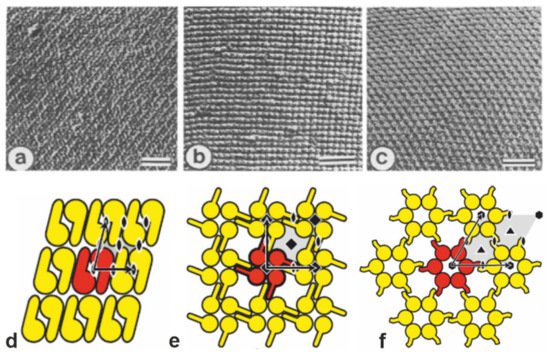
Figure 1. (a–c) Transmission electron micrographs of freeze-etched preparations of S-layer on intact cells showing three lattice types schematically determined in (d–f). (a,d) oblique (p2) lattice; (b,e) square (p4) lattice; (c,f) hexagonal (p6) lattice. The bars in a-c represent 50 nm. In d-f, the different S-layer lattice types, their base vectors, the unit cell (shaded in gray), and the corresponding symmetry axis are depicted. The proteins at one morphological unit are shown in red. (a–c: Adapted from [11]. Copyright © 1988 with permission from Elsevier Science Publishers B.V. Amsterdam, The Netherlands. d–f: With permission from Ref. [6] (CC BY-NC-ND 3.0)).

Figure 2. Schematic drawing of synthetic and S-layer ultrafiltration membrane. Left: The active ultrafiltration layers of most synthetic membranes show a porosity of up to 10%. Right: Crystalline S-layers reveal a porosity of up to approx. 70%. Although the active ultrafiltration layer is usually composed of several closed associated monolayers, the rejection characteristics is exclusively determined by the sieving properties of the individual S-layer protein monolayers. (Adapted from [8]. Copyright © 1986 with permission from Springer-Verlag).
2. Ultrastructure and Self-Assembly of S-Layer Proteins
Monomolecular arrays of protein or glycoprotein subunits forming S-layers are one of the most observed prokaryotic cell envelope components [6]. The isoporous lattices covering the entire cell surface provide organisms with various selection advantages including functioning as protective coats, molecular sieves, and ion traps, as structures involved in surface recognition and cell adhesion, and as antifouling layers [6]. S-layers completely cover the cell surface during all stages of cell growth and division [20][21][22][23][24][25][26]. The location and ultrastructure of S-layers of a great number of Bacteria and Archaea have initially been studied by transmission electron microscopy (TEM) (Figure 1a–c) [21][27][28][29][30][31][32][33] and more recently by atomic force microscopy (AFM) [34][35][36][37][38][39]. It turned out that S-layers exhibit either oblique, square, or hexagonal lattice symmetry, where the number of SLPs per morphological unit is either one or two (p1, p2; oblique), four (p4; square), or three or six (p3, p6; hexagonal) (Figure 1d,e). The unit cell dimension ranges from 3 to 30 nm for bacterial and archaeal S-layer lattices. The morphology of the S-layer lattice is asymmetric with commonly a smooth outer face and a more corrugated inner face (with respect to their orientation at the cell). Finally, bacterial S-layer lattices are highly porous protein mesh works (30 to 70% porosity) with pores of uniform morphology and size (between 2 to 8 nm) and a thickness between 5 and 20 nm [6][20][40][41][42][43][44].
Generally, S-layers are isolated from cell wall fragments, which were obtained by breaking up the cells and removing their content, including the cytoplasmic membrane. Most often, hydrogen-bond breaking agents like guanidine hydrochloride or urea are used to disintegrate and solubilize the SLPs. For a detailed compilation of protocols, see reference [45].
The capability of isolated SLPs to assemble into two-dimensional arrays in vivo and in vitro is one of their key properties exploited in basic and application-oriented research. It occurs upon dialysis of the disrupting agents [6][45][46][47]. The formation of the monomolecular self-assembled protein lattices is only determined by the amino acid sequence of the polypeptide chains, and consequently the tertiary structure of the SLP species [48].
The assembly process in solution shows multiphasic kinetics with a rapid initial and a slow consecutive phase [49]. The rapid phase may be attributed to the formation of oligomeric precursors. The latter fuse in the consecutive phase and reassemble into the finally formed S-layer self-assembly products [6][46][47][49][50][51]. SLPs are non-covalently linked to each other and, in the case of their adhesion to supporting structures (e.g., microfiltration membrane or polymeric solid surfaces) combinations of weak bonds (hydrophobic bonds, ionic bonds involving divalent cations or direct interaction of polar groups, and hydrogen bonds) are responsible for the structural integrity as well [6]. Once formed, SLPs were never observed to leave the lattice, and thus, it was concluded that lattice growth is irreversible. The reason for the latter may be that after the addition of the “last” protein monomer to the (incomplete) morphological unit, this monomer is locked into place and now has a low probability of leaving [34][52].
3. S-Layers as Molecular Sieves
To determine the size of pores in S-layer lattices of different Bacillaceae, permeability studies were performed. Native and glutaraldehyde-treated S-layer containers were prepared that resembled the shape of bacterial cell envelopes [53]. The solutions selected for the molecular sieving measurements were sugars, proteins, and dextrans of increasing molecular weights. It was clearly demonstrated that the S-layer lattices are isoporous molecular sieves that allow free passage for molecules with a molecular weight of up to 30.000 Da and showed sharp exclusion limits between molecular weights of 30.000 and 45.000 Da [8][11][53]. This finding suggests a limiting pore diameter in the range of 3–4.5 nm, which resembles the pore dimensions determined by high-resolution TEM and AFM [27][54]. This information on the structure and function of different S-layers of Bacillaceae makes it unlikely that their S-layers have the potential to function as an effective barrier against lysogenic enzymes [53]. Most important, a great variety of permeability studies on S-layers from Bacillaceae demonstrated that the surface and pore areas of the protein meshwork have a very low tendency for unspecific adsorption of (macro)molecules [10][12][18][55][56][57][58].
4. S-Layer Ultrafiltration Membrane (SUM)
SUMs are made by depositing either sheet like S-layer self-assembly products (see Section 2) or S-layer carrying cell wall fragments (approx. 0.5 to 1.5 µm size) on commercial microfiltration membranes with a spongy structure or radiation-track type membranes having pore sizes of 0.05 to 0.1 µm in a pressure dependent procedure (Figure 3) [10][11][59][60]. S-layer carrying cell wall fragments were prepared from whole cells by extracting the cytoplasmic membrane with Triton X-100 under conditions, which preserved the integrity of the S layer and the peptidoglycan-containing layer [53]. Like tiling a roof, this procedure generates a coherent coating, composed of multilayers of S-layer fragments arranged in random orientation. Interestingly, the pores of the constituent (monomolecular) S-layer lattice solely determined the MWCO of these composite multilayered structures.
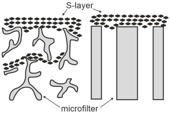
Figure 3. Schematic drawing of S-layer ultrafiltration membranes. Left: S-layer fragments are deposited on the surface and in the substructure of open-celled foam-like microfiltration membranes. Right: S-layer fragments are attached to the surface of radiation-track membranes. (Adapted from [8]. Copyright © 1986 with permission from Springer-Verlag).
After deposition, the SLP lattices are inter- and intramolecular crosslinked with glutaraldehyde and Schiff bases are reduced with sodium borohydride [10]. The nominal MWCO of SUMs from S-layers of mesophilic and thermophilic Bacillaceae revealed no significant difference and was in the range of 30.000 to 40.000 Da (Figure 4) [10][12][53]. As estimated by TEM and AFM, the porosity of the SLP lattice lies between ca. 30 and 70% and is therefore significantly higher than that determined for polymeric UF membranes with a maximum of 10%.

Figure 4. Rejection curve of S-layer ultrafiltration membranes. The isoporous S-layer fragments attached to a supporting microfiltration membrane form the active ultrafiltration layer responsible for the sieving properties. Note the steep increase between molecular weights 30.000 and 45.000 Da for S-layer fragments of Geobacillus stearothermophilus. (Adapted from [8]. Copyright © 1986 with permission from Springer-Verlag).
Native S-layers were found to be zwitterionic, thus preventing nonspecific adsorption of charged macromolecules, which would lead to pore blocking (fouling) [61]. After crosslinking the S-layer protein lattice with glutaraldehyde, which involves loss of positively-charged amino groups, SUMs revealed a net negative charge on the surface and inside the pores [10][17][19][62]. SUMs possessing a high density of free carboxylic acid groups did not adsorb negatively charged macromolecules but strongly interacted with proteins exhibiting a positive net charge [14][41][56]. In ultrafiltration processes, protein adsorption, which is considered as the first step in membrane fouling is expressed in terms of flux losses for particle free water after protein filtration [63][64]. By using SUMs composed of two-dimensional crystalline protein lattices with defined pore sizes and defined surface charge, it was possible for the first time to determine correlations between the pore size and the net charge of the active filtration layer, the molecular characteristics (dimensions, net charge) of adsorbed protein molecules and the flux losses caused by adsorption [19][62][65]. The flux of SUMs produced with S-layers from Geobacillus strains ranged from 150 to 250 L m−2 h−1 when measured at 0.2 MPa with water [12]. Although the initial flux of SUMS correlated with the number of S-layer fragments deposited on the microfiltration membrane, the dimension of the active filtration layer did not influence the MWCO [10][59][60]. As S-layers are periodic structures, they exhibit repetitive identical physicochemical properties down to the sub-nanometer scale not only on the surface but also in the pore area. Thus, surface properties and molecular sieving as well as antifouling characteristics of SUMs could be tuned by chemical modifications involving activation of carboxylic acid groups with carbodiimides and subsequently converting them with differently sized and charged nucleophiles [41][42][62][66]. Thereby, SUMs can be manufactured with different net charge, hydrophilic or hydrophobic surface properties and separation characteristics [62]. A correlation was observed between the molecular size of attached nucleophiles and the shift of the sharp rejection curve to the lower molecular weight range [17][62]. This confirmed that a pore size reduction had occurred due to an aperture like enlargement of modified carboxylic acid groups exposed in precise position and orientation on protein domains inside the pores. It was also demonstrated that both, the net charge of the S-layer lattice and that of the protein molecules used in filtration experiments determine the solute rejection characteristics of SUMs [42][62][67]. Thus, reams of chemical and/or genetically induced modifications allow the adaptation of SUMs to very specific process requirements. In comparison with conventional ultrafiltration membranes produced by amorphous polymers, SUMs exhibited an extremely low unspecific protein adsorption (membrane fouling) in buffer solutions. Concerning the chemical stability, SUMs composed of inter- and intramolecular crosslinked S-layer lattices were resistant towards many organic solvents (ketones, alcohols, chlorinated hydrocarbons, aromatic compounds), chaotropic agents, acidic and alkaline pH conditions (pH 1–13 at 20 °C for 160 h), and shear forces. These studies demonstrated that SUMs are equivalent in their chemical resistance to polyamide membranes [10][14][18][66][68].
References
- Pavanasam, A.; Abbas, A. Ultrafiltration and Virus Removal: A Mini Review of Recent Patents. Recent Pat. Chem. Eng. 2008, 1, 151–156.
- Pabby, A.K.; Rizvi, S.S.; Requena, A.M. (Eds.) Handbook of Membrane Separations: Chemical, Pharmaceutical, Food, and Biotechnological Applications, 2nd ed.; CRC Press: Boca Raton, FL, USA, 2015.
- Strathmann, H. Membrane separation processes. J. Membr. Sci. 1981, 9, 121–189.
- Fane, A.; Fell, C.; Waters, A. The relationship between membrane surface pore characteristics and flux for ultrafiltration membranes. J. Membr. Sci. 1981, 9, 245–262.
- Crowther, R.; Sleytr, U.B. An analysis of the fine structure of the surface layers from two strains of Clostridia, including correction for distorted images. J. Ultrastruct. Res. 1977, 58, 41–49.
- Sleytr, U.B.; Schuster, B.; Egelseer, E.M.; Pum, D. S-layers: Principles and applications. FEMS Microbiol. Rev. 2014, 38, 823–864.
- Sára, M.; Sleytr, U.B. Verwendung isoporer, kristalliner Bakterienzellwandschichten als Ultrafiltrationsmembranen. Lebensmittel Biotechnol. 1985, 4, 141–146.
- Sleytr, U.B.; Sára, M. Ultrafiltration membranes with uniform pores from crystalline bacterial cell envelope layers. Appl. Microbiol. Biotechnol. 1986, 25, 83–90.
- Sleytr, U.B.; Sára, M. Herstellung isoporer Ultrafiltrationsmembranen aus kristallinen Bakterienzellwandschichten, In Tech-nische Membranen in der Biotechnologie; Kula, M.-R., Schügerl, K., Wandrey, C., Eds.; GBF Monographien, Bd. 9; Verlag Chemie: Weinheim, Germany, 1986; pp. 225–229.
- Sára, M.; Sleytr, U.B. Production and characteristics of ultrafiltration membranes with uniform pores from two-dimensional arrays of proteins. J. Membr. Sci. 1987, 33, 27–49.
- Sára, M.; Manigley, C.; Wolf, G.; Sleytr, U.B. Isoporous ultrafiltration membranes from bacterial cell envelope layers. J. Membr. Sci. 1988, 36, 179–186.
- Sára, M.; Sleytr, U.B. Membrane Biotechnology: Two-dimensional Protein Crystals for Ultrafiltration Purposes. In Biotechnology; Rehm, H.J., Ed.; VCH: Weinheim, Germany, 1988; Volume 6b, pp. 615–636.
- Sleytr, U.B.; Sára, M.; Wolf, G.; Manigley, C. Isoporous ultrafiltration membranes from crystalline cell envelope layers. In The Influence of New Technology on Medical Practice; Paul, J.P., McCruden, A.B., Schuetz, P.W., Eds.; Macmillan Press: Strathclyde, UK, 1988; pp. 83–87.
- Manigley, C.; Wolf, G.; Sára, M.; Sleytr, U.B. Comparative studies on synthetic and S layer ultrafiltration membranes. In Crystalline Bacterial Cell Surface Layers; Sleytr, U.B., Messner, P., Pum, D., Sára, M., Eds.; Springer Verlag: Berlin, Germany, 1988; pp. 154–159.
- Sára, M.; Wolf, G.; Küpcü, S.; Pum, D.; Sleytr, U.B. Use of crystalline bacterial cell envelope layers as ultrafiltration membranes and supports for the immobilization of macromolecules. In Dechema Biotechnology Conferences; VCH: Weinheim, Germany, 1988; Volume 2, pp. 35–51.
- Sára, M.; Küpcü, S.; Sleytr, U.B. Crystalline bacterial cell surface layers used as ultrafiltration membranes and immobilization matrix. Gen. Eng. Biotechnol. 1990, 10, 10–13.
- Küpcü, S.; Sára, M.; Sleytr, U.B. Chemical modification of crystalline ultrafiltration membranes and immobilization of macromolecules. J. Membr. Sci. 1991, 61, 167–175.
- Sára, M.; Küpcü, S.; Weiner, C.; Weigert, S.; Sleytr, U.B. Crystalline protein layers as isoporous molecular sieves and immobi-lisation and affinity matrices. In Immobilised Macromolecules: Application Potentials; Sleytr, U.B., Messner, P., Pum, D., Sára, M., Eds.; Springer: London, UK, 1993; pp. 71–86.
- Weigert, S.; Sára, M. Surface modification of an ultrafiltration membrane with crystalline structure and studies on interactions with selected protein molecules. J. Membr. Sci. 1995, 106, 147–159.
- Sleytr, U.B.; Messner, P.; Pum, D.; Sára, M. Crystalline Bacterial Cell Surface Layers (S Layers): From Supramolecular Cell Structure to Biomimetics and Nanotechnology. Angew. Chem. Int. Ed. 1999, 38, 1034–1054.
- Sleytr, U.B.; Glauert, A.M. Analysis of regular arrays of subunits on bacterial surfaces: Evidence for a dynamic process of as-sembly. J. Ultrastruct. Res. 1975, 50, 103–116.
- Sleytr, U.B. Regular Arrays of Macromolecules on Bacterial Cell Walls: Structure, Chemistry, Assembly, and Function. Adv. Clin. Chem. 1978, 53, 1–64.
- Sleytr, U.B.; Messner, P. Self-assembly of crystalline bacterial cell surface layers (S-layers). In Electron Microscopy of Subcellular Dynamics; Plattner, H., Ed.; CRC Press: Boca Raton, FL, USA, 1989; pp. 13–31.
- Messner, P.; Sleytr, U.B. The use of freeze-etching and freeze-drying to evaluate crystalline cell surface layers (S-layers). In Microbial Cell Surface Analysis Structural and Physicochemical Methods; Mozes, N., Handley, P.S., Busscher, H.J., Rouxhet, P.G., Eds.; VCH Publishers: New York, NY, USA, 1991; pp. 109–125.
- Pum, D.; Messner, P.; Sleytr, U.B. Role of the S layer in morphogenesis and cell division of the archaebacterium Methanocorpusculum sinense. J. Bacteriol. 1991, 173, 6865–6873.
- Rachel, R. Cell envelopes of crenarchaeota and nanoarchaeota. In Prokaryotic Cell Wall Compounds—Structure and Biochemistry; König, H., Claus, H., Varma, A., Eds.; Springer: Heidelberg, Germany, 2010; Volume 9, pp. 271–291.
- Pavkov-Keller, T.; Howorka, S.; Keller, W. The Structure of Bacterial S-Layer Proteins. In Progress in Molecular Biology and Translational Science; Horworka, S., Ed.; Elsevier BV, Academic Press: Burlington, VT, USA, 2011; Volume 103, pp. 73–130.
- Thornley, M.J.; Glauert, A.M.; Sleytr, U.B. Structure and assembly of bacterial surface layers composed of regular arrays of subunits. Philos. Trans. R. Soc. B Biol. Sci. 1974, 268, 147–153.
- Sleytr, U.B.; Messner, P. Crystalline Surface Layers on Bacteria. Annu. Rev. Microbiol. 1983, 37, 311–339.
- Baumeister, W.; Engelhardt, H. Three-Dimensional Structure of Bacterial Surface Layers; Harris, J.R., Horne, R.W., Eds.; Academic Press, Inc.: London, UK, 1987; Volume 6, pp. 109–154.
- Sleytr, U.B.; Messner, P.; Pum, D. 2 Analysis of Crystalline Bacterial Surface Layers by Freeze-etching, Metal Shadowing, Negative Staining and Ultrathin Sectioning. Methods Microbiol. 1988, 20, 29–60.
- Beveridge, T.J. Bacterial S-layers. Curr. Opin. Struct. Biol. 1994, 4, 204–212.
- Sleytr, U.B.; Messner, P.; Pum, D.; Sára, M. Crystalline surface layers on eubacteria and archaeobacteria. In Crystalline Bacterial Cell Surface Proteins; Sleytr, U.B., Messner, P., Pum, D., Sára, M., Eds.; R.G. Landes Company and Academic Press, Inc.: Austin, TX, USA, 1996; pp. 211–225.
- Chung, S.; Shin, S.-H.; Bertozzi, C.R.; De Yoreo, J.J. Self-catalyzed growth of S layers via an amorphous-to-crystalline transition limited by folding kinetics. Proc. Natl. Acad. Sci. USA 2010, 107, 16536–16541.
- Müller, D.J.; Baumeister, W.; Engel, A. Conformational change of the hexagonally packed intermediate layer of Deinococcus radiodurans monitored by atomic force microscopy. J. Bacteriol. 1996, 178, 3025–3030.
- Müller, D.J.; Baumeister, W.; Engel, A. Controlled unzipping of a bacterial surface layer with atomic force microscopy. Proc. Natl. Acad. Sci. USA 1999, 96, 13170–13174.
- Ebner, A.; Kienberger, F.; Huber, C.; Kamruzzahan, A.S.M.; Pastushenko, V.P.; Tang, J.; Kada, G.; Gruber, H.J.; Sleytr, U.B.; Sára, M.; et al. Atomic-Force-Microscopy Imaging and Molecular-Recognition-Force Microscopy of Recrystallized Heterotetramers Comprising an S-Layer-Streptavidin Fusion Protein. ChemBioChem 2006, 7, 588–591.
- Moreno-Flores, S.; Kasry, A.; Butt, H.-J.; Vavilala, C.; Schmittel, M.; Pum, D.; Sleytr, U.B.; Toca-Herrera, J.L. From Native to Non-Native Two-Dimensional Protein Lattices through Underlying Hydrophilic/Hydrophobic Nanoprotrusions. Angew. Chem. Int. Ed. 2008, 47, 4707–4710.
- Lopez, A.E.; Pum, D.; Sleytr, U.B.; Toca-Herrera, J.L. Influence of surface chemistry and protein concentration on the adsorption rate and S-layer crystal formation. Phys. Chem. Chem. Phys. 2011, 13, 11905–11913.
- Sleytr, U.B. Basic and applied S-layer research: An overview. FEMS Microbiol. Rev. 1997, 20, 5–12.
- Sleytr, U.B.; Sára, M.; Pum, D.; Schuster, B. Characterization and use of crystalline bacterial cell surface layers. Prog. Surf. Sci. 2001, 68, 231–278.
- Sleytr, U.B.; Sára, M.; Pum, D.; Schuster, B. Molecular nanotechnology and nanobiotechnology with two-dimensional protein crystals (S-layers). In Nano-Surface Chemistry; Rosoff, M., Ed.; Marcel Dekker: New York, NY, USA; Basel, Switzerland, 2001; pp. 333–389.
- Sleytr, U.B.; Sára, M.; Pum, D.; Schuster, B.; Messner, P.; Schäffer, C. Self Assembly Protein Systems: Microbial S-Layers. In Biopolymers; Steinbüchel, A., Fahnenstock, S., Eds.; Wiley-VCH: Weinheim, Germany, 2002; Volume 7, pp. 285–338.
- Sleytr, U.B.; Sára, M.; Pum, D.; Schuster, B. Crystalline bacterial cell surface layers (S layers): A versatile self-assembly system. In Supramolecular Polymers, 2nd ed.; Ciferri, A., Ed.; CRC Press, Taylor & Francis Group: Boca Raton, FL, US, 2005; pp. 583–616.
- Schuster, B.; Sleytr, U.B. Nanotechnology with S-layer proteins. In Methods in Molecular Biology, Protein Nanotechnology, Pro-tocols, Instrumentation and Applications; Gerrard, J.A., Domigan, L.J., Eds.; Humana Press, Springer Science + Business Media: New York, NY, USA, 2020; Volume 2073, pp. 195–218. ISBN 978-1-4939-9868-5.
- Pum, D.; Sleytr, U.B. Reassembly of S-layer proteins. Nanotechnology 2014, 25, 312001.
- Pum, D.; Toca-Herrera, J.L.; Sleytr, U.B. S-Layer Protein Self-Assembly. Int. J. Mol. Sci. 2013, 14, 2484–2501.
- Sleytr, U.B. Heterologous reattachment of regular arrays of glycoproteins on bacterial surfaces. Nat. Cell Biol. 1975, 257, 400–402.
- Jaenicke, R.; Welsch, R.; Sára, M.; Sleytr, U.B. Stability and Self-Assembly of the S-Layer Protein of the Cell Wall of Bacillus stearothermophilus. Biol. Chem. Hoppe-Seyler 1985, 366, 663–670.
- Nomellini, J.F.; Küpcü, S.; Sleytr, U.B.; Smit, J. Factors controlling in vitro recrystallization of the Caulobacter crescentus paracrystalline S-layer. J. Bacteriol. 1997, 179, 6349–6354.
- Herrmann, J.; Li, P.-N.; Jabbarpour, F.; Chan, A.C.K.; Rajkovic, I.; Matsui, T.; Shapiro, L.; Smit, J.; Weiss, T.M.; Murphy, M.E.P.; et al. A bacterial surface layer protein exploits multistep crystallization for rapid self-assembly. Proc. Natl. Acad. Sci. USA 2020, 117, 388–394.
- Comolli, L.R.; Siegerist, C.E.; Shin, S.-H.; Bertozzi, C.; Regan, W.; Zettl, A.; De Yoreo, J. Conformational Transitions at an S-Layer Growing Boundary Resolved by Cryo-TEM. Angew. Chem. Int. Ed. 2013, 52, 4829–4832.
- Sára, M.; Sleytr, U.B. Molecular sieving through S layers of Bacillus stearothermophilus strains. J. Bacteriol. 1987, 169, 4092–4098.
- Messner, P.; Schäffer, C.; Egelseer, E.M.; Sleytr, U.B. Occurrence, structure, chemistry, genetics, morphogenesis, and function of S-layers. In Prokaryotic Cell Wall Compounds—Structure and Biochemistry; König, H., Claus, H., Varma, A., Eds.; Springer: Heidelberg, Germany, 2010; Volume 2, pp. 53–109.
- Sára, M.; Sleytr, U.B. Relevance of charged groups for the integrity of the S-layer from Bacillus coagulans E38-66 and for molecular interactions. J. Bacteriol. 1993, 175, 2248–2254.
- Sára, M.; Küpcü, S.; Sleytr, U.B. Biotechnological applications of S layers. In Crystalline Bacterial Cell Surface Layer Proteins (S Layers); Sleytr, U.B., Messner, P., Pum, D., Sára, M., Eds.; Academic Press, R.G. Landes Company: Austin, TX, USA, 1996; pp. 133–159.
- Weigert, S.; Sára, M. Ultrafiltration membranes prepared from crystalline bacterial cell surface layers as model systems for studying the influence of surface properties on protein adsorption. J. Membr. Sci. 1996, 121, 185–196.
- Sleytr, U.B.; Sára, M.; Küpcü, Z.; Messner, P. Structural and chemical characterization of S-layers of selected strains of Bacillus stearothermophilus and Desulfotomaculum nigrificans. Arch. Microbiol. 1986, 146, 19–24.
- Sleytr, U.B.; Sara, M. Structure with Membrane Having Continuous Pores. U.S. Patent 4,752,395, 21 June 1988.
- Sleytr, U.B.; Sara, M. Use of Structure with Membrane Having Continuous Pores. U.S. Patent 4,849,109, 18 July 1989.
- Sára, M.; Wolf, G.; Sleytr, U.B. Permeability properties and the use of S-layers for the production of ultrafiltration membranes, In Crystalline Bacterial Cell Surface Layers; Sleytr, U.B., Messner, P., Pum, D., Sára, M., Eds.; Springer: Berlin, Germany, 1988; pp. 149–153.
- Küpcü, S.; Sára, M.; Sleytr, U.B. Influence of covalent attachment of low molecular weight substances on the rejection and adsorption properties of crystalline proteinaceous ultrafiltration membranes. Desalination 1993, 90, 65–76.
- Jim, K.; Fane, A.; Fell, C.; Joy, D.J. Fouling mechanisms of membranes during protein ultrafiltration. J. Membr. Sci. 1992, 68, 79–91.
- Nilsson, J.L. Protein fouling of uf membranes: Causes and consequences. J. Membr. Sci. 1990, 52, 121–142.
- Sára, M.; Pum, D.; Sleytr, U.B. Permeability and charge-dependent adsorption properties of the S-layer lattice from Bacillus coagulans E38-66. J. Bacteriol. 1992, 174, 3487–3493.
- Moreno-Flores, S.; Friedmann, J.; Pum, D.; Sleytr, U.B. Chemical and thermal denaturation of crystalline bacterial S-layer proteins: An atomic force microscopy study. Microsc. Res. Tech. 2004, 65, 226–234.
- Sleytr, U.B.; Sára, M.; Pum, D. Crystalline bacterial cell surface layers (S-layers): A versatile self-assembly system. In Supramolecular Polymerization; Ciferri, A., Ed.; Marcel Dekker: New York, NY, USA; Basel, Switzerland, 2000; pp. 177–213.
- Weiner, C.; Sára, M.; Dasgupta, G.; Sleytr, U.B. Affinity cross-flow filtration: Purification of IgG with a novel protein a affinity matrix prepared from two-dimensional protein crystals. Biotechnol. Bioeng. 1994, 44, 55–65.

