Submitted Successfully!
To reward your contribution, here is a gift for you: A free trial for our video production service.
Thank you for your contribution! You can also upload a video entry or images related to this topic.
Version Summary Created by Modification Content Size Created at Operation
1 -- 4016 2024-03-26 10:26:49 |
2 format Meta information modification 4016 2024-03-27 04:01:35 |

Video Upload Options

Do you have a full video?

Confirm

Are you sure to Delete?
Cite
If you have any further questions, please contact Encyclopedia Editorial Office.
Stougiannou, T.M.; Christodoulou, K.C.; Dimarakis, I.; Mikroulis, D.; Karangelis, D. Stem and Progenitor Cell for Ischemic Heart Disease. Encyclopedia. Available online: https://encyclopedia.pub/entry/56484 (accessed on 27 April 2024).
Stougiannou TM, Christodoulou KC, Dimarakis I, Mikroulis D, Karangelis D. Stem and Progenitor Cell for Ischemic Heart Disease. Encyclopedia. Available at: https://encyclopedia.pub/entry/56484. Accessed April 27, 2024.
Stougiannou, Theodora M., Konstantinos C. Christodoulou, Ioannis Dimarakis, Dimitrios Mikroulis, Dimos Karangelis. "Stem and Progenitor Cell for Ischemic Heart Disease" Encyclopedia, https://encyclopedia.pub/entry/56484 (accessed April 27, 2024).
Stougiannou, T.M., Christodoulou, K.C., Dimarakis, I., Mikroulis, D., & Karangelis, D. (2024, March 26). Stem and Progenitor Cell for Ischemic Heart Disease. In Encyclopedia. https://encyclopedia.pub/entry/56484
Stougiannou, Theodora M., et al. "Stem and Progenitor Cell for Ischemic Heart Disease." Encyclopedia. Web. 26 March, 2024.
Stem and Progenitor Cell for Ischemic Heart Disease
Edit

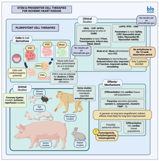
Despite improvements in contemporary medical and surgical therapies, cardiovascular disease (CVD) remains a significant cause of worldwide morbidity and mortality; more specifically, ischemic heart disease (IHD) may affect individuals as young as 20 years old. Typically managed with guideline-directed medical therapy, interventional or surgical methods, the incurred cardiomyocyte loss is not always completely reversible; however, recent research into various stem cell (SC) populations has highlighted their potential for the treatment and perhaps regeneration of injured cardiac tissue, either directly through cellular replacement or indirectly through local paracrine effects. Different stem cell (SC) types have been employed in studies of infarcted myocardium, both in animal models of myocardial infarction (MI) as well as in clinical studies of MI patients, including embryonic stem cells (ESCs) and induced pluripotent stem cells (iPSCs), Muse cells, multipotent stem cells such as bone marrow-derived cells, mesenchymal stem cells (MSCs) and cardiac stem and progenitor cells (CSC/CPCs). These have been delivered as is, in the form of cell therapies, or have been used to generate tissue-engineered (TE) constructs with variable results. 

cardiovascular disease coronary artery disease myocardial infarction cardiac surgery

1. Cell Therapies

Among the possible strategies employed to aid in cardiomyocyte regeneration after myocardial infarction (MI), local transplantation of stem or progenitor cell populations is one possible option. Different cell types may be utilized, while their effects and safety have been tested either with preclinical studies, clinical trials, or both.
Although several studies have been conducted thus far, detailed analysis and comparison of these is beyond the scope of this text; instead, some indicative studies shall be presented for each cell type, including studies where the therapy in question was administered in a manner that might favor its administration during an invasive or interventional cardiac procedure. This is to shed light on possible mechanisms of action and the general effects of such therapeutic interventions on ischemic and infarcted myocardial tissue post-MI.

1.1. Pluripotent Stem Cells in Animal Studies and Clinical Trials

Pluripotent stem cells (PSC), i.e., cells capable of generating tissue from all three germ layers that compose an adult organism [1], including induced PSCs (iPSCs) and embryonic stem cells (ESC), may be transplanted either directly or used for cardiomyocyte generation and subsequent administration. Several preclinical studies have tested the effect of PSC transplantation within infarcted myocardial tissue (Figure 1). In particular, murine ESCs (mESCs) derived from mouse blastocysts have been transplanted into murine models of MI through local intramyocardial injection. These transplanted mESCs prevented cardiomyocyte hypertrophy and mitigated the local post-MI collagen deposition [2]. Another study by Min et al., testing the effect of directly transplanted ESCs, reported positive results with improved ventricular function due to paracrine and angiogenic effects, as well as the generation of differentiated cardiomyocyte progeny within the site of injury [3].
Figure 1. Stem and progenitor cell therapies for ischemic heart disease—pluripotent stem cell therapies (created with BioRender.com, accessed on 24 February 2024) [4][5][6]. CM, Cardiomyocyte; TIMP-1, tissue inhibitor of metalloproteinase 1; LV, left ventricle; BF, blood flow; VTach, ventricular tachycardia; VWall, ventricular wall; LVEF, left ventricular ejection fraction; iPSC, induced pluripotent stem cells; hPSC, human pluripotent stem cells; MSC, mesenchymal stem cells; ESCs, embryonic stem cells; CHF, congestive heart failure; CABG, coronary artery bypass graft; MACE, major adverse cardiovascular events; PCI, percutaneous coronary intervention.
PSCs may also be used indirectly for the derivation of cardiomyocyte (CM) populations, or PSC-CMs, which can then be transplanted onto the target area [7]. Hatani and Yoshida have described protocols for the derivation and subsequent administration of hiPSC-derived cardiomyocytes in immunodeficient mice [8]. Animal studies using direct myocardial administration of stem cell populations from a starting PSC population, i.e., PSC-derived mesenchymal stem cells (MSC), have shown promising results with better efficacy compared to MSCs acquired from other locations [9]. Selecting PSC-derived cells with intact DNA structure has also facilitated the integration rate of transplanted cardiomyocytes onto target tissue [10]. Similar experiments have been carried out in non-human primates as well, with Kobayashi et al. describing protocols for the administration of PSC-derived cardiomyocytes in the Cynomolgus monkey after establishing a model of myocardial infarction [11]. Similarly executed experiments in the Macaca nemestrina species yielded promising results, including reduced infarct size and the generation of myocardial tissue in the injured area, although instances of ventricular arrhythmias were observed [11][12].
Direct transplantation of pluripotent cells onto damaged myocardial tissue has not been described in human subjects [13], perhaps due to concerns regarding oncogenicity owing to the vast differentiation potential of PSCs [14]. Numerous factors, such as the generation of heterogeneous cell populations that may include immature cells, concerns about arrhythmogenicity, and immunogenicity due to the expression of different human leukocyte antigen (HLA) markers, seem to be at the root of the difficulties in translating PSC-derived cell studies into the clinic. However, autologous patient-derived iPSCs or HLA-matching of iPSC populations (stem cell banks) may offer a solution, at least in terms of the immunogenicity of administered cell populations. Nonetheless, clinical trials have been or are currently being conducted to study the effects of PSC-derived cells in IHD patients. Two studies have been set up to test direct transplantation of PSC-derived cardiomyocytes [15]; HEAL-CHF has been evaluating the effect of epicardially injected PSC-generated cardiomyocytes on heart failure patients with indications for coronary artery bypass graft (CABG) [4], while LAPiS, a phase I/II clinical trial with an estimated completion date in 2024, will similarly be assessing the effect of iPSC-derived cardiomyocyte spheroids on heart failure patients, more than one month after an episode of MI with a low ventricular ejection fracture (LVEF), through injection [5].
More recently, Muse cells, i.e., cells expressing pluripotency markers, have been isolated and tested; they seem to carry a lower tumorigenic potential themselves since they express low quantities of tumorigenic transcription factors, attributed to increased let-7 (lethal-7) expression, which suppresses Lin28, a transcription factor contributing to the pluripotent phenotype and, as a result, tumorigenicity. Muse cells also exhibit reduced telomerase activity as well as increased capability for DNA repair [16]. Muse cells have been isolated from various locations, including the bone marrow (BM), connective tissues in various organs, as well as the amniotic membrane; these cells, like PSCs, are capable of differentiation into tissues from all three germ layers [17]. Use of Muse cells in animal models, including intravenous administration in a rabbit model of MI, revealed improvements in ejection fraction (EF), infarct scar size reduction and differentiation into relevant cell types [18]. The mechanism for this effect is thought to be sphingosine monophosphate (S1P)-sphingosine 1 monophosphate receptor 2 (S1PR2) signaling [18], an interaction already observed to facilitate the homing of certain immune and progenitor cells [19]. Similarly promising results were observed in larger animal models when Muse cells were administered in swine models of acute MI; infarct size seemed to be reduced, with associated improvements in ejection fraction and ventricular dimensions and no evidence of arrhythmias [20].
Muse cells have also been utilized in human MI patients in a clinical study commenced in 2018; results showed improved ventricular function and reduced scar size, most likely due to direct cardiomyocyte differentiation as well as local paracrine effects. Nevertheless, only three patients were enrolled in this clinical study and were only followed for 12 weeks; thus, any possible tumorigenicity was not properly assessed [6], even though in general, Muse cells have been described as non-tumorigenic compared to other PSC types [17][21].
In essence, the effects of pluripotent stem cell transplantation may be summarized as effects due to differentiation into local cell populations, observable through the identification of engrafted cells expressing cardiac markers (alpha Myosin heavy chain-aMHC, troponin I-TnI) [3]. Pluripotent cells may also be selected beforehand for their cardiomyogenic potential to better control their differentiation into cardiac cell lineages upon transplantation [3]. Another possible mechanism of action for pluripotent stem cell therapies might include the cardioprotective effects of extracellular vesicles (EV) secreted by PSC-derived cell populations; it seems that PSC-EV administration might not be associated with teratoma formation, as opposed to PSCs themselves, based on results from associated studies [22]. Some authors posit a possible paracrine effect of transplanted pluripotent populations, possibly through the secretion of factors such as cystatin C, osteopontin, clusterin or tissue inhibitor of metalloproteinase-1 (TIMP-1), capable of preventing local cardiomyocyte apoptosis and excess fibrosis. This improves local myocardial tissue viability, in turn enhancing cardiac function [2]. Furthermore, while most studies have shown positive effects after transplantation, some studies do not seem to report long-term engraftment of selected cells; it is thus thought that paracrine effects are most likely the reason for cardiac improvement in the long term [23].
Some drawbacks specifically encountered with pluripotent stem cell populations are immunogenicity and tumorigenicity; although the former may be amended through autologous derivation of iPSC populations, this might extend the therapeutic timeline significantly, mainly due to the time required for iPSC line derivation [23]. Tumorigenicity is another disadvantage frequently encountered with PSCs but not Muse cells; this may be attributed to a variety of factors, including residual, undifferentiated PSCs still capable of growth, genomic instability after multiple cell passages, or even persistent epigenetic modifications long after differentiation into target lineages. To this end, multiple methodologies for evaluating this tumorigenic potential have been created, and although a case-by-case testing basis is feasible, validation studies involving multiple sites have been organized as well [24].

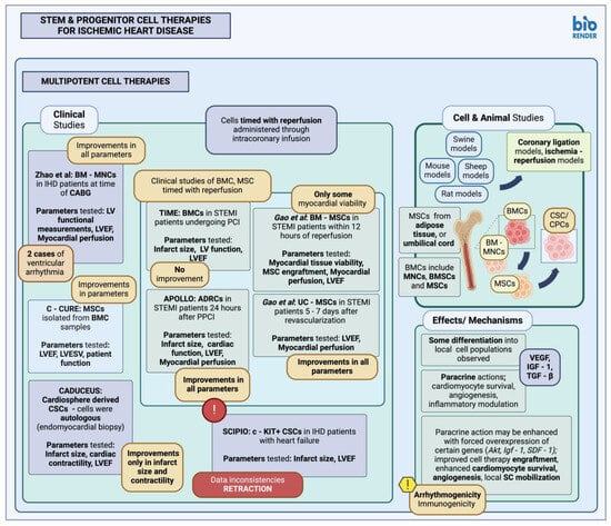
1.2. Multipotent Stem Cells in Animal Studies and Clinical Trials

The use of multipotent stem cells, i.e., cells capable of generating cells relevant to the tissue they are usually found in animal studies and in human patients, has been well described [25]. Examples of multipotent stem cell types used for this purpose include bone-marrow (BM)-derived cells (BMC), mesenchymal stem cells (MSCs) and cardiac stem/progenitor cells (CPC/CSC) (Figure 2). BMCs are perhaps among the earliest used multipotent stem cells for cardiac applications [26]. In fact, the first study reporting their use was published in 2001, where BMC transplantation seemed to cause new cardiac tissue formation 9 days post-implantation [27]. BMCs in general represent a mixed cell population; more specifically, in both small and large animal models, BM-derived mononuclear cells (BM-MNCs) have been used either through intramyocardial injection or intracoronary injection, with promising results and subsequent improvement in ventricular function as well as new local vessel formation, especially in the studies by Kamihata et al. [27][28]. Other studies, however, did not show significant improvement in function or any differentiation of transplanted cells into appropriate local phenotypes [29][30].
Figure 2. Stem and progenitor cell therapies for ischemic heart disease—multipotent stem cell therapies (created with BioRender.com, accessed on 24 February 2024) [31][32][33][34][35][36][37][38][39]. LV, left ventricle; LVESV, left ventricular end systolic volume; LVEF, left ventricular ejection fraction; MSC, mesenchymal stem cells; CABG, coronary artery bypass graft; STEMI, ST-elevation myocardial infarction; BM, bone marrow; BMC, bone marrow cells; BMSCs, bone marrow stem cells; BM-MNC, bone marrow mononuclear cells; MNC, mononuclear cells; c-KIT, tyrosine protein kinase kit; C-CURE, cardiopoietic stem cell therapy in heart failure; TIME, timing in myocardial infarction evaluation; PCI, percutaneous coronary intervention; PPCI, primary percutaneous coronary intervention; Akt, protein kinase B; Igf-1; Insulin-like growth factor 1; VEGF, vascular endothelial growth factor; SDF-1, stromal cell-derived factor 1; TGF-β, transforming growth factor beta; ADRC, adipose-derived regenerative cells; UC-MSC, umbilical cord mesenchymal stem cells; CSC, cardiac stem cell; CPC, cardiac progenitor cell; SCIPIO, stem cell infusion in patients with ischemic cardiomyopathy; CADUCEUS, cardiosphere-derived autologous stem cells to reverse ventricular dysfunction; SC, stem cell.
Clinical trials utilizing BMCs to try and tackle the cardiomyocyte loss associated with MI have been carried out as well. Some of these studies employed additional measures to select specifically for mesenchymal stem cells (MSC) within the bone marrow sample [31][32], while others used BMCs or BM-MNCs, either through intracoronary administration [33] or through intramyocardial injection along with coronary artery bypass graft (CABG) surgery, respectively [34]. While Bartunek et al. and Zhao et al. reported improvements in geometric and functional parameters after therapy [31], others reported no significant improvement after the employed cell therapy [32][33]. A published meta-analysis has shown that BMC administration may lead to improvement in ventricular function, infarct size, as well as the resulting cardiac remodeling [40], while another one, which evaluated the administration of BMCs specifically at the time of CABG, further exhibited the capacity for BMCs to improve left ventricle (LV) functional parameters when administered intraoperatively [41].
MSCs have also been used for the alleviation of post-MI myocardial tissue injury, MSCs possess abilities for the generation of differentiated connective tissue cell progeny, mitigation, and modulation of the inflammatory response [42]. Various animal studies have been conducted to test the effect of MSC on alleviating MI sequelae. Among them was a study of intramyocardial MSC injection in a rat model, which exhibited a reduction in local, post-infarct fibrosis, improvements in left ventricular wall compliance, and, as a result, better contractility, and function [43]. The improvement in the tested parameters, including infarct size and rate of cardiomyocyte apoptosis, is thought to occur not only through MSC differentiation towards cardiac cell lineages, which some studies did not report at all [43], but mostly through local paracrine effects that may induce angiogenesis or aid in cardiomyocyte protection during hypoxic conditions. In fact, MSCs overexpressing Akt were shown to act in a mostly paracrine manner by aiding local cardiomyocyte survival [44], while MSCs overexpressing Igf-1 aided in the local recruitment of stem and progenitor cells as well. In the latter, the main mechanism for MSC-associated cardiac tissue repair was observed to be activation of the stromal-derived factor-1 (SDF-1)/CXCR4 signaling pathway [44][45][46].
The effect of MSCs on post-MI cardiac tissue structure and function has also been examined in clinical studies. MSCs have been isolated from the BM, adipose tissue (AT) (AT-derived multipotent stem cells), and the umbilical cord (UC), with no difference in isolation rate, multipotency characteristics or immune characteristics between the different MSC types [46][47]. The APOLLO trial, which involved administration of MSC-like cells derived from adipose tissue, denoted as adipose tissue-derived regenerative cells (ADRC), through intracoronary injection 24 h after primary PCI revascularization. The study reported promising results, with regard to scar size reduction and overall cardiac function [35]. However, similar trials utilizing adipose-derived MSCs did not report any significant improvements in cardiac function [48]. Another trial utilized umbilical cord-derived MSCs (UC-MSC) administered through intracoronary infusion in patients with STEMI, five to seven days after revascularization; the results in these studies were promising here as well, with improvements in LV function and myocardial perfusion [36][46].
CSC/CPCs include many different cell types, with many also possessing the capacity for generating differentiated cardiomyocytes, as detailed previously. Procurement may be autologous [14][49]; however, this may prove cumbersome due to the limited number of such cells within cardiac tissue [50]. CSC/CPCs thus far have been used in various animal studies, including c-KIT+ cells introduced through intracoronary infusion in a swine model of MI. In this study, implanted cells exhibited differentiation into both cardiac and vascular phenotypes and led to improved left ventricular function [51][52]. In general, implanted c-KIT+ cells show better capacity for scar reduction compared to MSCs [52][53]. Other studies have also utilized cardiosphere-derived progenitors through intracoronary injection, with similarly promising results [52].
CSCs/CPCs have also been used in clinical trials. Two relevant clinical trials have utilized autologous c-KIT+ and cardiosphere-derived cells, namely the SCIPIO and CADUCEUS trials, respectively [53]. In the SCIPIO trial autologous c-KIT+ cardiac progenitors were administered through intracoronary injection; the study was later retracted in 2019 [38][39][54]. Another similar trial, the CADUCEUS trial, examined the effect of cardiosphere-derived cells, administered through intracoronary injection, on infarcted hearts; results included an observed reduction in scar size, although this trial reported no effect on left ventricular ejection fraction [37].
The action of multipotent stem cell populations mostly lies in their ability to produce appropriate local factors that might aid in local cardiomyocyte survival and apoptosis, angiogenesis, or influence inflammatory pathways; however, they may also exhibit some capability for differentiation into cardiomyocyte-like cells, although the functionality of this progeny may not always be as clear-cut, as is the case with adipose-derived stem cells, for example [55]. As for secretory activity, this may include factors such as vascular endothelial growth factor (VEGF), insulin-like growth factor 1 (IGF-1) or transforming growth factor β (TGF-β) [56]. A multitude of different multipotent stem cell populations have been utilized in a multitude of different animal studies and clinical trials, including BM-derived populations, MSCs derived from various locations, as well as CSC/CPC. Results have been variable, although in cases of successful studies, the main effects have been improvements in local perfusion and capillary density, myocardial tissue viability and cardiac function, as well as infarct size [57]. This secretory function may be augmented through forced overexpression of certain genes, including Igf-1, Akt and stromal derived growth factor-1 (Sdf-1); when overexpressed, these may further facilitate the local function of these stem cell therapies or aid in recruitment of local stem and progenitor cells, further aiding in cardiomyocyte survival, angiogenesis, and overall improvement in cardiac function. Paracrine secretion also seems to be the mechanism of action for CPC/CSC populations as well, as further proven by studies that do not seem to assign any role for differentiated cardiomyocyte generation by c-KIT+ and SCA-1+ cells [58]. Additional mechanisms in these cases seem to be activation of the SDF-1/CXCR4 pathway, at least in cases of IGF-1 overexpression [44][45]. In general, however, studies employing multipotent stem cell populations seem to exhibit variable results, mostly due to variation in the stem cell type administered, the dose, the method of administration, or any additional biological alterations that may improve their efficacy; this might also serve as an explanation for the lack of a unified mechanism of action across different studies since different therapeutic approaches employ different mechanisms [59].
Although not tumorigenic or immunogenic when derived from autologous sources or when immunomodulatory MSCs are specifically utilized, as evident from relevant studies where xenogeneic MSCs do not seem to trigger a significant immune reaction [60], administration of multipotent stem cells, as with pluripotent stem cells, may also result in arrhythmias (arrhythmogenicity). Various mechanisms may contribute to this observed arrhythmogenicity; one such mechanism is pacemaker-like activity, or depolarizations from implanted cells [61], as well as negative effects on cellular excitation of implanted cells due to secretion of local factors, frequently observed after MSC transplantation [62]. Another mechanism may be tissue heterogeneity due to the innate variability of cellular excitation in the cellular sample introduced, oftentimes owing to differences in their electrophysiological profile; this may include variations in ion currents or even differential expression of ion channel or gap junction proteins. Finally, increased cellular automaticity, exacerbated by the high proportion of fibroblasts within the infarct scar, might also play a role [58].

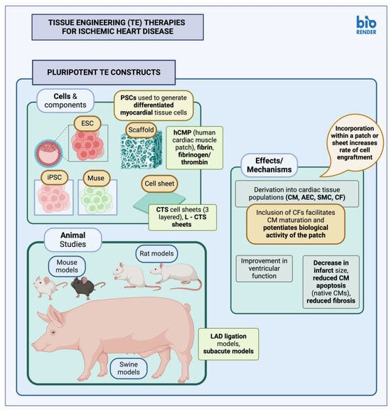
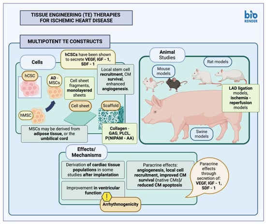
2. Tissue-Engineered Therapies

Stem cells may be delivered to the target area of post-MI ischemia, fibrosis, or necrosis in the form of a tissue-engineered (TE) construct. This may be in the form of a scaffold-derived product, generated from compatible biomaterials and infused with the appropriate cell types. These so-called myocardial patches may then be applied directly to the cardiac surface, either as monotherapy or combined with surgical revascularization. TE therapies for the treatment of post-MI cardiac tissue injury may also be delivered in the form of a cell sheet construct. In any case, delivery of appropriate cell types in a more rigid medium, compared to direct cell infusion, might address issues with cell engraftment and retention in the area, a problem frequently encountered with cell therapies [63][64].

2.1. Pluripotent Stem Cell Constructs

Human-derived iPSCs (hiPSCs) have been used to generate cell sheet in both small and large animal models of MI (Figure 3). iPSCs in these cases were used to derive tissue-specific cells and progenitors, although in some instances a mix of iPSCs and embryonic stem cells (ESC), collectively referred to as PSCs, have been utilized, as in the study by Lou et al. [65]. Amount of cell retention, new vessel formation, and alterations or improvements in myocardial function were some of the common parameters tested in most of these studies. Results in general seem to be positive [66][67][68], though cell engraftment observed in some swine models seems to be diminished [67][69]. iPSCs have the developmental potential for the generation of many different cell lineages; during iPSC reprogramming, however, some epigenetic modifications might persist in the starter cell populations. It has thus been proposed that it might be more favorable to use cardiac lineages for iPSC derivation instead [68].
Figure 3. Tissue engineering (TE) therapies for ischemic heart disease—Pluripotent TE constructs (created with BioRender.com, accessed on 24 February 2024). TE, tissue engineering; ESC, embryonic stem cells; iPSC, induced pluripotent stem cells; PSC, pluripotent stem cells; CTS, cardiac tissue sheet; L-CTS, large cardiac tissue sheet; CM, cardiomyocyte; LAD, left anterior descending coronary artery; hCMP, human cardiac muscle patch; hPSC, human pluripotent stem cell; hESC, human embryonic stem cell; SMC, smooth muscle cells; AEC, arterial endothelial cells; CF, cardiac fibroblast.
PSC-derived cells may also be delivered to target tissues within fibrin scaffolds; the addition of fibroblasts along with other commonly delivered, differentiated cell types may further enhance local tissue recovery. The addition of cardiac fibroblasts, for example, along with other cardiac-tissue-specific cell types, seems to enhance the maturation of cardiomyocytes within the patch, which is thought to occur through the enhanced intracellular cAMP signaling pathway, as well as further aid in local tissue recovery due to improvements in the rate of cardiomyocyte engraftment [65]. The observed positive effects may also be due to altered mechanical properties imparted by cardiac fibroblasts, or more specifically, due to their secretion of extracellular matrix (ECM) [65]. Myocardial patches of larger size have also been created and applied to larger animal models. In these cases, the results have been promising and may perhaps facilitate the translation of similar studies into a clinical setting [65][70].

2.2. Multipotent Stem Cell Constructs

Myocardial patches utilizing multipotent cells derived from the bone marrow (BM) or adipose tissue (AT) have also been generated (Figure 4); these have been applied in both small and larger animal models, suspended in various types of polymeric materials, including type I collagen [71][72], PLCL (poly(lactide-co-ε-caprolactone) [73], and poly(lactic-co-glycolic acid) [74], or generated as scaffold-free cell sheets [75], or alternatively, cell sheet fragments [76]. Cells may also be encapsulated in materials aiding in their survival when transplanted onto the target tissues; Tang et al., for example, have utilized a thermosensitive nanogel composed of P(NIPAM-AA) (poly(N-isopropylacrylamine-co-acrylic acid)), which has been shown to facilitate the favorable actions of multipotent stem cells when applied in both small and larger animal models [77].
Figure 4. Tissue engineering (TE) therapies for ischemic heart disease—Multipotent TE constructs (created with BioRender.com, accessed on 24 February 2024). TE, tissue engineering; CM, cardiomyocyte; LAD, left anterior descending coronary artery; MSC, mesenchymal stem cells; hCSC, human cardiac stem cells; hMSC, human mesenchymal stem cells; AD-MSC, adipose-derived mesenchymal stem cells; GAG, glycosaminoglycans; PLCL, poly(lactide-co-ε-caprolactone); P(NIPAM-AA), poly(N-isopropylacrylamine-co-acrylic acid); VEGF, vascular endothelial growth factor; IGF-1, insulin-like growth factor 1; SDF-1, stromal cell-derived factor-1.
Most of these constructs have generally led to improvements in cardiac function in relevant experiments, as well as local angiogenesis. In other studies, however, positive results were thought to be due to increased local generation of myofibroblasts [72]. Delivery of stem cells in a biomaterial matrix seems to aid in local cell survival since infarcted myocardial tissue is thought to be a poor environment for proper cellular growth and differentiation [73]. In addition, MSC administration seems to be favorable for inducing local biological processes aiding in tissue repair, including angiogenesis [75]. Some studies, however, reported arrhythmias after graft implantation [76]. It is also interesting to note that while pluripotent cells have been used indirectly in most studies, in order to derive relevant cell progeny suitable for cardiac implantation, multipotent cell constructs such as MSCs have also been administered as is, perhaps due to both their differentiation potential and their favorable paracrine effects [69]. Additional multipotent cell types used in similar studies include CPC/CSCs; these cells, although theoretically more inclined towards a cardiac differentiation lineage, have also been shown to mainly act through local effects, including secretion of proteins such as VEGF, IGF-1 and SDF-1, facilitating local angiogenesis, recruitment of endogenous stem cell lineages, and preventing marked cardiomyocyte apoptosis, thus enhancing overall local cellular survival [77][78].
Translation of preclinical studies involving TE constructs along with stem cells, or stem cell derivatives to a more clinical application seems to be difficult. There seem to be various issues arising due to inappropriate adhesion to underlying tissues (either not enough or inappropriately increased adhesion) and immunogenicity, not only of cellular products but of scaffold material as well. Arrhythmogenicity also seems to be a problem in some studies, which might require further research before a clinical application [78].

References

  1. Abu-Dawud, R.; Graffmann, N.; Ferber, S.; Wruck, W.; Adjaye, J. Pluripotent Stem Cells: Induction and Self-Renewal. Philos. Trans. R. Soc. B Biol. Sci. 2018, 373, 20170213.
  2. Singla, D.K.; Lyons, G.E.; Kamp, T.J. Transplanted Embryonic Stem Cells Following Mouse Myocardial Infarction Inhibit Apoptosis and Cardiac Remodeling. Am. J. Physiol. Heart Circ. Physiol. 2007, 293, H1308–H1314.
  3. Min, J.-Y.; Yang, Y.; Sullivan, M.F.; Ke, Q.; Converso, K.L.; Chen, Y.; Morgan, J.P.; Xiao, Y.-F. Long-Term Improvement of Cardiac Function in Rats after Infarction by Transplantation of Embryonic Stem Cells. J. Thorac. Cardiovasc. Surg. 2003, 125, 361–369.
  4. Help Therapeutics Epicardial Injection of Allogeneic Human Pluripotent Stem Cell-Derived Cardiomyocytes to Treat Severe Chronic Heart Failure. 2022. Available online: https://clinicaltrials.gov/study/NCT03763136#publications (accessed on 25 December 2023).
  5. Heartseed Inc. A Phase I/II Study of Human Induced Pluripotent Stem (iPS) Cell-Derived Cardiomyocyte Spheroids (HS-001) in Patients with Severe Heart Failure, Secondary to Ischemic Heart Disease. 2022. Available online: https://clinicaltrials.gov/study/NCT04945018 (accessed on 25 December 2023).
  6. Noda, T.; Nishigaki, K.; Minatoguchi, S. Safety and Efficacy of Human Muse Cell-Based Product for Acute Myocardial Infarction in a First-in-Human Trial. Circ. J. 2020, 84, 1189–1192.
  7. Soma, Y.; Tani, H.; Morita-Umei, Y.; Kishino, Y.; Fukuda, K.; Tohyama, S. Pluripotent Stem Cell-Based Cardiac Regenerative Therapy for Heart Failure. J. Mol. Cell Cardiol. 2024, 187, 90–100.
  8. Hatani, T.; Yoshida, Y. Transplantation of Human Induced Pluripotent Stem Cell-Derived CardiomyocytesHuman Induced Pluripotent Stem Cell-Derived Cardiomyocytes (iPSC-CMs) in a Mouse Myocardial InfarctionMyocardial Infarction Model. In Pluripotent Stem-Cell Derived Cardiomyocytes; Yoshida, Y., Ed.; Methods in Molecular Biology; Springer: New York, NY, USA, 2021; pp. 285–293. ISBN 978-1-07-161484-6.
  9. Thavapalachandran, S.; Le, T.Y.L.; Romanazzo, S.; Rashid, F.N.; Ogawa, M.; Kilian, K.A.; Brown, P.; Pouliopoulos, J.; Barry, A.M.; Fahmy, P.; et al. Pluripotent Stem Cell-Derived Mesenchymal Stromal Cells Improve Cardiac Function and Vascularity after Myocardial Infarction. Cytotherapy 2021, 23, 1074–1084.
  10. Kannappan, R.; Turner, J.F.; Miller, J.M.; Fan, C.; Rushdi, A.G.; Rajasekaran, N.S.; Zhang, J. Functionally Competent DNA Damage-Free Induced Pluripotent Stem Cell–Derived Cardiomyocytes for Myocardial Repair. Circulation 2019, 140, 520–522.
  11. Kobayashi, H.; Ichimura, H.; Ohashi, N.; Shiba, Y. Transplantation of Pluripotent Stem Cell-Derived Cardiomyocytes into a Myocardial Infarction Model of Cynomolgus Monkey. Methods Mol. Biol. 2021, 2320, 295–302.
  12. Chong, J.J.H.; Yang, X.; Don, C.W.; Minami, E.; Liu, Y.-W.; Weyers, J.J.; Mahoney, W.M.; Van Biber, B.; Cook, S.M.; Palpant, N.J.; et al. Human Embryonic Stem Cell-Derived Cardiomyocytes Regenerate Non-Human Primate Hearts. Nature 2014, 510, 273–277.
  13. Higuchi, A.; Ku, N.-J.; Tseng, Y.-C.; Pan, C.-H.; Li, H.-F.; Kumar, S.S.; Ling, Q.-D.; Chang, Y.; Alarfaj, A.A.; Munusamy, M.A.; et al. Stem Cell Therapies for Myocardial Infarction in Clinical Trials: Bioengineering and Biomaterial Aspects. Lab. Investig. 2017, 97, 1167–1179.
  14. Grimaldi, V.; Mancini, F.P.; Casamassimi, A.; Al-Omran, M.; Zullo, A.; Infante, T.; Napoli, C. Potential Benefits of Cell Therapy in Coronary Heart Disease. J. Cardiol. 2013, 62, 267–276.
  15. Selvakumar, D.; Reyes, L.; Chong, J.J.H. Cardiac Cell Therapy with Pluripotent Stem Cell-Derived Cardiomyocytes: What Has Been Done and What Remains to Do? Curr. Cardiol. Rep. 2022, 24, 445–461.
  16. Alanazi, R.F.; Alhwity, B.S.; Almahlawi, R.M.; Alatawi, B.D.; Albalawi, S.A.; Albalawi, R.A.; Albalawi, A.A.; Abdel-Maksoud, M.S.; Elsherbiny, N. Multilineage Differentiating Stress Enduring (Muse) Cells: A New Era of Stem Cell-Based Therapy. Cells 2023, 12, 1676.
  17. Ogawa, E.; Oguma, Y.; Kushida, Y.; Wakao, S.; Okawa, K.; Dezawa, M. Naïve Pluripotent-like Characteristics of Non-Tumorigenic Muse Cells Isolated from Human Amniotic Membrane. Sci. Rep. 2022, 12, 17222.
  18. Yamada, Y.; Wakao, S.; Kushida, Y.; Minatoguchi, S.; Mikami, A.; Higashi, K.; Baba, S.; Shigemoto, T.; Kuroda, Y.; Kanamori, H.; et al. S1P–S1PR2 Axis Mediates Homing of Muse Cells Into Damaged Heart for Long-Lasting Tissue Repair and Functional Recovery After Acute Myocardial Infarction. Circ. Res. 2018, 122, 1069–1083.
  19. Ratajczak, M.Z.; Suszynska, M.; Borkowska, S.; Ratajczak, J.; Schneider, G. The Role of Sphingosine-1 Phosphate and Ceramide-1 Phosphate in Trafficking of Normal Stem Cells and Cancer Cells. Expert. Opin. Ther. Targets 2014, 18, 95–107.
  20. Yamada, Y.; Minatoguchi, S.; Baba, S.; Shibata, S.; Takashima, S.; Wakao, S.; Okura, H.; Dezawa, M.; Minatoguchi, S. Human Muse Cells Reduce Myocardial Infarct Size and Improve Cardiac Function without Causing Arrythmias in a Swine Model of Acute Myocardial Infarction. PLoS ONE 2022, 17, e0265347.
  21. Kuroda, Y.; Wakao, S.; Kitada, M.; Murakami, T.; Nojima, M.; Dezawa, M. Isolation, Culture and Evaluation of Multilineage-Differentiating Stress-Enduring (Muse) Cells. Nat. Protoc. 2013, 8, 1391–1415.
  22. Gill, J.K.; Rehsia, S.K.; Verma, E.; Sareen, N.; Dhingra, S. Stem Cell Therapy for Cardiac Regeneration: Past, Present, and Future. Can. J. Physiol. Pharmacol. 2024, 102, 3.
  23. Yanamandala, M.; Zhu, W.; Garry, D.J.; Kamp, T.J.; Hare, J.M.; Jun, H.; Yoon, Y.; Bursac, N.; Prabhu, S.D.; Dorn, G.W.; et al. Overcoming the Roadblocks to Cardiac Cell Therapy Using Tissue Engineering. J. Am. Coll. Cardiol. 2017, 70, 766–775.
  24. Sato, Y.; Bando, H.; Piazza, M.D.; Gowing, G.; Herberts, C.; Jackman, S.; Leoni, G.; Libertini, S.; MacLachlan, T.; McBlane, J.W.; et al. Tumorigenicity Assessment of Cell Therapy Products: The Need for Global Consensus and Points to Consider. Cytotherapy 2019, 21, 1095–1111.
  25. Sobhani, A.; Khanlarkhani, N.; Baazm, M.; Mohammadzadeh, F.; Najafi, A.; Mehdinejadiani, S.; Sargolzaei Aval, F. Multipotent Stem Cell and Current Application. Acta Med. Iran 2017, 55, 6–23.
  26. Johnston, P.V. Gary Gerstenblith Stem Cells for Cardiac Surgical Disease. In Johns Hopkins Textbook of Cardiothoracic Surgery; McGraw-Hill Companies New York: New York, NY, USA, 2014; p. 1443. ISBN 978-0-07-166350-2.
  27. Orlic, D.; Kajstura, J.; Chimenti, S.; Jakoniuk, I.; Anderson, S.M.; Li, B.; Pickel, J.; McKay, R.; Nadal-Ginard, B.; Bodine, D.M.; et al. Bone Marrow Cells Regenerate Infarcted Myocardium. Nature 2001, 410, 701–705.
  28. Kamihata, H.; Matsubara, H.; Nishiue, T.; Fujiyama, S.; Tsutsumi, Y.; Ozono, R.; Masaki, H.; Mori, Y.; Iba, O.; Tateishi, E.; et al. Implantation of Bone Marrow Mononuclear Cells Into Ischemic Myocardium Enhances Collateral Perfusion and Regional Function via Side Supply of Angioblasts, Angiogenic Ligands, and Cytokines. Circulation 2001, 104, 1046–1052.
  29. Bel, A.; Messas, E.; Agbulut, O.; Richard, P.; Samuel, J.-L.; Bruneval, P.; Hagège, A.A.; Menasché, P. Transplantation of Autologous Fresh Bone Marrow Into Infarcted Myocardium: A Word of Caution. Circulation 2003, 108, II-247–II-252.
  30. de Silva, R.; Raval, A.N.; Hadi, M.; Gildea, K.M.; Bonifacino, A.C.; Yu, Z.-X.; Yau, Y.Y.; Leitman, S.F.; Bacharach, S.L.; Donahue, R.E.; et al. Intracoronary Infusion of Autologous Mononuclear Cells from Bone Marrow or Granulocyte Colony-Stimulating Factor-Mobilized Apheresis Product May Not Improve Remodelling, Contractile Function, Perfusion, or Infarct Size in a Swine Model of Large Myocardial Infarction. Eur. Heart J. 2008, 29, 1772–1782.
  31. Bartunek, J.; Behfar, A.; Dolatabadi, D.; Vanderheyden, M.; Ostojic, M.; Dens, J.; El, N.B.; Banovic, M.; Beleslin, B.; Vrolix, M.; et al. Cardiopoietic Stem Cell Therapy in Heart Failure. J. Am. Coll. Cardiol. 2013, 61, 2329–2338.
  32. Gao, L.R.; Pei, X.T.; Ding, Q.A.; Chen, Y.; Zhang, N.K.; Chen, H.Y.; Wang, Z.G.; Wang, Y.F.; Zhu, Z.M.; Li, T.C.; et al. A Critical Challenge: Dosage-Related Efficacy and Acute Complication Intracoronary Injection of Autologous Bone Marrow Mesenchymal Stem Cells in Acute Myocardial Infarction. Int. J. Cardiol. 2013, 168, 3191–3199.
  33. Traverse, J.H.; Henry, T.D.; Pepine, C.J.; Willerson, J.T.; Ellis, S.G. One-Year Follow-up of Intracoronary Stem Cell Delivery on Left Ventricular Function following ST-Elevation Myocardial Infarction. JAMA 2014, 311, 301–302.
  34. Zhao, Q.; Sun, Y.; Xia, L.; Chen, A.; Wang, Z. Randomized Study of Mononuclear Bone Marrow Cell Transplantation in Patients with Coronary Surgery. Ann. Thorac. Surg. 2008, 86, 1833–1840.
  35. Houtgraaf, J.H.; den Dekker, W.K.; van Dalen, B.M.; Springeling, T.; de Jong, R.; van Geuns, R.J.; Geleijnse, M.L.; Fernandez-Aviles, F.; Zijlsta, F.; Serruys, P.W.; et al. First Experience in Humans Using Adipose Tissue–Derived Regenerative Cells in the Treatment of Patients with ST-Segment Elevation Myocardial Infarction. J. Am. Coll. Cardiol. 2012, 59, 539–540.
  36. Gao, L.R.; Chen, Y.; Zhang, N.K.; Yang, X.L.; Liu, H.L.; Wang, Z.G.; Yan, X.Y.; Wang, Y.; Zhu, Z.M.; Li, T.C.; et al. Intracoronary Infusion of Wharton’s Jelly-Derived Mesenchymal Stem Cells in Acute Myocardial Infarction: Double-Blind, Randomized Controlled Trial. BMC Med. 2015, 13, 162.
  37. Makkar, R.R.; Smith, R.R.; Cheng, K.; Malliaras, K.; Thomson, L.E.; Berman, D.; Czer, L.S.; Marbán, L.; Mendizabal, A.; Johnston, P.V.; et al. Intracoronary Cardiosphere-Derived Cells for Heart Regeneration after Myocardial Infarction (CADUCEUS): A Prospective, Randomised Phase 1 Trial. Lancet 2012, 379, 895–904.
  38. Medscape Medical News: Harvard, Brigham Call for Retraction of 31 Papers by Disgraced Cardiac Stem Cell Doc. Available online: https://www.medscape.com/viewarticle/903475 (accessed on 26 February 2024).
  39. The BMJ News: NEJM Retracts Article from Former Researcher Once Hailed as Heart Stem Cell Pioneer. Available online: https://www.bmj.com/content/363/bmj.k4432 (accessed on 26 February 2024).
  40. Afzal, M.R.; Samanta, A.; Shah, Z.I.; Jeevanantham, V.; Abdel-Latif, A.; Zuba-Surma, E.K.; Dawn, B. Adult Bone Marrow Cell Therapy for Ischemic Heart Disease. Circ. Res. 2015, 117, 558–575.
  41. Ali-Hasan-Al-Saegh, S.; Mirhosseini, S.J.; Lotfaliani, M.-R.; Dehghan, H.R.; Sedaghat-Hamedani, F.; Kayvanpour, E.; Rezaeisadrabadi, M.; Ghaffari, N.; Vahabzadeh, V.; Jebran, A.F.; et al. Transplantation of Bone Marrow Stem Cells during Cardiac Surgery. Asian Cardiovasc. Thorac. Ann. 2015, 23, 363–374.
  42. Ala, M. The Beneficial Effects of Mesenchymal Stem Cells and Their Exosomes on Myocardial Infarction and Critical Considerations for Enhancing Their Efficacy. Aging Res. Rev. 2023, 89, 101980.
  43. Berry, M.F.; Engler, A.J.; Woo, Y.J.; Pirolli, T.J.; Bish, L.T.; Jayasankar, V.; Morine, K.J.; Gardner, T.J.; Discher, D.E.; Sweeney, H.L. Mesenchymal Stem Cell Injection after Myocardial Infarction Improves Myocardial Compliance. Am. J. Physiol. Heart Circ. Physiol. 2006, 290, H2196–H2203.
  44. Gnecchi, M.; He, H.; Liang, O.D.; Melo, L.G.; Morello, F.; Mu, H.; Noiseux, N.; Zhang, L.; Pratt, R.E.; Ingwall, J.S.; et al. Paracrine Action Accounts for Marked Protection of Ischemic Heart by Akt-Modified Mesenchymal Stem Cells. Nat. Med. 2005, 11, 367–368.
  45. Haider, H.K.; Jiang, S.; Idris, N.M.; Ashraf, M. IGF-1-Overexpressing Mesenchymal Stem Cells Accelerate Bone Marrow Stem Cell Mobilization via Paracrine Activation of SDF-1alpha/CXCR4 Signaling to Promote Myocardial Repair. Circ. Res. 2008, 103, 1300–1308.
  46. Yamada, Y.; Minatoguchi, S.; Kanamori, H.; Mikami, A.; Okura, H.; Dezawa, M.; Minatoguchi, S. Stem Cell Therapy for Acute Myocardial Infarction—Focusing on the Comparison between Muse Cells and Mesenchymal Stem Cells. J. Cardiol. 2022, 80, 80–87.
  47. Kern, S.; Eichler, H.; Stoeve, J.; Klüter, H.; Bieback, K. Comparative Analysis of Mesenchymal Stem Cells from Bone Marrow, Umbilical Cord Blood, or Adipose Tissue. Stem Cells 2006, 24, 1294–1301.
  48. The SCIENCE Investigators. Effect of Allogeneic Adipose Tissue-Derived Mesenchymal Stromal Cell Treatment in Chronic Ischaemic Heart Failure with Reduced Ejection Fraction—The Science Trial. Eur. J. Heart Fail. 2023, 25, 576–587.
  49. Eisenberg, L.M.; Eisenberg, C.A. Adult Stem Cells and Their Cardiac Potential. Anat. Rec. Part. A Discov. Mol. Cell. Evol. Biol. 2004, 276A, 103–112.
  50. Mehanna, R.A.; Essawy, M.M.; Barkat, M.A.; Awaad, A.K.; Thabet, E.H.; Hamed, H.A.; Elkafrawy, H.; Khalil, N.A.; Sallam, A.; Kholief, M.A.; et al. Cardiac Stem Cells: Current Knowledge and Future Prospects. World J. Stem Cells 2022, 14, 1–40.
  51. Bolli, R.; Tang, X.-L.; Sanganalmath, S.K.; Rimoldi, O.; Mosna, F.; Abdel-Latif, A.; Jneid, H.; Rota, M.; Leri, A.; Kajstura, J. Intracoronary Delivery of Autologous Cardiac Stem Cells Improves Cardiac Function in a Porcine Model of Chronic Ischemic Cardiomyopathy. Circulation 2013, 128, 122–131.
  52. Tompkins, B.A.; Balkan, W.; Winkler, J.; Gyöngyösi, M.; Goliasch, G.; Fernández-Avilés, F.; Hare, J.M. Preclinical Studies of Stem Cell Therapy for Heart Disease. Circ. Res. 2018, 122, 1006–1020.
  53. Mahmud, S.; Alam, S.; Emon, N.U.; Boby, U.H.; Kamruzzaman; Ahmed, F.; Monjur-Al-Hossain, A.S.M.; Tahamina, A.; Rudra, S.; Ajrin, M. Opportunities and Challenges in Stem Cell Therapy in Cardiovascular Diseases: Position Standing in 2022. Saudi Pharm. J. 2022, 30, 1360–1371.
  54. Expression of Concern: The SCIPIO Trial. Lancet 2014, 383, 1279.
  55. Xiao, W.; Shi, J. Application of Adipose-Derived Stem Cells in Ischemic Heart Disease: Theory, Potency, and Advantage. Front. Cardiovasc. Med. 2024, 11, 1324447.
  56. Wang, J.; An, M.; Haubner, B.J.; Penninger, J.M. Cardiac Regeneration: Options for Repairing the Injured Heart. Front. Cardiovasc. Med. 2023, 9, 981982.
  57. Guo, Q.-Y.; Yang, J.-Q.; Feng, X.-X.; Zhou, Y.-J. Regeneration of the Heart: From Molecular Mechanisms to Clinical Therapeutics. Mil. Med. Res. 2023, 10, 18.
  58. Chen, K.; Huang, Y.; Singh, R.; Wang, Z.Z. Arrhythmogenic Risks of Stem Cell Replacement Therapy for Cardiovascular Diseases. J. Cell Physiol. 2020, 235, 6257–6267.
  59. Carbone, R.G.; Negrini, S.; Murdaca, G.; Fontana, V.; Puppo, F. Stem Cells Treatment in Chronic Ischemic Heart Disease: A Narrative Review. Am. J. Stem Cells 2023, 12, 65–72.
  60. Harland, N.; Knoll, J.; Amend, B.; Abruzzese, T.; Abele, H.; Jakubowski, P.; Stenzl, A.; Aicher, W.K. Xenogenic Application of Human Placenta-Derived Mesenchymal Stromal Cells in a Porcine Large Animal Model. Cell Transpl. 2024, 33, 9636897241226737.
  61. Liu, Y.-W.; Chen, B.; Yang, X.; Fugate, J.A.; Kalucki, F.A.; Futakuchi-Tsuchida, A.; Couture, L.; Vogel, K.W.; Astley, C.A.; Baldessari, A.; et al. Human Embryonic Stem Cell–Derived Cardiomyocytes Restore Function in Infarcted Hearts of Non-Human Primates. Nat. Biotechnol. 2018, 36, 597–605, Erratum in Nat. Biotechnol. 2018, 36, 899.
  62. Askar, S.F.A.; Ramkisoensing, A.A.; Atsma, D.E.; Schalij, M.J.; de Vries, A.A.F.; Pijnappels, D.A. Engraftment Patterns of Human Adult Mesenchymal Stem Cells Expose Electrotonic and Paracrine Proarrhythmic Mechanisms in Myocardial Cell Cultures. Circ. Arrhythmia Electrophysiol. 2013, 6, 380–391.
  63. Chandrasekhar, S.K.; Thankam, F.G.; Ouseph, J.C.; Agrawal, D.K. Myocardial Tissue Engineering: Fundamentals and Future. In Tissue Engineering; Elsevier: Amsterdam, The Netherlands, 2022; pp. 33–51. ISBN 978-0-12-824064-9.
  64. Qasim, M.; Arunkumar, P.; Powell, H.M.; Khan, M. Current Research Trends and Challenges in Tissue Engineering for Mending Broken Hearts. Life Sci. 2019, 229, 233–250.
  65. Lou, X.; Tang, Y.; Ye, L.; Pretorius, D.; Fast, V.G.; Kahn-Krell, A.M.; Zhang, J.; Zhang, J.; Qiao, A.; Qin, G.; et al. Cardiac Muscle Patches Containing Four Types of Cardiac Cells Derived from Human Pluripotent Stem Cells Improve Recovery from Cardiac Injury in Mice. Cardiovasc. Res. 2023, 119, 1062–1076.
  66. Masumoto, H.; Ikuno, T.; Takeda, M.; Fukushima, H.; Marui, A.; Katayama, S.; Shimizu, T.; Ikeda, T.; Okano, T.; Sakata, R.; et al. Human iPS Cell-Engineered Cardiac Tissue Sheets with Cardiomyocytes and Vascular Cells for Cardiac Regeneration. Sci. Rep. 2014, 4, 6716.
  67. Ishigami, M.; Masumoto, H.; Ikuno, T.; Aoki, T.; Kawatou, M.; Minakata, K.; Ikeda, T.; Sakata, R.; Yamashita, J.K.; Minatoya, K. Human iPS Cell-Derived Cardiac Tissue Sheets for Functional Restoration of Infarcted Porcine Hearts. PLoS ONE 2018, 13, e0201650.
  68. Zhang, L.; Guo, J.; Zhang, P.; Xiong, Q.; Wu, S.C.; Xia, L.; Roy, S.S.; Tolar, J.; O’Connell, T.D.; Kyba, M.; et al. Derivation and High Engraftment of Patient-Specific Cardiomyocyte Sheet Using Induced Pluripotent Stem Cells Generated from Adult Cardiac Fibroblast. Circ. Heart Fail. 2015, 8, 156–166.
  69. Augustine, R.; Dan, P.; Hasan, A.; Khalaf, I.M.; Prasad, P.; Ghosal, K.; Gentile, C.; McClements, L.; Maureira, P. Stem Cell-Based Approaches in Cardiac Tissue Engineering: Controlling the Microenvironment for Autologous Cells. Biomed. Pharmacother. 2021, 138, 111425.
  70. Gao, L.; Gregorich, Z.R.; Zhu, W.; Mattapally, S.; Oduk, Y.; Lou, X.; Kannappan, R.; Borovjagin, A.V.; Walcott, G.P.; Pollard, A.E.; et al. Large Cardiac Muscle Patches Engineered from Human Induced-Pluripotent Stem Cell–Derived Cardiac Cells Improve Recovery from Myocardial Infarction in Swine. Circulation 2018, 137, 1712–1730.
  71. Xiang, Z.; Liao, R.; Kelly, M.S.; Spector, M. Collagen–GAG Scaffolds Grafted onto Myocardial Infarcts in a Rat Model: A Delivery Vehicle for Mesenchymal Stem Cells. Tissue Eng. 2006, 12, 2467–2478.
  72. Simpson, D.; Liu, H.; Fan, T.-H.M.; Nerem, R.; Dudley, S.C., Jr. A Tissue Engineering Approach to Progenitor Cell Delivery Results in Significant Cell Engraftment and Improved Myocardial Remodeling. Stem Cells 2007, 25, 2350–2357.
  73. Jin, J.; Jeong, S.I.; Shin, Y.M.; Lim, K.S.; Shin, H.S.; Lee, Y.M.; Koh, H.C.; Kim, K.-S. Transplantation of Mesenchymal Stem Cells within a Poly(Lactide-Co-ε-Caprolactone) Scaffold Improves Cardiac Function in a Rat Myocardial Infarction Model. Eur. J. Heart Fail. 2009, 11, 147–153.
  74. Zhang, J.; Li, J.; Qu, X.; Liu, Y.; Harada, A.; Hua, Y.; Yoshida, N.; Ishida, M.; Tabata, A.; Sun, L.; et al. Development of a Thick and Functional Human Adipose-Derived Stem Cell Tissue Sheet for Myocardial Infarction Repair in Rat Hearts. Stem Cell Res. Ther. 2023, 14, 380.
  75. Miyahara, Y.; Nagaya, N.; Kataoka, M.; Yanagawa, B.; Tanaka, K.; Hao, H.; Ishino, K.; Ishida, H.; Shimizu, T.; Kangawa, K.; et al. Monolayered Mesenchymal Stem Cells Repair Scarred Myocardium after Myocardial Infarction. Nat. Med. 2006, 12, 459–465.
  76. Huang, C.-C.; Tsai, H.-W.; Lee, W.-Y.; Lin, W.-W.; Chen, D.-Y.; Hung, Y.-W.; Chen, J.-W.; Hwang, S.-M.; Chang, Y.; Sung, H.-W. A Translational Approach in Using Cell Sheet Fragments of Autologous Bone Marrow-Derived Mesenchymal Stem Cells for Cellular Cardiomyoplasty in a Porcine Model. Biomaterials 2013, 34, 4582–4591.
  77. Tang, J.; Cui, X.; Caranasos, T.G.; Hensley, M.T.; Vandergriff, A.C.; Hartanto, Y.; Shen, D.; Zhang, H.; Zhang, J.; Cheng, K. Heart Repair Using Nanogel-Encapsulated Human Cardiac Stem Cells in Mice and Pigs with Myocardial Infarction. ACS Nano 2017, 11, 9738–9749.
  78. Mei, X.; Cheng, K. Recent Development in Therapeutic Cardiac Patches. Front. Cardiovasc. Med. 2020, 7, 610364.
More
Information
Contributors MDPI registered users' name will be linked to their SciProfiles pages. To register with us, please refer to https://encyclopedia.pub/register : , , , ,
View Times: 42
Revisions: 2 times (View History)
Update Date: 27 Mar 2024
1000/1000