Your browser does not fully support modern features. Please upgrade for a smoother experience.
Submitted Successfully!
 +1 credit
Thank you for your contribution! You can also upload a video entry or images related to this topic.
For video creation, please contact our Academic Video Service.
| Version | Summary | Created by | Modification | Content Size | Created at | Operation |
|---|---|---|---|---|---|---|
| 1 | Lalit Kaurani | -- | 3023 | 2024-03-14 08:18:16 | | | |
| 2 | Fanny Huang | Meta information modification | 3023 | 2024-03-15 10:25:27 | | |
Video Upload Options
We provide professional Academic Video Service to translate complex research into visually appealing presentations. Would you like to try it?
Cite
If you have any further questions, please contact Encyclopedia Editorial Office.
Kaurani, L. MicroRNAs in Depression. Encyclopedia. Available online: https://encyclopedia.pub/entry/56242 (accessed on 28 July 2026).
Kaurani L. MicroRNAs in Depression. Encyclopedia. Available at: https://encyclopedia.pub/entry/56242. Accessed July 28, 2026.
Kaurani, Lalit. "MicroRNAs in Depression" Encyclopedia, https://encyclopedia.pub/entry/56242 (accessed July 28, 2026).
Kaurani, L. (2024, March 14). MicroRNAs in Depression. In Encyclopedia. https://encyclopedia.pub/entry/56242
Kaurani, Lalit. "MicroRNAs in Depression." Encyclopedia. Web. 14 March, 2024.
Copy Citation
Depression is a major contributor to the overall global burden of disease. The discovery of biomarkers for diagnosis or prediction of treatment responses and as therapeutic agents is a current priority. Previous studies have demonstrated the importance of short RNA molecules in the etiology of depression. The most extensively researched of these are microRNAs, a major component of cellular gene regulation and function. MicroRNAs function in a temporal and tissue-specific manner to regulate and modify the post-transcriptional expression of target mRNAs. They can also be shuttled as cargo of extracellular vesicles between the brain and the blood, thus informing about relevant mechanisms in the CNS through the periphery.
microRNA
depression
biomarker
antidepressant
extracellular vesicles
1. Introduction
1.1. General Introduction to Depression
Depression, an overarching term encompassing a spectrum of mood disturbances, stands as a predominant global health concern, currently recognized as a principal contributor to disability across the world. An estimated 5% of the global adult population is impacted by depression, a number witnessing an alarming ascent each year [1][2][3]. Cumulative lifetime prevalence statistics, when evaluated on an international scale, suggest that about one in seven individuals will grapple with a depressive episode at some juncture in their lives, with rates ranging between 11.1% and 14.6% [4]. Notably, while depressive episodes can manifest at various life stages, spanning early adolescence to adulthood, the median onset age gravitates around the mid-20s [3][5]. Gender disparities are also evident; women exhibit nearly double the susceptibility to experiencing depression during their lifetimes compared to their male counterparts, though this ratio demonstrates considerable cross-national variation [6][7].
The etiology of depression is multifaceted, interwoven with both environmental contributors such as socioeconomic conditions and biological determinants, including genetic predispositions [3][8]. The interplay between genes and the environment is further modulated by epigenetic mechanisms, adding another layer to its complexity [9]. Among the various forms of depression, major depressive disorder (MDD) is particularly well-researched, although its pathophysiology is yet to be fully deciphered. Available evidence points towards molecular disruptions, including neurotransmitter imbalances and augmented inflammatory cytokine levels, underscoring their intricate molecular foundation [10].
1.2. Current Therapeutic Approaches
The main goal of treatment for depression is an accurate diagnosis and the remission of symptoms. Depression is clinically identified as ‘major depression’ when it meets the symptomatic criteria outlined in the Diagnostic and Statistical Manual of Mental Disorders, Fourth Edition, 2000 (DSM-V) [11]. Diagnostic measures are based on clinical observation, although no objective biological markers currently exist. The main approaches to treatment can be separated into four broad categories: generic psychosocial interventions, formulation-based interventions of psychological therapy, pharmacotherapy, and electroconvulsive therapy (ECT) [8]. In mild cases of depression, psychosocial and psychological intervention may be sufficient, while in more severe cases, pharmacotherapy is also required. Most pharmacological interventions for depression enhance the transmission of monoamines through different routes, with the most common first-line treatments being selective serotonin reuptake inhibitors and serotonin–norepinephrine reuptake inhibitors. Despite the range of psychological and pharmacological therapies available, many patients do not achieve remission [12][13].
1.3. General Introduction to miRNAs
MicroRNAs are small non-coding RNAs of approximately 19–22 nucleotides that function to inhibit the level of target genes within discrete regulatory networks [14]. Over 60% of human protein-coding genes contain conserved miRNA target sites [15]. The interaction networks of miRNAs are complex and vast; one miRNA can target many mRNAs, and one mRNA can be regulated by many different miRNAs [16][17]. Their identification in many different organisms also suggests their regulatory mechanisms are evolutionarily conserved [18]. According to recent work by the Functional Annotation of the Mammalian Genome (FANTOM5) consortium, approximately half of miRNAs show cell type specificity, and one-quarter are more broadly expressed [19].
In the central nervous system, miRNAs play a role in neurodevelopment, synaptic plasticity, and stress responses, thus providing a molecular link to psychiatric conditions [20][21][22]. Several lines of evidence have already shown their involvement in other neurological and mental health disorders [22].
In fact, miRNAs are differentially expressed in depression, as identified by a multitude of recent studies, pointing to their involvement here as well [23]. miRNAs have emerged as key players in depression due to their role as post-transcriptional regulators of gene expression and their ability to pass the blood–brain barrier [24]. Excitingly, they offer new avenues for understanding the intricate molecular underpinnings of depression. Their capacity to modulate multiple gene networks associated with neuroplasticity and stress response, as well as their shuttling between the blood and the brain, makes miRNAs compelling targets for both diagnostic and therapeutic interventions in depressive disorders.
2. MicroRNAs and Depression
2.1. miRNAs in the Pathophysiology of Depression
Emerging research underscores noticeable shifts in miRNA levels in both the brain and periphery of individuals with depression. Generally, the predominant research paradigms encompass the following: (a) a comparative study of miRNA profiles between patients and controls; (b) an exploration of miRNA modulations subsequent to antidepressant interventions. In the former approach, the prefrontal cortex emerges as a primary brain region of interest. In the periphery, researchers often rely on whole blood, plasma, or serum samples. The second methodology is predominantly engaged to discern miRNA shifts following therapeutic regimens. A less conventional, albeit more invasive, medium is cerebrospinal fluid to probe miRNA dysregulation. Downstream analysis usually involves bioinformatics-driven approaches to identify miRNA–mRNA interactions, facilitating an understanding of the broader regulatory networks at play. Furthermore, to decode the functional dynamics of miRNA regulation, in vitro evaluations and animal model studies are occasionally integrated, though they are less prevalent.
Some of the first studies to identify the role of microRNAs in the pathophysiology of MDD have examined the postmortem human brain. In one example study, the levels of miR-182 expression were disrupted in dentate gyrus granule cells of individuals with depression [25][26]. In the anterior cingulate cortex, a brain region critical for emotional and mood regulation, a depression-specific miRNA expression profile was identified compared to controls [27]. In a similar approach, miR-1202 was found to be downregulated in the prefrontal cortex of individuals with depression compared to controls [28]. This microRNA was found to be enriched in the brain ad negatively correlated with GRM4 expression, a metabotropic glutamate receptor. Importantly, this study also showed that in individuals who had attained remission, levels of miR-1202 were increased.
While the postmortem brain remains a key system for investigating the molecular mechanisms of MDD, a drawback remains that microRNAome profiling in brain tissue at the time of death or sample collection may not be reflective of the disease course [22]. In recent years, miRNAs have been assessed in the periphery of individuals with depression. An advantage of this approach is capturing real-time miRNA differences between patient and control populations and quantifying miRNA changes in patients in response to treatment. One of the first studies to take this approach showed decreased miR-135a levels in patients with depression compared to controls, which had increased after 3 months of selective serotonin reuptake inhibitor treatment or cognitive behavioral therapy [29]. Another study by Belzeaux and colleagues identified a distinct set of miRNAs that are dysregulated in patients with depression, which could predict clinical improvement and treatment response [30]. A further study showed that with electroconvulsive therapy for treatment-resistant depression, levels of miR-223-3p could predict treatment response [31].
Despite numerous studies, there remains a substantial lack of consensus on key miRNAs and their functions in depression. To address this, recent advancements in the field have developed methods for the isolation of cell-type-specific microRNAs in the periphery. For example, microRNAs originating in different brain cell types can now be selected. Pioneering work taking this approach has examined miRNAs within neuron-derived extracellular vesicles in the periphery and has found decreased miR-93 levels in individuals with depression [32]. In another study comparing the neuron-derived extracellular vesicle microRNAome before and after treatment with escitalopram, a signature of miR-21-5p, miR-30d-5p, and miR-486-5p was found to be associated with treatment response [33].
2.2. miRNAs as Diagnostic Biomarkers
The search for non-invasive peripheral biomarkers that mirror changes in the brain is a high priority in research on depression. Such biomarkers could substantially impact current clinical diagnosis practices and, if capable of predicting antidepressant response, the assignment of efficacious treatments. In this regard, circulating miRNAs in blood and other bodily fluids have emerged as promising candidates. Owing to their remarkable stability in circulation and the detectability of brain-specific miRNAs such as miR-9 and miR-223-3p [31][34], miRNAs offer a new avenue in biomarker research.
2.2.1. Blood-Derived miRNAs: Refining the Complex Landscape of Depression Biomarkers
Differential expression of miRNAs in the periphery has been consistently identified in depression and following treatment with antidepressants. Thus, it is clear that the etiology and therapeutic intervention in depression rely at least in part on regulation by miRNAs.
One seminal study highlighted the plasticity of miRNA profiles following treatment, showing that a twelve-week therapeutic regimen of escitalopram promoted the upregulation of 28 microRNAs while concomitantly downregulating miR-34c-5p and miR-770-5p in the blood of individuals with depression [35][36] (see Table 1). In another study, an eight-week citalopram treatment normalized blood concentrations of two deregulated miRNAs, miRNA-132 and miR-124 [37]. While a four-week intervention with citalopram normalized the levels of 10 previously deregulated miRNAs [38]. A particularly noteworthy observation common to these therapeutic interventions was the consistent upregulation of miR-335 [36][38], previously shown to regulate glutamate metabotropic receptor 4 (GRM4), implicated with anxiety-associated behaviors [38]. Another study observed that blood levels of miR-1202 decreased in individuals with depression, a trend that was rectified following an eight-week treatment course of either escitalopram or desvenlafaxine [28][39]. Notably, both miR-1202 and miR-335 have been implicated in regulating GRM4 signaling pathways, thereby influencing stress-related behaviors [28]. Further adding to the complexity, a different study indicated that treatment responses to escitalopram, desvenlafaxine, and duloxetine were associated with changes in miR-1202 and miR-16-5p levels [40].
Table 1. Comparative studies on peripheral miRNA expression in depression: pre- and post-antidepressant treatment effects.
| Refs. | Tissue Source | miRNAs | Status (Patient vs. Control) | Antidepressant Treatment | Regulation by Treatment | miRNAs |
|---|---|---|---|---|---|---|
| [28][39] | Whole blood | miR-1202 | Down | Citalopram | Up | miR-1202 |
| [35][36] | Whole blood | - | - | Escitalopram (12 weeks) |
Up | miR-130b, miR-505, miR-29b-2, miR-26b, miR-22, miR-26a, miR-664, miR-494, let-7d, let-7g, let-7e, let-7f, miR-629, miR-106b, miR-103, miR-191, miR-128, miR-502-3p, miR-374b, miR-132, miR-30d, miR-500, miR-589, miR-183, miR-574-3p, miR-140-3p, miR-335, miR-361-5p |
| Down | miR-34c-5p, miR-770-5p | |||||
| [37] | Whole blood | miR-132-3p, miR-124-3p | Up | Citalopram (8 weeks) |
Up | miR-124 |
| Down | miR-132-3p | |||||
| [41] | Whole blood | - | - | Duloxetine (8 weeks) |
Up | miR-3688, miR-5695 |
| [42] | Whole blood | - | - | Escitalopram (2 weeks) |
Up | miR-103a-3p, miR-103b, miR-106a-5p, miR-106b-3p, miR-140-3p, miR-145-5p, miR-148b-3p, miR-151a-5p, miR-15a-5p, miR-15b-5p, miR-17-5p, miR-182-5p, miR-185-3p, miR-185-5p, miR-186-5p, miR-20a-5p, miR-20b-5p, miR-210-3p, miR-25-3p, miR-30a-5p, miR-30b-5p, miR-3158-3p, miR-3158-5p, miR-324-5p, miR-331-5p, miR-500a-3p, miR-502-3p, miR-532-5p, miR-550a-3p, miR-584-5p, miR-589-5p, miR-660-5p, miR-93-5p |
| Down | miR-1301-3p, miR-191-3p, miR-200b-3p, miR-222-3p, miR-25-5p, miR-27a-3p, miR-30c-1-3p, miR-3168, miR-328-3p, miR-505-5p, miR-744-5p, miR-92a-1-5p | |||||
| [43] | Plasma | miR-144-5p | Down | Personalized (8 weeks) |
Up | miR-144-5p |
| [44] | Serum | miRNA-34a-5p, miRNA-221-3p | Up | Paroxetine (8 weeks) |
Down | miRNA-34a-5p, miRNA-221-3p |
| miRNA-451a | Down | Up | miRNA-451a | |||
| [33] | Neuron-derived EV | - | - | Escitalopram (8 weeks) |
Up (responders) |
miR-30d-5p and miR-486-5p |
In parallel to the analysis of discrete microRNAs, several studies have opted for a more comprehensive approach employing small RNA sequencing (sRNA-seq). Using this approach, Belzeaux et al. showed that miR-3688 and miR-5695 served as predictive markers for exacerbated suicidal ideation in individuals subjected to an eight-week course of duloxetine or a placebo [41]. Similarly, Yrondi et al. employed next-generation sequencing (NGS) and observed that pharmacological interventions with escitalopram affected levels of 45 microRNAs, with miR-185-5p being particularly correlated with the reduced incidence of nausea, a common side effect associated with selective serotonin reuptake inhibitors (SSRIs) [42]. In another study, Lopez et al. utilized small RNA sequencing to investigate miRNAs as potential biomarkers for antidepressant responses in MDD within a randomized placebo-controlled trial of duloxetine. The research, conducted before and 8 weeks post-treatment, identified that miR-146a-5p, miR-146b-5p, miR-425-3p, and miR-24-3p exhibited differential expression linked to treatment outcomes. Furthermore, these miRNAs were shown to regulate the MAPK/Wnt signaling pathways, suggesting their significant role in the antidepressant response [45].
In sum, these studies substantiate the proposed use of circulating miRNAs in blood as biomarkers for depressive disorders and treatment responses. Moreover, the findings highlight the complex molecular interplay underlying depression and pharmacological interventions, setting the stage for future research that can enable more targeted and efficacious therapeutic strategies.
2.2.2. Fluid-Based miRNA Landscapes: Serum, Plasma, and Cerebrospinal Fluid
The body of evidence linking specific miRNA profiles to depression extends beyond blood to encompass serum, plasma, and cerebrospinal fluid (CSF). For instance, two studies reported elevated serum and plasma levels of miR-182 and miR-132 in MDD, with both miRNAs found to modulate BDNF expression in neuronal cells [26][37]. Key studies show that inhibiting miR-182-5p alleviates depression-like behaviors in mouse models, suggesting its potential as a therapeutic target [46]. The upregulation of miR-182 reduces BDNF in the hippocampus, further underlining its role in depression pathophysiology [47]. A separate investigation by Wang et al. revealed that diminished plasma levels of miR-144-5p were correlated with depressive symptoms and reversed following eight weeks of personalized antidepressant treatment [43]. Rodent models also imply a role for miR-144-5p in hippocampal functioning, specifically through the regulation of the PTP1B protein, a known modulator of BDNF/TrkB signaling [48]. Several studies reported that many miRNAs, including miR-1202, miR-135a-5p, miR-184, let-7g-5p, miR-103a-3p, miR-107, miR-16-5p, miR-34a-5p, miR-451a, miR-221-3p, and let-7d-3p, found in CSF and serum, were dysregulated in depression and responded to antidepressant treatments [44][49][50][51][52].
2.2.3. Extracellular Vesicles: Emerging Protagonists in the Biomarker Landscape of Depression
In the exploration of biomarkers for depression, extracellular vesicles (EVs) have recently come under investigation [53]. As previously described, EVs serve as a robust reservoir of miRNAs, facilitating their circulation between the brain and peripheral blood, and vice versa. EVs have been implicated in inflammatory disorders [54][55], cancer [56], and neurodegenerative diseases [57], with their role in psychiatric conditions gaining increasing interest [58]. For example, high-throughput miRNA sequencing identified differentially expressed miRNAs in the serum exosomes of patients with depressive symptoms [24]. Upregulated miRNAs were linked to the modulation of neurotrophic signaling pathways, while downregulated ones were associated with the regulation of apoptosis and immune functions [24]. Notably, the levels of miR-139-5p in serum exosomes distinguished individuals with MDD from healthy controls and could also induce depressive-like behaviors in murine models [59].
Of particular interest is the potential for isolating brain-derived extracellular vesicles (BDEVs) from peripheral blood, which offers a window into central nervous system-specific molecular landscapes [60][61]. BDEVs can originate from many different cell types in the brain, including neurons, astrocytes, and microglia (Figure 1). Neuron-derived EVs have been isolated previously using a biotinylated antibody to L1 cell adhesion molecule (L1CAM), a transmembrane protein specific to neurons [62]. They could also be labeled using antibodies against the GluR2/3 subunits of AMPA receptors [63]. Using this approach, studies have already begun to assess the roles of neuron-derived EVs in depression. Elevated levels of miR-17-5p in neuron-derived EVs correlated with subthreshold depressive symptoms [61]. Moreover, neuronal EVs isolated from individuals with depression undergoing 8-week escitalopram treatment displayed brain-specific proteins and miRNAs (such as miR-30d-5p and miR-486-5p) and also exhibited size alterations reversible by the antidepressant therapy. Changes in miRNA cargo in these neuron-derived EVs also reflected varying responses to antidepressant interventions [33].
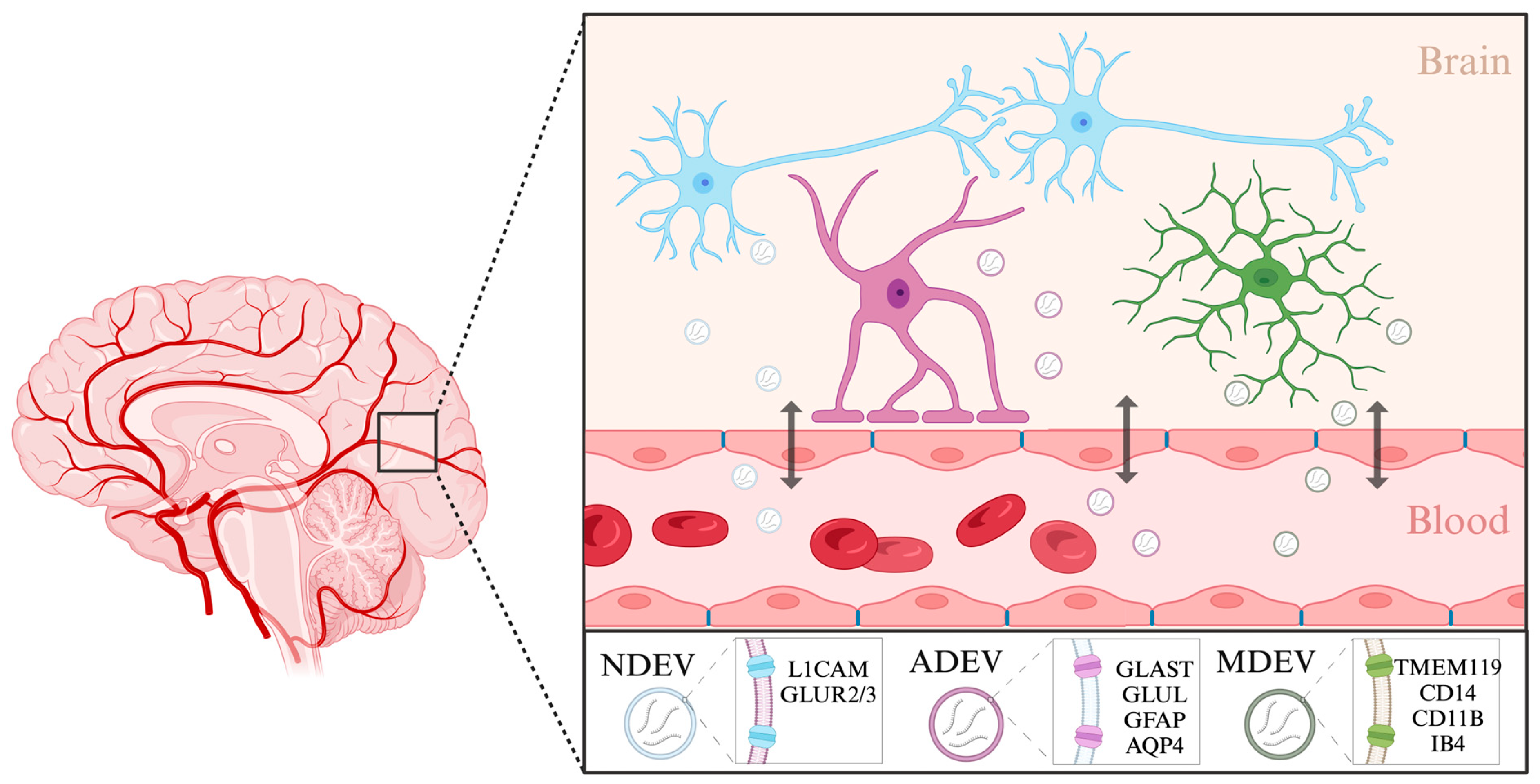
Figure 1. Characterization of brain-derived extracellular vesicles. Brain-derived extracellular vesicles enable cell-to-cell communication between different cell types in the brain as well as in the periphery. Extracellular vesicles can originate in neurons (NDEVs), astrocytes (ADEVs), and microglia (MDEVs). These vesicles can shuttle across the blood–brain barrier and enter the body’s circulation. Listed are the markers specific to each cell type that have been used to isolate extracellular vesicles from solution. Created with BioRender.com. (Retrieved from https://app.biorender.com/biorender-templates, accessed on 30 January 2024).
The isolation of astrocyte and microglia-derived EVs is also starting to be explored. One of the main forms of communication between neurons and astrocytes is through EVs, where they can have both neuroprotective and pathological properties, highlighting their importance [64]. Astrocyte-derived EVs (ADEVs) have previously been isolated from human blood using a biotinylated antibody to glutamine aspartate transporter (GLAST), an astrocyte-specific membrane protein, glutamine synthetase (GLUL) [65], as well as using glial fibrillary acidic protein (GFAP) and aquaporin 4 (AQP4) [66]. Using this approach, ADEVs have been implicated in modulating immune responses during stress and depression [67][68]. It has been shown that individuals with depression have larger numbers of GFAP and AQP4/GFAP-positive EVs in the blood than healthy controls, suggesting increased leakage of astrocyte-derived EVs through the blood–brain barrier. In a recent study, the miRNA content of ADEVs from individuals with depression was found to have significantly deregulated miR-9 levels, suggesting a specific involvement of this miRNA in the disease’s pathophysiology [69].
Similar to astrocytes, microglia also rely on EVs for cell-to-cell communication within the brain. Largely, the isolation of microglia-derived EVs from complex fluids such as blood samples has so far also been conducted using antibodies or lectins against microglial surface markers [70]. The heterogenous nature of microglial cells is inevitably reflected in their surface protein content and cargo and should thus be used to inform isolation protocols. For instance, in a stroke experimental model, EVs from microglia were isolated using markers characteristic of their specific activation state, namely TMEM119 and CD14 [71]. In another example, small EVs of microglial origin were isolated using CD11B, a more general marker of microglia [72]. Microglia or macrophage-derived EVs have also been isolated using myeloid marker IB4 [73]. Examining the contents of cell-type specific EVs enables a more targeted understanding of disease mechanisms. Further efforts to characterize extracellular vesicles and their components which originate in different brain cell types will improve researchers' understanding of disease mechanisms.
References
- Institute of Health Metrics and Evaluation. Global Health Data Exchange (GHDx); Institute of Health Metrics and Evaluation: Seattle, WA, USA, 2021.
- James, S.L.; Abate, D.; Abate, K.H.; Abay, S.M.; Abbafati, C.; Abbasi, N.; Abbastabar, H.; Abd-Allah, F.; Abdela, J.; Abdelalim, A.; et al. Global, Regional, and National Incidence, Prevalence, and Years Lived with Disability for 354 Diseases and Injuries for 195 Countries and Territories, 1990–2017: A Systematic Analysis for the Global Burden of Disease Study 2017. Lancet 2018, 392, 1789–1858.
- Kessler, R.C.; Bromet, E.J. The Epidemiology of Depression across Cultures. Annu. Rev. Public Health 2013, 34, 119–138.
- Bromet, E.; Andrade, L.H.; Hwang, I.; Sampson, N.A.; Alonso, J.; de Girolamo, G.; de Graaf, R.; Demyttenaere, K.; Hu, C.; Iwata, N.; et al. Cross-National Epidemiology of DSM-IV Major Depressive Episode. BMC Med. 2011, 9, 90.
- Nihalani, N.; Simionescu, M.; Dunlop, B.W. Depression: Phenomenology, Epidemiology, and Pathophysiology. In Depression; CRC Press: Boca Raton, FL, USA, 2010; ISBN 978-0-429-24993-8.
- Kuehner, C. Why Is Depression More Common among Women than among Men? Lancet Psychiatry 2017, 4, 146–158.
- Van de Velde, S.; Bracke, P.; Levecque, K. Gender Differences in Depression in 23 European Countries. Cross-National Variation in the Gender Gap in Depression. Soc. Sci. Med. 2010, 71, 305–313.
- Malhi, G.S.; Mann, J.J. Depression. Lancet 2018, 392, 2299–2312.
- Heim, C.; Binder, E.B. Current Research Trends in Early Life Stress and Depression: Review of Human Studies on Sensitive Periods, Gene–Environment Interactions, and Epigenetics. Exp. Neurol. 2012, 233, 102–111.
- Belmaker, R.H.; Agam, G. Major Depressive Disorder. N. Engl. J. Med. 2008, 358, 55–68.
- Hasin, D.S.; Sarvet, A.L.; Meyers, J.L.; Saha, T.D.; Ruan, W.J.; Stohl, M.; Grant, B.F. Epidemiology of Adult DSM-5 Major Depressive Disorder and Its Specifiers in the United States. JAMA Psychiatry 2018, 75, 336–346.
- Cuijpers, P.; Noma, H.; Karyotaki, E.; Vinkers, C.H.; Cipriani, A.; Furukawa, T.A. A Network Meta-Analysis of the Effects of Psychotherapies, Pharmacotherapies and Their Combination in the Treatment of Adult Depression. World Psychiatry 2020, 19, 92–107.
- Cuijpers, P.; Stringaris, A.; Wolpert, M. Treatment Outcomes for Depression: Challenges and Opportunities. Lancet Psychiatry 2020, 7, 925–927.
- Gurtan, A.M.; Sharp, P.A. The Role of miRNAs in Regulating Gene Expression Networks. J. Mol. Biol. 2013, 425, 3582.
- Friedman, R.C.; Farh, K.K.-H.; Burge, C.B.; Bartel, D.P. Most Mammalian mRNAs Are Conserved Targets of microRNAs. Genome Res. 2009, 19, 92–105.
- Gebert, L.F.R.; MacRae, I.J. Regulation of microRNA Function in Animals. Nat. Rev. Mol. Cell Biol. 2019, 20, 21–37.
- Selbach, M.; Schwanhäusser, B.; Thierfelder, N.; Fang, Z.; Khanin, R.; Rajewsky, N. Widespread Changes in Protein Synthesis Induced by microRNAs. Nature 2008, 455, 58–63.
- Griffiths-Jones, S.; Grocock, R.J.; van Dongen, S.; Bateman, A.; Enright, A.J. miRBase: MicroRNA Sequences, Targets and Gene Nomenclature. Nucleic Acids Res. 2006, 34, D140–D144.
- de Rie, D.; Abugessaisa, I.; Alam, T.; Arner, E.; Arner, P.; Ashoor, H.; Åström, G.; Babina, M.; Bertin, N.; Burroughs, A.M.; et al. An Integrated Expression Atlas of miRNAs and Their Promoters in Human and Mouse. Nat. Biotechnol. 2017, 35, 872–878.
- Smalheiser, N.R.; Lugli, G.; Zhang, H.; Rizavi, H.; Cook, E.H.; Dwivedi, Y. Expression of microRNAs and Other Small RNAs in Prefrontal Cortex in Schizophrenia, Bipolar Disorder and Depressed Subjects. PLoS ONE 2014, 9, e86469.
- Wang, W.; Kwon, E.J.; Tsai, L.-H. MicroRNAs in Learning, Memory, and Neurological Diseases. Learn. Mem. 2012, 19, 359–368.
- Issler, O.; Chen, A. Determining the Role of microRNAs in Psychiatric Disorders. Nat. Rev. Neurosci. 2015, 16, 201–212.
- Dwivedi, Y. MicroRNAs in Depression and Suicide: Recent Insights and Future Perspectives. J. Affect. Disord. 2018, 240, 146–154.
- Zhang, Y.; Zhao, Y.; Tian, C.; Wang, J.; Li, W.; Zhong, C. Differential Exosomal microRNA Profile in the Serum of a Patient with Depression. Eur. J. Psychiatry 2018, 32, 105–112.
- Kohen, R.; Dobra, A.; Tracy, J.H.; Haugen, E. Transcriptome Profiling of Human Hippocampus Dentate Gyrus Granule Cells in Mental Illness. Transl. Psychiatry 2014, 4, e366.
- Li, Y.-J.; Xu, M.; Gao, Z.-H.; Wang, Y.-Q.; Yue, Z.; Zhang, Y.-X.; Li, X.-X.; Zhang, C.; Xie, S.-Y.; Wang, P.-Y. Alterations of Serum Levels of BDNF-Related miRNAs in Patients with Depression. PLoS ONE 2013, 8, e63648.
- Yuta, Y.; Roy, B.; Dwivedi, Y. Altered miRNA Landscape of the Anterior Cingulate Cortex Is Associated with Potential Loss of Key Neuronal Functions in Depressed Brain. Eur. Neuropsychopharmacol. J. Eur. Coll. Neuropsychopharmacol. 2020, 40, 70.
- Lopez, J.P.; Lim, R.; Cruceanu, C.; Crapper, L.; Fasano, C.; Labonte, B.; Maussion, G.; Yang, J.P.; Yerko, V.; Vigneault, E.; et al. miR-1202 Is a Primate-Specific and Brain-Enriched microRNA Involved in Major Depression and Antidepressant Treatment. Nat. Med. 2014, 20, 764–768.
- Issler, O.; Haramati, S.; Paul, E.D.; Maeno, H.; Navon, I.; Zwang, R.; Gil, S.; Mayberg, H.S.; Dunlop, B.W.; Menke, A.; et al. MicroRNA 135 Is Essential for Chronic Stress Resiliency, Antidepressant Efficacy, and Intact Serotonergic Activity. Neuron 2014, 83, 344–360.
- Belzeaux, R.; Bergon, A.; Jeanjean, V.; Loriod, B.; Formisano-Tréziny, C.; Verrier, L.; Loundou, A.; Baumstarck-Barrau, K.; Boyer, L.; Gall, V.; et al. Responder and Nonresponder Patients Exhibit Different Peripheral Transcriptional Signatures during Major Depressive Episode. Transl. Psychiatry 2012, 2, e185.
- Kaurani, L.; Besse, M.; Methfessel, I.; Methi, A.; Zhou, J.; Pradhan, R.; Burkhardt, S.; Kranaster, L.; Sartorius, A.; Habel, U.; et al. Baseline Levels of miR-223-3p Correlate with the Effectiveness of Electroconvulsive Therapy in Patients with Major Depression. Transl. Psychiatry 2023, 13, 294.
- Burrows, K.; Figueroa-Hall, L.; Stewart, J.; Alarbi, A.; Kuplicki, R.; Hannafon, B.; Tan, C.; Risbrough, V.; McKinney, B.; Ramesh, R.; et al. Exploring the Role of Neuronal-Enriched Extracellular Vesicle miR-93 and Interoception in Major Depressive Disorder. Res. Sq. 2023; preprint.
- Saeedi, S.; Nagy, C.; Ibrahim, P.; Théroux, J.-F.; Wakid, M.; Fiori, L.M.; Yang, J.; Rotzinger, S.; Foster, J.A.; Mechawar, N.; et al. Neuron-Derived Extracellular Vesicles Enriched from Plasma Show Altered Size and miRNA Cargo as a Function of Antidepressant Drug Response. Mol. Psychiatry 2021, 26, 7417–7424.
- He, C.; Bai, Y.; Wang, Z.; Fan, D.; Wang, Q.; Liu, X.; Zhang, H.; Zhang, H.; Zhang, Z.; Yao, H.; et al. Identification of microRNA-9 Linking the Effects of Childhood Maltreatment on Depression Using Amygdala Connectivity. Neuroimage 2021, 224, 117428.
- Żurawek, D.; Turecki, G. The miRNome of Depression. Int. J. Mol. Sci. 2021, 22, 11312.
- Bocchio-Chiavetto, L.; Maffioletti, E.; Bettinsoli, P.; Giovannini, C.; Bignotti, S.; Tardito, D.; Corrada, D.; Milanesi, L.; Gennarelli, M. Blood microRNA Changes in Depressed Patients during Antidepressant Treatment. Eur. Neuropsychopharmacol. 2013, 23, 602–611.
- Fang, Y.; Qiu, Q.; Zhang, S.; Sun, L.; Li, G.; Xiao, S.; Li, X. Changes in miRNA-132 and miR-124 Levels in Non-Treated and Citalopram-Treated Patients with Depression. J. Affect. Disord. 2018, 227, 745–751.
- Li, J.; Meng, H.; Cao, W.; Qiu, T. MiR-335 Is Involved in Major Depression Disorder and Antidepressant Treatment through Targeting GRM4. Neurosci. Lett. 2015, 606, 167–172.
- Fiori, L.M.; Lopez, J.P.; Richard-Devantoy, S.; Berlim, M.; Chachamovich, E.; Jollant, F.; Foster, J.; Rotzinger, S.; Kennedy, S.H.; Turecki, G. Investigation of miR-1202, miR-135a, and miR-16 in Major Depressive Disorder and Antidepressant Response. Int. J. Neuropsychopharmacol. 2017, 20, 619–623.
- Funatsuki, T.; Ogata, H.; Tahara, H.; Shimamoto, A.; Takekita, Y.; Koshikawa, Y.; Nonen, S.; Higasa, K.; Kinoshita, T.; Kato, M. Changes in Multiple microRNA Levels with Antidepressant Treatment Are Associated with Remission and Interact with Key Pathways: A Comprehensive microRNA Analysis. Int. J. Mol. Sci. 2023, 24, 12199.
- Belzeaux, R.; Fiori, L.M.; Lopez, J.P.; Boucekine, M.; Boyer, L.; Blier, P.; Farzan, F.; Frey, B.N.; Giacobbe, P.; Lam, R.W.; et al. Predicting Worsening Suicidal Ideation With Clinical Features and Peripheral Expression of Messenger RNA and MicroRNA During Antidepressant Treatment. J. Clin. Psychiatry 2019, 80, 18m12556.
- Yrondi, A.; Fiori, L.M.; Frey, B.N.; Lam, R.W.; MacQueen, G.M.; Milev, R.; Müller, D.J.; Foster, J.A.; Kennedy, S.H.; Turecki, G. Association Between Side Effects and Blood microRNA Expression Levels and Their Targeted Pathways in Patients With Major Depressive Disorder Treated by a Selective Serotonin Reuptake Inhibitor, Escitalopram: A CAN-BIND-1 Report. Int. J. Neuropsychopharmacol. 2020, 23, 88–95.
- Wang, X.; Sundquist, K.; Hedelius, A.; Palmér, K.; Memon, A.A.; Sundquist, J. Circulating microRNA-144-5p Is Associated with Depressive Disorders. Clin. Epigenet. 2015, 7, 69.
- Kuang, W.-H.; Dong, Z.-Q.; Tian, L.-T.; Li, J. MicroRNA-451a, microRNA-34a-5p, and microRNA-221-3p as Predictors of Response to Antidepressant Treatment. Braz. J. Med. Biol. Res. 2018, 51, e7212.
- Lopez, J.P.; Fiori, L.M.; Cruceanu, C.; Lin, R.; Labonte, B.; Cates, H.M.; Heller, E.A.; Vialou, V.; Ku, S.M.; Gerald, C.; et al. MicroRNAs 146a/b-5 and 425-3p and 24-3p Are Markers of Antidepressant Response and Regulate MAPK/Wnt-System Genes. Nat. Commun. 2017, 8, 15497.
- Zheng, Y.-B.; Sheng, X.-M.; Jin, X.; Guan, W. MiR-182-5p: A Novel Biomarker in the Treatment of Depression in CSDS-Induced Mice. Int. J. Neuropsychopharmacol. 2023, 27, pyad064.
- Li, Y.; Li, S.; Yan, J.; Wang, D.; Yin, R.; Zhao, L.; Zhu, Y.; Zhu, X. miR-182 (microRNA-182) Suppression in the Hippocampus Evokes Antidepressant-like Effects in Rats. Prog. Neuropsychopharmacol. Biol. Psychiatry 2016, 65, 96–103.
- Li, Y.; Wang, N.; Pan, J.; Wang, X.; Zhao, Y.; Guo, Z. Hippocampal miRNA-144 Modulates Depressive-Like Behaviors in Rats by Targeting PTP1B. Neuropsychiatr. Dis. Treat. 2021, 17, 389–399.
- Gheysarzadeh, A.; Sadeghifard, N.; Afraidooni, L.; Pooyan, F.; Mofid, M.R.; Valadbeigi, H.; Bakhtiari, H.; Keikhavani, S. Serum-Based microRNA Biomarkers for Major Depression: MiR-16, miR-135a, and miR-1202. J. Res. Med. Sci. 2018, 23, 69.
- Roy, B.; Ochi, S.; Dwivedi, Y. Potential of Circulating miRNAs as Molecular Markers in Mood Disorders and Associated Suicidal Behavior. Int. J. Mol. Sci. 2023, 24, 4664.
- Shi, Y.; Wang, Q.; Song, R.; Kong, Y.; Zhang, Z. Non-Coding RNAs in Depression: Promising Diagnostic and Therapeutic Biomarkers. eBioMedicine 2021, 71, 103569.
- Wan, Y.; Liu, Y.; Wang, X.; Wu, J.; Liu, K.; Zhou, J.; Liu, L.; Zhang, C. Identification of Differential microRNAs in Cerebrospinal Fluid and Serum of Patients with Major Depressive Disorder. PLoS ONE 2015, 10, e0121975.
- Saeedi, S.; Israel, S.; Nagy, C.; Turecki, G. The Emerging Role of Exosomes in Mental Disorders. Transl. Psychiatry 2019, 9, 122.
- Buzas, E.I.; György, B.; Nagy, G.; Falus, A.; Gay, S. Emerging Role of Extracellular Vesicles in Inflammatory Diseases. Nat. Rev. Rheumatol. 2014, 10, 356–364.
- Hussain, M.T.; Iqbal, A.J.; Norling, L.V. The Role and Impact of Extracellular Vesicles in the Modulation and Delivery of Cytokines during Autoimmunity. Int. J. Mol. Sci. 2020, 21, 7096.
- Kalluri, R.; McAndrews, K.M. The Role of Extracellular Vesicles in Cancer. Cell 2023, 186, 1610–1626.
- Raghav, A.; Singh, M.; Jeong, G.-B.; Giri, R.; Agarwal, S.; Kala, S.; Gautam, K.A. Extracellular Vesicles in Neurodegenerative Diseases: A Systematic Review. Front. Mol. Neurosci. 2022, 15, 1061076.
- Kong, L.; Zhang, D.; Huang, S.; Lai, J.; Lu, L.; Zhang, J.; Hu, S. Extracellular Vesicles in Mental Disorders: A State-of-Art Review. Int. J. Biol. Sci. 2023, 19, 1094–1109.
- Wei, Z.-X.; Xie, G.-J.; Mao, X.; Zou, X.-P.; Liao, Y.-J.; Liu, Q.-S.; Wang, H.; Cheng, Y. Exosomes from Patients with Major Depression Cause Depressive-like Behaviors in Mice with Involvement of miR-139-5p-Regulated Neurogenesis. Neuropsychopharmacology 2020, 45, 1050–1058.
- Levine, A.; Strawn, J.R. Blood Tests of Brain Function: Neuronal Extracellular Vesicles. Biomark. Neuropsychiatry 2022, 7, 100058.
- Mizohata, Y.; Toda, H.; Koga, M.; Saito, T.; Fujita, M.; Kobayashi, T.; Hatakeyama, S.; Morimoto, Y. Neural Extracellular Vesicle-Derived miR-17 in Blood as a Potential Biomarker of Subthreshold Depression. Hum. Cell 2021, 34, 1087–1092.
- Sun, B.; Dalvi, P.; Abadjian, L.; Tang, N.; Pulliam, L. Blood Neuron-Derived Exosomes as Biomarkers of Cognitive Impairment in HIV. AIDS 2017, 31, F9–F17.
- Lachenal, G.; Pernet-Gallay, K.; Chivet, M.; Hemming, F.J.; Belly, A.; Bodon, G.; Blot, B.; Haase, G.; Goldberg, Y.; Sadoul, R. Release of Exosomes from Differentiated Neurons and Its Regulation by Synaptic Glutamatergic Activity. Mol. Cell Neurosci. 2011, 46, 409–418.
- Datta Chaudhuri, A.; Dasgheyb, R.M.; DeVine, L.R.; Bi, H.; Cole, R.N.; Haughey, N.J. Stimulus-Dependent Modifications in Astrocyte-Derived Extracellular Vesicle Cargo Regulate Neuronal Excitability. Glia 2020, 68, 128–144.
- Goetzl, E.J.; Mustapic, M.; Kapogiannis, D.; Eitan, E.; Lobach, I.V.; Goetzl, L.; Schwartz, J.B.; Miller, B.L. Cargo Proteins of Plasma Astrocyte-Derived Exosomes in Alzheimer’s Disease. FASEB J. 2016, 30, 3853–3859.
- Wallensten, J.; Nager, A.; Åsberg, M.; Borg, K.; Beser, A.; Wilczek, A.; Mobarrez, F. Leakage of Astrocyte-Derived Extracellular Vesicles in Stress-Induced Exhaustion Disorder: A Cross-Sectional Study. Sci. Rep. 2021, 11, 2009.
- Han, J.; Cho, H.-J.; Park, D.; Han, S. DICAM in the Extracellular Vesicles from Astrocytes Attenuates Microglia Activation and Neuroinflammation. Cells 2022, 11, 2977.
- Long, X.; Yao, X.; Jiang, Q.; Yang, Y.; He, X.; Tian, W.; Zhao, K.; Zhang, H. Astrocyte-Derived Exosomes Enriched with miR-873a-5p Inhibit Neuroinflammation via Microglia Phenotype Modulation after Traumatic Brain Injury. J. Neuroinflamm. 2020, 17, 89.
- Luarte, A.; Nardocci, G.; Chakraborty, A.; Batiz, L.F.; Pino-Lagos, K.; Wyneken, Ú. Astrocyte-Derived Extracellular Vesicles in Stress-Associated Mood Disorders. Does the Immune System Get Astrocytic? Pharmacol. Res. 2023, 194, 106833.
- Gabrielli, M.; Raffaele, S.; Fumagalli, M.; Verderio, C. The Multiple Faces of Extracellular Vesicles Released by Microglia: Where Are We 10 Years After? Front. Cell. Neurosci. 2022, 16, 984690.
- Roseborough, A.D.; Myers, S.J.; Khazaee, R.; Zhu, Y.; Zhao, L.; Iorio, E.; Elahi, F.M.; Pasternak, S.H.; Whitehead, S.N. Plasma Derived Extracellular Vesicle Biomarkers of Microglia Activation in an Experimental Stroke Model. J. Neuroinflamm. 2023, 20, 20.
- Cohn, W.; Melnik, M.; Huang, C.; Teter, B.; Chandra, S.; Zhu, C.; McIntire, L.B.; John, V.; Gylys, K.H.; Bilousova, T. Multi-Omics Analysis of Microglial Extracellular Vesicles From Human Alzheimer’s Disease Brain Tissue Reveals Disease-Associated Signatures. Front. Pharmacol. 2021, 12, 766082.
- Scaroni, F.; Visconte, C.; Serpente, M.; Golia, M.T.; Gabrielli, M.; Huiskamp, M.; Hulst, H.E.; Carandini, T.; De Riz, M.; Pietroboni, A.; et al. miR-150-5p and Let-7b-5p in Blood Myeloid Extracellular Vesicles Track Cognitive Symptoms in Patients with Multiple Sclerosis. Cells 2022, 11, 1551.
More
Information
Subjects:
Biology
Contributor
MDPI registered users' name will be linked to their SciProfiles pages. To register with us, please refer to https://encyclopedia.pub/register
:
View Times:
384
Revisions:
2 times
(View History)
Update Date:
15 Mar 2024
Table of Contents


Notice
You are not a member of the advisory board for this topic. If you want to update advisory board member profile, please contact office@encyclopedia.pub.
OK
Confirm
Only members of the Encyclopedia advisory board for this topic are allowed to note entries. Would you like to become an advisory board member of the Encyclopedia?
Yes
No
${ textCharacter }/${ maxCharacter }
Submit
Cancel
Back
Comments
${ item }
|
${ item.createdUser.fullName }
${ item.createdAt }
${ item.vote }
${ item.reply }
Delete
${ reply.createdUser.fullName }
${ reply.createdAt }
${ reply.vote }
Delete
There is no reply to this comment~
${ item.replyTextCharacter }/${ item.replyMaxCharacter }
Submit
Cancel
More
No more~
There is no comment~
${ textCharacter }/${ maxCharacter }
Submit
Cancel
${ selectedItem.replyTextCharacter }/${ selectedItem.replyMaxCharacter }
Submit
Cancel
Confirm
Are you sure to Delete?
Yes
No