Submitted Successfully!
To reward your contribution, here is a gift for you: A free trial for our video production service.
Thank you for your contribution! You can also upload a video entry or images related to this topic.
Version Summary Created by Modification Content Size Created at Operation
1 -- 8317 2024-03-06 07:52:56 |
2 Reference format revised. Meta information modification 8317 2024-03-07 01:50:50 |

Video Upload Options

Do you have a full video?

Confirm

Are you sure to Delete?
Cite
If you have any further questions, please contact Encyclopedia Editorial Office.
Mecca, M.; Picerno, S.; Cortellino, S. Interconnection between Inflammation, Epigenetics and Nutrition in Cancer. Encyclopedia. Available online: https://encyclopedia.pub/entry/55902 (accessed on 09 May 2024).
Mecca M, Picerno S, Cortellino S. Interconnection between Inflammation, Epigenetics and Nutrition in Cancer. Encyclopedia. Available at: https://encyclopedia.pub/entry/55902. Accessed May 09, 2024.
Mecca, Marisabel, Simona Picerno, Salvatore Cortellino. "Interconnection between Inflammation, Epigenetics and Nutrition in Cancer" Encyclopedia, https://encyclopedia.pub/entry/55902 (accessed May 09, 2024).
Mecca, M., Picerno, S., & Cortellino, S. (2024, March 06). Interconnection between Inflammation, Epigenetics and Nutrition in Cancer. In Encyclopedia. https://encyclopedia.pub/entry/55902
Mecca, Marisabel, et al. "Interconnection between Inflammation, Epigenetics and Nutrition in Cancer." Encyclopedia. Web. 06 March, 2024.
Interconnection between Inflammation, Epigenetics and Nutrition in Cancer
Edit

Inflammation is a key contributor to both the initiation and progression of tumors, and it can be triggered by genetic instability within tumors, as well as by lifestyle and dietary factors. The inflammatory response plays a critical role in the genetic and epigenetic reprogramming of tumor cells, as well as in the cells that comprise the tumor microenvironment. Cells in the microenvironment acquire a phenotype that promotes immune evasion, progression, and metastasis.

inflammation epigenetic DNA repair nutrition cancer

1. Introduction

Cancer is a complex disease that is influenced by both intrinsic and extrinsic factors. The accumulation of genetic mutations is a key trigger in the development and progression of tumors, but it is not sufficient on its own. In the early stages of cancer, the immune system acts as a guardian and limits tumor growth. However, tumor cells can develop mutations that allow them to evade recognition by the immune system, leading to their expansion and growth (immunoediting theory) [1]. To evade the immune response, tumor cells use various strategies. Recently, it has been discovered that the genetic instability of tumor cells induces an inflammatory response due to the release of nuclear DNA into the cytoplasm, caused by DNA repair defects [2][3]. One of the contributing factors to cancer progression is persistent inflammation, which is accompanied by the production of proinflammatory cytokines that recruit myeloid cells to the tumor microenvironment. The myeloid cells then differentiate into immunosuppressive cells, thereby fostering an environment that encourages tumor growth [4]. In addition, inflammation contributes to the epigenetic remodeling of both the tumor cells and the immune system, leading to the exhaustion of T lymphocytes and the acquisition of a malignant and aggressive phenotype by the tumor cells [5]. In this context, nutrition can play a crucial role in preventing tumor onset, reducing inflammation, and influencing the genetic and epigenetic reprogramming of tumor cells [6].

2. Inflammation

Inflammation is a highly intricate process that is regulated by the immune system in response to external or internal stimuli that threaten tissue integrity [7]. The innate immune system is primarily responsible for triggering this response. It recognizes molecules or portions of molecules that are released upon cellular stress or tissue injury, such as damage-associated molecular patterns (DAMPs), or are specific to pathogens, such as pathogen-associated molecular patterns (PAMPs). PAMPs and DAMPs bind to pattern recognition receptors (PRRs) located on the cytoplasmic or endosome membrane, activating intracellular signaling cascades that result in the expression of proinflammatory factors [8]. PRRs include toll-like receptors (TLRs), retinoic acid-inducible gene 1-(RIG1)-like receptors (RLRs), cytosolic DNA sensor cyclic GMP–AMP synthase (cGAS) stimulator of interferon genes (STINGs), C-type lectin receptors (CLRs), and nucleotide-binding oligomerization domain (NOD)-like receptors (NLRs) (Figure 1).
Figure 1. Inflammatory signaling pathways. cGAS is activated upon the recognition of cytosolic double-stranded DNA derived from viruses, bacteria, dead cells, or mislocalized endogenous DNA. Activated cGAS synthesizes 2′-3′ cyclic GMP-AMP (cGAMP), which promotes the translocation of stimulator of interferon gene (STING) from the endoplasmic reticulum (ER) membrane to the ER-Golgi intermediate and Golgi compartments. STING forms a complex with TANK-binding kinase 1 (TBK1), which recruits and activates interferon regulatory factor 3 (IRF3), leading to the transcription of genes encoding inflammatory cytokines such as interleukin 6 (IL-6), interleukin 12 (IL-12), and interferons (IFNs). RIG-I-like receptors (RLRs), including melanoma differentiation-associated gene 5 (MDA5), laboratory of genetics and physiology 2 (LGP2), and retinoic acid-inducible gene I (RIG-I), interact with viral double-stranded RNA (dsRNA) and 5′-triphosphate single-stranded RNA and bind to mitochondrial antiviral-signaling protein (MAVS), which activates IRF3 and 7 via TBK1 and IκB Kinase ε (IKKε). Phospho-IRF3 and phospho-IRF7 transcribe genes encoding IFNs and immunoregulatory genes. Toll-like receptors (TLRs) recruit adapter protein myeloid differentiation primary response 88 (MyD88) and TIR domain-containing adaptor protein (TRIF) upon recognition of pathogen-associated molecular patterns (PAMPs) or damage-associated molecular patterns (DAMPs) on the plasma membrane or in endosomes (not depicted in the caption). MyD88 and TRIF initiate a signaling cascade resulting in the activation of NLR inflammasome, nuclear factor kappa B (NF-κB), interferon regulatory factors (IRFs), or mitogen-activated protein kinases (MAPK) which result in transcription of proinflammatory cytokines and inflammasome component NLR family pyrin domain containing 3 (NLRP3). The stimulation of NLRP3 and apoptosis-associated speck-like protein containing a CARD (ASC) primes the assembly of the inflammasome complex, which triggers caspase-1 mediated cleavage of pro-IL-1β and pro-IL-18 and Gasdermin D precursor, which forms a pore on the plasma membrane through IL-1β and IL-18 being released into the extracellular matrix. C-type lectin receptors (CLRs) play a crucial role in modulating Toll-like receptor (TLR) signaling. They achieve this through two distinct mechanisms: either by activating NF-kB via RAS-RAF1 dependent signaling, or by recruiting spleen tyrosine kinase (SYK) to the phosphorylated immunoreceptor tyrosine-based activation motif (ITAM) of the paired signaling adaptor Fc receptor γ-chain (FcRγ). The recruitment of SYK to FcRγ inhibits the recruitment of MYD88, thereby reducing the production of TLR-induced cytokines (created with BioRender.com).
TLRs, transmembrane proteins, are located on the cytoplasmic membrane or intracellular compartments. They recognize PAMPs or DAMPs, recruit MyD88 and TRIF adapter proteins, and activate the signaling cascade, leading to nuclear factor kappaB (NF-κB), interferon regulatory factors (IRFs), or mitogen-activated protein kinase (MAPK) activation and cytokines, chemokines, and type I interferon (IFN) expression [9] (Figure 1).
The RLR family encompasses melanoma differentiation-associated factor 5 (MDA5), laboratory of genetics and physiology 2 (LGP2), and RIG-1 [10][11]. Upon engagement with double-stranded RNA (dsRNA) or 5′-triphosphate single-stranded RNA, MDA5, LGP2, and RIG-I bind to mitochondrial antiviral signaling proteins (MAVS), activating interferon regulatory factor 3 (IRF3) and 7 (IRF7) through TANK-binding kinase 1 (TBK1) and IκB kinase ε (IKKε) [10][12]. IRF3 and IRF7, in combination with NF-κB and activator protein-1 (AP-1), promote the transcription of type I IFNs and other antiviral or immunoregulatory genes [10][12]. Unlike MDA5, RIG-I also binds to double-stranded viral DNA and leads to the expression of type I IFNs through the transcription of IRF3 [13]. The RIG-I-mediated inflammatory response can also be induced by lipopolysaccharide (LPS), interferon gamma (IFN-γ), interleukin (IL)-1β, and tumor necrosis factor-alpha (TNF-α) [14][15][16][17] (Figure 1).
The cGAS-STING signaling pathway plays a crucial role in mediating the inflammatory response to infections, cellular stress, and tissue damage by binding to pathogenic and nonpathogenic DNA [18]. Upon binding to dsDNA, activated cGAS synthesizes 2′3′ cyclic GMP-AMP (cGAMP), which promotes the translocation of STING from the ER membrane to the ER-Golgi intermediate and Golgi compartments [19]. STING then forms a complex with TBK1, which activates IRF3 and NFkB, leading to the transcription of dozens of genes encoding inflammatory cytokines such as IL6 and IL12, as well as antiviral type I interferons [20][21][22] (Figure 1).
CLRs are soluble and transmembrane proteins expressed by myeloid cells, particularly by macrophages and dendritic cell (DC) subsets, which are divided into two groups: the mannose receptor family [23][24] and the asialoglycoprotein receptor family [25][26]. CLRs recognize mannose, fucose, and glucan structures expressed by pathogens [27][28] and promote pathogen internalization and degradation, leading to subsequent antigen processing and presentation [28]. CLRs modulate cytokine production by inducing multiple signal transduction cascades mediated by immunoreceptor tyrosine-based activation motif (ITAM)-containing adapter molecules such as the Fc receptor γ chain (FcRγ), or by activating protein kinases such as RAS and RAF and phosphatases such as SHP1 or SHP2 [25][29].
NLRs form a family of PRR proteins, characterized by the presence of a C-terminal leucine-rich repeat (LRR), a central nucleotide-binding domain (NOD), and an N-terminal effector domain [30]. Based on their N-terminal structure, NLRs are classified into five subgroups: NLRA, NLRB, NLRC, NLRP, and NLRX [31]. Since only some NLRs, such as NOD1 and NOD2, recognize and bind specific microbial products, NLRs serve as modulators of the TLR, RLR, and CLR signaling pathways. NOD1 and NOD2 activate NF-kB by interacting with RIP2, a receptor-interacting protein kinase. The activation of NF-kB by NOD1 occurs through the formation of a transient NOD1-RIP2-IKK complex, while NOD2 activates NF-kB via RIP2-dependent ubiquitination of NEMO, resulting in the release of pro-inflammatory cytokines, such as TNF-a, IL-1b, and IL-6, by monocytes and dendritic cells (DCs) [32][33].
The inflammatory response involves several NLRs, including NLRP1, NLRP3, and NLRC4, which form part of the inflammasome, a multiprotein complex. The inflammasome consists of sensor proteins, such as NLRs, absent in melanoma 2 (AIM2), and pyrin, as well as scaffold proteins, including apoptosis-associated speck-like protein containing a CARD (ASC) and the cysteine protease procaspase-1 [34][35]. The expression of pro-interleukin-1β (pro-IL-1β), pro-IL-18, and nod-like receptor protein 3 (NLRP3) is regulated by NF-κB, which primes inflammasome activation. In the canonical pathway, sensor proteins bind to DAMPs and PAMPs, leading to oligomerization and the recruitment of ASC to form a multimeric complex called a “speck”. The ASC speck complex promotes the recruitment and activation of caspase 1, resulting in inflammasome activation and the conversion of pro-IL-1β and pro-IL-18 into active IL-1β and IL-18. The inflammasome also cleaves the precursor of Gasdermin D (GSDMD), releasing the amino-terminal domain of GSDMD, which forms a pore on the plasma membrane. Mature IL-1β and IL-18 are released through this pore [36][37][38][39][40]. The secondary self-cleavage of CAS1 inactivates its enzymatic activity and inhibits inflammasome function [41][42].
In addition to the canonical caspase 1-dependent pathway, the inflammasome can be activated through caspase 4- and 5-dependent pathways, known as the non-canonical pathway. This non-canonical caspase-4/5 inflammasome is activated by intracellular LPS and requires NLRP3 for its activation. It plays a crucial role in supervising cytosolic Gram-negative bacteria by sensing lipopolysaccharide (LPS). Extracellular LPS generated by Gram-negative bacteria is internalized via TLR4- or RAGE/HMGB1-mediated endocytosis. 
Inflammasome activation can have mild or severe consequences on cell fate. Mild activation of the inflammasome allows the endosomal sorting complex required for transport (ESCRT) machinery to remove GSDMD pores and repair the plasma membrane, whereas robust activation of the inflammasome can generate a number of pores, exceeding the capacity of the ESCRT machinery to resolve them. 
Inflammasome activation triggers the release of proinflammatory cytokines and DAMPs, including high-mobility group box 1 (HMGB1). HMGB1 can be released into the extracellular space both actively secreted by inflammatory and immune cells and passively secreted by damaged or necrotic cells with compromised plasma membranes. These proinflammatory cytokines and DAMPs act as chemoattractants for neutrophils, macrophages, monocytes, dendritic cells, and T cells. 
The activation of inflammasomes results in the production of pro-inflammatory cytokines, such as IL-1α, IL-1β, and IL-18, as well as the maturation of anti-inflammatory cytokines like IL-37. IL-1α is responsible for inducing the release of pro-inflammatory cytokines such as TNFα and IL-2, and acts as a chemoattractant signal for neutrophils. IL-1β stimulates inflammatory responses by releasing pro-inflammatory cytokines such as IL-6 and IL-17a, as well as recruiting macrophages [43]. The pro-inflammatory cytokine IL-18 enhances adaptive immune responses by regulating leukocyte trafficking via chemokine production and promoting the secretion of proinflammatory cytokines IFNγ, IL-2, and IL-12 [44][45]. IL-37, secreted in response to inflammatory signaling, downregulates the production of pro-inflammatory cytokines to restrain immune cell toxicity and prevent tissue damage [46].
Generally, organisms possess intricate systems that identify and eradicate pathogens, protect against external infections, and maintain tissue homeostasis. The activation of these systems triggers an acute inflammatory response that involves the remodeling of local tissue through the release of cytokines that attract immune cells and regulate their functions. Acute inflammation is beneficial because it can quickly resolve tissue damage caused by pathogens or internal stimuli. However, if acute inflammation fails to eliminate the causes of inflammation and repair tissue damage, the inflammatory response persists, becomes chronic, and develops new characteristics that can damage DNA and compromise tissue health [47]

3. DNA Damage Response in Inflammation

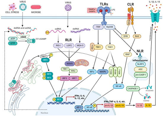
3.1. Mechanism Underlying DNA Damage-Induced Inflammation

DNA repair mechanisms and signaling pathways are closely linked to the inflammatory response. DNA damage activates the cGAS-STING pathway, which induces the expression of type I interferons and inflammatory factors. cGAS triggers this process by detecting endogenous DNA released from the nucleus, mitochondria, or micronuclei into the cytoplasm as well as potentially exogenous DNA derived from pathogenic microorganisms [48][49][50]. Micronuclei, which are small, membrane-enclosed structures composed of acentric chromosomal fragments, are generated as a result of defects in DNA repair processes, chromosomal segregation, and non-disjunction. These defects can be caused by mutations in genes involved in the DNA damage response (DDR) and the regulation of the mitotic spindle, or they can be induced by DNA-damaging treatments [51][52][53]. Unrepaired or misrepaired DNA double-strand breaks (DSBs) lead to the formation of acentric chromatids that segregate improperly during anaphase, are excluded from the nucleus, and are enveloped by the nuclear membrane [51].
Although the c-GAS-STING pathway serves as the primary mechanism to induce inflammatory responses to genotoxic stress, DDR proteins can also trigger inflammation by directly or indirectly activating NF-kB. Damaged DNA, induced by genotoxic agents, recruits and activates ATM and ATM and RAD3 related (ATR), which can stimulate NF-kB activation through the following: (1) stabilization of GATA4 through inhibition of p62 and autophagic degradation of GATA4 [54]; (2) assembly of an alternative STING signaling complex including the tumor suppressor p53 and the E3 ubiquitin ligase TRAF6 [55]; (3) degradation of IκBα through the formation of the IκBα-β-TrCP-ubiquitin ligase complex and phosphorylation of RELA (also known as p65) through interaction with protein kinase A (PKA) [56].

3.2. DNA Repair Deficiency Disorder and Inflammation

The DDR is a vital mechanism that ensures the preservation of genomic integrity and prevents tumor formation by employing an intricate signaling network that detects, signals, and repairs DNA damage. Numerous recent investigations have pointed out that the deletion or mutation of DDR genes, both inherited and spontaneous, as well as the genotoxic stress induced by DNA-damaging treatments, can trigger inflammation by activating the cGAS-STING pathway and other associated signaling cascades [48][57][58][59][60][61].
The deficiency of BRCA1 or BRCA2 tumor suppressors, which play a crucial role in repairing DNA breaks through the promotion of the homologous recombination (HR) DNA repair pathway, and in maintaining the stability of newly synthesized DNA strands by safeguarding stalled replication forks from degradation, also triggers type I IFN signaling and anti-tumor immunity. Cells lacking BRCA1 or BRCA2 exhibit elevated levels of cytosolic DNA and the constitutively active viral response cGAS/STING/TBK1/IRF3 pathway [60][61].
The c-GAS-STING pathway can also be activated by other proteins involved in DNA repair. For instance, the inactivation of BLM, a DNA helicase involved in the repair of DNA double-strand breaks through the homologous recombination (HR) pathway, can compromise the integrity of DNA by blocking the restart of stalled replication forks [62], as well as DNA double-strand break resection [63] and Holliday junction dissolution. BLM mutations are responsible for Bloom syndrome, a genetic disorder characterized by genetic instability and an increased risk of cancer.
Even the inhibition of proteins involved in base excision repair (BER) can elicit an interferon response through activation of the cGAS pathway. Mutations in the DNA polymerase beta (POLB) protein, which is involved in BER and fills in single-nucleotide gaps, result in replication-associated genomic instability and inflammation-associated carcinogenesis in mice. This occurs because failure to repair the damage leads to the formation of single-strand breaks (SSBs), which are converted into double-strand breaks (DSBs) during the S-phase of DNA replication. The accumulation of DSBs causes mitotic dysfunction, leading to an increase in micronuclei formation due to the mis-segregation of broken chromosomes during mitosis. This, in turn, triggers a c-GAS-mediated inflammatory response by releasing cytosolic DNA [64].

3.3. Inflammation Promoted by Transposable Element (TE) Instability

Dysfunctions in the DNA damage response pathway can result in a strong inflammatory response by accelerating the activation of transposable elements (TEs), which are DNA sequences capable of self-replication and relocating within the same genome [65]. The eukaryotic genome is primarily composed of repetitive DNA sequences, including satellite DNA and TEs. TEs are divided into two groups: retrotransposable elements (RTEs), such as long interspersed nuclear elements (LINEs) and short interspersed nuclear elements (SINEs), and endogenous retroviruses (ERVs) [66][67][68]. Active RTEs produce cytosolic DNA (cDNAs) that activate the cGAS-STING pathway and initiate the production of pro-inflammatory cytokines, such as IFNα, IFNβ, IL-6, and TNF, through NF-κB and IRF3 stimulation. Although the mechanism underlying RTE-induced inflammation remains unclear, it is believed that abnormal DNA damage repair mechanisms can result in the accumulation and dispersion of cDNA in the cytoplasm by affecting the integration of RTE DNA into the genome.

3.4. Inflammation Triggered by R-Loops Resolving Defects

Inflammation can arise from disruptions in the mechanisms responsible for maintaining and resolving R-loops. R-loops are nucleic acid structures comprising DNA-RNA hybrids and displaced single-stranded DNA resulting from transcription and replication [69]. If these structures persist for an extended period, endonucleases cut the exposed single-stranded DNA, leading to single- and double-strand breaks. Alterations in helicases, such as SENATAXIN, BLM, and WRN, and endonucleases, such as ERCC1 and XPG, can impair R-loop resolution [70][71][72][73][74]. The loss of tumor suppressors BRCA1 and BRCA2, which are involved in repairing double-strand breaks, can contribute to an abnormal accumulation of R-loops and cytosolic DNA, resulting in a heightened inflammatory response [75][76].

3.5. Anti-Tumor Cytotoxicity Mediated by Inflammatory Response

The effectiveness of chemotherapeutic agents, including anthracyclines, oxaliplatin, and doxorubicin, depends, in part, on their ability to induce anti-tumor immune responses through type I IFN signaling and ISGs [77][78]. Similarly, the anti-tumor immunity triggered by ionizing radiation (IR) is dependent on the cGAS-STING pathway [79]. Recent studies have identified the accumulation of cytosolic DNA following chemotherapy or radiation as the trigger for cGAS-STING activation, leading to the induction of IFN and ISGs [52][53][80]. Cytosolic DNA is generated as a result of double-strand break repair processes triggered by ionizing radiation or chemotherapy. Specifically, enzymes such as BLM helicase and exonuclease 1 (EXO1), which play a crucial role in the final resection of DNA during double-strand break repair, generate these DNA fragments that can either be enclosed in micronuclei or migrate to the cytoplasm, where they activate the cGAS-STING pathway [53][80].
Inhibiting or deactivating DDR pathways can enhance the inflammatory response and improve the immune response induced by genotoxic stress agents. PARP inhibitors (PARPi) increase endogenous genomic instability in BRCA1/2-deficient cells and tumors by blocking DNA repair mechanisms and replication fork progression, which leads to an increase in cytosolic DNA and activates the cGAS-STING pathway [81][82][83]. As a result, ISGs are expressed in BRCA1/2-deficient tumor cells and stimulate T-cell infiltration and activation, ultimately leading to tumor eradication in BRCA1/2-deficient mouse models of ovarian and breast cancer [84].

4. Epigenetics and Inflammation

4.1. Epigenetic Regulatory Mechanisms

Epigenetic modulators are divided into (1) writers, which add covalent modifications to histones or DNA; (2) erasers, which remove histones and DNA modifications; and (3) readers, which recognize and bind epigenetic modifications [85][86][87] (Figure 2).
Figure 2. Key epigenetic mechanisms controlling cancer, inflammation, and nutrition. Epigenetic mechanisms such as DNA methylation, histone modifications, and non-coding RNAs play a significant role in regulating the expression of genes associated with inflammation, cancer, and nutrient conditions. In cancer cells, alterations in metabolic pathways occur during tumorigenesis and progression. These metabolic changes are often closely linked to epigenetic modifications and are influenced by inflammation and nutrition. The balance of histone and DNA modifications is crucial for proper regulation. The level of histone acetylation is regulated by the opposing activities of histone acetyltransferases (HATs) and histone deacetylases (HDACs). High levels of histone acetylation promote gene transcription by opening chromatin and facilitating the recruitment of transcription factors and the transcriptional machinery. Histone methyltransferase (KMT) enzymes possess the ability to monomethylate, dimethylate, or trimethylate histone tail lysine and arginine residues, which can subsequently be removed by histone demethylase (KDM) enzymes. The methylation of histones at specific lysine and arginine residues is critical for determining the structure of chromatin and the recruitment of transcriptional repressors or activators. The enzyme DNA methyltransferases (DNMTs) catalyze the DNA methylation of the 5-carbon of cytosine, resulting in the formation of 5-methylcytosine. On the other hand, the removal of the methyl group is carried out by the activity of the ten-eleven translocation (TET) methylcytosine dioxygenase enzymes, which progressively oxidize 5-methylcytosine to 5-hydroxymethylcytosine, 5-formylcytosine, and 5-carboxylcytosine. Thymine-DNA glycosylase (TDG) removes 5-formylcytosine and 5-carboxylcytosine, which are then replaced by cytosine. In human cancers, CpG methylation promotes transcriptional silencing and malignant transformation, while hypomethylation of the transposable element DNA leads to genomic instability and an inflammatory response. Long non-coding RNAs (lncRNAs) can interact with chromatin modifiers and recruit them to the promoters of target genes, where they can activate or suppress transcription. Additionally, lncRNAs can sequester chromatin modifiers away from the promoters of target genes and regulate transcription. Mature microRNAs (miRNAs) are incorporated into a large protein complex known as RNA-induced silencing complex (RISC). This complex either cleaves messenger RNA (mRNA) or induces translational repression by binding to the 3′ untranslated region (UTR) of the target mRNA. Alternatively, it can induce translational activation if it binds to open reading frame (ORF) sequences or the 5′-UTR. miRNAs can modulate gene transcription through direct binding or by altering methylation patterns at the promoter level of the target gene (created with BioRender.com).
Histone modifications. Histone acetylation occurs on lysine and arginine residues and is carried out by histone acetyl transferases (HATs), which act as epigenetic regulators. The addition of an acetyl group neutralizes the positive charge of lysine and weakens the bond between the histone and negatively charged DNA, thus allowing for more open chromatin and more accessible transcription factors [88]. Histone deacetylases (HDACs) counteract the function of HATs by removing acetyl groups from histones, thus promoting chromatin compaction and silencing gene expression. HDACs include four classes; classes I, II, and IV are zinc-dependent, whereas class III HDACs, also known as sirtuins, depend on nicotinamide adenine dinucleotide (NAD+) [89] (Figure 2).
Histone lysine methyl transferases (KMTs) label histones by adding mono-/di-/or tri-methyl groups to the lysine residue, thus generating a histone methylation pattern known as histone codes [90]. Histone methylation can repress or activate gene transcription, depending on the position of the methylated lysine. The trimethylation of lysine 27 on histone 3 (H3K27me3) is known to result in the silencing of gene expression, whereas the dimethylation of lysine 4 on histone 3 (H3K4me) promotes gene transcription by increasing DNA accessibility [91]. Histone-specific demethylases, like lysine-specific histone demethylase (LSD)1 and Jumonji-C (JMJC) families, remove methyl groups from histones and modify the chromatin structure, promoting epigenetic plasticity and gene transcription modulation in response to internal and external stimuli [92] (Figure 2).
Histone acetylation and methylation play a significant role in chromatin marking, controlling the accessibility of chromatin to epigenetic readers, such as bromodomain and extra-terminal domain (BET) proteins. BET proteins bind to acetylated histones via the bromodomain and enhance transcriptional activation by facilitating chromatin opening and recruiting coactivators and transcription factors to gene promoters [93].
DNA methylation. DNA methylation is a mechanism of epigenetic regulation that involves the transfer of a methyl group from S-adenosyl methionine (SAM) to cytosine on carbon 5, which is catalyzed by DNA methyltransferases (DNMTs), such as DNMT1, DNMT3A, and DNMT3B. This results in the formation of 5-methyl-cytosine (5-mC) [94]. Cytosine methylation within CpG islands, which are genomic regions composed of multiple repeats of CpG dinucleotides positioned upstream of the promoter, can repress gene transcription by preventing the recruitment of transcription factors or promoting the binding of repressor complexes to methylated DNA [95].
Transcriptional regulation is tightly controlled by the coordinated action of histone and DNA modification proteins. Methylated DNA interacts with the methyl-CpG-binding protein (MBD) family, which facilitates histone deacetylation by attracting various transcriptional corepressor complexes that contain HDACs [96]. DNMT1 and DNMT3a limit gene expression by binding to SUV39H1, an enzyme that methylates H3K9 [97]

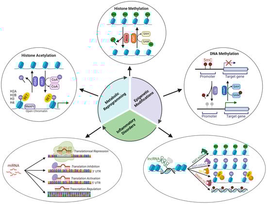
4.2. Epigenetic Signatures Underlying Inflammation

Histone acetylation. Histone acetyltransferases (HATs) and histone deacetylases (HDACs) regulate the expression of several genes involved in the inflammatory response [98]. NF-κB-dependent gene expression requires the involvement of histone acetylase, which will decompress the repressed chromatin environment through histone acetylation [99][100]. Several inflammatory lung diseases are associated with increased H3 acetylation in a specific promoter region that is regulated by NF-κB. Histone modifications at specific acetylation sites (H3K9ac, H3K14ac, H3K18ac, H3K23ac, H3K27ac, H3K36ac, H2B1KK120ac, H2B2BK20ac, H2BK16ac, H2BK20ac, H2BK108ac, H2BK116ac, and H2BK120ac upregulation; H2BK5, H2BK11 downregulation) are involved in the pathogenesis of asthma [101]. CBP/p300 acetyltransferase promotes the transcription of various proinflammatory cytokines, such as IL-1, IL-2, IL-8, and IL-12, by acetylating histones associated with the promoter region of these genes [98]. The enhanced expression of TNF-α upon LPS stimulation is associated with epigenetic remodeling of the TNF-α locus, which is characterized by increased histone 4 acetylation [102]. In paraquat-induced pulmonary fibrosis, elevated IL-6 expression depends on acetylation of H3K9ac in the IL6 promoter region [103].

In contrast, certain proteins like promyelocytic leukemia zinc finger (PLZF) play a role in suppressing the inflammatory response triggered by the activation of TLR- and TNFα-dependent signaling pathways. PLZF does so by promoting chromatin remodeling, which, in turn, represses the NF-kB response by ensuring the stability of a co-repressor complex that comprises HDAC3 histone deacetylase and the NF-kB p50 subunit [104].
Histone Methylation. The regulation of epigenetic histone methylation is crucial in the inflammatory response and has a significant impact on the transcription of various genes, including those involved in the production of pro-inflammatory cytokines. The histone demethylase LSD1 has various effects on the expression of pro-inflammatory cytokines depending on the cell type and immune signals. In smooth muscle cells (SMCs) and hematopoietic stem cells (HSCs), LSD1-mediated demethylation of H3K4me2 suppresses cytokine gene expression but triggers an inflammatory response in conditions such as rheumatoid arthritis (RA) and sepsis [105][106].
Histone methyltransferases also suppress PRR signal transduction. Following papilloma virus infection, the E7 oncoprotein forms a complex with NF-κBp50–p65 and recruits histone demethylase JARID1B and histone deacetylase HDAC1. This complex binds to a specific region on the TLR9 promoter, leading to decreased methylation and acetylation of histones upstream of the TLR9 transcriptional start site [107]. The overexpression of KDM5B/5C histone demethylases has been found to have a detrimental impact on the innate immune response in breast cancer.
DNA methylation. The methylation status of CpG islands significantly affects the inflammatory response and the progression of various inflammatory diseases. In systemic lupus erythematosus (SLE), the overexpression of CD11a, CD40L, CD70, KIR2DL4, and PRF1 in T lymphocytes is associated with DNA hypomethylation in promoter regions [108]. In pulmonary inflammatory diseases such as asthma and chronic obstructive pulmonary disease (COPD), hypomethylated CpG patterns are correlated with disease severity. Specific DNA methylation signatures are present in ulcerative colitis patients [109]. DNA hypermethylation in the promoters of CXCL14, CXCL5, GATA3, IL17C, and IL4R and hypomethylation in the promoters of CCL25, IL13, IL12B, CXCL5, IL4R, IL17A, and IL17RA are associated with the onset or aggravation of ulcerative colitis [110].
Non-coding RNA. Several microRNAs (miRNAs), including miR-126, miR-132, miR-146, miR-155, and miR-221, have been implicated in the regulation of inflammatory genes [111]. miR-10a, in particular, has been shown to suppress the expression of inflammatory cytokines, such as IL-6, IL-8, MCP1, MMP1, and MMP13, in fibroblast-like synoviocytes (FLSs), thereby preventing the release of proinflammatory interleukins and chemokines [112].
Long non-coding RNAs (lncRNAs) also play a role in modulating the inflammatory response by regulating the ubiquitination, stability, and activity of proteins and remodeling chromatin. For instance, the MaIL1 lncRNA and lncRNA NRIR have been shown to promote IFN-I production by activating the ISG pathway upon LPS stimulation [113].

4.3. Epigenetic Modulation of the Immune Function

In addition to their role in the inflammatory response, epigenetic modifications are crucial in the differentiation and maintenance of T-lymphocyte functions. In the inactive or naive state, DNMT1 methylates the promoter regions of transcription factors that define the T-cell lineage, thereby suppressing their expression. Upon antigenic stimulation, TET proteins assist in removing the methyl group from gene promoters, triggering the transcription of specific transcription factors, such as T-bet, GATA-3, RORgt, and Foxp3, which are expressed in each T-cell subset [114][115]. The differentiation of T lymphocytes into specific subsets, such as Th1 and TH2, involves epigenetic modifications. For instance, helper T-lymphocyte polarization into the Th1 subset is characterized by an increase in H4 acetylation on the Ifn-γ promoter [116], while polarization into the Th2 subset results in the repression of Ifn-γ via H3K27me3 methylation by EZH2 [117]. The differentiation of CD8 T cells into effector T cells, such as cytotoxic T cells, is accompanied by increased expression of IFN-γ, perforin (PRF), and granzyme B (GZB) genes due to demethylation of their promoter regions [118][119]. LncRNAs also play a significant role in the activation and differentiation of lymphocytes. Several lncRNAs, such as NRON, NKILA, BCALM, GAS5, and PVT1, regulate the activation of T lymphocytes and the TCR/BCR signaling pathway by modulating the activity of NFAT, NFκB, and MYC [120][121][122][123][124].

5. Nutrition, Inflammation, and Epigenetics

5.1. Role of Nutrition in Inflammation

Nutrition exerts a critical influence on the inflammatory response, as it regulates diverse genetic and epigenetic mechanisms involved in this process. The inflammatory response is dependent on the activity of eicosanoid hormones, specialized pro-resolution mediators (SPMs), and modulatory genes [125].
Eicosanoids are primarily derived from arachidonic acids, which are membrane phospholipids that are released upon the activation of phospholipase A2 by an inflammatory insult. The intensity of the inflammatory response is proportional to the amount of arachidonic acid present in the membrane [126]. The production of arachidonic acid is contingent upon the activity and regulation of two enzymes, delta-6-desaturase and delta-5-desaturase, which are responsible for converting linoleic acid to arachidonic acid. Elevated levels of insulin activate these enzymes, and insulin resistance induced by cytokines, such as TNF-α, can contribute to the production of arachidonic acid [127][128].
Specialized pro-resolving mediators (SPMs) are hormones derived from EPA, DHA, and docosapentaenoic acid (DPA), which play a crucial role in the resolution of residual inflammation [129]. These SPMs include resolvins, maresins, and protectins and exert their anti-inflammatory effects by inhibiting the migration of neutrophils to the site of injury, promoting the transition of macrophages from the pro-inflammatory M1 subtype to the anti-inflammatory M2 subtype and facilitating the removal of apoptotic cells by professional and non-professional phagocytes [130].
The 5′-adenosine monophosphate-activated protein kinase (AMPK) gene is the most critical regulator of inflammation modulation, as it inhibits NF-kB. AMPK regulates cellular energy homeostasis and is activated in response to energy stress, leading to an increase in the AMP/ATP ratio. Activation of AMPK facilitates the metabolic shift from anabolism to catabolism, initiates autophagy, which is vital for supplying energy, metabolites, and biosynthetic intermediates necessary for healing inflammatory lesions, and stimulates mitophagy to replace damaged mitochondria with functional organelles capable of providing the energy required for resolving inflammatory damage [131][132][133][134]. AMPK activity is positively regulated by the SPM signaling pathway, as the binding of SPM, such as Resolvin D1 (RvD1), to SPM receptors, such as FPR2/ALX, promotes AMPK activation [135].
Polyphenols, natural compounds derived from plants, fruits, and vegetables with antioxidant properties, can reduce the inflammatory response and promote resolution of the inflammatory state through the indirect activation of AMPK. Polyphenols, particularly anthocyanins, have been demonstrated to exert a positive regulatory effect on AMPK through the activation of sirtuins. Sirtuins, in turn, stimulate the transcription of hepatic kinase B1 (LKB1), which subsequently promotes the transcription of AMPK by deacetylating the AMPK promoter [136][137].
However, an essential requirement for an effective anti-inflammatory diet is a low-calorie intake. Calorie restriction (CR) exerts potent anti-inflammatory effects through the activation of the anti-stress response, which leads to a reduction in oxidative stress and the protection of DNA from oxidative damage by activating AMPK and suppressing protein kinase A (PKA) and mammalian target of rapamycin (mTOR) signaling pathways, which are triggered by glucose and amino acids, such as threonine and valine [138] (Figure 3). 
Figure 3. Nutrient-induced metabolic and epigenetic changes. The regulation of cellular genetics and epigenetics by calorie restriction (CR) and periodic fasting (PF) is achieved through the modulation of metabolism and hormonal systems. By reducing the levels of tumor growth-promoting nutrients and factors, including glucose, insulin like growth factor 1 (IGF1), and insulin, these interventions inhibit the IGF1-PI3K-AKT-mTOR and protein kinase A (PKA) signaling pathways, activate AMP-activated protein kinase (AMPK), and lead to the activation of stress-resistance genes that protect against the onset of age-related diseases. The decreased availability of glucose results in the liberation of amino acids and fatty acids from muscle and adipose tissue, respectively. Amino acids are utilized for glucose production through the process of gluconeogenesis, while fatty acids in the liver are converted into ketone bodies, which serve as the primary source of energy during periods of nutritional restriction. These metabolic adjustments shift cellular metabolism from glycolysis to oxidative phosphorylation. These modifications result in the formation of acetyl-CoA, which serves as an acetyl group donor for histone acetyltransferase (HAT)-dependent acetylation of nucleosomal histones. The elevation of NAD+ resulting from energy scarcity or the augmentation of de novo synthesis from amino acids, such as tryptophan, triggers the activation of histone deacetylase (HDAC) sirtuins (SIRTs). These sirtuins subsequently deacetylate histone H3, which in turn modulates the expression of metabolic genes and pathways, including glycolysis, gluconeogenesis, mitochondrial respiration, fatty acid oxidation, and lipogenesis. α-ketoglutarate (α-KG), which is produced from glutamine and participates in the TCA cycle, functions as a cofactor for the catalytic activity of lysine demethylase (JMJc) and ten-eleven translocation (TETs) enzymes, which play a role in DNA demethylation. Variations in glutamine and glucosamine glucose levels have an impact on the biosynthesis of uridine diphosphate N-acetylglucosamine (UDP-GlcNAc) and, consequently, the O-GlcNAcylation (OGT) of histones and proteins, which subsequently influence epigenetic reshaping. FAD+ levels generated by the KREBs cycle modulate the activity of LSD1 histone demethylases (created with BioRender.com).

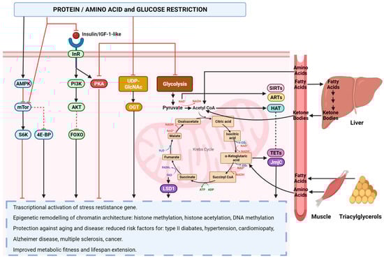
5.2. Role of Nutrition in Epigenetic Reprogramming

Epigenetic mechanisms are influenced by nutrition, which regulates metabolic pathways and produces intermediate metabolites that serve as coactivators for the enzymes involved in the modification of chromatin and DNA. A diet low in carbohydrates and proteins, including CR and PF, causes a metabolic shift from glycolysis to ketone bodies and fatty acid-based oxidative phosphorylation (OXPHOS). This metabolic change results in alterations in the metabolites produced by glycolysis and the Krebs cycle, such as NAD+, flavin adenine dinucleotide (FAD+), lactate, and α-ketoglutarate, which impacts gene expression by regulating epigenetic writers involved in the DNA methylation and histone acetylation, ADP ribosylation, methylation, O-GlcNAcylation, and lactylation [138][139][140][141].
NAD+ serves as a substrate for both sirtuin deacetylases and ADP-ribosyltransferases (ARTs). By adding ADP-ribose to histone proteins, these enzymes promote chromatin relaxation and increase the accessibility of transcription complexes to genomic DNA. As a result, the increase in NAD+ induced by energy stress from CR leads to epigenetic remodeling and a rewiring of transcription through the deacetylation of histones by sirtuins at certain gene loci and the ADP-ribosylation of other loci, resulting in gene transcriptional reprogramming [142].
α-Ketoglutarate is a crucial metabolic intermediate of the Krebs cycle that functions as a cofactor for various chromatin-modifying enzymes, such as histone demethylases (KDMs) and the TET family of enzymes, which are involved in DNA demethylation with TDG. The metabolic changes that occur due to calorie restriction and fasting result in an increase in α-Ketoglutarate, which alters the chromatin structure and DNA accessibility by regulating the activity of KDM and TETs [142] (Figure 3).
FAD+, a crucial redox cofactor that plays a vital role in numerous metabolic reactions, operates as a coactivator of histone demethylases (JmjC). Reduced caloric intake, which is associated with caloric restriction and fasting, leads to increased levels of FAD+, thereby promoting the demethylation of histones and ultimately resulting in the remodeling of the chromatin structure through the activation of histone demethylases [142] (Figure 3).
Acetyl-CoA, a product of the oxidative metabolism of glycolytic pyruvate, free fatty acids, branched-chain amino acids, and ketone bodies within the tricarboxylic acid cycle, plays a crucial role in epigenetic signaling. It serves as a vital cofactor for HAT acetyltransferase. During periods of fasting or caloric restriction, the scarcity of nutrients leads to an enhancement in autophagy and mitochondrial metabolism, which results in a decrease in metabolic cofactors, including acetyl coenzyme A. This essential compound is crucial for epigenetic remodeling and cell differentiation, leading to a decrease in histone acetylation and subsequent alteration in transcription.

6. Inflammation, Epigenetics, and Cancer

6.1. Inflammation in Tumor Onset and Progression

Chronic inflammation and tumor-induced inflammatory responses play a key role in promoting tumorigenesis and tumor progression by modulating the differentiation of immune and stromal cells towards immunosuppressive subtypes that support immune evasion and tumor growth [4][143]. The release of proinflammatory cytokines, such as IL-1, IL-6, IL-8, and TNF-α, by immune, stromal, and tumor cells activates tumor proliferative and pro-survival signaling pathways, such as NF-κB and STAT3 [144]. Reactive oxygen species (ROS) produced by myeloid cells stimulate the tumor secretion of TNFα, which, in turn, drives the release of proinflammatory cytokines, creating a vicious loop of tumor-promoting factors [145]. The production of ROS and oxidative nitrogen species, such as superoxide, hydroxyl radical, and peroxynitrite, by myeloid cells during inflammation in the tumor microenvironment causes DNA damage. This, in turn, activates the previously mentioned pro-inflammatory pathways, further fueling inflammation. This crosstalk between inflammation and DNA damage creates a feedback loop that promotes tumor progression [146].
The inflammatory state within the tumor microenvironment could potentially be linked to the dysregulation of the inflammasome as a result of genetic mutations that accumulate within tumor cells. Gain-of-function mutations have been identified in NLRP1 and NLRP3 in various cancer types, including self-healing palmoplantar carcinomas, nodular melanoma, lung adenocarcinoma, small-cell lung cancer, bladder, gastric, and pancreatic cancer. AIM2 and GSDMD are overexpressed in non-squamous non-small-cell lung cancer, while GSDMD downregulation is associated with gastric cancer [147][148][149][150][151][152].
Inflammasomes are activated in tumor cells, tumor-associated macrophages, tumor-associated fibroblasts, and bone-marrow-derived suppressive cells, resulting in the secretion of IL-1β and IL-18 in the TME [153][154][155][156]. The release of IL-1β promotes the migration, invasion, and metastasis of melanoma and gastric cancer cells by increasing the expression of matrix metalloproteases MMP-2 and MMP-9 [157]. The activation of inflammasomes in myeloid cells, cancer-associated fibroblasts, and tumor-associated macrophages has been found to be positively correlated with the metastasis and poor survival rates of patients with breast and lung cancer [156][158][159][160].

6.2. Epigenetic Alterations in Cancer

The onset and progression of cancer are closely associated with changes in the chromatin structure, resulting in distinct gene expression states and channel-specific phenotypic differentiation [161]. Specific genetic mutations can alter the signaling pathway that drives cells to adopt specific genetic and epigenetic states, ultimately conferring a cancer-related phenotype [162].
The process of epigenetic reprogramming in cancer is not solely attributable to mutations in oncogenes or tumor suppressor genes. Instead, it is influenced by alterations in genes that regulate epigenetic modifications. These changes in epigenetic regulation provide tumor cells with phenotypic plasticity and heterogeneity, allowing genetically identical cells to exhibit distinct phenotypes and temporarily modify their expression. These characteristics of tumor cells contribute to drug resistance, facilitate epithelial–mesenchymal transition (EMT), or enable immune evasion [163][164].
Histone alterations in cancer. Abnormal patterns of histone and DNA modifications are prevalent in numerous tumor types. In human cancer cells, decreased global monoacetylation and trimethylation of histone H4 (H4K16ac and H4K20me3), altered methylation status of H3K9 and H3K27, and hypomethylation of repetitive DNA sequences are often observed [165]
Somatic alterations in H3, particularly at amino acids K27, K36, and G34, promote the tumor initiation and progression of pediatric high-grade gliomas, including glioblastomas (GBMs) [166]. These amino acid substitutions affect H3 methylation and acetylation, leading to an abnormal chromatin structure and gene expression.
Altered expression or mutation of epigenetic writers is frequently associated with several tumor types, including melanoma and breast, bladder, endometrial, renal cell, liver, and lung cancers, and it is often involved in tumor progression and metastasis [167].
HATs are crucial in tumor development, as demonstrated by recurrent chromosomal translocations such as MLL-CBP and MOZ-TIF2, coding mutations (e.g., p300/CBP), and altered expression in solid and hematological malignancies [168][169].
Somatic mutations in HDACs do not seem to be a significant factor in the development of cancer, yet the expression levels of various HDACs are altered in many types of malignancies. Cancer-related chimeric fusion proteins, such as PML-RARa, PLZF-RARa, and AML1-ETO, have been found to contribute to leukemogenesis by bringing HDACs to inappropriately silence genes, thereby promoting the development of leukemia [170].
EZH2 is a component of the polycomb repressive complex 2 (PRC2), which is responsible for silencing gene transcription through the methylation of H3. Thus, both gain-of-function and loss-of-function mutations in EZH2 can result in chromatin remodeling, which in turn promotes tumor development by either repressing tumor suppressor genes or expressing oncogenes [171]. In epithelioid sarcoma, EZH2 is constitutively active due to the inactivation of the SWI/SNF chromatin remodeling complex, which functions antagonistically to EZH2 by promoting active transcription through chromatin decompaction [172]. The gain of function of EZH2 in mesothelioma is attributed to the mutation of BAP1, a deubiquitinating enzyme and component of the PR-DUB complex. The inactivation of BAP1 results in an increase in ubiquitinated H2AK119ub1, which then recruits the PRC2 complex and facilitates the methylation of H3K27me3 via EZH2. This leads to increased chromatin compaction and gene repression [173].
Cytogenetics and next-generation sequencing analysis of diverse cancer genomes have consistently revealed frequent translocations and/or coding mutations in numerous KMT genes. In acute lymphoblastic leukemia, rearrangements of the histone lysine N-methyltransferase 2A (KMT2A) gene result in the production of aberrant fusion proteins that recruit DOT1-like histone lysine methyltransferase (DOT1L) to incorrect sites and methylates H3K79, causing the expression of oncogenes such as HOXA9 and MEIS1 [174].
Alterations in the expression of the lysine-specific histone demethylase LSD1 are frequently observed in various hematopoietic malignancies and solid tumors, including breast, lung, colorectal, and prostate cancers [175][176][177][178].
Histone and DNA reader alterations in cancer. BET proteins have been implicated in a variety of diseases, including cancer, inflammation, and metabolic disorders. These proteins have been found to promote cancer development by altering gene expression in both tumor and TME cells [179]
Multiple studies have reported alterations in the histone methylation lysine reader sequence or expression in both solid and hematological tumors [180]. Nevertheless, the precise mechanisms that connect the disruption of methyl-lysine binding to chromatin to cancer development are not yet fully understood [181].
DNA methyl binding proteins (MBDs) serve as epigenetic readers because they bind to methylated DNA and recruit enzymes that modify histones to coordinate chromatin processes. Mutations in MBD proteins have been identified in various human cancers and have been shown to be critical in the development of neoplasms [182][183][184][185].
Alteration in chromatin remodeler in cancer. Chromatin remodelers are complexes that utilize ATP to shape the chromatin structure by moving, evicting, and exchanging histones. They act in response to the epigenetic modifications of chromatin and DNA generated by various epigenetic writers. There are four main families of chromatin remodelers: switch defective/sucrose non-fermenting (SWI/SNF), imitation switch (ISWI), nucleosome remodeling deacetylase/Mi-2/chromodomain helicase family (NuRD/Mi-2/CHD), and inositol requiring 80 (INO80) family [186]. Chromatin remodeling complexes play a crucial role in granting access to condensed genomic DNA to regulatory transcription, thereby functioning in opposition to polycomb complexes, which repress the chromatin structure. Alterations and mutations in chromatin remodeler complexes can lead to an imbalance in the chromatin structure, which can promote tumor malignancy by disrupting the balance between cell self-renewal and differentiation genes and upregulating the expression of genes involved in cell cycle progression, cell motility, and nuclear hormone signaling [187].

6.3. Inflammation-Induced Epigenetic Alterations in Tumor Immune Microenvironment

Chronic inflammation can lead to changes in the TME that trigger metabolic, genetic, and epigenetic alterations in both the tumor cells and the TME cells [188]. Epigenetic changes can have a significant impact on gene expression and cell identity, potentially leading to the development of malignant and metastatic tumor cells. In addition to cancer cells, abnormal epigenetic modifications also take place in immune cells present in the tumor microenvironment. These modifications contribute to the creation of an immune-tolerant state by inducing T-cell exhaustion, enhancing the immunosuppressive activity of myeloid-derived suppressor cells (MDSCs) and T-regulatory cells (Tregs) and decreasing tumor-associated antigens and T-cell co-stimulatory signals.
Epigenetic regulation of innate immune cells. The function of dendritic cells, which are essential for T-cell-mediated immunity, is altered in the TME. The epigenetic changes in the TME impede the maturation of dendritic cells, leading to immunotolerant phenotypes with a low expression of the major histocompatibility complex (MHC, a key component in antigen-presenting machinery) and co-stimulatory molecules [189]. In pancreatic and colon cancer, FOXM1 overexpression, a transcription factor that suppresses DC maturation and function, is due to increased H3K79me2 methylation caused by dysregulated DOT1L methyltransferase activity [190]. In the TME, KLF4 activation in dendritic cells inhibits their maturation by promoting IL-6 release, a dendritic maturation inhibitor, through hyperacetylation of histones in the IL-6 promoter [191][192]
The microenvironment of tumors alters the function of macrophages, which are vital for both innate and adaptive immune responses, by promoting their polarization from a pro-inflammatory M1 phenotype with anti-tumor properties to an anti-inflammatory M2 phenotype with pro-tumor properties. Epigenetic mechanisms play a key role in regulating the polarization of M1/M2 macrophages through the control of their key transcription factors [193]. The release of IL-4 in the TME promotes the expression of histone demethylase JMJD3 in macrophages, which then promotes the transcription of M2 gene markers (such as Arg1 and Retnla) by demethylating dimethyl and trimethyl H3K27 (H3K27me2/3) in their promoter regions [194]
Myeloid-derived suppressor cells (MDSCs), including immature neutrophils and monocytes, are negative prognostic markers. They infiltrate the tumor microenvironment (TME) and create an immunosuppressive environment that permits tumor escape. The accumulation and immunosuppression functions of MDSCs in the TME rely on epigenetic processes. The immunosuppressive activity and proliferation of MDSCs depend on the production of immunosuppressive agents Arg1 and S100A8, which are regulated by the activation of STAT3. STAT3 activation relies on DNMT3a and DNMT3b, which suppress STAT3 expression by methylating its promoter region [195]. The inhibitory effects of HDAC11 and HDAC6 on IL-10 (an activator of STAT3) resulted in the suppression of MDSC proliferation [196]
The status of natural killer (NK) cells, which play a crucial role in the immune response against tumors by eliminating target cells and secreting cytokines, is often altered in tumors and is typically associated with reduced expression of activating receptors, such as NKG2D, NKp46, and KIR2DS, as well as an increase in the expression of inhibitory receptors, such as NKG2A and TIGIT. This is mainly due to aberrant DNA methylation and histone acetylation–methylation in the promoter regions of these receptors. In patients with hepatocellular carcinoma (HCC), it has been observed that the repression of NKG2D is linked to the hypermethylation and hypoacetylation of histone H3K9Ac in the NKG2D promoter region [197].
Epigenetic regulation of adaptive immune cells. CD8+ T cells are crucial in limiting tumor growth and preventing its progression. The differentiation of T-naive cells into T-effector cells, central memory cells, effector memory cells, and exhausted T cells depends on epigenetic mechanisms. However, the tumor and tumor microenvironment manipulate these mechanisms to render T cells dysfunctional and exhausted. T-cell exhaustion is characterized by an augmentation of transcriptional activity in genes associated with exhaustion, which is attributed to chromatin remodeling and relaxation. The expression of exhaustion markers, such as Tim-3, HMG box transcription factor Tox, and Tox2, is closely tied to the demethylation of their respective promoter regions [198]. The lack of memory T cells in the tumor immune infiltration is due to specific demethylation at tissue-resident effector gene loci, such as CD39 and CD103 [199]. In pancreatic cancer, tumor-associated macrophages (TAMs) have the potential to impact the epigenetic landscape of tumor-infiltrating lymphocytes (TILs).

6.4. Nutrition and Cancer

Nutrition and diet have recently received considerable attention because of their potential impact on the development of neoplasms and as a complementary therapy to enhance the effectiveness of anti-tumor treatments. Despite numerous epidemiological studies assessing the effects of different dietary habits on health and cancer incidence, the results remain inconclusive. These studies found statistical differences among various dietary patterns, but the differences were small and difficult to reproduce [200][201][202]. Confounding factors, such as socioeconomic status, age, physical activity, food quality, and cooking methods, may also influence the observed benefits of different dietary habits.
Nutrition can affect tumorigenesis in several ways, including modulating the immune system and inflammatory state; regulating endocrine factors, such as circulating insulin, insulin-like growth factor, leptin, and adiponectin [203]; and influencing the gut microbiota [204]. Although the mechanisms by which dietary macronutrients and micronutrients affect the inflammatory state are not yet fully understood, vitamin deficiencies, excessive food intake, and food deprivation have been shown to modulate the immune system and inflammatory processes [205].
Overnutrition refers to the intake of excessive amounts of both macro- and micronutrients, which are then stored in the body’s tissues, especially adipose tissue. When there is no longer any space to store these excess nutrients, adipocytes swell and undergo changes that result in chronic inflammation, which can lead to noncommunicable diseases, such as diabetes mellitus, coronary artery disease, and stroke.
The overconsumption of food can lead to obesity, hyperlipidemia, and metabolic syndrome, which can cause chronic inflammation and predispose one to cancer [206]. These conditions are associated with an increase in pro-inflammatory macrophage M1 and a decrease in anti-inflammatory M2 macrophages in adipose tissue, as well as an imbalance in the intestinal microbiota that leads to an accumulation of bacteria that produce procarcinogenic metabolites [203][207]. Additionally, free fatty acids can activate Toll-like receptors, leading to the activation of the pro-inflammatory NF-κB and JNK1 pathways [208]. Overnutrition also leads to an increase in acetyl-CoA, overactivation of mTOR, and reduction in the autophagic protein ATG7, all of which cooperate in blocking autophagy [209]. The inhibition of autophagy can lead to the accumulation of damaged organelles that favor the aging process, activation of the NLRP3 inflammasome [210], and accumulation of oncogenic P62 [211].
An unbalanced diet (excess or deficiency of macro- or micronutrients) may increase the risk of developing metabolic syndrome, which is characterized by elevated levels of various markers, such as C-reactive protein, glucose, IL-6, insulin, leptin, triglycerides, and low adiponectin. These changes can lead to inflammation and impair the immune system, increasing the risk of certain types of cancer. 
Micronutrients, such as vitamin B6 and 25-hydroxyvitamin D, have been found to exhibit anti-tumor activity and to enhance anti-tumor immunosurveillance by preventing DNA damage and inflammation. High consumption of vitamin B6 and elevated plasma concentrations of its metabolite, pyridoxal-5′-phosphate (PLP), have been linked to a reduced risk of cancer, including gastrointestinal tumors [212]. Increased levels of 25-hydroxyvitamin D in serum have also been associated with a decrease in pro-inflammatory markers and favorable prognosis in patients with breast cancer, prostate cancer, or colorectal cancer [213][214][215].

References

  1. Dunn, G.P.; Bruce, A.T.; Ikeda, H.; Old, L.J.; Schreiber, R.D. Cancer immunoediting: From immunosurveillance to tumor escape. Nat. Immunol. 2002, 3, 991–998.
  2. Bakhoum, S.F.; Ngo, B.; Laughney, A.M.; Cavallo, J.A.; Murphy, C.J.; Ly, P.; Shah, P.; Sriram, R.K.; Watkins, T.B.K.; Taunk, N.K.; et al. Chromosomal instability drives metastasis through a cytosolic DNA response. Nature 2018, 553, 467–472.
  3. Zhao, Y.; Simon, M.; Seluanov, A.; Gorbunova, V. DNA damage and repair in age-related inflammation. Nat. Rev. Immunol. 2023, 23, 75–89.
  4. Cortellino, S.; Longo, V.D. Metabolites and Immune Response in Tumor Microenvironments. Cancers 2023, 15, 3898.
  5. Hanahan, D.; Weinberg, R.A. The hallmarks of cancer. Cell 2000, 100, 57–70.
  6. Martínez-Garay, C.; Djouder, N. Dietary interventions and precision nutrition in cancer therapy. Trends Mol. Med. 2023, 29, 489–511.
  7. Chen, L.; Deng, H.; Cui, H.; Fang, J.; Zuo, Z.; Deng, J.; Li, Y.; Wang, X.; Zhao, L. Inflammatory responses and inflammation-associated diseases in organs. Oncotarget 2018, 9, 7204–7218.
  8. Balka, K.R.; De Nardo, D. Understanding early TLR signaling through the Myddosome. J. Leukoc. Biol. 2019, 105, 339–351.
  9. Kawai, T.; Akira, S. The role of pattern-recognition receptors in innate immunity: Update on Toll-like receptors. Nat. Immunol. 2010, 11, 373–384.
  10. Goubau, D.; Deddouche, S.; Reis e Sousa, C. Cytosolic sensing of viruses. Immunity 2013, 38, 855–869.
  11. Yoneyama, M.; Kikuchi, M.; Natsukawa, T.; Shinobu, N.; Imaizumi, T.; Miyagishi, M.; Taira, K.; Akira, S.; Fujita, T. The RNA helicase RIG-I has an essential function in double-stranded RNA-induced innate antiviral responses. Nat. Immunol. 2004, 5, 730–737.
  12. Hartmann, G. Nucleic Acid Immunity. Adv. Immunol. 2017, 133, 121–169.
  13. Ablasser, A.; Bauernfeind, F.; Hartmann, G.; Latz, E.; Fitzgerald, K.A.; Hornung, V. RIG-I-dependent sensing of poly(dA:dT) through the induction of an RNA polymerase III-transcribed RNA intermediate. Nat. Immunol. 2009, 10, 1065–1072.
  14. Imaizumi, T.; Aratani, S.; Nakajima, T.; Carlson, M.; Matsumiya, T.; Tanji, K.; Ookawa, K.; Yoshida, H.; Tsuchida, S.; McIntyre, T.M.; et al. Retinoic acid-inducible gene-I is induced in endothelial cells by LPS and regulates expression of COX-2. Biochem. Biophys. Res. Commun. 2002, 292, 274–279.
  15. Imaizumi, T.; Hatakeyama, M.; Yamashita, K.; Yoshida, H.; Ishikawa, A.; Taima, K.; Satoh, K.; Mori, F.; Wakabayashi, K. Interferon-gamma induces retinoic acid-inducible gene-I in endothelial cells. Endothelium 2004, 11, 169–173.
  16. Imaizumi, T.; Matsumiya, T.; Yoshida, H.; Naraoka, T.; Uesato, R.; Ishibashi, Y.; Ota, K.; Toh, S.; Fukuda, S.; Satoh, K. Tumor-necrosis factor-alpha induces retinoic acid-inducible gene-I in rheumatoid fibroblast-like synoviocytes. Immunol. Lett. 2009, 122, 89–93.
  17. Sakaki, H.; Imaizumi, T.; Matsumiya, T.; Kusumi, A.; Nakagawa, H.; Kubota, K.; Nishi, N.; Nakamura, T.; Hirashima, M.; Satoh, K.; et al. Retinoic acid-inducible gene-I is induced by interleukin-1beta in cultured human gingival fibroblasts. Oral. Microbiol. Immunol. 2005, 20, 47–50.
  18. Ablasser, A.; Chen, Z.J. cGAS in action: Expanding roles in immunity and inflammation. Science 2019, 363, eaat8657.
  19. Ishikawa, H.; Ma, Z.; Barber, G.N. STING regulates intracellular DNA-mediated, type I interferon-dependent innate immunity. Nature 2009, 461, 788–792.
  20. Sun, L.; Wu, J.; Du, F.; Chen, X.; Chen, Z.J. Cyclic GMP-AMP synthase is a cytosolic DNA sensor that activates the type I interferon pathway. Science 2013, 339, 786–791.
  21. Liu, S.; Cai, X.; Wu, J.; Cong, Q.; Chen, X.; Li, T.; Du, F.; Ren, J.; Wu, Y.T.; Grishin, N.V.; et al. Phosphorylation of innate immune adaptor proteins MAVS, STING, and TRIF induces IRF3 activation. Science 2015, 347, aaa2630.
  22. Wu, J.; Dobbs, N.; Yang, K.; Yan, N. Interferon-Independent Activities of Mammalian STING Mediate Antiviral Response and Tumor Immune Evasion. Immunity 2020, 53, 115–126.e5.
  23. Gazi, U.; Martinez-Pomares, L. Influence of the mannose receptor in host immune responses. Immunobiology 2009, 214, 554–561.
  24. Miller, J.L.; de Wet, B.J.; Martinez-Pomares, L.; Radcliffe, C.M.; Dwek, R.A.; Rudd, P.M.; Gordon, S. The mannose receptor mediates dengue virus infection of macrophages. PLoS Pathog. 2008, 4, e17.
  25. Gringhuis, S.I.; den Dunnen, J.; Litjens, M.; van Het Hof, B.; van Kooyk, Y.; Geijtenbeek, T.B. C-type lectin DC-SIGN modulates Toll-like receptor signaling via Raf-1 kinase-dependent acetylation of transcription factor NF-kappaB. Immunity 2007, 26, 605–616.
  26. Hodges, A.; Sharrocks, K.; Edelmann, M.; Baban, D.; Moris, A.; Schwartz, O.; Drakesmith, H.; Davies, K.; Kessler, B.; McMichael, A.; et al. Activation of the lectin DC-SIGN induces an immature dendritic cell phenotype triggering Rho-GTPase activity required for HIV-1 replication. Nat. Immunol. 2007, 8, 569–577.
  27. Rothfuchs, A.G.; Bafica, A.; Feng, C.G.; Egen, J.G.; Williams, D.L.; Brown, G.D.; Sher, A. Dectin-1 interaction with Mycobacterium tuberculosis leads to enhanced IL-12p40 production by splenic dendritic cells. J. Immunol. 2007, 179, 3463–3471.
  28. Van Kooyk, Y.; Rabinovich, G.A. Protein-glycan interactions in the control of innate and adaptive immune responses. Nat. Immunol. 2008, 9, 593–601.
  29. Richard, M.; Thibault, N.; Veilleux, P.; Gareau-Pagé, G.; Beaulieu, A.D. Granulocyte macrophage-colony stimulating factor reduces the affinity of SHP-2 for the ITIM of CLECSF6 in neutrophils: A new mechanism of action for SHP-2. Mol. Immunol. 2006, 43, 1716–1721.
  30. Schroder, K.; Tschopp, J. The inflammasomes. Cell 2010, 140, 821–832.
  31. Ting, J.P.; Lovering, R.C.; Alnemri, E.S.; Bertin, J.; Boss, J.M.; Davis, B.K.; Flavell, R.A.; Girardin, S.E.; Godzik, A.; Harton, J.A.; et al. The NLR gene family: A standard nomenclature. Immunity 2008, 28, 285–287.
  32. Inohara, N.; Koseki, T.; Lin, J.; del Peso, L.; Lucas, P.C.; Chen, F.F.; Ogura, Y.; Núñez, G. An induced proximity model for NF-kappa B activation in the Nod1/RICK and RIP signaling pathways. J. Biol. Chem. 2000, 275, 27823–27831.
  33. Abbott, D.W.; Wilkins, A.; Asara, J.M.; Cantley, L.C. The Crohn’s disease protein, NOD2, requires RIP2 in order to induce ubiquitinylation of a novel site on NEMO. Curr. Biol. 2004, 14, 2217–2227.
  34. Lamkanfi, M.; Dixit, V.M. Mechanisms and functions of inflammasomes. Cell 2014, 157, 1013–1022.
  35. Broz, P.; Dixit, V.M. Inflammasomes: Mechanism of assembly, regulation and signalling. Nat. Rev. Immunol. 2016, 16, 407–420.
  36. Cai, X.; Chen, J.; Xu, H.; Liu, S.; Jiang, Q.X.; Halfmann, R.; Chen, Z.J. Prion-like polymerization underlies signal transduction in antiviral immune defense and inflammasome activation. Cell 2014, 156, 1207–1222.
  37. Lu, A.; Magupalli, V.G.; Ruan, J.; Yin, Q.; Atianand, M.K.; Vos, M.R.; Schröder, G.F.; Fitzgerald, K.A.; Wu, H.; Egelman, E.H. Unified polymerization mechanism for the assembly of ASC-dependent inflammasomes. Cell 2014, 156, 1193–1206.
  38. Afonina, I.S.; Müller, C.; Martin, S.J.; Beyaert, R. Proteolytic Processing of Interleukin-1 Family Cytokines: Variations on a Common Theme. Immunity 2015, 42, 991–1004.
  39. He, W.T.; Wan, H.; Hu, L.; Chen, P.; Wang, X.; Huang, Z.; Yang, Z.H.; Zhong, C.Q.; Han, J. Gasdermin D is an executor of pyroptosis and required for interleukin-1β secretion. Cell Res. 2015, 25, 1285–1298.
  40. Chan, A.H.; Schroder, K. Inflammasome signaling and regulation of interleukin-1 family cytokines. J. Exp. Med. 2020, 217, e20190314.
  41. Rühl, S.; Shkarina, K.; Demarco, B.; Heilig, R.; Santos, J.C.; Broz, P. ESCRT-dependent membrane repair negatively regulates pyroptosis downstream of GSDMD activation. Science 2018, 362, 956–960.
  42. Aglietti, R.A.; Estevez, A.; Gupta, A.; Ramirez, M.G.; Liu, P.S.; Kayagaki, N.; Ciferri, C.; Dixit, V.M.; Dueber, E.C. GsdmD p30 elicited by caspase-11 during pyroptosis forms pores in membranes. Proc. Natl. Acad. Sci. USA 2016, 113, 7858–7863.
  43. Rider, P.; Carmi, Y.; Guttman, O.; Braiman, A.; Cohen, I.; Voronov, E.; White, M.R.; Dinarello, C.A.; Apte, R.N. IL-1α and IL-1β recruit different myeloid cells and promote different stages of sterile inflammation. J. Immunol. 2011, 187, 4835–4843.
  44. Dinarello, C.A. Overview of the IL-1 family in innate inflammation and acquired immunity. Immunol. Rev. 2018, 281, 8–27.
  45. Schroder, K.; Hertzog, P.J.; Ravasi, T.; Hume, D.A. Interferon-gamma: An overview of signals, mechanisms and functions. J. Leukoc. Biol. 2004, 75, 163–189.
  46. Nold, M.F.; Nold-Petry, C.A.; Zepp, J.A.; Palmer, B.E.; Bufler, P.; Dinarello, C.A. IL-37 is a fundamental inhibitor of innate immunity. Nat. Immunol. 2010, 11, 1014–1022.
  47. McLane, L.M.; Abdel-Hakeem, M.S.; Wherry, E.J. CD8 T Cell Exhaustion During Chronic Viral Infection and Cancer. Annu. Rev. Immunol. 2019, 37, 457–495.
  48. Härtlova, A.; Erttmann, S.F.; Raffi, F.A.; Schmalz, A.M.; Resch, U.; Anugula, S.; Lienenklaus, S.; Nilsson, L.M.; Kröger, A.; Nilsson, J.A.; et al. DNA damage primes the type I interferon system via the cytosolic DNA sensor STING to promote anti-microbial innate immunity. Immunity 2015, 42, 332–343.
  49. Li, T.; Chen, Z.J. The cGAS-cGAMP-STING pathway connects DNA damage to inflammation, senescence, and cancer. J. Exp. Med. 2018, 215, 1287–1299.
  50. Van Vugt, M.A.; Parkes, E.E. When breaks get hot: Inflammatory signaling in BRCA1/2-mutant cancers. Trends Cancer 2022, 8, 174–189.
  51. Fenech, M.; Kirsch-Volders, M.; Natarajan, A.T.; Surralles, J.; Crott, J.W.; Parry, J.; Norppa, H.; Eastmond, D.A.; Tucker, J.D.; Thomas, P. Molecular mechanisms of micronucleus, nucleoplasmic bridge and nuclear bud formation in mammalian and human cells. Mutagenesis 2011, 26, 125–132.
  52. Mackenzie, K.J.; Carroll, P.; Martin, C.A.; Murina, O.; Fluteau, A.; Simpson, D.J.; Olova, N.; Sutcliffe, H.; Rainger, J.K.; Leitch, A.; et al. cGAS surveillance of micronuclei links genome instability to innate immunity. Nature 2017, 548, 461–465.
  53. Erdal, E.; Haider, S.; Rehwinkel, J.; Harris, A.L.; McHugh, P.J. A prosurvival DNA damage-induced cytoplasmic interferon response is mediated by end resection factors and is limited by Trex1. Genes. Dev. 2017, 31, 353–369.
  54. Kang, C.; Xu, Q.; Martin, T.D.; Li, M.Z.; Demaria, M.; Aron, L.; Lu, T.; Yankner, B.A.; Campisi, J.; Elledge, S.J. The DNA damage response induces inflammation and senescence by inhibiting autophagy of GATA4. Science 2015, 349, aaa5612.
  55. Hinz, M.; Stilmann, M.; Arslan, S.; Khanna, K.K.; Dittmar, G.; Scheidereit, C. A cytoplasmic ATM-TRAF6-cIAP1 module links nuclear DNA damage signaling to ubiquitin-mediated NF-κB activation. Mol. Cell 2010, 40, 63–74.
  56. Fang, L.; Choudhary, S.; Zhao, Y.; Edeh, C.B.; Yang, C.; Boldogh, I.; Brasier, A.R. ATM regulates NF-κB-dependent immediate-early genes via RelA Ser 276 phosphorylation coupled to CDK9 promoter recruitment. Nucleic Acids Res. 2014, 42, 8416–8432.
  57. Chabanon, R.M.; Rouanne, M.; Lord, C.J.; Soria, J.C.; Pasero, P.; Postel-Vinay, S. Targeting the DNA damage response in immuno-oncology: Developments and opportunities. Nat. Rev. Cancer 2021, 21, 701–717.
  58. Coquel, F.; Silva, M.J.; Técher, H.; Zadorozhny, K.; Sharma, S.; Nieminuszczy, J.; Mettling, C.; Dardillac, E.; Barthe, A.; Schmitz, A.L.; et al. SAMHD1 acts at stalled replication forks to prevent interferon induction. Nature 2018, 557, 57–61.
  59. Gratia, M.; Rodero, M.P.; Conrad, C.; Bou Samra, E.; Maurin, M.; Rice, G.I.; Duffy, D.; Revy, P.; Petit, F.; Dale, R.C.; et al. Bloom syndrome protein restrains innate immune sensing of micronuclei by cGAS. J. Exp. Med. 2019, 216, 1199–1213.
  60. Parkes, E.E.; Walker, S.M.; Taggart, L.E.; McCabe, N.; Knight, L.A.; Wilkinson, R.; McCloskey, K.D.; Buckley, N.E.; Savage, K.I.; Salto-Tellez, M.; et al. Activation of STING-Dependent Innate Immune Signaling By S-Phase-Specific DNA Damage in Breast Cancer. J. Natl. Cancer Inst. 2017, 109, djw199.
  61. Heijink, A.M.; Talens, F.; Jae, L.T.; van Gijn, S.E.; Fehrmann, R.S.N.; Brummelkamp, T.R.; van Vugt, M.A.T.M. BRCA2 deficiency instigates cGAS-mediated inflammatory signaling and confers sensitivity to tumor necrosis factor-alpha-mediated cytotoxicity. Nat. Commun. 2019, 10, 100.
  62. Davies, S.L.; North, P.S.; Hickson, I.D. Role for BLM in replication-fork restart and suppression of origin firing after replicative stress. Nat. Struct. Mol. Biol. 2007, 14, 677–679.
  63. Nimonkar, A.V.; Genschel, J.; Kinoshita, E.; Polaczek, P.; Campbell, J.L.; Wyman, C.; Modrich, P.; Kowalczykowski, S.C. BLM-DNA2-RPA-MRN and EXO1-BLM-RPA-MRN constitute two DNA end resection machineries for human DNA break repair. Genes. Dev. 2011, 25, 350–362.
  64. Zhao, S.; Goewey Ruiz, J.A.; Sebastian, M.; Kidane, D. Defective DNA polymerase beta invoke a cytosolic DNA mediated inflammatory response. Front. Immunol. 2022, 13, 1039009.
  65. Yamaguchi, K.; Kajikawa, M.; Okada, N. LINE retrotransposition and host DNA repair machinery. Mob. Genet. Elem. 2015, 5, 92–97.
  66. Lander, E.S.; Linton, L.M.; Birren, B.; Nusbaum, C.; Zody, M.C.; Baldwin, J.; Devon, K.; Dewar, K.; Doyle, M.; FitzHugh, W.; et al. Initial sequencing and analysis of the human genome. Nature 2001, 409, 860–921.
  67. Kazazian, H.H.; Moran, J.V. Mobile DNA in Health and Disease. N. Engl. J. Med. 2017, 377, 361–370.
  68. Gorbunova, V.; Seluanov, A.; Mita, P.; McKerrow, W.; Fenyö, D.; Boeke, J.D.; Linker, S.B.; Gage, F.H.; Kreiling, J.A.; Petrashen, A.P.; et al. The role of retrotransposable elements in ageing and age-associated diseases. Nature 2021, 596, 43–53.
  69. Niehrs, C.; Luke, B. Regulatory R-loops as facilitators of gene expression and genome stability. Nat. Rev. Mol. Cell Biol. 2020, 21, 167–178.
  70. Skourti-Stathaki, K.; Proudfoot, N.J.; Gromak, N. Human senataxin resolves RNA/DNA hybrids formed at transcriptional pause sites to promote Xrn2-dependent termination. Mol. Cell 2011, 42, 794–805.
  71. Goulielmaki, E.; Tsekrekou, M.; Batsiotos, N.; Ascensão-Ferreira, M.; Ledaki, E.; Stratigi, K.; Chatzinikolaou, G.; Topalis, P.; Kosteas, T.; Altmüller, J.; et al. The splicing factor XAB2 interacts with ERCC1-XPF and XPG for R-loop processing. Nat. Commun. 2021, 12, 3153.
  72. Chang, E.Y.; Novoa, C.A.; Aristizabal, M.J.; Coulombe, Y.; Segovia, R.; Chaturvedi, R.; Shen, Y.; Keong, C.; Tam, A.S.; Jones, S.J.M.; et al. RECQ-like helicases Sgs1 and BLM regulate R-loop-associated genome instability. J. Cell Biol. 2017, 216, 3991–4005.
  73. Marabitti, V.; Lillo, G.; Malacaria, E.; Palermo, V.; Sanchez, M.; Pichierri, P.; Franchitto, A. ATM pathway activation limits R-loop-associated genomic instability in Werner syndrome cells. Nucleic Acids Res. 2019, 47, 3485–3502.
  74. Barnhoorn, S.; Uittenboogaard, L.M.; Jaarsma, D.; Vermeij, W.P.; Tresini, M.; Weymaere, M.; Menoni, H.; Brandt, R.M.; de Waard, M.C.; Botter, S.M.; et al. Cell-autonomous progeroid changes in conditional mouse models for repair endonuclease XPG deficiency. PLoS Genet. 2014, 10, e1004686.
  75. Bhatia, V.; Barroso, S.I.; García-Rubio, M.L.; Tumini, E.; Herrera-Moyano, E.; Aguilera, A. BRCA2 prevents R-loop accumulation and associates with TREX-2 mRNA export factor PCID2. Nature 2014, 511, 362–365.
  76. Hatchi, E.; Skourti-Stathaki, K.; Ventz, S.; Pinello, L.; Yen, A.; Kamieniarz-Gdula, K.; Dimitrov, S.; Pathania, S.; McKinney, K.M.; Eaton, M.L.; et al. BRCA1 recruitment to transcriptional pause sites is required for R-loop-driven DNA damage repair. Mol. Cell 2015, 57, 636–647.
  77. Sistigu, A.; Yamazaki, T.; Vacchelli, E.; Chaba, K.; Enot, D.P.; Adam, J.; Vitale, I.; Goubar, A.; Baracco, E.E.; Remédios, C.; et al. Cancer cell-autonomous contribution of type I interferon signaling to the efficacy of chemotherapy. Nat. Med. 2014, 20, 1301–1309.
  78. Zitvogel, L.; Kepp, O.; Kroemer, G. Immune parameters affecting the efficacy of chemotherapeutic regimens. Nat. Rev. Clin. Oncol. 2011, 8, 151–160.
  79. Deng, L.; Liang, H.; Xu, M.; Yang, X.; Burnette, B.; Arina, A.; Li, X.D.; Mauceri, H.; Beckett, M.; Darga, T.; et al. STING-Dependent Cytosolic DNA Sensing Promotes Radiation-Induced Type I Interferon-Dependent Anti-tumor Immunity in Immunogenic Tumors. Immunity 2014, 41, 843–852.
  80. Harding, S.M.; Benci, J.L.; Irianto, J.; Discher, D.E.; Minn, A.J.; Greenberg, R.A. Mitotic progression following DNA damage enables pattern recognition within micronuclei. Nature 2017, 548, 466–470.
  81. Ding, L.; Kim, H.J.; Wang, Q.; Kearns, M.; Jiang, T.; Ohlson, C.E.; Li, B.B.; Xie, S.; Liu, J.F.; Stover, E.H.; et al. PARP Inhibition Elicits STING-Dependent Anti-tumor Immunity in Brca1-Deficient Ovarian Cancer. Cell Rep. 2018, 25, 2972–2980.e5.
  82. Pantelidou, C.; Sonzogni, O.; De Oliveria Taveira, M.; Mehta, A.K.; Kothari, A.; Wang, D.; Visal, T.; Li, M.K.; Pinto, J.; Castrillon, J.A.; et al. PARP Inhibitor Efficacy Depends on CD8. Cancer Discov. 2019, 9, 722–737.
  83. Reisländer, T.; Lombardi, E.P.; Groelly, F.J.; Miar, A.; Porru, M.; Di Vito, S.; Wright, B.; Lockstone, H.; Biroccio, A.; Harris, A.; et al. BRCA2 abrogation triggers innate immune responses potentiated by treatment with PARP inhibitors. Nat. Commun. 2019, 10, 3143.
  84. Wang, Z.; Sun, K.; Xiao, Y.; Feng, B.; Mikule, K.; Ma, X.; Feng, N.; Vellano, C.P.; Federico, L.; Marszalek, J.R.; et al. Niraparib activates interferon signaling and potentiates anti-PD-1 antibody efficacy in tumor models. Sci. Rep. 2019, 9, 1853.
  85. Zhang, T.; Cooper, S.; Brockdorff, N. The interplay of histone modifications—Writers that read. EMBO Rep. 2015, 16, 1467–1481.
  86. Zhang, Y.; Sun, Z.; Jia, J.; Du, T.; Zhang, N.; Tang, Y.; Fang, Y.; Fang, D. Overview of Histone Modification. Adv. Exp. Med. Biol. 2021, 1283, 1–16.
  87. Cheung, P.; Lau, P. Epigenetic regulation by histone methylation and histone variants. Mol. Endocrinol. 2005, 19, 563–573.
  88. Yang, X.J.; Seto, E. HATs and HDACs: From structure, function and regulation to novel strategies for therapy and prevention. Oncogene 2007, 26, 5310–5318.
  89. Narlikar, G.J.; Fan, H.Y.; Kingston, R.E. Cooperation between complexes that regulate chromatin structure and transcription. Cell 2002, 108, 475–487.
  90. Youn, H.D. Methylation and demethylation of DNA and histones in chromatin: The most complicated epigenetic marker. Exp. Mol. Med. 2017, 49, e321.
  91. Igolkina, A.A.; Zinkevich, A.; Karandasheva, K.O.; Popov, A.A.; Selifanova, M.V.; Nikolaeva, D.; Tkachev, V.; Penzar, D.; Nikitin, D.M.; Buzdin, A. H3K4me3, H3K9ac, H3K27ac, H3K27me3 and H3K9me3 Histone Tags Suggest Distinct Regulatory Evolution of Open and Condensed Chromatin Landmarks. Cells 2019, 8, 1034.
  92. Shi, Y.G.; Tsukada, Y. The discovery of histone demethylases. Cold Spring Harb. Perspect. Biol. 2013, 5, a017947.
  93. Filippakopoulos, P.; Knapp, S. Targeting bromodomains: Epigenetic readers of lysine acetylation. Nat. Rev. Drug Discov. 2014, 13, 337–356.
  94. Flores, K.B.; Wolschin, F.; Amdam, G.V. The role of methylation of DNA in environmental adaptation. Integr. Comp. Biol. 2013, 53, 359–372.
  95. Lim, W.J.; Kim, K.H.; Kim, J.Y.; Jeong, S.; Kim, N. Identification of DNA-Methylated CpG Islands Associated with Gene Silencing in the Adult Body Tissues of the Ogye Chicken Using RNA-Seq and Reduced Representation Bisulfite Sequencing. Front. Genet. 2019, 10, 346.
  96. Boyes, J.; Bird, A. DNA methylation inhibits transcription indirectly via a methyl-CpG binding protein. Cell 1991, 64, 1123–1134.
  97. Fuks, F.; Hurd, P.J.; Deplus, R.; Kouzarides, T. The DNA methyltransferases associate with HP1 and the SUV39H1 histone methyltransferase. Nucleic Acids Res. 2003, 31, 2305–2312.
  98. Villagra, A.; Sotomayor, E.M.; Seto, E. Histone deacetylases and the immunological network: Implications in cancer and inflammation. Oncogene 2010, 29, 157–173.
  99. Seto, E.; Yoshida, M. Erasers of histone acetylation: The histone deacetylase enzymes. Cold Spring Harb. Perspect. Biol. 2014, 6, a018713.
  100. Calao, M.; Burny, A.; Quivy, V.; Dekoninck, A.; Van Lint, C. A pervasive role of histone acetyltransferases and deacetylases in an NF-kappaB-signaling code. Trends Biochem. Sci. 2008, 33, 339–349.
  101. Ren, Y.; Li, M.; Bai, S.; Kong, L.; Su, X. Identification of histone acetylation in a murine model of allergic asthma by proteomic analysis. Exp. Biol. Med. 2021, 246, 929–939.
  102. Stender, J.D.; Pascual, G.; Liu, W.; Kaikkonen, M.U.; Do, K.; Spann, N.J.; Boutros, M.; Perrimon, N.; Rosenfeld, M.G.; Glass, C.K. Control of proinflammatory gene programs by regulated trimethylation and demethylation of histone H4K20. Mol. Cell 2012, 48, 28–38.
  103. Hu, L.; Yu, Y.; Huang, H.; Fan, H.; Yin, C.; Li, K.; Fulton, D.J.; Chen, F. Epigenetic Regulation of Interleukin 6 by Histone Acetylation in Macrophages and Its Role in Paraquat-Induced Pulmonary Fibrosis. Front. Immunol. 2016, 7, 696.
  104. Sadler, A.J.; Suliman, B.A.; Yu, L.; Yuan, X.; Wang, D.; Irving, A.T.; Sarvestani, S.T.; Banerjee, A.; Mansell, A.S.; Liu, J.P.; et al. The acetyltransferase HAT1 moderates the NF-κB response by regulating the transcription factor PLZF. Nat. Commun. 2015, 6, 6795.
  105. Shi, Y.; Lan, F.; Matson, C.; Mulligan, P.; Whetstine, J.R.; Cole, P.A.; Casero, R.A. Histone demethylation mediated by the nuclear amine oxidase homolog LSD1. Cell 2004, 119, 941–953.
  106. Zhao, Z.; Shilatifard, A. Epigenetic modifications of histones in cancer. Genome Biol. 2019, 20, 245.
  107. Hasan, U.A.; Zannetti, C.; Parroche, P.; Goutagny, N.; Malfroy, M.; Roblot, G.; Carreira, C.; Hussain, I.; Müller, M.; Taylor-Papadimitriou, J.; et al. The human papillomavirus type 16 E7 oncoprotein induces a transcriptional repressor complex on the Toll-like receptor 9 promoter. J. Exp. Med. 2013, 210, 1369–1387.
  108. Balada, E.; Castro-Marrero, J.; Felip, L.; Ordi-Ros, J.; Vilardell-Tarrés, M. Associations between the expression of epigenetically regulated genes and the expression of DNMTs and MBDs in systemic lupus erythematosus. PLoS ONE 2012, 7, e45897.
  109. Vucic, E.A.; Chari, R.; Thu, K.L.; Wilson, I.M.; Cotton, A.M.; Kennett, J.Y.; Zhang, M.; Lonergan, K.M.; Steiling, K.; Brown, C.J.; et al. DNA methylation is globally disrupted and associated with expression changes in chronic obstructive pulmonary disease small airways. Am. J. Respir. Cell Mol. Biol. 2014, 50, 912–922.
  110. Karatzas, P.S.; Mantzaris, G.J.; Safioleas, M.; Gazouli, M. DNA methylation profile of genes involved in inflammation and autoimmunity in inflammatory bowel disease. Medicine 2014, 93, e309.
  111. Akbaba, T.H.; Akkaya-Ulum, Y.Z.; Tavukcuoglu, Z.; Bilginer, Y.; Ozen, S.; Balci-Peynircioglu, B. Inflammation-related differentially expressed common miRNAs in systemic autoinflammatory disorders patients can regulate the clinical course. Clin. Exp. Rheumatol. 2021, 39 (Suppl. S132), 109–117.
  112. Hussain, N.; Zhu, W.; Jiang, C.; Xu, J.; Wu, X.; Geng, M.; Hussain, S.; Cai, Y.; Xu, K.; Xu, P.; et al. Down-regulation of miR-10a-5p in synoviocytes contributes to TBX5-controlled joint inflammation. J. Cell Mol. Med. 2018, 22, 241–250.
  113. Aznaourova, M.; Janga, H.; Sefried, S.; Kaufmann, A.; Dorna, J.; Volkers, S.M.; Georg, P.; Lechner, M.; Hoppe, J.; Dökel, S.; et al. Noncoding RNA. Proc. Natl. Acad. Sci. USA 2020, 117, 9042–9053.
  114. Kersh, E.N.; Fitzpatrick, D.R.; Murali-Krishna, K.; Shires, J.; Speck, S.H.; Boss, J.M.; Ahmed, R. Rapid demethylation of the IFN-gamma gene occurs in memory but not naive CD8 T cells. J. Immunol. 2006, 176, 4083–4093.
  115. Feng, Y.; Arvey, A.; Chinen, T.; van der Veeken, J.; Gasteiger, G.; Rudensky, A.Y. Control of the inheritance of regulatory T cell identity by a cis element in the Foxp3 locus. Cell 2014, 158, 749–763.
  116. Chang, S.; Collins, P.L.; Aune, T.M. T-bet dependent removal of Sin3A-histone deacetylase complexes at the Ifng locus drives Th1 differentiation. J. Immunol. 2008, 181, 8372–8381.
  117. Tumes, D.J.; Onodera, A.; Suzuki, A.; Shinoda, K.; Endo, Y.; Iwamura, C.; Hosokawa, H.; Koseki, H.; Tokoyoda, K.; Suzuki, Y.; et al. The polycomb protein Ezh2 regulates differentiation and plasticity of CD4(+) T helper type 1 and type 2 cells. Immunity 2013, 39, 819–832.
  118. Scharer, C.D.; Barwick, B.G.; Youngblood, B.A.; Ahmed, R.; Boss, J.M. Global DNA methylation remodeling accompanies CD8 T cell effector function. J. Immunol. 2013, 191, 3419–3429.
  119. Weng, N.P.; Araki, Y.; Subedi, K. The molecular basis of the memory T cell response: Differential gene expression and its epigenetic regulation. Nat. Rev. Immunol. 2012, 12, 306–315.
  120. Willingham, A.T.; Orth, A.P.; Batalov, S.; Peters, E.C.; Wen, B.G.; Aza-Blanc, P.; Hogenesch, J.B.; Schultz, P.G. A strategy for probing the function of noncoding RNAs finds a repressor of NFAT. Science 2005, 309, 1570–1573.
  121. Huang, D.; Chen, J.; Yang, L.; Ouyang, Q.; Li, J.; Lao, L.; Zhao, J.; Liu, J.; Lu, Y.; Xing, Y.; et al. NKILA lncRNA promotes tumor immune evasion by sensitizing T cells to activation-induced cell death. Nat. Immunol. 2018, 19, 1112–1125.
  122. Pyfrom, S.C.; Quinn, C.C.; Dorando, H.K.; Luo, H.; Payton, J.E. BCALM (AC099524.1) Is a Human B Lymphocyte-Specific Long Noncoding RNA That Modulates B Cell Receptor-Mediated Calcium Signaling. J. Immunol. 2020, 205, 595–607.
  123. Li, J.; Tian, J.; Lu, J.; Wang, Z.; Ling, J.; Wu, X.; Yang, F.; Xia, Y. LncRNA GAS5 inhibits Th17 differentiation and alleviates immune thrombocytopenia via promoting the ubiquitination of STAT3. Int. Immunopharmacol. 2020, 80, 106127.
  124. Fu, J.; Shi, H.; Wang, B.; Zhan, T.; Shao, Y.; Ye, L.; Wu, S.; Yu, C.; Zheng, L. LncRNA PVT1 links Myc to glycolytic metabolism upon CD4. J. Autoimmun. 2020, 107, 102358.
  125. Serhan, C.N. Pro-resolving lipid mediators are leads for resolution physiology. Nature 2014, 510, 92–101.
  126. Sears, B.; Lawren, B. The Zone: A Dietary Road Map; ReganBooks: New York, NY, USA, 1995; p. xvii. 328p.
  127. Brenner, R.R. Hormonal modulation of delta6 and delta5 desaturases: Case of diabetes. Prostaglandins Leukot. Essent. Fatty Acids 2003, 68, 151–162.
  128. Akash, M.S.H.; Rehman, K.; Liaqat, A. Tumor Necrosis Factor-Alpha: Role in Development of Insulin Resistance and Pathogenesis of Type 2 Diabetes Mellitus. J. Cell Biochem. 2018, 119, 105–110.
  129. Serhan, C.N.; Levy, B.D. Resolvins in inflammation: Emergence of the pro-resolving superfamily of mediators. J. Clin. Investig. 2018, 128, 2657–2669.
  130. Dalli, J.; Serhan, C.N. Pro-Resolving Mediators in Regulating and Conferring Macrophage Function. Front. Immunol. 2017, 8, 1400.
  131. Jeon, S.M. Regulation and function of AMPK in physiology and diseases. Exp. Mol. Med. 2016, 48, e245.
  132. Day, E.A.; Ford, R.J.; Steinberg, G.R. AMPK as a Therapeutic Target for Treating Metabolic Diseases. Trends Endocrinol. Metab. 2017, 28, 545–560.
  133. Hardie, D.G. Keeping the home fires burning: AMP-activated protein kinase. J. R. Soc. Interface 2018, 15, 20170774.
  134. Herzig, S.; Shaw, R.J. AMPK: Guardian of metabolism and mitochondrial homeostasis. Nat. Rev. Mol. Cell Biol. 2018, 19, 121–135.
  135. McArthur, S.; Juban, G.; Gobbetti, T.; Desgeorges, T.; Theret, M.; Gondin, J.; Toller-Kawahisa, J.E.; Reutelingsperger, C.P.; Chazaud, B.; Perretti, M.; et al. Annexin A1 drives macrophage skewing to accelerate muscle regeneration through AMPK activation. J. Clin. Investig. 2020, 130, 1156–1167.
  136. Chung, S.; Yao, H.; Caito, S.; Hwang, J.W.; Arunachalam, G.; Rahman, I. Regulation of SIRT1 in cellular functions: Role of polyphenols. Arch. Biochem. Biophys. 2010, 501, 79–90.
  137. Shackelford, D.B.; Shaw, R.J. The LKB1-AMPK pathway: Metabolism and growth control in tumour suppression. Nat. Rev. Cancer 2009, 9, 563–575.
  138. Cheng, C.W.; Adams, G.B.; Perin, L.; Wei, M.; Zhou, X.; Lam, B.S.; Da Sacco, S.; Mirisola, M.; Quinn, D.I.; Dorff, T.B.; et al. Prolonged fasting reduces IGF-1/PKA to promote hematopoietic-stem-cell-based regeneration and reverse immunosuppression. Cell Stem Cell 2014, 14, 810–823.
  139. Cortellino, S.; Raveane, A.; Chiodoni, C.; Delfanti, G.; Pisati, F.; Spagnolo, V.; Visco, E.; Fragale, G.; Ferrante, F.; Magni, S.; et al. Fasting renders immunotherapy effective against low-immunogenic breast cancer while reducing side effects. Cell Rep. 2022, 40, 111256.
  140. Nencioni, A.; Caffa, I.; Cortellino, S.; Longo, V.D. Fasting and cancer: Molecular mechanisms and clinical application. Nat. Rev. Cancer 2018, 18, 707–719.
  141. Etchegaray, J.P.; Mostoslavsky, R. Interplay between Metabolism and Epigenetics: A Nuclear Adaptation to Environmental Changes. Mol. Cell 2016, 62, 695–711.
  142. Keating, S.T.; El-Osta, A. Epigenetics and metabolism. Circ. Res. 2015, 116, 715–736.
  143. Greten, F.R.; Grivennikov, S.I. Inflammation and Cancer: Triggers, Mechanisms, and Consequences. Immunity 2019, 51, 27–41.
  144. Heichler, C.; Scheibe, K.; Schmied, A.; Geppert, C.I.; Schmid, B.; Wirtz, S.; Thoma, O.M.; Kramer, V.; Waldner, M.J.; Büttner, C.; et al. STAT3 activation through IL-6/IL-11 in cancer-associated fibroblasts promotes colorectal tumour development and correlates with poor prognosis. Gut 2020, 69, 1269–1282.
  145. Canli, Ö.; Nicolas, A.M.; Gupta, J.; Finkelmeier, F.; Goncharova, O.; Pesic, M.; Neumann, T.; Horst, D.; Löwer, M.; Sahin, U.; et al. Myeloid Cell-Derived Reactive Oxygen Species Induce Epithelial Mutagenesis. Cancer Cell 2017, 32, 869–883.e5.
  146. Kay, J.; Thadhani, E.; Samson, L.; Engelward, B. Inflammation-induced DNA damage, mutations and cancer. DNA Repair 2019, 83, 102673.
  147. Drutman, S.B.; Haerynck, F.; Zhong, F.L.; Hum, D.; Hernandez, N.J.; Belkaya, S.; Rapaport, F.; de Jong, S.J.; Creytens, D.; Tavernier, S.J.; et al. Homozygous. Proc. Natl. Acad. Sci. USA 2019, 116, 19055–19063.
  148. Zhong, F.L.; Mamaï, O.; Sborgi, L.; Boussofara, L.; Hopkins, R.; Robinson, K.; Szeverényi, I.; Takeichi, T.; Balaji, R.; Lau, A.; et al. Germline NLRP1 Mutations Cause Skin Inflammatory and Cancer Susceptibility Syndromes via Inflammasome Activation. Cell 2016, 167, 187–202.e17.
  149. Verma, D.; Bivik, C.; Farahani, E.; Synnerstad, I.; Fredrikson, M.; Enerbäck, C.; Rosdahl, I.; Söderkvist, P. Inflammasome polymorphisms confer susceptibility to sporadic malignant melanoma. Pigment. Cell Melanoma Res. 2012, 25, 506–513.
  150. Castaño-Rodríguez, N.; Kaakoush, N.O.; Goh, K.L.; Fock, K.M.; Mitchell, H.M. The NOD-like receptor signalling pathway in Helicobacter pylori infection and related gastric cancer: A case-control study and gene expression analyses. PLoS ONE 2015, 10, e0117870.
  151. Miskiewicz, A.; Szparecki, G.; Durlik, M.; Rydzewska, G.; Ziobrowski, I.; Górska, R. The Q705K and F359L Single-Nucleotide Polymorphisms of NOD-Like Receptor Signaling Pathway: Association with Chronic Pancreatitis, Pancreatic Cancer, and Periodontitis. Arch. Immunol. Ther. Exp. 2015, 63, 485–494.
  152. Zhao, X.; Zhang, C.; Hua, M.; Wang, R.; Zhong, C.; Yu, J.; Han, F.; He, N.; Zhao, Y.; Liu, G.; et al. NLRP3 inflammasome activation plays a carcinogenic role through effector cytokine IL-18 in lymphoma. Oncotarget 2017, 8, 108571–108583.
  153. Chai, D.; Zhang, Z.; Shi, S.Y.; Qiu, D.; Zhang, C.; Wang, G.; Fang, L.; Li, H.; Tian, H.; Zheng, J. Absent in melanoma 2-mediating M1 macrophages facilitate tumor rejection in renal carcinoma. Transl. Oncol. 2021, 14, 101018.
  154. Theivanthiran, B.; Evans, K.S.; DeVito, N.C.; Plebanek, M.; Sturdivant, M.; Wachsmuth, L.P.; Salama, A.K.; Kang, Y.; Hsu, D.; Balko, J.M.; et al. A tumor-intrinsic PD-L1/NLRP3 inflammasome signaling pathway drives resistance to anti-PD-1 immunotherapy. J. Clin. Investig. 2020, 130, 2570–2586.
  155. Das, S.; Shapiro, B.; Vucic, E.A.; Vogt, S.; Bar-Sagi, D. Tumor Cell-Derived IL1β Promotes Desmoplasia and Immune Suppression in Pancreatic Cancer. Cancer Res. 2020, 80, 1088–1101.
  156. Ershaid, N.; Sharon, Y.; Doron, H.; Raz, Y.; Shani, O.; Cohen, N.; Monteran, L.; Leider-Trejo, L.; Ben-Shmuel, A.; Yassin, M.; et al. NLRP3 inflammasome in fibroblasts links tissue damage with inflammation in breast cancer progression and metastasis. Nat. Commun. 2019, 10, 4375.
  157. Huang, Q.; Lan, F.; Wang, X.; Yu, Y.; Ouyang, X.; Zheng, F.; Han, J.; Lin, Y.; Xie, Y.; Xie, F.; et al. IL-1β-induced activation of p38 promotes metastasis in gastric adenocarcinoma via upregulation of AP-1/c-fos, MMP2 and MMP9. Mol. Cancer 2014, 13, 18.
  158. Lee, H.E.; Lee, J.Y.; Yang, G.; Kang, H.C.; Cho, Y.Y.; Lee, H.S. Inhibition of NLRP3 inflammasome in tumor microenvironment leads to suppression of metastatic potential of cancer cells. Sci. Rep. 2019, 9, 12277.
  159. Bent, R.; Moll, L.; Grabbe, S.; Bros, M. Interleukin-1 Beta-A Friend or Foe in Malignancies? Int. J. Mol. Sci. 2018, 19, 2155.
  160. Jin, H.; Ko, Y.S.; Kim, H.J. P2Y2R-mediated inflammasome activation is involved in tumor progression in breast cancer cells and in radiotherapy-resistant breast cancer. Int. J. Oncol. 2018, 53, 1953–1966.
  161. Cavalli, G.; Heard, E. Advances in epigenetics link genetics to the environment and disease. Nature 2019, 571, 489–499.
  162. Jia, D.; Jolly, M.K.; Kulkarni, P.; Levine, H. Phenotypic Plasticity and Cell Fate Decisions in Cancer: Insights from Dynamical Systems Theory. Cancers 2017, 9, 70.
  163. Sacchetti, A.; Teeuwssen, M.; Verhagen, M.; Joosten, R.; Xu, T.; Stabile, R.; van der Steen, B.; Watson, M.M.; Gusinac, A.; Kim, W.K.; et al. Phenotypic plasticity underlies local invasion and distant metastasis in colon cancer. eLife 2021, 10, e61461.
  164. Sharma, S.V.; Lee, D.Y.; Li, B.; Quinlan, M.P.; Takahashi, F.; Maheswaran, S.; McDermott, U.; Azizian, N.; Zou, L.; Fischbach, M.A.; et al. A chromatin-mediated reversible drug-tolerant state in cancer cell subpopulations. Cell 2010, 141, 69–80.
  165. Zhao, S.; Allis, C.D.; Wang, G.G. The language of chromatin modification in human cancers. Nat. Rev. Cancer 2021, 21, 413–430.
  166. Schwartzentruber, J.; Korshunov, A.; Liu, X.Y.; Jones, D.T.; Pfaff, E.; Jacob, K.; Sturm, D.; Fontebasso, A.M.; Quang, D.A.; Tönjes, M.; et al. Driver mutations in histone H3.3 and chromatin remodelling genes in paediatric glioblastoma. Nature 2012, 482, 226–231.
  167. Chase, A.; Cross, N.C. Aberrations of EZH2 in cancer. Clin. Cancer Res. 2011, 17, 2613–2618.
  168. Avvakumov, N.; Côté, J. The MYST family of histone acetyltransferases and their intimate links to cancer. Oncogene 2007, 26, 5395–5407.
  169. Iyer, N.G.; Ozdag, H.; Caldas, C. p300/CBP and cancer. Oncogene 2004, 23, 4225–4231.
  170. Johnstone, R.W.; Licht, J.D. Histone deacetylase inhibitors in cancer therapy: Is transcription the primary target? Cancer Cell 2003, 4, 13–18.
  171. Laugesen, A.; Højfeldt, J.W.; Helin, K. Molecular Mechanisms Directing PRC2 Recruitment and H3K27 Methylation. Mol. Cell 2019, 74, 8–18.
  172. Modena, P.; Lualdi, E.; Facchinetti, F.; Galli, L.; Teixeira, M.R.; Pilotti, S.; Sozzi, G. SMARCB1/INI1 tumor suppressor gene is frequently inactivated in epithelioid sarcomas. Cancer Res. 2005, 65, 4012–4019.
  173. LaFave, L.M.; Béguelin, W.; Koche, R.; Teater, M.; Spitzer, B.; Chramiec, A.; Papalexi, E.; Keller, M.D.; Hricik, T.; Konstantinoff, K.; et al. Loss of BAP1 function leads to EZH2-dependent transformation. Nat. Med. 2015, 21, 1344–1349.
  174. Okada, Y.; Feng, Q.; Lin, Y.; Jiang, Q.; Li, Y.; Coffield, V.M.; Su, L.; Xu, G.; Zhang, Y. hDOT1L links histone methylation to leukemogenesis. Cell 2005, 121, 167–178.
  175. Lim, S.; Janzer, A.; Becker, A.; Zimmer, A.; Schüle, R.; Buettner, R.; Kirfel, J. Lysine-specific demethylase 1 (LSD1) is highly expressed in ER-negative breast cancers and a biomarker predicting aggressive biology. Carcinogenesis 2010, 31, 512–520.
  176. Lv, T.; Yuan, D.; Miao, X.; Lv, Y.; Zhan, P.; Shen, X.; Song, Y. Over-expression of LSD1 promotes proliferation, migration and invasion in non-small cell lung cancer. PLoS ONE 2012, 7, e35065.
  177. Ding, J.; Zhang, Z.M.; Xia, Y.; Liao, G.Q.; Pan, Y.; Liu, S.; Zhang, Y.; Yan, Z.S. LSD1-mediated epigenetic modification contributes to proliferation and metastasis of colon cancer. Br. J. Cancer 2013, 109, 994–1003.
  178. Kahl, P.; Gullotti, L.; Heukamp, L.C.; Wolf, S.; Friedrichs, N.; Vorreuther, R.; Solleder, G.; Bastian, P.J.; Ellinger, J.; Metzger, E.; et al. Androgen receptor coactivators lysine-specific histone demethylase 1 and four and a half LIM domain protein 2 predict risk of prostate cancer recurrence. Cancer Res. 2006, 66, 11341–11347.
  179. Belkina, A.C.; Denis, G.V. BET domain co-regulators in obesity, inflammation and cancer. Nat. Rev. Cancer 2012, 12, 465–477.
  180. De Koning, L.; Savignoni, A.; Boumendil, C.; Rehman, H.; Asselain, B.; Sastre-Garau, X.; Almouzni, G. Heterochromatin protein 1alpha: A hallmark of cell proliferation relevant to clinical oncology. EMBO Mol. Med. 2009, 1, 178–191.
  181. Coles, A.H.; Jones, S.N. The ING gene family in the regulation of cell growth and tumorigenesis. J. Cell Physiol. 2009, 218, 45–57.
  182. Klose, R.J.; Bird, A.P. Genomic DNA methylation: The mark and its mediators. Trends Biochem. Sci. 2006, 31, 89–97.
  183. Tricarico, R.; Cortellino, S.; Riccio, A.; Jagmohan-Changur, S.; Van der Klift, H.; Wijnen, J.; Turner, D.; Ventura, A.; Rovella, V.; Percesepe, A.; et al. Involvement of MBD4 inactivation in mismatch repair-deficient tumorigenesis. Oncotarget 2015, 6, 42892–42904.
  184. Riccio, A.; Aaltonen, L.A.; Godwin, A.K.; Loukola, A.; Percesepe, A.; Salovaara, R.; Masciullo, V.; Genuardi, M.; Paravatou-Petsotas, M.; Bassi, D.E.; et al. The DNA repair gene MBD4 (MED1) is mutated in human carcinomas with microsatellite instability. Nat. Genet. 1999, 23, 266–268.
  185. Nejati-Koshki, K.; Roberts, C.T.; Babaei, G.; Rastegar, M. The Epigenetic Reader Methyl-CpG-Binding Protein 2 (MeCP2) Is an Emerging Oncogene in Cancer Biology. Cancers 2023, 15, 2683.
  186. Wang, G.G.; Allis, C.D.; Chi, P. Chromatin remodeling and cancer, Part II: ATP-dependent chromatin remodeling. Trends Mol. Med. 2007, 13, 373–380.
  187. Wilson, B.G.; Roberts, C.W. SWI/SNF nucleosome remodellers and cancer. Nat. Rev. Cancer 2011, 11, 481–492.
  188. Del Poggetto, E.; Ho, I.L.; Balestrieri, C.; Yen, E.Y.; Zhang, S.; Citron, F.; Shah, R.; Corti, D.; Diaferia, G.R.; Li, C.Y.; et al. Epithelial memory of inflammation limits tissue damage while promoting pancreatic tumorigenesis. Science 2021, 373, eabj0486.
  189. Stoitzner, P.; Green, L.K.; Jung, J.Y.; Price, K.M.; Atarea, H.; Kivell, B.; Ronchese, F. Inefficient presentation of tumor-derived antigen by tumor-infiltrating dendritic cells. Cancer Immunol. Immunother. 2008, 57, 1665–1673.
  190. Zhou, Z.; Chen, H.; Xie, R.; Wang, H.; Li, S.; Xu, Q.; Xu, N.; Cheng, Q.; Qian, Y.; Huang, R.; et al. Epigenetically modulated FOXM1 suppresses dendritic cell maturation in pancreatic cancer and colon cancer. Mol. Oncol. 2019, 13, 873–893.
  191. Kitamura, H.; Ohno, Y.; Toyoshima, Y.; Ohtake, J.; Homma, S.; Kawamura, H.; Takahashi, N.; Taketomi, A. Interleukin-6/STAT3 signaling as a promising target to improve the efficacy of cancer immunotherapy. Cancer Sci. 2017, 108, 1947–1952.
  192. Rosenzweig, J.M.; Glenn, J.D.; Calabresi, P.A.; Whartenby, K.A. KLF4 modulates expression of IL-6 in dendritic cells via both promoter activation and epigenetic modification. J. Biol. Chem. 2013, 288, 23868–23874.
  193. Ivashkiv, L.B. Epigenetic regulation of macrophage polarization and function. Trends Immunol. 2013, 34, 216–223.
  194. Ishii, M.; Wen, H.; Corsa, C.A.; Liu, T.; Coelho, A.L.; Allen, R.M.; Carson, W.F.; Cavassani, K.A.; Li, X.; Lukacs, N.W.; et al. Epigenetic regulation of the alternatively activated macrophage phenotype. Blood 2009, 114, 3244–3254.
  195. Vasquez-Dunddel, D.; Pan, F.; Zeng, Q.; Gorbounov, M.; Albesiano, E.; Fu, J.; Blosser, R.L.; Tam, A.J.; Bruno, T.; Zhang, H.; et al. STAT3 regulates arginase-I in myeloid-derived suppressor cells from cancer patients. J. Clin. Investig. 2013, 123, 1580–1589.
  196. Cheng, F.; Lienlaf, M.; Perez-Villarroel, P.; Wang, H.W.; Lee, C.; Woan, K.; Woods, D.; Knox, T.; Bergman, J.; Pinilla-Ibarz, J.; et al. Divergent roles of histone deacetylase 6 (HDAC6) and histone deacetylase 11 (HDAC11) on the transcriptional regulation of IL10 in antigen presenting cells. Mol. Immunol. 2014, 60, 44–53.
  197. Zhao, N.H.; Qian, Y.; Wu, C.S.; Wang, J.W.; Fang, Y.; Fan, X.P.; Gao, S.; Fan, Y.C.; Wang, K. Diagnostic value of NKG2D promoter methylation in hepatitis B virus-associated hepatocellular carcinoma. Biomark. Med. 2019, 13, 1093–1105.
  198. Stephen, T.L.; Payne, K.K.; Chaurio, R.A.; Allegrezza, M.J.; Zhu, H.; Perez-Sanz, J.; Perales-Puchalt, A.; Nguyen, J.M.; Vara-Ailor, A.E.; Eruslanov, E.B.; et al. SATB1 Expression Governs Epigenetic Repression of PD-1 in Tumor-Reactive T Cells. Immunity 2017, 46, 51–64.
  199. Yang, R.; Cheng, S.; Luo, N.; Gao, R.; Yu, K.; Kang, B.; Wang, L.; Zhang, Q.; Fang, Q.; Zhang, L.; et al. Distinct epigenetic features of tumor-reactive CD8+ T cells in colorectal cancer patients revealed by genome-wide DNA methylation analysis. Genome Biol. 2019, 21, 2.
  200. Key, T.J.; Allen, N.E.; Spencer, E.A.; Travis, R.C. The effect of diet on risk of cancer. Lancet 2002, 360, 861–868.
  201. Newmark, H.L.; Yang, K.; Kurihara, N.; Fan, K.; Augenlicht, L.H.; Lipkin, M. Western-style diet-induced colonic tumors and their modulation by calcium and vitamin D in C57Bl/6 mice: A preclinical model for human sporadic colon cancer. Carcinogenesis 2009, 30, 88–92.
  202. Trichopoulou, A.; Bamia, C.; Lagiou, P.; Trichopoulos, D. Conformity to traditional Mediterranean diet and breast cancer risk in the Greek EPIC (European Prospective Investigation into Cancer and Nutrition) cohort. Am. J. Clin. Nutr. 2010, 92, 620–625.
  203. Font-Burgada, J.; Sun, B.; Karin, M. Obesity and Cancer: The Oil that Feeds the Flame. Cell Metab. 2016, 23, 48–62.
  204. O’Keefe, S.J. Diet, microorganisms and their metabolites, and colon cancer. Nat. Rev. Gastroenterol. Hepatol. 2016, 13, 691–706.
  205. Ames, B.N. DNA damage from micronutrient deficiencies is likely to be a major cause of cancer. Mutat. Res. 2001, 475, 7–20.
  206. Iyengar, N.M.; Gucalp, A.; Dannenberg, A.J.; Hudis, C.A. Obesity and Cancer Mechanisms: Tumor Microenvironment and Inflammation. J. Clin. Oncol. 2016, 34, 4270–4276.
  207. Louis, P.; Hold, G.L.; Flint, H.J. The gut microbiota, bacterial metabolites and colorectal cancer. Nat. Rev. Microbiol. 2014, 12, 661–672.
  208. Pal, D.; Dasgupta, S.; Kundu, R.; Maitra, S.; Das, G.; Mukhopadhyay, S.; Ray, S.; Majumdar, S.S.; Bhattacharya, S. Fetuin-A acts as an endogenous ligand of TLR4 to promote lipid-induced insulin resistance. Nat. Med. 2012, 18, 1279–1285.
  209. Mariño, G.; Pietrocola, F.; Eisenberg, T.; Kong, Y.; Malik, S.A.; Andryushkova, A.; Schroeder, S.; Pendl, T.; Harger, A.; Niso-Santano, M.; et al. Regulation of autophagy by cytosolic acetyl-coenzyme A. Mol. Cell 2014, 53, 710–725.
  210. Yang, L.; Li, P.; Fu, S.; Calay, E.S.; Hotamisligil, G.S. Defective hepatic autophagy in obesity promotes ER stress and causes insulin resistance. Cell Metab. 2010, 11, 467–478.
  211. Umemura, A.; He, F.; Taniguchi, K.; Nakagawa, H.; Yamachika, S.; Font-Burgada, J.; Zhong, Z.; Subramaniam, S.; Raghunandan, S.; Duran, A.; et al. p62, Upregulated during Preneoplasia, Induces Hepatocellular Carcinogenesis by Maintaining Survival of Stressed HCC-Initiating Cells. Cancer Cell 2016, 29, 935–948.
  212. Mocellin, S.; Briarava, M.; Pilati, P. Vitamin B6 and Cancer Risk: A Field Synopsis and Meta-Analysis. J. Natl. Cancer Inst. 2017, 109, djw230.
  213. Dou, R.; Ng, K.; Giovannucci, E.L.; Manson, J.E.; Qian, Z.R.; Ogino, S. Vitamin D and colorectal cancer: Molecular, epidemiological and clinical evidence. Br. J. Nutr. 2016, 115, 1643–1660.
  214. Goodwin, P.J.; Ennis, M.; Pritchard, K.I.; Koo, J.; Hood, N. Prognostic effects of 25-hydroxyvitamin D levels in early breast cancer. J. Clin. Oncol. 2009, 27, 3757–3763.
  215. Tretli, S.; Hernes, E.; Berg, J.P.; Hestvik, U.E.; Robsahm, T.E. Association between serum 25(OH)D and death from prostate cancer. Br. J. Cancer 2009, 100, 450–454.
More
Information
Subjects: Immunology
Contributors MDPI registered users' name will be linked to their SciProfiles pages. To register with us, please refer to https://encyclopedia.pub/register : , ,
View Times: 73
Revisions: 2 times (View History)
Update Date: 07 Mar 2024
1000/1000