Submitted Successfully!
To reward your contribution, here is a gift for you: A free trial for our video production service.
Thank you for your contribution! You can also upload a video entry or images related to this topic.
Version Summary Created by Modification Content Size Created at Operation
1 -- 4447 2024-03-04 09:14:49 |
2 Reference format revised. + 15 word(s) 4462 2024-03-05 01:47:18 |

Video Upload Options

Do you have a full video?

Confirm

Are you sure to Delete?
Cite
If you have any further questions, please contact Encyclopedia Editorial Office.
Alsubaie, M.G.; Luo, S.; Shaukat, K. Deep Learning for Alzheimer’s Disease Detection. Encyclopedia. Available online: https://encyclopedia.pub/entry/55803 (accessed on 01 July 2024).
Alsubaie MG, Luo S, Shaukat K. Deep Learning for Alzheimer’s Disease Detection. Encyclopedia. Available at: https://encyclopedia.pub/entry/55803. Accessed July 01, 2024.
Alsubaie, Mohammed G, Suhuai Luo, Kamran Shaukat. "Deep Learning for Alzheimer’s Disease Detection" Encyclopedia, https://encyclopedia.pub/entry/55803 (accessed July 01, 2024).
Alsubaie, M.G., Luo, S., & Shaukat, K. (2024, March 04). Deep Learning for Alzheimer’s Disease Detection. In Encyclopedia. https://encyclopedia.pub/entry/55803
Alsubaie, Mohammed G, et al. "Deep Learning for Alzheimer’s Disease Detection." Encyclopedia. Web. 04 March, 2024.
Deep Learning for Alzheimer’s Disease Detection
Edit

Deep learning has become a prominent approach in Alzheimer’s disease (AD) detection using medical image data, incorporating modalities like positron emission tomography (PET) and magnetic resonance imaging (MRI). These advances in deep learning and multimodal imaging have improved AD detection accuracy and effectiveness, leveraging convolutional neural networks (CNNs), recurrent neural networks (RNNs), and generative modelling techniques. 

Alzheimer’s disease AD detection convolutional neural network deep learning

1. Introduction

Advancements in medical sciences and healthcare have led to improved health indicators and increased life expectancy, contributing to a global population projected to reach around 11.2 billion by 2100 [1]. With a substantial rise in the elderly population, projections suggest that by 2050, approximately 21% of the population will be over 60, resulting in a significant elderly demographic of two billion [2]. As the elderly population grows, age-related diseases, including Alzheimer’s disease (AD), have become more prevalent. AD, the most common form of dementia, is a progressive and incurable neurodegenerative disorder characterized by memory loss, cognitive decline, and difficulties in daily activities [3]. While the exact cause of AD remains unknown, genetic factors are believed to play a significant role [4]. Pathologically, the spread of neurofibrillary tangles and amyloid plaques in the brain disrupts neuronal communication and leads to the death of nerve cells, resulting in a smaller cerebral cortex and enlarged brain ventricles [5].
AD is an irreversible and progressive neurodegenerative disorder that gradually impairs memory, communication, and daily activities like speech and mobility [6]. It is the most prevalent form of dementia, accounting for approximately 60–80% of all dementia cases [7]. Mild cognitive impairment (MCI) represents an early stage of AD, characterized by mild cognitive changes that are noticeable to the affected individual and their loved ones while still allowing for the performance of daily tasks. However, not all individuals with MCI will progress to AD. Approximately 15–20% of individuals aged 65 or older have MCI, and within five years, around 30–40% of those with MCI will develop AD [8]. The conversion period from MCI to AD can vary between 6 to 36 months but typically lasts around 18 months. MCI patients are then classified as MCI converters (MCIc) or non-converters (MCInc) based on whether they transition to AD within 18 months. Additionally, there are other less commonly mentioned subtypes of MCI, such as early or late MCI [9].
To address the need for unbiased clinical decision making and the ability to differentiate AD and its stages from normal controls (NCs), a multi-class classification system is necessary [10][11][12]. While predicting conversion to mild cognitive impairment (MCI) is more valuable than solely classifying AD patients from normal controls, research often focuses on distinguishing AD from normal controls, providing insights into the early signs of AD [13][14][15][16][17][18][19][20]. The key challenge lies in accurately determining MCI and predicting disease progression [16][21]. Although computer-aided systems cannot replace medical expertise, they can offer supplementary information to enhance the accuracy of clinical decisions. Furthermore, studies have also considered other stages of the disease, including early or late MCI [22][23].
Detecting AD using artificial intelligence presents several challenges for researchers. Firstly, there is often a limitation in the quality of medical image acquisition and errors in preprocessing and brain segmentation [24]. The quality of medical images can be compromised by noise, artefacts, and technical limitations [25], which can affect the accuracy of AD detection algorithms. Additionally, pre-processing and segmentation technique errors further hinder the reliable analysis of these images.
Another challenge lies in the unavailability of comprehensive datasets encompassing a wide range of subjects and biomarkers. Building robust AD detection models requires access to diverse datasets that cover different stages of the disease and include various biomarkers [26]. However, obtaining such comprehensive datasets with a large number of subjects can be difficult, limiting the ability to train and evaluate AI models effectively.

2. Alzheimer’s Disease Detection System
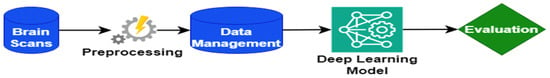
Figure 1 illustrates the AD detection system, an intricate and comprehensive framework designed to facilitate the efficient detection of AD. This system harnesses the synergistic integration of essential components, including brain scans, preprocessing techniques, data management strategies, deep learning models, and evaluation. Together, these elements establish a robust foundation for the system, ensuring its effectiveness, reliability, and precision.
Figure 1. Illustration depicting the interconnected elements of the AD detection system.

2.1. Brain Scans

Brain scans play a fundamental role in the AD detection system, as they provide critical information about structural and functional changes associated with AD [27].
Various imaging techniques are used to obtain detailed images of the brain, including magnetic resonance imaging (MRI), positron emission tomography (PET), and diffusion tensor imaging (DTI) [28]. MRI uses magnetic fields and radio waves to generate high-resolution images, revealing anatomical features of the brain [29]. PET involves injecting a radioactive tracer into the body, which highlights specific areas of the brain associated with AD pathology [30]. DTI measures the diffusion of water molecules in brain tissue, which allows for the visualization of white matter pathways and assessment of the integrity of neuronal connections [31].
Brain scans provide valuable information about structural changes, neurochemical abnormalities, and functional alterations in people with AD [32]. These scans can detect the presence of amyloid plaques and neurofibrillary tangles, the characteristic pathologies of AD, and reveal patterns of brain atrophy and synaptic dysfunction [33].
The data acquired by the brain scan serve as the basis for further analysis and interpretation [34]. However, it is important to note that interpreting brain scans requires expertise and knowledge of neuroimaging. Radiologists and neurologists often collaborate to ensure accurate and reliable interpretation of scans [35].
In the context of the AD detection system, brain scans serve as the primary input data, capturing the unique characteristics of each individual’s brain. These scans undergo further preprocessing and analysis to extract meaningful features and patterns that can contribute to the detection and classification of Alzheimer’s disease [36].

2.2. Preprocessing

Preprocessing plays a critical role in the AD detection system by applying essential steps to enhance the quality and reliability of data obtained from brain scans. This subsection focuses on the key preprocessing techniques used to prepare acquired imaging data before further analysis and interpretation.
One of the initial preprocessing steps is image registration, which involves aligning brain scans to a common reference space. This alignment compensates for variations in positioning and orientation, ensuring consistent analyses across different individuals and time points [37]. Commonly used techniques for image registration include affine and non-linear transformations.
Following image registration, intensity normalization techniques are applied to address variations in signal intensity between scans. These techniques aim to normalize intensity levels, facilitating more accurate and reliable comparisons among different brain regions and subjects [38]. Common normalization methods include z-score normalization and histogram matching.
Another important preprocessing step is noise reduction, which aims to minimize unwanted artefacts and noise that can interfere with subsequent analyses. Techniques such as Gaussian filtering and wavelet denoising are commonly employed to reduce noise while preserving important features in brain images [39].
Spatial smoothing is an additional preprocessing technique that involves applying a smoothing filter to the data. This process reduces local variations and improves the signal-to-noise ratio, facilitating the identification of relevant patterns and structures in brain scans [40]. Furthermore, motion correction is performed to address motion-related artefacts that may occur during brain scan acquisition. Motion correction algorithms can detect and correct head movements, ensuring that the data accurately represent the structural and functional characteristics of the brain [41].
It is important to note that preprocessing techniques may vary depending on the imaging modality used, such as MRI or PET. Each modality may require specific preprocessing steps tailored to its characteristics and challenges.

2.3. Data Management

Data management is a crucial component of the Alzheimer’s disease detection system, as it involves the efficient organization, storage, and handling of large quantities of imaging and clinical data. This subsection focuses on the key aspects of data management in Alzheimer’s disease research, including data acquisition, storage, integration, and quality control.
Data acquisition involves the collection of imaging data from various modalities such as MRI, PET, or CT scans, as well as clinical data, including demographic information, cognitive assessments, and medical history. Standardized protocols and validated assessment tools are used to ensure consistent data collection procedures [34].
Once data has been acquired, the storage of large-scale imaging and clinical datasets requires efficient and scalable storage solutions. Various database management systems, such as relational databases or NO Structured Query Language (NoSQL) databases, can be used to organize and store data securely and provide efficient retrieval and query capabilities [42].
The integration of heterogeneous data from different sources is crucial to enable comprehensive analysis and interpretation. Data integration techniques, such as data fusion or data harmonization, aim to combine data from multiple modalities or studies into a unified format to ensure compatibility and enable holistic analysis [43].
Data quality control is an essential step in guaranteeing the reliability and validity of the data collected. It involves identifying and correcting anomalies, missing values, outliers, or artefacts that could affect the accuracy and integrity of subsequent analyses. Quality control procedures, including data cleaning and validation checks, are applied to maintain data consistency and accuracy [44].
Effective data management also involves adherence to ethical and privacy guidelines to protect participant confidentiality and ensure data security. Compliance with regulatory requirements, such as obtaining informed consent and anonymizing data, is essential to protect participants’ rights and maintain data integrity.

2.4. Deep Learning Model

Deep learning models have emerged as powerful tools in Alzheimer’s disease detection, leveraging their ability to learn complex patterns and representations from large-scale imaging datasets. This subsection explores the application of deep learning models in Alzheimer’s disease detection, highlighting their architectures, training strategies, and performance evaluation.
Convolutional neural networks (CNNs) have been widely adopted in Alzheimer’s disease research due to their effectiveness in analyzing spatial relationships within brain images. CNNs consist of multiple convolutional and pooling operations layers, followed by fully connected layers for classification [45]. These architectures enable automatic feature extraction and hierarchical learning, capturing local and global brain scan patterns.
To train deep learning models, large annotated datasets are required. The Alzheimer’s Disease Neuroimaging Initiative (ADNI) and other publicly available datasets, such as the Open Access Series of Imaging Studies (OASIS) and the Australian Imaging, Biomarkers and Lifestyle (AIBL) study, have played crucial roles in facilitating the development and evaluation of deep learning models for Alzheimer’s disease detection [46].
Training deep learning models involves optimizing their parameters using labelled data. Stochastic gradient descent (SGD) and its variants, such as Adam and RMSprop, are commonly used optimization algorithms for deep learning [47]. Additionally, regularization techniques like dropout or batch normalization are employed to prevent overfitting and improve generalization performance [48].
The performance of deep learning models in Alzheimer’s disease detection is typically evaluated using metrics such as accuracy, sensitivity, specificity, and area under the receiver operating characteristic curve (AUC-ROC). Cross-validation or independent test sets are used to assess the generalization ability of the models [49].

3. Deep Learning for Alzheimer’s Disease Detection

3.1. Convolutional Neural Networks for AD Detection

Convolutional neural networks (CNNs) have gained prominence in medical imaging for AD detection, as they excel at learning hierarchical features from raw image data, enabling accurate predictions [50]. CNNs’ promising results in AD detection have garnered attention from researchers and clinicians.

3.1.1. Neuroimaging and CNNs for AD Analysis

CNNs have been employed for AD diagnosis, classification, prediction, and image generation. Neuroimaging data, including T1-weighted MRI scans and PET images, serve as foundational inputs [51]. CNNs adeptly extract features from these images, enabling precise AD classification and prediction.

3.1.2. CNN-3D Architecture for AD Classification

CNN-3D architectures, designed to harness the 3D nature of neuroimaging data, excel in capturing spatial relationships and fine details. Across various datasets, CNN-3D models exhibit robust performance in AD classification, capitalizing on their ability to extract discriminative features from 3D brain images.

3.1.3. GANs for Data Augmentation and Enhancement

Generative adversarial networks (GANs) generate synthetic brain images resembling real ones, alleviating limited labelled data availability. By enhancing downstream tasks like AD classification through realistic image synthesis, GANs contribute to improved model generalization and performance.

5.1.4. Transfer Learning and Multimodal Fusion

Transfer learning fine-tunes pre-trained CNN models from general image datasets to AD tasks, compensating for limited AD-specific data. Multimodal approaches, merging data from diverse imaging modalities, enhance classification by capturing complementary AD pathology facets.

3.1.5. Temporal Convolutional Networks (TCNs)

TCNs are another type of neural network architecture that can capture temporal dependencies in sequential data. Unlike LSTMs, TCNs utilize 1D convolutional layers with dilated convolutions to extract features from the temporal sequences. TCNs have been applied to various AD-related tasks, including disease classification, progression prediction, and anomaly detection. They offer computational efficiency and can capture both short-term and long-term temporal dependencies in the data.

3.1.6. Dataset Quality and Interpretable Models

Dataset size and diversity critically impact a CNN’s performance. Standardized, broad AD datasets are pivotal for robust models. Addressing interpretability, efforts should focus on unveiling learned features and decision-making processes to enhance trustworthiness and clinical applicability.

3.1.7. Convolutional Neural Network (CNN) Studies for AD Detection

Convolutional neural networks (CNNs) have demonstrated remarkable achievements in tasks such as organ segmentation [52][53][54] and disease detection [55][56][57] within the field of medical imaging. By leveraging neuroimaging data, these models can uncover hidden representations, establish connections between different components of an image, and identify patterns related to diseases [58]. They have been successfully applied to diverse medical imaging modalities, encompassing structural MRI [59], functional MRI [60][61] (fMRI), PET [62][63], and diffusion tensor imaging (DTI) [64][65][66]. Consequently, researchers have begun exploring the potential of deep learning models in detecting AD using medical images [67][68][69][70]

3.1.8. Performance Comparison

When assessing the performance of different CNN-based approaches in Alzheimer’s disease (AD) research, their remarkable potential becomes evident. Traditional CNN architectures, such as LeNet-5 and AlexNet, offer robust feature extraction capabilities, enabling them to capture intricate AD-related patterns. However, it is essential to note that LeNet-5′s relatively shallow architecture may limit its ability to discern complex features, while the high parameter count in AlexNet can lead to overfitting concerns.
Transfer learning, a strategy where pre-trained models like VGG16 are fine-tuned for AD detection, has emerged as a highly effective approach. By leveraging the insights gained from extensive image datasets, transfer learning significantly enhances AD detection accuracy.
The introduction of 3D CNNs has further expanded the capabilities of CNN-based methods, particularly in the analysis of volumetric data, such as MRI scans. These models excel at learning nuanced features, a critical advantage given the temporal progression of AD.
In terms of performance evaluation, CNN-based methods are typically assessed using various metrics, including accuracy, sensitivity, specificity, precision, and the F1-score. While these metrics effectively gauge performance, interpretability remains a challenge. Nevertheless, ongoing efforts, such as attention mechanisms and visualization tools, aim to address this issue.
Despite their promise, CNNs face limitations, primarily related to data availability. To ensure the generalization of CNN-based AD detection models across diverse populations, acquiring and curating large, representative datasets remains a priority for future research. In summary, CNN-based methodologies have demonstrated their mettle in AD research, showcasing strengths across traditional and 3D architectures, transfer learning, and ongoing interpretability enhancements. To realize their full potential for real-world clinical applications, addressing data limitations and improving generalization are critical objectives.

3.1.9. Meaningful Insights

The application of convolutional neural networks (CNNs) in Alzheimer’s disease (AD) detection has unveiled several meaningful insights. CNNs, particularly 3D architectures, have showcased their prowess in deciphering complex patterns within volumetric neuroimaging data.
One remarkable insight is the ability of CNNs to extract hierarchical features from brain images. Traditional CNN architectures, like LeNet-5 and AlexNet, excel in capturing intricate structural information but may struggle with deeper, more abstract features. In contrast, transfer learning, where pre-trained models are fine-tuned for AD detection, has proven highly effective. This approach capitalizes on the wealth of knowledge acquired from diverse image datasets, offering a robust foundation for AD-related feature extraction. The introduction of 3D CNNs has further illuminated the importance of spatial context in AD diagnosis. These models excel in capturing nuanced patterns across multiple image slices, aligning with the progressive nature of AD.
Performance metrics, including accuracy, sensitivity, specificity, and precision, have substantiated CNN’s effectiveness. These metrics provide quantitative evidence of CNNs’ diagnostic capabilities. Additionally, ongoing efforts in developing attention mechanisms and visualization tools aim to enhance model interpretability.
However, the ultimate insight gleaned from CNN-based AD detection is the need for substantial data. Generalizability across diverse populations demands large, representative datasets. This challenge underscores the importance of data acquisition and curation efforts.
In conclusion, CNNs have illuminated the path towards more accurate, data-driven AD detection. Leveraging hierarchical feature extraction, embracing 3D architectures, and ensuring interpretability are pivotal in harnessing CNNs’ potential for earlier and more reliable AD diagnosis.

3.2. Recurrent Neural Networks (RNN) for AD Detection

Recurrent neural networks (RNNs) have acquired considerable attention in medical imaging for AD detection. These deep learning models are well-suited for capturing temporal dependencies and sequential patterns in data, making them particularly useful for analyzing time series or sequential data in AD detection tasks. In recent years, RNNs have shown promising results in capturing complex relationships within longitudinal neuroimaging data and aiding in the early diagnosis of AD.

3.2.1. Long Short-Term Memory (LSTM) Networks

LSTM is a type of RNN that can effectively capture long-term dependencies in sequential data. Several studies have employed LSTM networks for AD diagnosis and prediction. These models typically take sequential data, such as time-series measurements from brain imaging or cognitive assessments, as input, and learn temporal patterns to classify or predict AD progression. LSTM-based models have demonstrated promising results in accurately diagnosing AD and predicting cognitive decline.

3.2.2. Encoder–Decoder Architectures

Encoder–decoder architectures, often combined with attention mechanisms, have been used in AD research to address tasks such as predicting disease progression or generating informative features. These models encode input sequences into latent representations and decode them to generate predictions or reconstructed sequences. Encoder–decoder architectures with attention mechanisms allow for the network to focus on relevant temporal information, improving prediction accuracy and interpretability.

3.2.3. Hybrid Models

Some studies have combined RNNs with other deep learning architectures, such as convolutional neural networks (CNNs) or generative adversarial networks (GANs), to leverage their respective strengths. These hybrid models aim to capture both spatial and temporal information from brain imaging data, leading to improved performance in AD diagnosis, progression prediction, or generating synthetic data for augmentation.

3.2.4. Recurrent Neural Network (RNN) Studies for AD Detection

Recurrent neural networks (RNNs) have emerged as a popular deep learning technique for analyzing temporal data, making them well-suited for Alzheimer’s disease research. This discussion section will highlight the various methods that have utilized RNNs in AD research, provide an overview of their approaches, compare their performance, and present meaningful insights for further discussion.

3.2.5. Performance Comparison

Comparing the performance of different RNN-based methods in AD research can be challenging due to variations in datasets, evaluation metrics, and experimental setups. However, several studies have reported high accuracy, sensitivity, and specificity in AD diagnosis and prediction tasks using RNNs. For example, LSTM-based models have achieved accuracies ranging from 80% to over 90% in AD classification. TCNs have demonstrated competitive performance in predicting cognitive decline, with high AUC scores. Encoder–decoder architectures with attention mechanisms have shown improvements in disease progression prediction compared to traditional LSTM models. Hybrid models combining RNNs with other architectures have reported enhanced performance by leveraging spatial and temporal information.

3.2.6. Meaningful Insights

RNNs, such as LSTMs, are well-suited for capturing long-term dependencies in sequential data. In the context of AD research, this capability allows for identifying subtle temporal patterns and predicting disease progression. By analyzing longitudinal data, RNNs can potentially detect early signs of cognitive decline and facilitate early intervention strategies.
RNNs can also be effectively used for data augmentation in AD research. Synthetic sequences can be generated using generative models, such as variational autoencoders (VAEs) or GANs, to increase the diversity and size of the training dataset. This augmented data can enhance the robustness and generalizability of RNN models, leading to improved diagnostic accuracy and generalization to unseen data.
In addition, RNNs offer interpretability and explainability in AD research. By analyzing the temporal patterns learned by the models, researchers can gain insights into the underlying disease progression mechanisms. This information can aid in understanding the neurobiological processes associated with AD and provide valuable clues for potential therapeutic interventions.
Moreover, RNNs can handle multimodal data sources, such as combining brain imaging (e.g., MRI, PET scans) with clinical assessments or genetic information. Integrating multiple modalities can provide a more comprehensive understanding of AD, capturing both structural and functional changes in the brain along with clinical markers. RNN-based models enable the fusion of diverse data sources to improve the accuracy and reliability of AD diagnosis and prognosis.
RNNs trained on large-scale datasets can learn robust representations that generalize well to unseen data. Pre-training RNN models on large cohorts or external datasets and finetuning them on specific AD datasets can facilitate knowledge transfer and enhance the performance of AD classification and prediction tasks. Transfer learning approaches enable the utilization of existing knowledge and leverage the expertise gained from related tasks or domains.
While RNNs have shown promise in AD research, there are still challenges to address. One major challenge is the limited availability of large-scale, longitudinal AD datasets. Acquiring and curating diverse datasets with longitudinal follow-up is crucial for training RNN models effectively. Additionally, incorporating uncertainty estimation and quantifying model confidence in predictions can further enhance the reliability and clinical applicability of RNN-based methods.
Furthermore, exploring the combination of RNNs with other advanced techniques, such as attention mechanisms, graph neural networks, or reinforcement learning, holds promise for improving AD diagnosis, understanding disease progression, and guiding personalized treatment strategies. Integrating multimodal data sources, such as imaging, genetics, and omics data, can provide a more comprehensive view of AD pathophysiology.

3.3. Generative Modeling for AD Detection

Generative modelling techniques have gained attention in medical imaging for Alzheimer’s disease (AD) detection. These models are capable of generating new samples that follow the distribution of the training data, enabling them to capture the underlying patterns and variations in AD-related imaging data. By leveraging generative models, researchers aim to enhance early detection, improve classification accuracy, and gain insights into the underlying mechanisms of AD.

3.3.1. GANs for Image Generation

One prominent application of generative modelling in Alzheimer’s disease is the generation of synthetic brain images for diagnostic and research purposes. GANs have been used to generate realistic brain images that mimic the characteristics of Alzheimer’s disease, such as the presence of amyloid beta plaques and neurofibrillary tangles. These synthetic images can be valuable for augmenting datasets, addressing data scarcity issues, and improving classification performance.

5.3.2. Conditional GANs for Disease Progression Modeling

Conditional GANs (cGANs) have been employed to model the progression of Alzheimer’s disease over time. By conditioning the generator on longitudinal data, cGANs can generate synthetic brain images that capture disease progression stages, ranging from normal to mild cognitive impairment (MCI) and finally to Alzheimer’s disease. This enables the generation of realistic images representing the transition from healthy to pathological brain states.

3.3.3. Variational Autoencoders (VAEs) for Feature Extraction

In addition to GANs, variational autoencoders (VAEs) have been utilized to extract informative features from brain images for Alzheimer’s disease classification. VAEs can learn a compressed representation of the input images, known as latent space, which captures relevant features associated with the disease. By sampling from the latent space, new images can be generated, and the extracted features can be used for classification tasks.

3.3.4. Hybrid Approaches

Some studies have explored hybrid approaches that combine different generative models to leverage their respective advantages. For example, combining GANs and VAEs can harness the generative power of GANs while benefiting from the probabilistic nature and interpretability of VAEs. These hybrid models aim to generate high-quality images while preserving the meaningful representations learned by VAEs.

3.3.5. Generative Modeling Studies (GAN) for AD Detection

Generative modelling, particularly through approaches like generative adversarial networks (GANs), has emerged as a promising technique in the field of Alzheimer’s disease research. This discussion section will provide an overview of the various methods used in generative modelling for Alzheimer’s disease, compare their strengths and limitations, and highlight meaningful insights for further exploration and discussion.

3.3.6. Comparative Analysis

When comparing the different generative modelling methods in Alzheimer’s disease research, several factors should be considered:
  • Image Quality: The primary goal of generative modelling is to generate high-quality brain images that closely resemble real data. GANs have demonstrated remarkable success in producing visually realistic images, while VAEs tend to produce slightly blurred images due to the nature of their probabilistic decoding process.
  • Feature Extraction: While GANs excel in image generation, VAEs are more suitable for feature extraction and latent space representation. VAEs can capture meaningful features that reflect disease progression and provide interpretability, making them valuable for understanding the underlying mechanisms of Alzheimer’s disease.
  • Data Scarcity: Alzheimer’s disease datasets are often limited in size, posing challenges for training deep learning models. Generative modelling techniques, especially GANs, can help address data scarcity by generating synthetic samples that augment the training data and improve model generalization.
  • Interpretability: VAEs offer an advantage in terms of interpretability because they learn a structured latent space that captures meaningful variations in the data. This can aid in understanding disease patterns and identifying potential biomarkers.

3.3.7. Meaningful Insights

Generative modelling in Alzheimer’s disease research holds great promise for advancing diagnosis, disease progression modelling, and understanding the underlying mechanisms of the disease. By generating realistic brain images and capturing disease-related features, these techniques can complement traditional diagnostic methods and provide new avenues for personalized treatment and intervention strategies.
One meaningful insight from the application of generative modelling is the potential to address data scarcity issues. Alzheimer’s disease datasets are often limited in size and subject to variability in imaging protocols and data acquisition. By using generative models like GANs and VAEs, researchers can generate synthetic data that closely resemble real brain images. This augmentation of the dataset not only increases the sample size but also captures a wider range of disease characteristics and progression patterns. Consequently, it enhances the robustness and generalizability of machine learning models trained on these augmented datasets.
Moreover, generative modelling techniques provide a unique opportunity to simulate disease progression and explore hypothetical scenarios. By conditioning the generative models on various disease stages, researchers can generate synthetic brain images that represent different pathological states, from early stages of mild cognitive impairment to advanced Alzheimer’s disease. This capability allows for the investigation of disease progression dynamics, identification of critical biomarkers, and evaluation of potential intervention strategies.
Furthermore, the combination of generative models with other deep learning techniques, such as convolutional neural networks (CNNs) or recurrent neural networks (RNNs), can further enhance the performance of Alzheimer’s disease classification and prediction tasks. These hybrid models can leverage the strengths of different architectures and generate more accurate and interpretable results. For example, combining GANs for image generation with CNNs for feature extraction and classification can lead to improved diagnostic accuracy and a better understanding of the underlying disease mechanisms.

References

  1. Vaupel, J.W. Biodemography of human ageing. Nature 2010, 464, 536–542.
  2. Samir, K.C.; Lutz, W. The human core of the shared socioeconomic pathways: Population scenarios by age, sex and level of education for all countries to 2100. Glob. Environ. Chang. 2017, 42, 181–192.
  3. Godyń, J.; Jończyk, J.; Panek, D.; Malawska, B. Therapeutic strategies for Alzheimer’s disease in clinical trials. Pharmacol. Rep. 2016, 68, 127–138.
  4. Blaikie, L.; Kay, G.; Lin, P.K.T. Current and emerging therapeutic targets of Alzheimer’s disease for the design of multi-target directed ligands. MedChemComm 2019, 10, 2052–2072.
  5. Gutierrez, B.A.; Limon, A. Synaptic disruption by soluble oligomers in patients with Alzheimer’s and Parkinson’s disease. Biomedicines 2022, 10, 1743.
  6. Zvěřová, M. Clinical aspects of Alzheimer’s disease. Clin. Biochem. 2019, 72, 3–6.
  7. Bronzuoli, M.R.; Iacomino, A.; Steardo, L.; Scuderi, C. Targeting neuroinflammation in Alzheimer’s disease. J. Inflamm. Res. 2016, 9, 199–208.
  8. Aljunid, S.M.; Maimaiti, N.; Ahmed, Z.; Nur, A.M.; Nor, N.M.; Ismail, N.; Haron, S.A.; Shafie, A.A.; Salleh, M.; Yusuf, S.; et al. Development of clinical pathway for mild cognitive impairment and dementia to quantify cost of age-related cognitive disorders in Malaysia. Malays. J. Public Health Med. 2014, 14, 88–96.
  9. Hu, K.; Wang, Y.; Chen, K.; Hou, L.; Zhang, X. Multi-scale features extraction from baseline structure MRI for MCI patient classification and AD early diagnosis. Neurocomputing 2016, 175, 132–145.
  10. Ebrahimighahnavieh, A.; Luo, S.; Chiong, R. Deep learning to detect Alzheimer’s disease from neuroimaging: A systematic literature review. Comput. Methods Programs Biomed. 2020, 187, 105242.
  11. Fathi, S.; Ahmadi, M.; Dehnad, A. Early diagnosis of Alzheimer’s disease based on deep learning: A systematic review. Comput. Biol. Med. 2022, 146, 105634.
  12. Binaco, R.; Calzaretto, N.; Epifano, J.; McGuire, S.; Umer, M.; Emrani, S.; Wasserman, V.; Libon, D.J.; Polikar, R. Machine learning analysis of digital clock drawing test performance for differential classification of mild cognitive impairment subtypes versus Alzheimer’s disease. J. Int. Neuropsychol. Soc. 2020, 26, 690–700.
  13. Karikari, T.K.; Ashton, N.J.; Brinkmalm, G.; Brum, W.S.; Benedet, A.L.; Montoliu-Gaya, L.; Lantero-Rodriguez, J.; Pascoal, T.A.; Suárez-Calvet, M.; Rosa-Neto, P.; et al. Blood phospho-tau in Alzheimer disease: Analysis, interpretation, and clinical utility. Nat. Rev. Neurol. 2022, 18, 400–418.
  14. Wang, M.; Song, W.M.; Ming, C.; Wang, Q.; Zhou, X.; Xu, P.; Krek, A.; Yoon, Y.; Ho, L.; Orr, M.E.; et al. Guidelines for bioinformatics of single-cell sequencing data analysis in Alzheimer’s disease: Review, recommendation, implementation and application. Mol. Neurodegener. 2022, 17, 1–52.
  15. Zhang, Y.; Wu, K.-M.; Yang, L.; Dong, Q.; Yu, J.-T. Tauopathies: New perspectives and challenges. Mol. Neurodegener. 2022, 17, 28.
  16. Klyucherev, T.O.; Olszewski, P.; Shalimova, A.A.; Chubarev, V.N.; Tarasov, V.V.; Attwood, M.M.; Syvänen, S.; Schiöth, H.B. Advances in the development of new biomarkers for Alzheimer’s disease. Transl. Neurodegener. 2022, 11, 1–24.
  17. Zhao, K.; Duka, B.; Xie, H.; Oathes, D.J.; Calhoun, V.; Zhang, Y. A dynamic graph convolutional neural network framework reveals new insights into connectome dysfunctions in ADHD. NeuroImage 2022, 246, 118774.
  18. Jiang, Y.; Zhou, X.; Ip, F.C.; Chan, P.; Chen, Y.; Lai, N.C.; Cheung, K.; Lo, R.M.; Tong, E.P.; Wong, B.W.; et al. Large-scale plasma proteomic profiling identifies a high-performance biomarker panel for Alzheimer’s disease screening and staging. Alzheimer’s Dement. 2022, 18, 88–102.
  19. Gómez-Isla, T.; Frosch, M.P. Lesions without symptoms: Understanding resilience to Alzheimer disease neuropathological changes. Nat. Rev. Neurol. 2022, 18, 323–332.
  20. Liu, C.; Xiang, X.; Han, S.; Lim, H.Y.; Li, L.; Zhang, X.; Ma, Z.; Yang, L.; Guo, S.; Soo, R.; et al. Blood-based liquid biopsy: Insights into early detection and clinical management of lung cancer. Cancer Lett. 2022, 524, 91–102.
  21. Hernandez, M.; Ramon-Julvez, U.; Ferraz, F.; with the ADNI Consortium. Explainable AI toward understanding the performance of the top three TADPOLE Challenge methods in the forecast of Alzheimer’s disease diagnosis. PLoS ONE 2022, 17, e0264695.
  22. Lydon, E.A.; Nguyen, L.T.; Shende, S.A.; Chiang, H.-S.; Spence, J.S.; Mudar, R.A. EEG theta and alpha oscillations in early versus late mild cognitive impairment during a semantic Go/NoGo task. Behav. Brain Res. 2022, 416, 113539.
  23. Yadav, S.; Zhou Shu, K.; Zachary, Z.; Yueyang, G.; Lana, X.; ADNI Consortium. Integrated Metabolomics and Transcriptomics Analysis Identifies Molecular Subtypes within the Early and Late Mild Cognitive Impairment Stages of Alzheimer’s Disease. medRxiv 2023.
  24. Jeyavathana, R.B.; Balasubramanian, R.; Pandian, A.A. A survey: Analysis on pre-processing and segmentation techniques for medical images. Int. J. Res. Sci. Innov. (IJRSI) 2016, 3, 113–120.
  25. James, A.P.; Dasarathy, B.V. Medical image fusion: A survey of the state of the art. Inf. Fusion 2014, 19, 4–19.
  26. Balagurunathan, Y.; Mitchell, R.; El Naqa, I. Requirements and reliability of AI in the medical context. Phys. Medica 2021, 83, 72–78.
  27. Waldemar, G.; Dubois, B.; Emre, M.; Georges, J.; McKeith, I.G.; Rossor, M.; Scheltens, P.; Tariska, P.; Winblad, B. Recommendations for the diagnosis and management of Alzheimer’s disease and other disorders associated with dementia: EFNS guideline. Eur. J. Neurol. 2007, 14, e1–e26.
  28. Shenton, M.E.; Hamoda, H.M.; Schneiderman, J.S.; Bouix, S.; Pasternak, O.; Rathi, Y.; Vu, M.-A.; Purohit, M.P.; Helmer, K.; Koerte, I.; et al. A review of magnetic resonance imaging and diffusion tensor imaging findings in mild traumatic brain injury. Brain Imaging Behav. 2012, 6, 137–192.
  29. Matthews, P.M.; Peter, J. Functional magnetic resonance imaging. J. Neurol. Neurosurg. Psychiatry 2004, 75, 6–12.
  30. Klunk, W.E.; Mathis, C.A.; Price, J.C.; DeKosky, S.T.; Lopresti, B.J.; Tsopelas, N.D.; Judith, A.S.; Robert, D.N. Amyloid imaging with PET in Alzheimer’s disease, mild cognitive impairment, and clinically unimpaired subjects. In PET in the Evaluation of Alzheimer’s Disease and Related Disorders; Springer: New York, NY, USA, 2009; pp. 119–147.
  31. Basser, P.J. Inferring microstructural features and the physiological state of tissues from diffusion-weighted images. NMR Biomed. 1995, 8, 333–344.
  32. Braak, H.; Alafuzoff, I.; Arzberger, T.; Kretzschmar, H.; Del Tredici, K. Staging of Alzheimer disease-associated neurofibrillary pathology using paraffin sections and immunocytochemistry. Acta Neuropathol. 2006, 112, 389–404.
  33. Dubois, B.; Feldman, H.H.; Jacova, C.; DeKosky, S.T.; Barberger-Gateau, P.; Cummings, J.L.; Delacourte, A.; Galasko, D.; Gauthier, S.; Jicha, G.A.; et al. Research criteria for the diagnosis of Alzheimer’s disease: Revising the NINCDS–ADRDA criteria. Lancet Neurol. 2007, 6, 734–746.
  34. Mueller, S.G.; Weiner, M.W.; Thal, L.J.; Petersen, R.C.; Jack, C.; Jagust, W.; Trojanowski, J.Q.; Toga, A.W.; Beckett, L. The Alzheimer’s disease neuroimaging initiative. Neuroimaging Clin. 2005, 15, 869–877.
  35. Jagust, W. Imaging the evolution and pathophysiology of Alzheimer disease. Nat. Rev. Neurosci. 2018, 19, 687–700.
  36. Weiner, M.W.; Veitch, D.P.; Aisen, P.S.; Beckett, L.A.; Cairns, N.J.; Green, R.C.; Harvey, D.; Jack, C.R.; Jagust, W.; Liu, E.; et al. The Alzheimer’s Disease Neuroimaging Initiative: A review of papers published since its inception. Alzheimer’s Dement. 2013, 9, e111–e194.
  37. Klein, A.; Andersson, J.; Ardekani, B.A.; Ashburner, J.; Avants, B.; Chiang, M.-C.; Christensen, G.E.; Collins, D.L.; Gee, J.; Hellier, P.; et al. Evaluation of 14 nonlinear deformation algorithms applied to human brain MRI registration. NeuroImage 2009, 46, 786–802.
  38. Ashburner, J.; Friston, K.J. Voxel-based morphometry—The methods. Neuroimage 2000, 11, 805–821.
  39. Choy, G.; Khalilzadeh, O.; Michalski, M.; Synho, D.; Samir, A.E.; Pianykh, O.S.; Geis, J.R.; Pandharipande, P.V.; Brink, J.A.; Dreyer, K.J. Current applications and future impact of machine learning in radiology. Radiology 2018, 288, 318–328.
  40. Smith, S.M.; Jenkinson, M.; Woolrich, M.W.; Beckmann, C.F.; Behrens, T.E.; Johansen-Berg, H.; Bannister, P.R.; De Luca, M.; Drobnjak, I.; Flitney, D.E.; et al. Advances in functional and structural MR image analysis and implementation as FSL. NeuroImage 2004, 23, S208–S219.
  41. Jenkinson, M.; Bannister, P.; Brady, M.; Smith, S. Improved optimization for the robust and accurate linear registration and motion correction of brain images. Neuroimage 2002, 17, 825–841.
  42. Hashem IA, T.; Yaqoob, I.; Anuar, N.B.; Mokhtar, S.; Gani, A.; Khan, S.U. The rise of “big data” on cloud computing: Review and open research issues. Inf. Syst. 2015, 47, 98–115.
  43. Gorgolewski, K.J.; Auer, T.; Calhoun, V.D.; Craddock, R.C.; Das, S.; Duff, E.P.; Flandin, G.; Ghosh, S.S.; Glatard, T.; Halchenko, Y.O.; et al. The brain imaging data structure, a format for organizing and describing outputs of neuroimaging experiments. Sci. Data 2016, 3, 160044.
  44. Kandel, S.; Heer, J.; Plaisant, C.; Kennedy, J.; van Ham, F.; Riche, N.H.; Weaver, C.; Lee, B.; Brodbeck, D.; Buono, P. Research directions in data wrangling: Visualizations and transformations for usable and credible data. Inf. Vis. 2011, 10, 271–288.
  45. LeCun, Y.; Bengio, Y.; Hinton, G. Deep learning. Nature 2015, 521, 436–444.
  46. Madrid, L.; Labrador, S.C.; González-Pérez, A.; Sáez, M.E.; Alzheimer’s Disease Neuroimaging Initiative (ADNI and others). Integrated Genomic, Transcriptomic and Proteomic Analysis for Identifying Markers of Alzheimer’s Disease. Diagnostics 2021, 11, 2303.
  47. Konur, O.; Kingma, D.; Ba, J. Adam: A method for stochastic optimization. In Proceedings of the International Conference on Learning Representations, ICLR 2015, San Diego, CA, USA, 7–9 May 2015; pp. 1–15.
  48. Srivastava, N.; Hinton, G.; Krizhevsky, A.; Sutskever, I.; Salakhutdinov, R. Dropout: A simple way to prevent neural networks from overfitting. J. Mach. Learn. Res. 2014, 15, 1929–1958.
  49. Jiang, X.; Chang, L.; Zhang, Y.-D. Classification of Alzheimer’s disease via eight-layer convolutional neural network with batch normalization and dropout techniques. J. Med. Imaging Health Inform. 2020, 10, 1040–1048.
  50. Li, Z.; Liu, F.; Yang, W.; Peng, S.; Zhou, J. A survey of convolutional neural networks: Analysis, applications, and prospects. IEEE Trans. Neural Netw. Learn. Syst. 2021, 33, 6999–7019.
  51. Bin Bae, J.; Lee, S.; Jung, W.; Park, S.; Kim, W.; Oh, H.; Han, J.W.; Kim, G.E.; Kim, J.S.; Kim, J.H.; et al. Identification of Alzheimer’s disease using a convolutional neural network model based on T1-weighted magnetic resonance imaging. Sci. Rep. 2020, 10, 22252.
  52. Ilesanmi, A.E.; Ilesanmi, T.; Idowu, O.P.; Torigian, D.A.; Udupa, J.K. Organ segmentation from computed tomography images using the 3D convolutional neural network: A systematic review. Int. J. Multimed. Inf. Retr. 2022, 11, 315–331.
  53. Irshad, S.; Gomes, D.P.S.; Kim, S.T. Improved abdominal multi-organ segmentation via 3d boundary-constrained deep neural networks. IEEE Access 2023, 11, 35097–35110.
  54. Rickmann, A.-M.; Senapati, J.; Kovalenko, O.; Peters, A.; Bamberg, F.; Wachinger, C. AbdomenNet: Deep neural network for abdominal organ segmentation in epidemiologic imaging studies. BMC Med. Imaging 2022, 22, 1–11.
  55. Hassan, S.M.; Maji, A.K. Plant disease identification using a novel convolutional neural network. IEEE Access 2022, 10, 5390–5401.
  56. Ashwinkumar, S.; Rajagopal, S.; Manimaran, V.; Jegajothi, B. Automated plant leaf disease detection and classification using optimal MobileNet based convolutional neural networks. Mater. Today Proc. 2022, 51, 480–487.
  57. Jiang, J.; Liu, H.; Zhao, C.; He, C.; Ma, J.; Cheng, T.; Zhu, Y.; Cao, W.; Yao, X. Evaluation of Diverse Convolutional Neural Networks and Training Strategies for Wheat Leaf Disease Identification with Field-Acquired Photographs. Remote Sens. 2022, 14, 3446.
  58. Nirthika, R.; Manivannan, S.; Ramanan, A.; Wang, R. Pooling in convolutional neural networks for medical image analysis: A survey and an empirical study. Neural Comput. Appl. 2022, 34, 5321–5347.
  59. Mattia, G.M.; Sarton, B.; Villain, E.; Vinour, H.; Ferre, F.; Buffieres, W.; Le Lann, M.-V.; Franceries, X.; Peran, P.; Silva, S. Multimodal MRI-Based Whole-Brain Assessment in Patients In Anoxoischemic Coma by Using 3D Convolutional Neural Networks. Neurocritical Care 2022, 37 (Suppl. S2), 303–312.
  60. Warren, S.L.; Moustafa, A.A. Functional magnetic resonance imaging, deep learning, and Alzheimer’s disease: A systematic review. J. Neuroimaging 2023, 33, 5–18.
  61. Dan, T.; Huang, Z.; Cai, H.; Laurienti, P.J.; Wu, G. Learning brain dynamics of evolving manifold functional MRI data using geometric-attention neural network. IEEE Trans. Med. Imaging 2022, 41, 2752–2763.
  62. Guo, X.; Zhou, B.; Pigg, D.; Spottiswoode, B.; Casey, M.E.; Liu, C.; Dvornek, N.C. Unsupervised inter-frame motion correction for whole-body dynamic PET using convolutional long short-term memory in a convolutional neural network. Med. Image Anal. 2022, 80, 102524.
  63. Bin Tufail, A.; Anwar, N.; Ben Othman, M.T.; Ullah, I.; Khan, R.A.; Ma, Y.-K.; Adhikari, D.; Rehman, A.U.; Shafiq, M.; Hamam, H. Early-stage Alzheimer’s disease categorization using PET neuroimaging modality and convolutional neural networks in the 2d and 3d domains. Sensors 2022, 22, 4609.
  64. Estudillo-Romero, A.; Haegelen, C.; Jannin, P.; Baxter, J.S.H. Voxel-based diktiometry: Combining convolutional neural networks with voxel-based analysis and its application in diffusion tensor imaging for Parkinson’s disease. Hum. Brain Mapp. 2022, 43, 4835–4851.
  65. Liu, S.; Liu, Y.; Xu, X.; Chen, R.; Liang, D.; Jin, Q.; Liu, H.; Chen, G.; Zhu, Y. Accelerated cardiac diffusion tensor imaging using deep neural network. Phys. Med. Biol. 2022, 68, 025008.
  66. Park, S.; Yu, J.; Woo, H.-H.; Park, C.G. A novel network architecture combining central-peripheral deviation with image-based convolutional neural networks for diffusion tensor imaging studies. J. Appl. Stat. 2022, 50, 3294–3311.
  67. AlSaeed, D.; Omar, S.F. Brain MRI analysis for Alzheimer’s disease diagnosis using CNN-based feature extraction and machine learning. Sensors 2022, 22, 2911.
  68. Ghazal, T.M.; Abbas, S.; Munir, S.; Ahmad, M.; Issa, G.F.; Zahra, S.B.; Khan, M.A.; Hasan, M.K. Alzheimer disease detection empowered with transfer learning. Comput. Mater. Contin. 2022, 70, 5005–5019.
  69. Liu, S.; Masurkar, A.V.; Rusinek, H.; Chen, J.; Zhang, B.; Zhu, W.; Fernandez-Granda, C.; Razavian, N. Generalizable deep learning model for early Alzheimer’s disease detection from structural MRIs. Sci. Rep. 2022, 12, 17106.
  70. Loddo, A.; Buttau, S.; Di Ruberto, C. Deep learning based pipelines for Alzheimer’s disease diagnosis: A comparative study and a novel deep-ensemble method. Comput. Biol. Med. 2021, 141, 105032.
More
Information
Contributors MDPI registered users' name will be linked to their SciProfiles pages. To register with us, please refer to https://encyclopedia.pub/register : , ,
View Times: 102
Revisions: 2 times (View History)
Update Date: 05 Mar 2024
1000/1000
Video Production Service