Colorectal cancer (CRC) is a common malignant tumor of the gastrointestinal tract, which has become a serious threat to human health worldwide. CRC is a complex disease due to its extensive heterogeneity; thus, effective treatment could be enhanced by the implementation of a personalized medicine approach. Despite constantly improved diagnostic and individualized therapeutic methods, CRC remains one of the biggest problems of contemporary medicine. Knowledge of the basic risk factors, early clinical symptoms, and available screening tests, as well as the preservation of oncological alert, allow the proper targeting of the diagnostic process and, consequently, the earlier diagnosis of the disease. Undoubtedly, new research at the molecular and genetic level allows us to precisely understand the process of initiation and progression of cancerous diseases and, consequently, precise, personalized prevention and treatment.
1. Introduction
The recent World Health Organization (WHO) evaluations show a significant increase in the incidence of malignant tumors within the aging population
[1][2]. Up to 2035, deaths from colorectal cancer (CRC) and rectal cancer are predicted to increase. In Latin American and Caribbean countries, the number of deaths is predicted to double by 2035
[3]. Cancer is the second-leading cause of death in the world and is one of the most significant health and economic issues for society
[1]. CRC carcinogenesis—a transformation from adenoma to cancer—is a long-lasting process
[4]. On the one hand, it simplifies successful screening in risk groups; on the other hand, early diagnostics at the time of initial, non-characteristic symptoms may lead to the detection of this disease in a fully curable stage. The morbidity and mortality associated with CRC can be reduced by implementing preventive measures such as targeted screening programs and early therapeutic intervention. Adapting one’s lifestyle, following an appropriate diet, regular physical activity, and the maintenance of weight all play a key role in minimizing the risk of CRC
[5].
2. The State of Knowledge from Symptoms to Novel Molecular Biology Achievements
2.1. Incidence of Colorectal Cancer and Its Geographic Variations
Throughout the world, CRC is nowadays a major cause of cancer-related morbidity and mortality, with nearly 1.9 million new cases diagnosed and almost 935,000 deaths in 2020
[1].
The global distribution of CRC has large geographic differences, with the number of new cases rapidly increasing due to population growth, changes in demographics, and the Westernization of lifestyle habits
[6].
The highest incidence of CRC occurs in Europe, Australia/New Zealand, and North America
[1]. CRC is both the third most commonly diagnosed cancer and the third most common cause of cancer-related deaths in both men and women in the United States
[5]. In 2020, CRC was the most common type of cancer incidence among men in Slovakia, the countries of the Arabian Peninsula, Ethiopia, and Southeast Asian countries such as Singapore and Brunei Darussalam. It was also reported that CRC is the cancer with the highest mortality among men in countries of the Arabian Peninsula and Ethiopia and among women in Spain, Croatia, Belarus, Estonia, and Japan
[1]. Two-thirds of CRC cases occur in countries characterized by high or very high indices of development and/or income. A pathological examination in approximately 95% of CRC cases reveals adenocarcinomas and other types of cancers that occur, including mucinous carcinomas and adenosquamous carcinomas
[1][6].
2.2. Temporal Trends in Colorectal Cancer Incidence, Mortality and Survival Rates
The incidence of CRC worldwide is constantly increasing, especially in countries characterized by a high-income economy and high human development index (HDI)
[5][6][7]. CRC is considered one of the clearest markers of the cancer transition, replacing infection-related cancers in countries undergoing rapid social and economic changes. Such changes in the causes of cancer are predominantly linked to Western lifestyles and are, thus, frequently found in high-income countries. Given the temporal profiles and demographic projection, it is expected that by 2030, the global number of CRC cases will increase by 60% to more than 2.2 million new cases and approximately 1.1 million deaths annually
[6].
Although over the past few decades, effective therapeutic strategies for CRC have been developed, the five-year overall survival is unsatisfactory. This is caused by the presence of the following poor prognostic factors: vascular, neural invasion, a low lymphocyte-to-monocyte ratio (LMR), late diagnosis, and tumor stage
[8]. It is estimated that approximately 20% of CRC patients have already progressed into a metastatic state at the time of presentation, and more than 30% of patients with early CRC eventually develop metastatic disease
[9][10]. CRC treatment’s long-term costs remain a huge social and economic burden
[11].
2.3. Symptoms of Colorectal Cancer
The course of CRC can be asymptomatic for a long time; the appearance of clinical symptoms is often indicated with an advanced stage of cancer. CRC symptomatology depends on the tumor localization. The classic symptoms of CRC include explicit or latent bleeding from the gastrointestinal tract (rectal bleeding or blood in the stool), abdominal pain, a palpably perceptible tumor, the rhythm of bowel movement disturbance (alternating constipation and diarrhea), gastrointestinal tract obstruction, unintended weight loss, fatigue, anemia, and febrile states
[12][13]. Explicit bleeding from the lower digestive tract is common (in 50–60% of patients with CRC), and an easily observable symptom (per rectum examination) requires the determination of its cause in each case
[14][15]. Another very common symptom is a bowel movement disorder (constipation/diarrhea), which occurs in about 50% of CRC cases; if it appears for more than 6 weeks without apparent cause, this becomes an alarming symptom
[16]. An important symptom for CRC patients, which has been stated by various authors at a frequency of 30% to more than 75%, is anemia
[13][17]. When diagnosing men over 40 years of age, as well as post-menopausal women with iron deficiency, such as anemia, the exclusion of a proliferative process is required. Weight loss, a palpable tumor, and abdominal pain are significantly associated with high levels of colorectal cancer.
2.4. Pathological Evaluation of Colorectal Carcinogenesis
The WHO pathologic classification divides CRC into the following histologic subtypes: glandular cancers (classic adenocarcinomas, AC, the highest percentage of diagnoses), mucinous adenocarcinomas (MAC), signet cell carcinomas (SRCC), and variants including adenosquamous carcinomas, squamous cell carcinomas, medullary carcinomas, neuroendocrine, undifferentiated and others
[18]. CRC is a cancer of epithelial origin, usually developing based on conventional adenomas.
In total, 70% of all CRCs stem from adenomas. Progressions from adenoma to carcinoma take more than 10 years in sporadic cancer, whereas much shorter intervals can be observed in the Lynch syndrome
[19]. Adenomas are distributed relatively throughout the colon; those with a flat or depressed morphology are distributed more in the proximal colon, and pedunculated lesions are more in the distal colon. Adenomas are, by definition, dysplastic, with the majority being low-grade; they can be characterized by tubular or villous histology, with the overwhelming majority being tubular. The increasing size of adenomas is associated with villous elements and invasive cancer (invasive cancer in adenomas ≤5 mm is extremely rare). An “advanced” stage is defined as a lesion ≥1 cm in size or having high-grade dysplasia or villous elements
[20].
2.5. Epigenetics of CRC
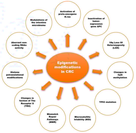
Evidence from scientific reports suggests an important role of epigenetic modifications in the development of CRC (
Figure 1)
[13][21][22][23][24][25][26]. Epigenetic modifications consist of changes in the methylation of cytosine-guanine (CpG) dinucleotides (DNA methylation), histone-tail post-translational modifications, and the expression of non-coding RNAs (ncRNA).
Figure 1. Epigenetic modifications associated with the development of CRC
[13][21][22][23][24][25][26].
3. Risk Factors for Colorectal Cancer
3.1. Colorectal Cancer Non-Modifiable Risk Factors
Among nonmodifiable factors, the most important role for CRC’s development includes age, sex, and individual background of adenomatous polyps or inflammatory bowel disease (IBD), a familial history of CRC or adenomatous polyps, and inherited genetic risk
[27][28].
The likelihood of a CRC diagnosis commonly affects males and is strongly age-related, increasing after the age of 40 and continues to rise with increasing age. More than 90% of CRC cases are diagnosed in patients aged 50 or older, and the incidence rate is more than 50 times higher in persons aged 60 to 79 years. However, CRC appears to also be increasing among younger people aged 20 to 49 years: a trend observed in high HDI economies
[29].
Well-known precursor lesions of CRC are tubular and villous colorectal adenomas. The statistics show that nearly 95% of sporadic CRCs come from adenomas. Individuals who have suffered from adenomas have an increased risk of developing CRC. A long latency period estimated up to 20 years, is usually required for the development of malignancy from adenomas
[30].
IBD is a group of colon and small intestine inflammatory conditions; Crohn’s disease and ulcerative colitis are the principal types of IBD. The differences between ulcerative colitis and Crohn’s disease can be seen in the areas affected by those conditions. Ulcerative colitis only causes the inflammation of the mucosa of the colon and rectum, whereas Crohn’s disease affects deeper tissues causing inflammation of the full thickness of the bowel wall and may involve any part of the digestive tract from the mouth to the anus. Individuals with IBD suffer from symptoms such as pain, vomiting, and diarrhea, which worsen their quality of life. The progression of IBD leads to complications such as a toxic megacolon and bowel perforation.
The majority of people affected by CRC do not have a family history of CRC or a predisposing illness. Nevertheless, up to 30% of those suffering from CRC have other family members who have been affected by this disease
[31]. One or more first-degree relatives who have had CRC or adenomatous polyps put people at increased risk; the correlation between these is not fully understood, and scientists speculate it is a combination of genetic factors and shared environmental factors.
3.2. Environmental Risk Factors for Colorectal Cancer
CRC is widely considered to be an environmental disease with a wide range of ill-defined cultural, social, and lifestyle factors. CRC is one of the major cancers for which modifiable causes may be identified, and a large number of cases are theoretically prevented
[6].
The research indicates that diet strongly influences the risk of CRC
[24][28][32][33]. Fortunately, changes in nutritional habits might reduce up to 70% of this cancer burden. A diet that is substantial in animal fat is a major risk for CRC (with a stronger association for colon cancer than rectal cancer)
[33]. Potential underlying mechanisms for a positive association between red meat consumption and CRC incidence include the presence of heme iron in red meat. Moreover, some meats are cooked at high temperatures, which results in the accumulation of heterocyclic amines and polycyclic aromatic hydrocarbons, which are both believed to have carcinogenic properties. Some studies also suggest that a diet poor in fruits, vegetables, and fiber may result in a higher risk of CRC. Epidemiological and experimental evidence highlights the preventive role of folate in carcinogenesis and shows that a higher intake of folic acid is associated with a lower risk of CRC
[34][35]. Folic acid participates in DNA biosynthesis, repair, and methylation and plays an important role in cellular homeostasis
[34]. It has been confirmed that a colon microbiome may influence the progression of CRC
[36].
Physical inactivity and obesity are reported to be modifiable and interrelated risk factors that account for about a fourth to a third of CRC cases
[2]. There is abundant evidence that higher levels of physical activity are associated with a lower risk of CRC. The biologic mechanisms responsible for this association are elucidated. Physical activity raises the metabolic rate and increases gut motility and maximal oxygen which increases the body’s metabolic efficiency and capacity and reduces blood pressure and insulin resistance.
Cigarette smoking associated with lung cancer evidence is well established
[2][32][33]. Smoking is also extremely harmful to the colon and rectum—12% of CRC deaths are attributed to smoking. The carcinogens found in tobacco increase CRC and adenomatous polyps’ growth.
3.3. Inherited Genetic Risk for Colorectal Cancer
CRC is considered a complex disease, with both inherited and environmental factors involved in its predisposition. In total, 5 to 10% of CRC cases are a consequence of recognized hereditary conditions. Genome-wide association studies (GWASs) have identified over 40 genetic loci associated with CRC and adenoma risk in the general population so far. Familial adenomatous polyposis (FAP) and hereditary nonpolyposis colorectal cancer (HNPCC, LS—Lynch syndrome) are the most common inherited conditions. LS is an autosomal dominant condition, the most common cause of inherited CRC, accounting for about 3% of newly diagnosed cases of colorectal malignancy.
Mutations in the MLH-1 and MSH2 genes involved in the DNA repair pathway are associated with HNPCC; FAP is caused by mutations in the tumor suppressor gene APC
[25][37][38]. In 1991, the International Collaborative Group on Hereditary Non-Polyposis Colon Cancer published the Amsterdam I criteria (AC-I) for defining HNPCC
[39][40]. The AC-I was revised in 1996 and fulfilled if the following conditions were met:
- (1)
-
Three or more relatives (one of whom is a first-degree relative) with CRC or with HNPCC-s associated cancers (endometrial carcinoma, small bowel adenocarcinoma, ureter or renal pelvis carcinoma);
- (2)
-
Two successive generations affected;
- (3)
-
FAP excluded;
- (4)
-
Tumors confirmed in histology;
- (5)
-
One or more HNPCC-related cancers diagnosed before the age of 50 years.
Half of the families that fulfill the original Amsterdam Criteria have a hereditary DNA mismatch repair gene mutation as well as the Lynch Syndrome. The other HNPCC families have no evidence of DNA mismatch repair deficiency, and studies now show that these families are different from Lynch Syndrome families. The name used to refer to the “other half of HNPCC” is Familial Colorectal Cancer Type X (FCCTX), which is undoubtedly a heterogenous grouping
[39]. It likely includes some families that have a random aggregation of a common tumor; some families may be attributable to shared lifestyle factors and/or a polygenic predisposition, and some families likely have a yet-to-be-defined syndrome or an undiagnosed single-gene disorder
[40].
3.4. COX Role in Colorectal Cancer Carcinogenesis
In recent decades, the role of cyclooxygenase 2 (COX-2) has been appreciated in cancer development and progression. Cyclooxygenase converts arachidonic acid to prostaglandin H2. There are two main COX isoforms that are known: the “constitutive” isoform COX-1 and the “inducible” isoform COX-2. Since the early 1990s, there have been many publications confirming that COX-2 promotes pro-tumorigenic activity through several mechanisms: angiogenesis development and resistance to apoptosis, the modulation of host immune surveillance, increasing DNA mutagenesis, activity peroxidase activity and xenobiotic carcinogens, and promoting invasiveness.
In CRC, there is an overexpression of the COX-2 protein or mRNA compared to the surrounding normal mucosa. COX-2 overexpression is observed in up to 90% of CRCs. It is interesting that COX-2 expression is increased in adenoma and carcinoma; the COX-2 expression is higher in larger tumors and deep invasions
[41][42].
The human COX-2 gene, mapped to chromosome 1q25.2-q25.3, is 8.3 kb in size, contains 10 exons, and produces an mRNA of 4.6 kb. The COX-2 gene is polymorphic, and contains a large number of single-nucleotide polymorphisms (SNPs), such as −765 G>C (rs20417), −1195 G>A (rs689466), −8473 T>C (rs5275), −1759 G>A (rs3218625), −202 C>T (rs2745557), and −1290 A>G (rs689466). COX-2 expression and COX-2 functional polymorphisms are thought to be an early event involved in colorectal cancer development.
4. Colorectal Cancer Treatment—A Multidisciplinary Approach
4.1. Colorectal Cancer Surgery
The contemporary treatment of CRC is based on the combined usage of different methods of therapy. At certain levels of disease advancement, surgical treatment, radiotherapy, and chemotherapy are administrated according to the TNM classification. There is no doubt that surgery remains the gold standard of treatment that allows the optimal goal to be achieved, which is a complete cure for the disease.
The location of the tumor in the large intestine and the severity of the neoplastic disease implies the possible method of treatment (surgery, radiotherapy, chemotherapy). The advances in surgical treatment in recent decades allow us to ensure that the most severe surgical complications can be avoided and the continuity of the gastrointestinal tract is maintained. The extent of intestinal resection depends on tumor localization. There are no established standards of management among patients with grade IV, and the method of surgical treatment depends on the goal that is possible to achieve. In small-group patients with the presence of peritoneal metastases as the only cancer dissemination site, hyperthermic intraperitoneal chemotherapy (HIPEC) can also be applied with a chance of success only when it is combined with the maximum cytoreductive surgical procedure (the removal of all tumor sites with a diameter above 0.5–1 cm) and in the absence of distant metastases
[43][44].
4.2. The Length of the Gut Resection Margins
CRC can spread an absorbent through the blood vessels through the continuity and exfoliation of tumor cells. The head principle of oncological surgeons is to not separate/split the macroscopically visible infiltration of the tumor in adjacent organs but to remove it entirely—this strategy is called “en-block resection”. However, it is of huge importance in the case of CRC infiltration that there are no cancer cells in the surgical cut line (R0 resection). It is considered that a proximal and distal intestinal margin of 5 cm in length is enough to ensure the radicality of the procedure
[44][45]. This rule, however, does not apply to the distal margin length in the case of rectal cancer’s low-resection: pathomorphological evidence showed that despite the presence of tumor infiltration below the lower tumor border (DIS = distal intramural spread) states in about 2–50% of patients, only in about 5% of cases did the length of infiltration exceed 10 mm. In the case of low resection-located rectal cancer, a distal bowel margin of more than 1 cm was considered to be sufficient (under investigation)
[46].
4.3. Lymph Node Removal in CRC Surgery
The number of lymph nodes found during surgical preparation is one of the measurable parameters of the quality of CRC surgery. There are convincing data that show that, during CRC resection, a minimum of 12 lymph nodes should be removed. However, this recommendation does not apply to rectal cancer previously under radiotherapy or preoperative chemoradiotherapy because lymph nodes may become fibrotic.
4.4. Complete Removal of Mesorectum (TME)
The implementation of the standard of CRC treatment, a new surgical technique involving the complete removal of the mesentery of the rectum (TME,
total mesorectal excision), allowed for the reduction in the local recurrence rate after the resection of rectal cancer (from more than 30% to below 10%). Among patients with upper rectum cancer, a full cut-out of the mesorectum at the length of 5 cm below the lower tumor border (subtotal mesorectal excision) is sufficient. A complete mesenteric excision allows us to keep an optimal margin around the tumor. It is known that an excision margin length up to 1 mm is an independent unfavorable prognostic; in such cases, the postoperative pathomorphological report should contain data on both circular margin length and the macroscopic assessment of mesenteric excision according to the so-called Quirck scale
[44][45][47].
4.5. Colorectal Cancer Systemic Treatment
Therapeutic strategies for rectal cancer have greatly progressed over the last three decades. Preoperative radiotherapy or neoadjuvant chemoradiotherapy (CRT) followed by complete tumor resection (total mesorectal excision—TME) is a standard treatment leading to a reduction in the local recurrence rates in locally advanced rectal cancer. Radiotherapy followed by TME is recommended in intermediate cases (cT2, cT3 without threatened factors, some cT4a)
[47][48][49]. In locally advanced cases, and less often in unresectable cases, preoperative chemoradiotherapy followed by radical surgery 6–8 weeks later should be administrated.
5. Contemporary Diagnostics and Screening Methods—Guidelines for Colorectal Cancer
5.1. Screening for Colorectal Cancer
5.1.1. Colonoscopy
Colonoscopy is the most well-known and popular screening technique; it has an advantage over other screening methods because of its ability to detect and, at the same time, remove lesions suspected of a neoplastic process
[50]. It is characterized by very high sensitivity in detecting any visible changes in the large intestine. This applies to both cancer and precancerous lesions. It is worth emphasizing that if no pathological changes are found during the examination, another examination may be performed after 10 years (long intervals). Reports from the USA and Germany highlight the impact of colonoscopy on a reduction in CRC incidence and mortality: 80% in the distal colon and 60% in the proximal colon
[51].
5.1.2. Fecal Immunochemical Tests (FITs)
Fecal immunochemical tests (FITs) for hemoglobin (Hb) are increasingly recommended for colorectal cancer (CRC) screening. An estimated 1–5% of large, tested populations have a positive fecal occult blood test. Of those, about 2–10% have cancer, while 20–30% have adenomas. The advantages of FIT include its non-invasive nature (easy to deliver and affordable), high sensitivity for cancer (79% in 1 meta-analysis), and low costs. The disadvantage of FIT is it needs to be repeated with poor or no sensitivity for serrated class precursor lesions
[11][19].
5.1.3. FIT-Fecal DNA Test
The test approved by the FDA (The U.S. Food and Drug Administration) for CRC screening is a combination of FIT and markers for abnormal DNA (aberrantly methylated promoter, regions, mutant K-ras, and β-actin). FIT-DNA has demonstrated a higher sensitivity than FIT for advanced adenomas (42% vs. 23%) and CRC (92% vs. 72%)
[51].
5.1.4. CT Colonography (Virtual Colonoscopy)
CT colonography (CTC) is a tool to evaluate the bowel for CRC for initial bowel screening or after FIT. It requires bowel cleansing preparation. Carbon dioxide is insufflated into the bowel using a small rectal catheter. The advantages of CT colonography include a lower risk of perforation compared with colonoscopy and a sensitivity from 82% to 92% for adenomas >1 cm in size (but for smaller lesions, the sensitivity of CTC drops to 50%). A meta-analysis suggested that symptomatic patients preferred colonoscopy as opposed to screening patients who demonstrated a preference for CTC
[19][51].
5.1.5. Flexible Sigmoidoscopy (FS)
FS screens for rectum and sigmoid adenomas use a flexible endoscope inserted into the distal colon. Reductions in the distal colon or rectosigmoid cancer incidence and/or mortality from 29% to 76% with FS have been confirmed through randomized trials. Flexible sigmoidoscopy has several advantages, including lower cost and risk compared with colonoscopy, more limited bowel preparation, and usually no sedation. Flexible sigmoidoscopy is recommended at 5-year intervals
[19][51].
5.1.6. Colon Capsule Colonoscopy (CCE)
CCE is approved for average-risk screening and is dedicated to imaging the proximal colon among patients with previous incomplete colonoscopies and for those who need colorectal imaging but have contraindications to sedation. The advantages of capsule colonoscopy are as follows: avoiding invasive procedures and avoiding the risks of colonoscopy. The disadvantage is that bowel preparation is more extensive than that for colonoscopy (European guidelines recommend the use of 4 L of polypethyleneglycol for preparation).
5.2. Per Rectum Examination
The oldest and the simplest method for rectum examination is easy to apply in primary care services, with 70% sensitivity for rectal cancer
[52].
5.3. Blood Enzymes Testing
The Septin9 assay is a blood serum assay.
SEPT9 is located at chromosome 17q25.3. It is a conservative skeletal protein gene involved in cytokinesis and cytoskeletal organization.
SEPT9 is closely related to CRC carcinogenesis when the promoter region is hypermethylated. Once hypermethylated within CRC cells, the septin-9 protein is released into the bloodstream and can be detected via an assay. Sensitivities and specificities for detecting CRC have been reported between 52–73% and 84–91%, respectively. These detection rates were higher for late-stage cancers
[19][53]. The method using the magnetic properties of nanomaterials seems to be promising in this context
[54].
The CEA tumor marker is an oncofetal antigen generated from endodermal epithelial tumor cells and is primarily used for the tumor detecting and monitoring response to therapy: (1) monitoring patients with CRC, (2) the rapid recognition of the recurrence or spread of CRC, (3) determining the survival time before palliative chemotherapy as an independent prognostic factor, (4) monitoring treatment during palliative chemotherapy, (5) determining the survival time of patients with lymph node metastases as an independent prognostic factor, (6) and the diagnosis of liver metastases (CEA growth > 20 ng/mL is a high probability of metastases in the liver within 3 months)
[53][55].
5.4. Recommendations for CRC Screening
The most commonly used methods for CRC screening are a fecal occult blood test, colonoscopy, and sigmoidoscopy, but there is also information on the virtual colonoscopy, magnetic resonance imaging, rectal examination, or rectal enema with double contrast usage in the literature
[19][51][52][53].
In Europe, the fecal occult blood test (FOBT) performed every year or every 2 years is the most frequently recommended screening test—confirmed as effective and useful in reducing CRC’s mortality rate
[56]. A positive FOBT result (3–5% of subjects) is obligatorily verified with colonoscopy. To properly perform FOBT, 6 stool samples should be collected (2 specimens from 3 consecutive stools)—a positive result in at least 1/6 of the samples is an indication for a colonoscopy. The disadvantage of this method in comparison to endoscopic methods is undoubtedly its lower sensitivity and specificity in the detection of colorectal cancer, as well as lower sensitivity (11–56%) and zero specificity in the diagnosis of adenomas.
6. The Role of General Practitioner and Prophylaxis in Colorectal Cancer Management
The knowledge of CRC risk factors provides opportunities for intervention and early prevention. The leading role in early prevention belongs to primary healthcare
[57]. It is part of a novel approach to early prevention to start the anticancer battle in the patient’s environment and direct neighborhood. It seems the best place to introduce medicine, a healthy lifestyle approach, information about risk factors, and early symptoms in carcinogenesis is the institution of the General Practitioner (GP). Traditionally, the management of cancer is delivered by in-hospital specialists. In order to provide personalized and integrated care, increase cost-effectiveness, and meet the patient’s needs and expectations, policymakers, patients, and professionals advocate a transfer of cancer care from the hospital environment to the primary care setting. In countries where the GP is the gatekeeper in the care system (e.g., the Netherlands), the GP has a good personal relationship with the patient, with their current state of health and history of previous treatment. The patients often trust their GP in health matters. A good relationship between the GP and the patient has been confirmed in the GRIP study
[58]. The nature of the GP’s work and working conditions create opportunities to improve the continuous and personalized care of the ever-growing number of cancer patients. All specialists involved in cancer treatment, as well as cancer patients and politicians involved in the healthcare system, point to the extremely significant position of GPs in the period of cancer management and during the follow-up
[58].
 +1 credit