Submitted Successfully!
To reward your contribution, here is a gift for you: A free trial for our video production service.
Thank you for your contribution! You can also upload a video entry or images related to this topic.
Version Summary Created by Modification Content Size Created at Operation
1 -- 1357 2024-02-19 12:26:25 |
2 Edited + 2 word(s) 1359 2024-02-19 12:33:29 | |
3 layout Meta information modification 1359 2024-02-28 09:48:26 | |
4 Edited typing errors + 1 word(s) 1360 2024-03-20 15:43:03 |

Video Upload Options

Do you have a full video?

Confirm

Are you sure to Delete?
Cite
If you have any further questions, please contact Encyclopedia Editorial Office.
De Giorgio, G.; Matera, B.; Vurro, D.; Manfredi, E.; Galstyan, V.; Tarabella, G.; Ghezzi, B.; D’angelo, P. The Composition and Structure of Silk Fibroin. Encyclopedia. Available online: https://encyclopedia.pub/entry/55169 (accessed on 11 May 2024).
De Giorgio G, Matera B, Vurro D, Manfredi E, Galstyan V, Tarabella G, et al. The Composition and Structure of Silk Fibroin. Encyclopedia. Available at: https://encyclopedia.pub/entry/55169. Accessed May 11, 2024.
De Giorgio, Giuseppe, Biagio Matera, Davide Vurro, Edoardo Manfredi, Vardan Galstyan, Giuseppe Tarabella, Benedetta Ghezzi, Pasquale D’angelo. "The Composition and Structure of Silk Fibroin" Encyclopedia, https://encyclopedia.pub/entry/55169 (accessed May 11, 2024).
De Giorgio, G., Matera, B., Vurro, D., Manfredi, E., Galstyan, V., Tarabella, G., Ghezzi, B., & D’angelo, P. (2024, February 19). The Composition and Structure of Silk Fibroin. In Encyclopedia. https://encyclopedia.pub/entry/55169
De Giorgio, Giuseppe, et al. "The Composition and Structure of Silk Fibroin." Encyclopedia. Web. 19 February, 2024.
The Composition and Structure of Silk Fibroin
Edit

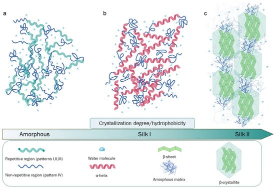
Silk fibroin is the principal component of raw silk and represents an extensively studied and utilized biopolymer. Silk fibroin is composed by three chains, light, heavy, and P25 protein. Heavy chain is rigorously organized in redundant aminoacidic sequences rich in glycine and alanine, secondary structure is organized in anti-parallel β-sheets that in turn form β-crystallites stacked in nano-fibrils. Those peculiar fibroin’s structural and compositional elements are crucial to determine the excellent physical properties, such as strength and toughness. Besides these characteristics, the processability and biocompatibility have attracted significant attention for the fabrication of several biomaterials suitable in many fields of application.

silk fibroin tissue engineering regenerative medicine biomedicine bone regeneration

1. Introduction

The use of silk dates back to the Roman Empire, when it was used not only as a luxury garment or in jewelry but also as a real medical device in the form of suture thread, thus highlighting silk’s exceptional body compatibility [1]. Silk is a natural fibrous protein produced by several arthropods, such as Lepidoptera, principally represented by Bombyx mori (silkworms), Araneae (spiders), and species of Trichoptera and Myriapoda [2]. The main differences between arthropods able to produce silk are attributable to the development of different spinning systems, embodied by evolutionary biological variants of silk gland typologies. Silk can be generated in several districts, such as Malpighian tubules, labial glands, or dermal glands, and the biological production purposes are distinct. For example, silkworms spin silk into cocoons to protect themselves from the external environment during the pupal stage, while spiders and other species produce silk webs for predatory purposes [3]. Silk is used also for reproductive finalities by Thysanoptera, Diptera, etc. [2]. Despite miscellaneous biological functions, sources, spinning systems, and consequent protein structures, these silks have typically high levels of protein crystallinity and similar aminoacidic compositions [4].

2. The Composition and Structure of Silk Fibroin

The composition of raw silkworm silk is as follows: approximately 70–80% fibroin, 20–30% sericin (SR), and a small percentage of other components such as wax, inorganic matter, and pigments [5]. SR encompasses a family of glycoproteins generated by alternative splicing events that take place in the larvae-secreting organs (silk glands). The aminoacidic composition of these glycoproteins is characterized by a high serin abundance [6]. SR’s serin-rich domains allow for interaction with water molecules, making SR sticky and imbuing it with a glue-like consistency from a macroscopic/mechanical point of view. Due to these peculiar properties, during the cocooning process, two fibroin filaments are naturally “glued” together with SR, protecting the complex from biological and atmospheric agents [7][8][9].
Silk fibroin (SF) belongs to a distinct class of glycoproteins, and its structure is characterized by a heavy chain (Hc) with an approximate molecular weight (MW) of 350 kDa and a light chain (Lc) of about 25 kDa, which are linked together by a disulfide bond, forming the (H-L) complex. This complex, in turn, interacts with a glycosylated protein of 27 kDa, named P25, with hydrophobic interactions in a 6:6:1 molar ratio, forming an elementary unit that is crucial for SF secretion, the spinning process, and fiber formation. In particular, during the secretion process, Lc and P25 exert structural control to SF, playing a protective role in the silk glands [10][11]. Lc has a non-repetitive and amorphous structure that is more hydrophilic than Hc. For this reason, during the secretion step, Lc protects against excessive crystallization, while Hc prevents premature SF denaturation. During secretion, a further contribution is given by P25, whose biological role is to maintain the stability of the H-L complex. An additional role for the P25 protein has also been proposed. Specifically, it may induce molecular chaperone activity, assisting the correct folding state of SF. P25 is less stable than the H-L complex, and it degrades after the SF fiber formation process [12][13].
The primary structure of Hc is the key for the structural and biological roles of SF, being characterized by a complex aminoacidic composition and patterning. The four most represented amino acids are as follows: glycine (Gly, 46%), alanine (Ala, 30%), serine (Ser, 12%), and tyrosine (Tyr, 5%) [14]. The highly repetitive patterns present in Hc can be classified into four principal typologies: (I) GAGAGS, a pattern playing a critical role as a core component of the SF crystalline region; (II) GAGAGVGY–GAGAGY–GAGAGV, which are three sequences rich in aromatic and hydrophobic residues, principally located in the semicrystalline region of the protein; (III) this pattern is highly similar to (I) but has the presence of an “AAS” β-sheet breaking pattern located at the c-terminal; (IV) these hydrophilic, non-repetitive patterns, which are also called linkers, lack a high-ordered structure, and hence, they may be found in combination with crystallized regions in SF. Patterns such as (IV), in particular, have different aminoacidic compositions and are interspersed through the (I, II, III) patterns [14][15][16]. Globally, 12 (I, II, or III) repetitive patterns interspaced by 11 (IV) non-repetitive amorphous regions are present in the primary structure of Hc (Figure 1).
Figure 1. Representation of the patterns of Hc’s repetitive or non-repetitive regions and an illustration of Hc’s crystallization degrees relationship with spatial organization, conformational state, and hydrophobicity. (a) Amorphous and unstructured SF conformational states typical of SF aqueous solutions. (b) Silk I form, characterized by the predominance of α-helix structures naturally present in silk glands and reproducible in the laboratory by slow drying, freezing-induced crystallization, and other methods. (c) Silk II form, with a high density of β-sheet crystallites, is produced by silkworms during the spinning process; this SF form can be obtained by several techniques due to the spontaneous tendency of SF to assume this conformation. Created using Biorender.com.
Thanks to analyses based on x-ray diffraction [17][18], cDNA sequencing, nuclear magnetic resonance (NMR), and mass spectrometry [10][14][19][20], different studies have examined the primary structure of SF and estimated the spatial arrangements of its secondary and tertiary structure, characterizing the protein from a molecular point of view.
In SF, β-sheets are organized into β-crystallites stacked in nano-fibrils interspersed in an amorphous matrix formed by non-repetitive domains [21]. SF’s mechanical strength and elasticity are attributable not only to the spatial organization of crystallites but also by that of nano-scaled fibers. In this respect, the nano-fibrillar arrangement of this network represents a pivotal factor for the outstanding density/strength ratio showed by SF fibers; the strength given by the molecular bonds of β-sheet crystallites is, in fact, counterbalanced by the extensibility and tenacity of the amorphous matrix, which, inter alia, is fundamental for other mechanical properties such as elasticity, toughness, and lightness [16][21][22]. The alternance between flexible amorphous regions and crystallizable repetitive regions determines a conformational protein polymorphism. This attribute is enhanced by the high Gly content, since Gly has a low steric hindrance and allows for a secondary structure switch between α-helices and β-sheets (as will be discussed in the following) [23].
Additionally, an amorphous SF state, lacking in high-order structures, can be obtained in the laboratory after the SF degumming and purification process, which leads to the production of an amorphous aqueous SF solution [24] (Figure 1a). On the other hand, Silk I and Silk II are the two main conformational states of SF and are strictly dependent on the β-sheet packing density or the presence of α-helix structures. In Silk I, there is an important presence of water molecules stuck between SF chains that, together with the formation of a high percentage of α-helix structures, impede close β-sheet packing and extensive β-sheet crystallization. Silk I is a metastable form of SF naturally present before the secretion step in silk glands, but it can also be experimentally obtained and exploited in different ways for the synthesis and tuning of different SF-based materials (Figure 1b). Silk II is characterized by a high density of antiparallel β-sheet crystals organized in β-crystallites and lacking α-helix structures. Here, the dominant crystalized conformation allows for an improvement in the stability and hydrophobicity of the related fibers and materials (Figure 1c). Silk II is formed by silkworms during the spinning process and is derived from Silk I; however, it can be obtained experimentally from amorphous SF aqueous solution by thermal treatments or exposure to methanol or ethanol [10][25]. Another SF conformation is Silk III, which consists of a left-handled triple helix conformation; this SF structure has been exclusively observed at the air/water interface of SF solutions [26].

References

  1. Bucciarelli, A.; Motta, A. Use of Bombyx Mori Silk Fibroin in Tissue Engineering: From Cocoons to Medical Devices, Challenges, and Future Perspectives. Biomater. Adv. 2022, 139, 212982.
  2. Craig, C.L. Evolution of arthropod silks. Annu. Rev. Èntomol. 1997, 42, 231–267.
  3. Saad, M.; El-Samad, L.M.; Gomaa, R.A.; Augustyniak, M.; Hassan, M.A. A Comprehensive Review of Recent Advances in Silk Sericin: Extraction Approaches, Structure, Biochemical Characterization, and Biomedical Applications. Int. J. Biol. Macromol. 2023, 250, 126067.
  4. Sutherland, T.D.; Young, J.H.; Weisman, S.; Hayashi, C.Y.; Merritt, D.J. Insect Silk: One Name, Many Materials. Annu. Rev. Entomol. 2010, 55, 171–188.
  5. Kwak, H.W.; Ju, J.E.; Shin, M.; Holland, C.; Lee, K.H. Sericin Promotes Fibroin Silk i Stabilization Across a Phase-Separation. Biomacromolecules 2017, 18, 2343–2349.
  6. Michaille, J.J.; Garel, A.; Prudhomme, J.C. The expression of five middle silk gland specific genes is territorially regulated during the larval development of Bombyx mori. Insect Biochem. 1989, 19, 19–27.
  7. Mondal, M.; Trivedy, K.; Nirmal Kumar, S. The Silk Proteins, Sericin and Fibroin in Silkworm, Bombyx Mori Linn—A Review. Casp. J. Environ. Sci. 2007, 5, 63–76.
  8. Chen, F.; Porter, D.; Vollrath, F. Structure and Physical Properties of Silkworm Cocoons. J. R. Soc. Interface 2012, 9, 2299–2308.
  9. Kunz, R.I.; Brancalhão, R.M.C.; Ribeiro, L.D.F.C.; Natali, M.R.M. Silkworm Sericin: Properties and Biomedical Applications. BioMed Res. Int. 2016, 2016, 1–19.
  10. Asakura, T.; Okushita, K.; Williamson, M.P. Analysis of the Structure of Bombyx Mori Silk Fibroin by NMR. Macromolecules 2015, 48, 2345–2357.
  11. Inoue, S.; Tanaka, K.; Arisaka, F.; Kimura, S.; Ohtomo, K.; Mizuno, S. Silk Fibroin of Bombyx Mori Is Secreted, Assembling a High Molecular Mass Elementary Unit Consisting of H-Chain, L-Chain, and P25, with a 6:6:1 Molar Ratio. J. Biol. Chem. 2000, 275, 40517–40528.
  12. Tanaka, K.; Inoue, S.; Mizuno, S. Hydrophobic interaction of P25, containing Asn-linked oligosaccharide chains, with the H-L complex of silk fibroin produced by Bombyx mori. Insect Biochem. Mol. Biol. 1999, 29, 269–276.
  13. Tanaka, K.; Kajiyama, N.; Ishikura, K.; Waga, S.; Kikuchi, A.; Ohtomo, K.; Takagi, T.; Mizuno, S. Determination of the site of disulfide linkage between heavy and light chains of silk fibroin produced by Bombyx mori. Biochim. Biophys. Acta (BBA)-Protein Struct. Mol. Enzym. 1999, 1432, 92–103.
  14. Zhou, C.-Z.; Confalonieri, F.; Medina, N.; Zivanovic, Y.; Esnault, C.; Yang, T.; Jacquet, M.; Janin, J.; Duguet, M.; Perasso, R.; et al. Fine Organization of Bombyx Mori Fibroin Heavy Chain Gene. Nucleic Acids Res. 2000, 28, 2413–2419.
  15. Foo, C.W.P.; Bini, E.; Hensman, J.; Knight, D.P.; Lewis, R.V.; Kaplan, D.L. Role of PH and Charge on Silk Protein Assembly in Insects and Spiders. Appl. Phys. A Mater. Sci. Process 2006, 82, 223–233.
  16. Zhou, C.Z.; Confalonieri, F.; Jacquet, M.; Perasso, R.; Li, Z.G.; Janin, J. Silk Fibroin: Structural Implications of a Remarkable Amino Acid Sequence. Proteins: Struct. Funct. Genet. 2001, 44, 119–122.
  17. Lotz, B.; Cesari, F.C. The Chemical Structure and the crystalline structures of Bombyx Mori Silk Fibroin. Biochimie 1979, 61, 205–214.
  18. Marsh, R.E.; Corey, R.B.; Pauling, L. An Investigation of the Structure of Silk Fibroin. Biochimica Biophysica Acta 1955, 16, 1–34.
  19. Yamaguchi’, K.; Kikuchi’, Y.; Takagi’, T.; Kikuchi’, A.; Oyama’, F.; Shimural, K.; Mizuno’s, S. Primary Structure of the Silk Fibroin Light Chain Determined by CDNA Sequencing and Peptide Analysis. J. Mol. Biol. 1989, 210, 127–139.
  20. Asakura, T.; Ohgo, K.; Komatsu, K.; Kanenari, M.; Okuyama, K. Refinement of Repeated β-Turn Structure for Silk I Conformation of Bombyx Mori Silk Fibroin Using13C Solid-State NMR and X-Ray Diffraction Methods. Macromolecules 2005, 38, 7397–7403.
  21. Du, N.; Yang, Z.; Liu, X.Y.; Li, Y.; Xu, H.Y. Structural Origin of the Strain-Hardening of Spider Silk. Adv. Funct. Mater. 2011, 21, 772–778.
  22. Nguyen, A.T.; Huang, Q.L.; Yang, Z.; Lin, N.; Xu, G.; Liu, X.Y. Crystal Networks in Silk Fibrous Materials: From Hierarchical Structure to Ultra Performance. Small 2014, 11, 1039–1054.
  23. Wilson, D.; Valluzzi, R.; Kaplan, D. Conformational Transitions Model Silk Peptides. Biophys. J. 2000, 78, 2690–2701.
  24. Matsumoto, A.; Lindsay, A.; Abedian, B.; Kaplan, D.L. Silk Fibroin Solution Properties Related to Assembly and Structure. Macromol. Biosci. 2008, 8, 1006–1018.
  25. Cebe, P.; Partlow, B.P.; Kaplan, D.L.; Wurm, A.; Zhuravlev, E.; Schick, C. Silk I and Silk II Studied by Fast Scanning Calorimetry. Acta Biomater. 2017, 55, 323–332.
  26. Valluzzi, R.; Gido, S.P.; Muller, W.; Kaplan, D.L. Orientation of silk III at the air-water interface. Int. J. Biol. Macromol. 1999, 24, 237–242.
More
Information
Contributors MDPI registered users' name will be linked to their SciProfiles pages. To register with us, please refer to https://encyclopedia.pub/register : , , , , , , ,
View Times: 455
Revisions: 4 times (View History)
Update Date: 20 Mar 2024
1000/1000