Submitted Successfully!
To reward your contribution, here is a gift for you: A free trial for our video production service.
Thank you for your contribution! You can also upload a video entry or images related to this topic.
Version Summary Created by Modification Content Size Created at Operation
1 -- 1898 2024-02-19 11:21:49 |
2 references update and layout Meta information modification 1898 2024-02-20 04:15:29 |

Video Upload Options

Do you have a full video?

Confirm

Are you sure to Delete?
Cite
If you have any further questions, please contact Encyclopedia Editorial Office.
Andreucci, C.A. Innovation in Biomechanical Orthognathic Surgery. Encyclopedia. Available online: https://encyclopedia.pub/entry/55164 (accessed on 28 April 2024).
Andreucci CA. Innovation in Biomechanical Orthognathic Surgery. Encyclopedia. Available at: https://encyclopedia.pub/entry/55164. Accessed April 28, 2024.
Andreucci, Carlos Aurelio. "Innovation in Biomechanical Orthognathic Surgery" Encyclopedia, https://encyclopedia.pub/entry/55164 (accessed April 28, 2024).
Andreucci, C.A. (2024, February 19). Innovation in Biomechanical Orthognathic Surgery. In Encyclopedia. https://encyclopedia.pub/entry/55164
Andreucci, Carlos Aurelio. "Innovation in Biomechanical Orthognathic Surgery." Encyclopedia. Web. 19 February, 2024.
Innovation in Biomechanical Orthognathic Surgery
Edit

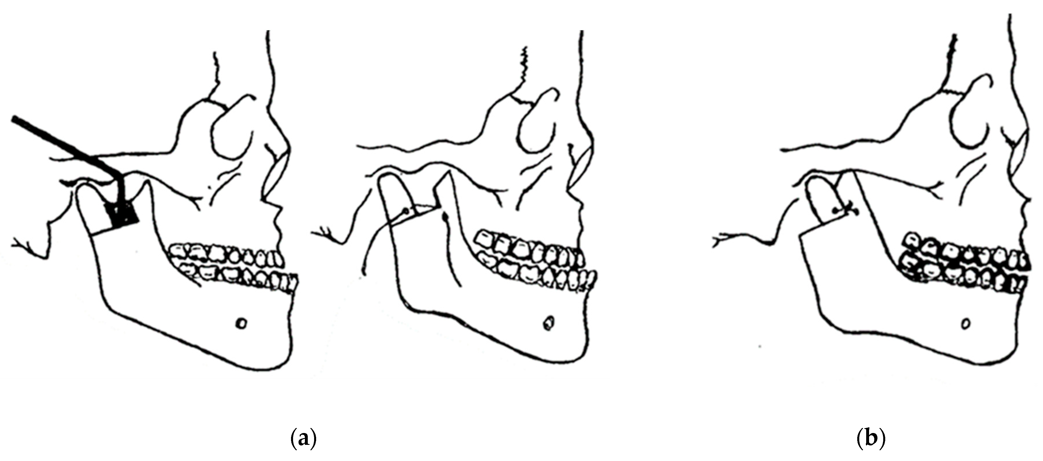
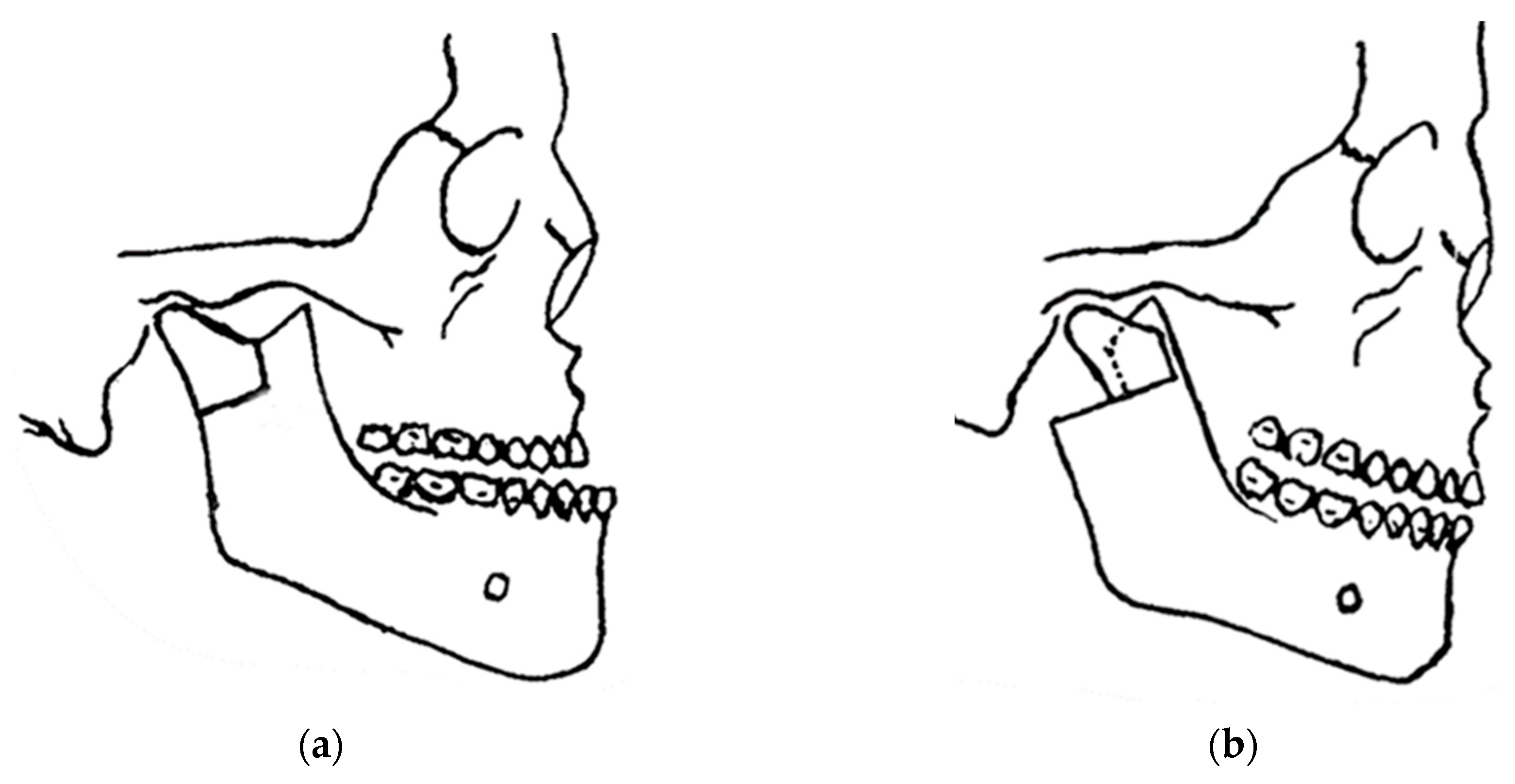
Craniofacial surgery is proposed and performed for a variety of reasons, ranging from congenital or acquired malformations to emotional disorders and parafunctions of the masticatory, respiratory, auditory, and visual systems. Surgery of the mandible and its orthostatic repositioning is the most common of these corrections of craniofacial anomalies. Throughout the history of these procedures, various techniques have been proposed and perfected, but always with a high rate of minor and major complications. The recurrence rate of mandibular malposition is high, as is the temporary loss of facial sensitivity and motor skills. These outcomes are often related to the choice of surgical technique rather than the skill of the surgeon, which is considered to be one of the most important factors in the final outcome. Surgical techniques involving direct manipulation of the vascular-nervous bundles, such as bilateral sagittal split osteotomy, clearly present the possibility of major or minor complications.

orthognathic surgery craniofacial surgery

1. Introduction

The aim of orthognathic surgery is to reposition the maxilla, mandible, and chin, and commonly performed procedures include LeFort I osteotomy and bilateral sagittal split osteotomy (BSSO) with or without osseous genioplasty. The recent history of mandibular orthognathic surgery began with Hullihen in 1846 [1], who performed a mandibular body osteotomy to correct prognathism in a case of mandibular elongation and distortion of the face and neck, caused by a burn, successfully treated [2]. He was a general surgeon with dental training, like other examples of general surgeons of the time who reported on maxillofacial surgery: von Langenbeck, Cheever, Billroth, and Dufourmentel. At the beginning of the 20th century, Blair performed a horizontal ramus osteotomy [3][4], which was described and published by Blair and Angle, who were the first to propose a classification of mandibular deformities, stating: “An almost ideal occlusion would rarely be accompanied by the best facial result”, which is still valid today (Figure 1).
Figure 1. Pioneers of mandible surgery: (a) the first operation (Hullihen’s procedure) for the correction of malocclusion carried out in 1849; and (b) osteotomy of the mandibular body performed by Blair in 1897.
Berger in 1897 introduced condylar osteotomy to correct prognathism, which was practiced in France until 1950, when Dufourmentel and Mouly in 1959 described good results with this technique. Babcock in 1909 and, a few years later, Bruhn and Lindemann in 1921 described a horizontal osteotomy just between the sigmoid notch and the mandibular foramen [3][4].
This operative technique was modified a few years later by Kostecka in 1931, who described his technique as a “blind procedure” in which the osteotomy was made with a Gigli saw through a stab incision. Limberg and Wassmund performed further modifications of external approaches to ramus osteotomies in the 1920s and 1930s with a high recurrence rate [5].
Perthes in 1922, following Schlossmann’s suggestion, tried a type of sagittal splitting of the ramus using an extraoral approach with an oblique transverse osteotomy. Kazanjian suggested a horizontal oblique osteotomy in 1951. In 1942, Schuchardt described the first intra-oral approach for mandibular ramus osteotomy [4][6]. In 1954, Caldwell and Letterman described a vertical ramus osteotomy technique in an attempt to preserve the inferior alveolar neurovascular bundle [7][8].
In 1957, Trauner and Obwegeser described what became the current bilateral sagittal split osteotomy or BSSO transoral approach [9]. Improvements and modifications to this surgical procedure have always been aimed at reducing relapse, improving healing, and reducing complications. Contributions have been made by Dal Pont in 1961, Hunsuck in 1968, and Epker in 1977. Dal Pont [10][11] modified the inferior horizontal incision to a vertical osteotomy in the buccal cortex between the first and second molars, which allowed larger contact surfaces and required minimal muscle displacement.
Hunsuck [12] modified the technique with a shorter medial horizontal cut, just posterior to the lingual, to minimize soft tissue dissection. His anterior vertical incision was similar to that of Dal Pont (Figure 2) [11]. Epker (1977) proposed several improvements and refinements to the intrabuccal technique [13], including less removal of the masseter muscle with limited medial dissection to reduce postoperative edema from hemorrhage and manipulation of the neurovascular bundle. Spiessel introduced rigid internal fixation in 1976 in an attempt to restore function early and reduce relapse [14]. The introduction of rigid internal fixation, rather than 5–6 weeks of intermaxillary fixation, had the objective benefit of improving patient comfort [2][15][16].
Figure 2. Development of modern jaw surgery for facial discrepancies.
In 1948, maxillofacial surgery was almost non-existent in most dental schools and universities around the world. In the few places where specialists were available, it was a series of unsatisfactory procedures, mainly to correct mandibular advancement or prognathism [7]. These included the Blair and Kostecka procedures (Figure 1b and Figure 3). Throughout history, other authors have contributed to the improvement of facial surgery and mandibular osteotomy [17][18]. Oswaldo de Castro [18][19] developed a modification of Smith’s technique (Figure 3) with an L-shaped osteotomy under the sigmoid notch and condylar neck, in a neurovascular safety zone, described by Hensel, for mandibular osteotomy. This procedure allows dorsal displacement of the mandible and bone apposition without the need to wire the fragments (Figure 4).
Figure 3. Smith’s technique in 1956: (a) a guide (in black) corresponding to the bone quadrangle to be removed with a line corresponding to the osteotomy under the condylar neck; and (b) using metal wire for osteosynthesis.
Figure 4. Castro’s in 1964 surgery: (a) inverted “L” osteotomy; and (b) dorsal sliding of the mandible and bone apposition without the need to wire the fragments.
The mandible is fixed with interdental wires for eight weeks with a pre-auricular incision approach. Gino Emilio Lasco, a pioneering oral and maxillofacial surgeon in Sao Paulo, Brazil, who developed various techniques and modifications of surgical techniques, such as surgery to correct benign hypertrophy of the masseter muscle, and surgery for free repositioning of the meniscus in temporomandibular disorders, and Oswaldo de Castro’s modified orthognathic surgery with the possibility of a submandibular extra-buccal approach [19][20][21][22][23][24][25] are also described here with the latest modifications and improvements, with a 20-year follow-up describing what Lasco’s surgical team, coordinated by the first author, has been doing since 1999.
Most of the techniques described here, with the exception of the modification proposed by Castro and Lasco [19], have tried to maintain the option of intra-oral surgery, making modifications to the sagittal split of the mandible, innovating with the expectation of reducing vasculo-nervous lesions, bleeding, recurrences, enhancing comfort and efficiency for the surgeon, and improving patient acceptance of the proposed treatment. Today, it is very clear that there is no single effective technique for all cases of facial discrepancies [15][16]. Therefore, there is an opportunity to develop an innovative, simplified technique that is faster to perform, preserves anatomical structures, and uses the musculoskeletal system to promote healing, repair, and remodeling, prevent relapse, and provide stability for anatomical positioning of the mandibular joint.
In 1969, Obwegeser [7][26] repositioned the mandible and maxilla simultaneously with a sagittal ramus splitting and a LeFort I osteotomy, making orthognathic surgery a separate subspecialty, leaving the cranio-orbital region to be defined by Paul Tessier. In 1967, Tessier introduced the transcranial and subcranial LeFort III procedures to correct cranial and orbital deformities [27]. Hans Luhr (1968) then published his work on internal plate and screw fixation [28][29][30][31], attempting to limit the need for prolonged intermaxillary fixation and increase stability with less reliance on complex interlocking joints and bone grafts. What could be achieved with a rotary burr was also improved using instruments with thin saw blades, allowing more-refined osteotomies [7]. The use of combined maxillo-mandibular orthognathic surgery, as well as other facial procedures to correct craniofacial deformities, has been a great improvement for patients.
The complications of these procedures cannot be ignored, and therefore the preservation of life and the correct indication of cases in which this type of surgery will really produce the expected result and be in accordance with the patient’s expectations is a priority [32][33][34][35]. Many cases fall between the line of surgical necessity and clinical management alone, and it is up to the surgeon to decide what is best for each individual patient. In the more than 60 years of experience of Professor Lasco’s team [36][37][38][39], satisfactory long-term results were obtained with isolated mandibular surgery without the need for Le Fort I maxillary surgery, which was limited to extreme maxillary discrepancies, and segmental and partial osteotomies were routinely performed. There were no aesthetic complaints from patients regarding the final result and the indelible submandibular scar created by the incision along the facial lines (Figure 5).
Figure 5. Follow-up after 20 years of orthognathic surgery: (a) incision performed extra-orally via the submandibular region; (b) it is almost impossible to identify the indelible scar.
The treatment of malocclusions associated with minor skeletal discrepancies is possible via orthodontic compensation of the dentition, with the risk of unsatisfactory facial aesthetics. Borderline cases should be carefully assessed before deciding on orthodontic treatment alone or in combination with orthognathic surgery. The treatment plan should be discussed with the patient, explaining the advantages and disadvantages of each approach in accordance with the patient’s attitude and preferences.

2. Innovation in Biomechanical Orthognathic Surgery

The occlusion planned preoperatively and adjusted by prior orthodontic treatment proved to be satisfactory intra-operatively and in the immediate and late postoperative periods. Small occlusal adjustments were made with carbon paper to analyze premature tooth contacts, which were removed with spherical diamond burs until final occlusal stability was achieved during the first weeks after surgery. The orthodontic brace was maintained for six months after surgery, at which time an acrylic retaining plate was placed in the maxilla and a lingual bonded retainer was fitted, which remains in place to this day at the discretion of the orthodontist. The patient showed no sensory or motor changes throughout the postoperative period.
There have been no respiratory or apnea problems in the last 20 years, with a great improvement in phonetics and adaptation of the tongue to the reduced space after surgery. The patient reported sporadic pain and clicking in her temporomandibular joint and weekly headaches prior to surgery. A clinical diagnosis of temporomandibular joint dysfunction (TMD) was made preoperatively and was associated with an anterior crossbite as an aggravating factor. Immediately after functional surgery and during more than twenty years of follow-up, the patient has not experienced any of the previously reported symptoms, in this case describing oral rehabilitation, including orthognathic surgery, as a surgical treatment for TMD. The aesthetic result desired by the patient was achieved without major changes to the natural facial biomechanics due to the characteristics of the surgical technique.
The modification proposed by Professor Lasco, which have been adopted, eliminates the risk of damaging the marginal mandibular nerve through the submandibular approach. In five other patients, not included in this study group, treated during the same period, the pre-auricular approach and surgery without prior orthodontic treatment was chosen because of their individual anatomical characteristics, using exactly the same surgical technique as mandibular osteotomy, and three of them showed marginal mandibular nerve injury for a maximum of 21 days. It is clear that not using the extra-oral approach because of fear of scarring is more related to the surgeon’s personal experience with the technique. In all 64 osteotomies performed, there were no recurrences, no malocclusions that could not be corrected with occlusal adjustments, no significant neurovascular lesions, and no significant scarring.
By using the modified submandibular technique, the masseter muscle is incised at its origin and therefore there are no complications with its displacement, including the careful detachment of the entire mandibular periosteum, which improves bone repair and healing. Caution should be exercised in extra-oral indications in patients with a history and signs of scar hypertrophy. The average operative time for the proposed surgical technique is 72 min.

References

  1. Hullihen, S.P. Case of elongation of the underjaw and distortion of the face and neck, caused by a burn, successfully treated. Am. J. Dent. Sci. 1849, 9, 157–161.
  2. Aziz, S.R.; Simon, P. Hullihen and the origin of orthognathic surgery. J. Oral Maxillofac. Surg. 2004, 62, 1303–1307.
  3. Blair, V.P. Surgery and Diseases of the Mouth and Jaws; The C.V. Mosby Co.: St. Louis, MO, USA, 1914.
  4. Steinhäuser, E.W. Historical development of orthognathic surgery. J. Cranio-Maxillofac. Surg. 1996, 24, 195–204.
  5. Kazanjian, V.H. The treatment of mandibular prognathism with special reference to edentulous patients. J. Oral Surg. Oral Med. Oral Pathol. 1951, 4, 680–688.
  6. Schuchardt, K. Die Chirurgie als Helferin der Kieferorthopädie . Fortschr. Kieferorthop. 1954, 15, 1–25.
  7. Obwegeser, H.L. Orthognathic surgery and a tale of how three procedures came to be: A letter to the next generations of surgeons. Clin. Plast. Surg. 2007, 34, 331–355.
  8. Caldwell, J.B.; Letterman, G.S. Vertical osteotomy in the mandibular rami for correction of prognathism. J. Oral. Surg. 1954, 12, 185–202.
  9. Trauner, R.; Obwegeser, H. Zur Operationstechnik bei der Progenia und anderen Unterkieferanomalien. Dtsch. Zahn. Mund. Kieferhlkd. 1955, 23, 11–25.
  10. Dal Pont, G. L’osteotomia retromolare per la correzione della progenia. Minerva. Chir. 1959, 14, 1138–1141.
  11. Dal Pont, G. Retromolar osteotomy for the correction of prognathism. J. Oral Surg. Anesth. Hosp. Dent. Serv. 1961, 19, 42–47.
  12. Hunsuck, E.E. A modified intraoral sagittal splitting technic for correction of mandibular prognathism. J. Oral Surg. 1968, 26, 250–253.
  13. Epker, B.N. Modifications in the sagittal osteotomy of the mandible. J. Oral Surg. 1977, 35, 157–159.
  14. Schmoker, R.; Spiessl, B.; Tschopp, H.M.; Prein, J.; Jaques, W.A. Die funktionsstablie Osteosynthese am Unterkiefer mittels exzentrisch-dynamischer Kompressionsplatte (EDCP). Ergebnisse einer Nachuntersuchung der ersten 25 Fälle . SSO Schweiz. Monatsschr. Zahnheilkd. 1976, 86, 167–185.
  15. Ow, A.; Cheung, L.K. Skeletal stability and complications of bilateral sagittal split osteotomies and mandibular distraction osteogenesis: An evidence-based review. J. Oral Maxillofac. Surg. 2009, 67, 2344–2353.
  16. Saman, M.; Abramowitz, J.M.; Buchbinder, D. Mandibular osteotomies and distraction osteogenesis: Evolution and current advances. JAMA Facial Plast. Surg. 2013, 15, 167–173.
  17. Hofer, O. Operation del Prognathie und Microgenie. Dtsch. Zahn. Mund. Kleferhlkd. 1942, 9, 121–132.
  18. Babcock, K.W. Surgical treatment of certain deformities of jaw associated with malocclusion of teeth. J Am Med Assoc. 1909, 53, 833–839.
  19. Castro, O. Mandibular prognathism. Mandibuloplasty by Smith’s technique. Rev. Lat. Am. Cir. Plast. 1964, 8, 24.
  20. Castro, O. Surgical correction of prognathism: Angled osteoplasty of the mandibular rami. Br. J. Plast. Surg. 1967, 20, 57–60.
  21. Castro, O.; Mortari, S.; Colares, H.J. Significant details regarding treatment of prognathism using angled osteotomy of the mandibular rami. Aesth. Plast. Surg. 1980, 4, 349–355.
  22. Smith, A.E.; Robinson, M. The evaluation of physiologic result from submandibular-notch osteotomy, condylotomy operation for prognathism. Plast. Reconstr. Surg. 1955, 15, 196.
  23. Rehrmann, A. Horizontal osteotomy of the mandibular rami with wire suture for the correction of prognathism with preservation of the original position of the condyles. Dtsch. Zahn. Mund. Kieferheilkd. Zentralbl. Gesamte. 1967, 49, 72–76.
  24. Rehrmann, A.; Schettler, D. Treatment of bilateral fractures of the temporomandibular joint in infants and young children with reposition and fixation according to Rehrmann’s method. Dtsch. Zahnarztl. Z. 1966, 21, 777–782.
  25. Rehrmann, A. Reposition and retention of the mandible in fractures of the articular process and condyle by extraoral elastic traction in the chin region. Dtsch. Zahn. Mund. Kieferheilkd. Zentralbl. Gesamte. 1971, 57, 379–392.
  26. Obwegeser, H. Die einzeitige Vorbewegung des Oberkiefers und R flick bewegung des Unterkiefers zur Korrektur der extremen ‘Progenie’. Schweiz Mschr. Zahnheilk. 1970, 80, 305.
  27. Tessier, P.; Guiot, G.; Rougerie, J. Cranio-naso-orbito-facial osteotomies (hypertelorism). Ann. Chir. Plast. 1967, 12, 103–118.
  28. Luhr, H.G. The compression osteosynthesis of mandibular fracture in dogs. A histological contribution to “primary bone healing”. Eur. Sur. Res. 1967, 1, 3.
  29. Luhr, H.G. Zur stabilen osteosyntheses bei unterkiefer-frakturen. Dtsch. Zahnarztl. Z 1968, 23, 754.
  30. Luhr, H.G. Stabile Fixation von Oberkiefer-Mittelgesichtsfrakturen durch Mini-Kompressionsplatten. Dtsch. Zahnarztl. Z 1979, 34, 851.
  31. Luhr, H.G. Indications for use of a microsystem for internal fixation in craniofacial surgery. J. Craniofac. Surg. 1990, 1, 35–52.
  32. Dreiseidler, T.; Bergmann, J.; Zirk, M.; Rothamel, D.; Zöller, J.E.; Kreppel, M. Three-dimensional fracture pattern analysis of the Obwegeser and Dal Pont bilateral sagittal split osteotomy. Int. J. Oral Maxillofac. Surg. 2016, 45, 1452–1458.
  33. Mensink, G.; Verweij, J.P.; Frank, M.D.; Bergsma, J.E.; van Merkesteyn, J.P.R. Bad split during bilateral sagittal split osteotomy of the mandible with separators: A retrospective study of 427 patients. Br. J. Oral Maxillofac. Surg. 2013, 51, 525–529.
  34. Salzano, G.; Audino, G.; Friscia, M.; Vaira, L.A.; Biglio, A.; Maglitto, F.; Committeri, U.; Piombino, P.; Bonavolontà, P.; Petrocelli, M.; et al. Bad splits in bilateral sagittal split osteotomy: A retrospective comparative analysis of the use of different tools. J. Cranio-Maxillofac. Surg. 2022, 50, 543–549.
  35. Li, F.; Li, S.; Wu, S.; Le, Y.; Tan, J.; Wan, Q. Effect of the lateral bone cut end on pattern of lingual split during bilateral sagittal split osteotomy in patients with skeletal class III malocclusion. Br. J. Oral Maxillofac. Surg. 2023, 61, 309–314.
  36. Lasco, G.E. 20 anos de cirurgia maxilo-facial (20 years of maxillofacial surgery). In Atualizaçäo Clínica em Odontologia; Marco Antonio, B., Christa, F., Eds.; Artes Médicas: Säo Paulo, Brazil, 1984; pp. 219–230.
  37. Lasco, G.E.; Mello, J.B. Contribuiçäo ao tratamento cirúrgico das luxaçöes recidivantes da ATM (Contribution to surgical treatment of temporomandibular joint recidivant dislocation). In Atualizaçäo Clínica em Odontologia; Marco Antonio, B., Christa, F., Eds.; Artes Médicas: Säo Paulo, Brazil, 1984; pp. 231–236.
  38. Lasco, G.E.; Mello, J.B. Disfunçöes dolorosas da ATM (TMJ pain dysfunctions). In Atualizaçäo Clínica em Odontologia; Marco Antonio, B., Christa, F., Eds.; Artes Médicas: Säo Paulo, Brazil, 1984; pp. 237–242.
  39. Lasco, G.E. Contribuição ao estudo da patologia e terapêutica da Articulação Temporomandibular (Contribution to the study of the pathology and therapy of the Temporomandibular Joint). Rev. APCD 1967, 2, 264.
More
Information
Contributor MDPI registered users' name will be linked to their SciProfiles pages. To register with us, please refer to https://encyclopedia.pub/register :
View Times: 86
Revisions: 2 times (View History)
Update Date: 20 Feb 2024
1000/1000