Submitted Successfully!
To reward your contribution, here is a gift for you: A free trial for our video production service.
Thank you for your contribution! You can also upload a video entry or images related to this topic.
Version Summary Created by Modification Content Size Created at Operation
1 -- 1578 2024-02-09 11:01:25 |
2 format correct Meta information modification 1578 2024-02-18 07:15:46 |

Video Upload Options

Do you have a full video?

Confirm

Are you sure to Delete?
Cite
If you have any further questions, please contact Encyclopedia Editorial Office.
Sala-Luis, A.; Oliveira-Urquiri, H.; Bosch-Roig, P.; Martín-Rey, S. Eco-Sustainable Approaches for Fungal Biodeterioration on Easel Painting. Encyclopedia. Available online: https://encyclopedia.pub/entry/54961 (accessed on 27 April 2024).
Sala-Luis A, Oliveira-Urquiri H, Bosch-Roig P, Martín-Rey S. Eco-Sustainable Approaches for Fungal Biodeterioration on Easel Painting. Encyclopedia. Available at: https://encyclopedia.pub/entry/54961. Accessed April 27, 2024.
Sala-Luis, Agustí, Haizea Oliveira-Urquiri, Pilar Bosch-Roig, Susana Martín-Rey. "Eco-Sustainable Approaches for Fungal Biodeterioration on Easel Painting" Encyclopedia, https://encyclopedia.pub/entry/54961 (accessed April 27, 2024).
Sala-Luis, A., Oliveira-Urquiri, H., Bosch-Roig, P., & Martín-Rey, S. (2024, February 09). Eco-Sustainable Approaches for Fungal Biodeterioration on Easel Painting. In Encyclopedia. https://encyclopedia.pub/entry/54961
Sala-Luis, Agustí, et al. "Eco-Sustainable Approaches for Fungal Biodeterioration on Easel Painting." Encyclopedia. Web. 09 February, 2024.
Eco-Sustainable Approaches for Fungal Biodeterioration on Easel Painting
Edit

Cultural Heritage (CH) materials are susceptible to being complexly damaged physically, chemically, and aesthetically by the growing and metabolic activities of living beings, as investigators know as biodeterioration. Historical easel paintings have a mixture of materials and layers, increasing their conservation complexity. Materials such as cellulose on the support, rabbit skin glue, egg yolk, linseed oil, or varnishes on the polychromy are some organic materials that can compound an easel painting, although the painting layer also contains inorganic pigments. These organic materials can be degraded by fungi if the environmental conditions are favorable. Eliminating and controlling fungal biodeterioration is one of the most important challenges of easel painting conservation.

biodeterioration fungi easel paintings wood textile fibers

1. Introduction

Cultural Heritage (CH) materials are susceptible to being complexly damaged physically, chemically, and aesthetically by the growing and metabolic activities of living beings, as investigators know as biodeterioration [1]. Many studies have been performed concerning vegetal or microbiological biodeterioration on inorganic materials, such as mural paintings [2][3][4][5], stone buildings [6][7][8][9], or stone sculptures [10][11][12][13][14]. Easel painting biodeterioration is a less-studied topic, although, in the last ten years, there has been more interest in their biological control and prevention [15][16][17][18][19][20].
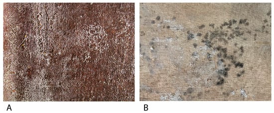
Historical easel paintings have a mixture of materials and layers, increasing their conservation complexity. Materials such as cellulose on the support, rabbit skin glue, egg yolk, linseed oil, or varnishes on the polychromy are some organic materials that can compound an easel painting, although the painting layer also contains inorganic pigments [21][22]. These organic materials can be degraded by fungi if the environmental conditions are favorable (Figure 1). More common fungi are mesophiles, whose ideal conditions revolve around 20–30 °C and a Relative Humidity (RH) higher than 70%, and pH conditions between 4 and 6 [18][20][23][24][25].
Figure 1. (A) Filamentous fungi growth on the obverse a Bartolomé Mongrell’s oil on canvas painting (Museu Municipal d’Alzira, València, Spain). (B) Fungal growth on the reverse of Santos Juanes Church’s (València, Spain) oil on canvas marouflage.
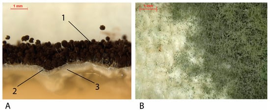
In the specific literature about fungal biodeterioration of easel painting, there are many fungal families that can grow on this kind of surface, of which the most prevalent are exposed in Table 1. All those fungi are mostly from the phylum Ascomycota, although there are fungi from the phylum Basidiomycota or Mucoromycota, which are three types of filamentous fungi. Filamentous fungi are formed by hyphae—large cell filaments—that generally have walls between them and are called coenocytic hyphae. Depending on their function, this kind of fungi has two different types of structures: vegetative mycelium, which penetrates the substrate and absorbs nutrients, and aerial mycelium and fruitful bodies, which are responsible for reproductive function [26] (Figure 2).
Figure 2. (A) Cross-section of A. niger cultivated in agar. (1) Fruitful bodies (dark parts): spores-production structures. (2) Aerial mycelium: white hyphae on surface. (3). Vegetative mycelium: white hyphae into the agar medium. (B) Penicillium spp. fruitful bodies and areal mycelium on a cotton canvas mock-up.
Table 1. Most common fungal species on easel painting.
Species Genus Order Phylum References
Aerobasidium pullulans Aerobasidium Dothideales Ascomycota [22][27][28][29][30][31][32]
Alternaria alternata Alternaria Pleosporales Ascomycota [22][29][31][32][33][34]
Aspergillus clavatus Aspergillus Eurotiales Ascomycota [22]
Aspergillus flavus Aspergillus Eurotiales Ascomycota [29][30][35][36]
Aspergillus fumigatus Aspergillus Eurotiales Ascomycota [32]
Aspergillus niger Apergillus Eurotiales Ascomycota [27][28][31][32][33][34][35][37][38]
Aspergillus sp. Apergillus Eurotiales Ascomycota [25][39]
Aspergillus versicolor Aspergillus Eurotiales Ascomycota [27][29][32][34]
Bjerkandera adusta Bjerkandera Polyporales Basidiomycota [22]
Chaetonium globosum Chaetonium Sordariales Ascomycota [28][31][32][36][40]
Cladosporium cladosporioides Cladosporium Capnodiales Ascomycota [27][28][29][30][32][34]
Cladosporium sp. Cladosporium Capnodiales Ascomycota [25]
Filobasidium magnum Filobasidium Filobasidiales Basidiomycota [22][25][29]
Mucor spp. Mucor Mucorales Mucoromycota
Penicillium chrysogenum Penicillium Eurotiales Ascomycota [20][22][27][28][29][34][36][37][38]
Penicillium sp. Penicillium Eurotiales Ascomycota [20][25]
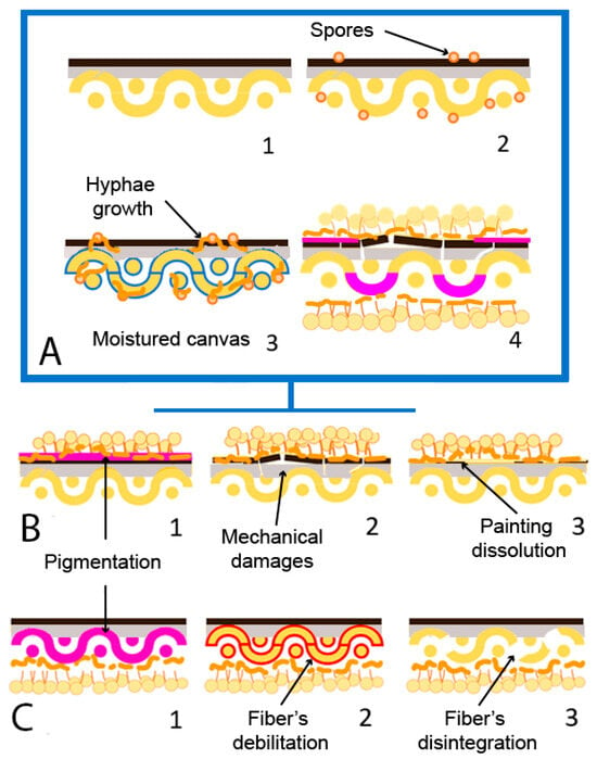
The development of fungal strains damages easel painting materials in different ways, causing a multitude of pathologies depending on the substrate and the strain [41]. Generally, fungal growth produces acidification, chromatic changes, structural damage across chemical mechanisms—liberation of fatty acids and enzymatic action— and mycelium growth [19] (Figure 3). Microbial deterioration usually affects the painting support in the first instance, reaching and colonizing the pictorial layers in later phases [42]. The organic nature of these materials presents a large carbon source for microorganisms, which makes them susceptible to biological attack. In addition, the presence of glues or coating pastes applied as sizing treatments increases that susceptibility. Aesthetical damages are generally related to pigment production. Ascomycota strains such as A. niger produces azanigerones B and C—yellow [43]—or other colors from pale yellow to brown [44]. Cladosporium sp. and Fusarium sp. produce anhydrofusarubin—red [45][46]—or A. versicolor produces asperversin—yellow [47]. Red-orange, β-carotene, is the most produced pigment by Mucoromycota as a preventive measure for stress oxidative damages [48].
Figure 3. Fungal biodeterioration mechanism and damages on canvas painting (adapted from Poyatos et al., 2018 [19]). (A) (1) Non-biodeteriorated oil on canvas painting; (2) presence of fungal spores (orange) on the surface; (3) moisture of canvas (blue) due to RH increase and fungal development. (4) Biodeteriorated painting. (B) Damages on painting strata; (1) Pigmentation (pink); (2) mechanical damage of painting film; (3) dissolution of painting film. (C) Damages on canvas support; (1) pigmentation (pink); (2) fiber’s debilitation (red); (3) fiber’s disintegration.

2. Fungal Biodeterioration of Wood on Easel Paintings

Wood was the most important painting support until canvas popularization in the 16th century. It presents well-known biodeterioration problems due to its use in different industries or as a construction material [49]. Wood fungal biodeterioration can be classified as white rot, brown rot (exclusively by Basidiomycota), and soft rot (generally Ascomycota), depending on the produced damages [50][51]. Brown rot fungi feed on cellulose and produce brown pigmentation and structural damage on wood, while white rot fungi also feed on lignin and produce discoloration [52]. Soft rot fungi can produce lignin and cellulose, penetrating the surface at various centimeters, producing cavities and erosion [51]. Recently, Afifi et al., 2023 [53] detected biodeterioration damages produced by Aspergillus sp., such as discoloration or hyphae penetration into the wooden support of mock-ups based on a historical stucco (Sultan al-Ashraf Qaytbay Mausoleum, Egypt).

3. Fungal Biodeterioration of Textile Fibers on Easel Paintings

Starting in the 17th century, textile fibers began to expand as the main artistic support due to their lower cost or technical characteristics, such as the possibility of producing larger and lighter formats, the contribution of texture, or the ease of transport and storage [19][54]. The historical textiles usually used are made of natural fiber fabrics, such as cotton, hemp, linen, or jute. These fabrics’ main component is cellulose, which comes from the plant fibers from which the materials are obtained [42][55]. For example, cotton comprises seed fibers, while flax or jute are made from phloem fibers [42]. The main component of the cell walls of tissues of plant origin is cellulose. Cellulose is a polysaccharide formed by a crystalline region with great resistance to biodeterioration and an amorphous region easily attacked by microorganisms, in which the biological attack usually begins [56]. The fungal deterioration of tissues happens mainly due to the production capacity of intracellular and extracellular hydrolytic enzymes and organic acids, using natural fibers as a carbon source for their growth and development. These enzymes are cellulases, breaking cellulose’s intramolecular bonds and obtaining glucose molecules [55][56]. That is, they decrease the degree of polymerization of the cellulose molecules [57]. This can cause fiber breakage and thinning and, therefore, a decrease in the individual resistance of the fibers and the textile [56].
In addition, organic acids produce minerals for some phototrophs and have a wide variety of pigments, being the first alteration of fungal biodeterioration and complex to eliminate. Therefore, the amount of cellulose in the tissues will be decisive regarding their vulnerability to being colonized by micro-organisms. Of the fabrics widely used as pictorial textile supports, cotton is the material that has the highest amount of cellulose (90%), followed by linen (80%), hemp (77%), and finally, jute (60%) [42][55]. Factors such as composition or fiber and the structural and chemical characteristics of the textile influence the risk of biodeterioration. Increasing the amount of cellulose—in addition to pectins or pentoses—on the textile composition will increase the biodeterioration susceptibility, too. However, some substances are present in textiles that few microorganisms cannot metabolize, such as lignin [55][58]. Structurally and chemically, the cellulose chains’ length, degree of polymerization and crystallinity, the fibers’ orientation or the threads’ thickness, and their chemical, photochemical, and mechanical degradation must be considered [19][55][59][60].

4. Fungal Biodeterioration of Oil Binders in Easel Paintings

On polychromies, fungal biodeterioration affects the chemical and physical stability: while fatty acids acidify the surface, esterases and lipases disaggregate the pictorial layer, damaging the oil medium and producing resistance loss, fracture, and detachments in extreme cases [19][61][62]. Therefore, enzyme liberation supposes triacylglycerols hydrolyzation by lipases and the aqueous part hydrolyzation by esterases, favoring the mycelium growth, which produces microcracks [38][63]. As ideal conditions for fungal growth—high RH—are also prejudicial for easel paintings because of the different mechanical behavior between layers [64], a synergy between biodeterioration agents and other types of alteration is caused, such as moisture, dust, or environmental contaminants [65]. Contrary to support, pictorial films have more factors in the biodeterioration mechanisms, such as pigments. Paintings with Pb, Zn, Cr, or Cd are more resistant to fungal biodeterioration, while earth pigments are more susceptible [66][67]. Different investigations studied the effect of fungal strains isolated from easel paintings. Salvador et al., 2017 [15] analyzed the biodeterioration of different Giorgio Marini (1836–1905) portraits conserved in museums or private collections in Évora, Portugal. Strains of Aspergillus sp., Cladosporium sp., Penicillium sp., and Mucor sp. were isolated, attributing their growth to the presence of proteinaceous binders and inadequate climate conditions. Even in outdoor environments, Poyatos-Jiménez et al., 2021 [25] evaluated the biodeterioration of a collection of paintings placed on different Cloisters of Quito, Ecuador. In their study, researchers isolated strains of Aspergillus sp., Mucor sp., Cladosporium sp., or Penicillium sp. They attributed their biodeterioration pathologies to the semi-tropical environment where the paintings are exhibited.

References

  1. Russo, R.; Palla, F. Plant Essential Oils as Biocides in Sustainable Strategies for the Conservation of Cultural Heritage. Sustainability 2023, 15, 8522.
  2. Cennamo, P.; Luca, D.D. A Metabarcoding Approach for the Study of Biodeterioration of Ancient Wall Paintings in an Italian Cave. J. Phys. Conf. Ser. 2022, 2204, 012011.
  3. Zucconi, L.; Canini, F.; Isola, D.; Caneva, G. Fungi Affecting Wall Paintings of Historical Value: A Worldwide Meta-Analysis of Their Detected Diversity. Appl. Sci. 2022, 12, 2988.
  4. Fiorillo, F.; Fiorentino, S.; Montanari, M.; Roversi Monaco, C.; del Bianco, A.; Vandini, M. Learning from the Past, Intervening in the Present: The Role of Conservation Science in the Challenging Restoration of the Wall Painting Marriage at Cana by Luca Longhi (Ravenna, Italy). Herit. Sci. 2020, 8, 10.
  5. Suphaphimol, N.; Suwannarach, N.; Purahong, W.; Jaikang, C.; Pengpat, K.; Semakul, N.; Yimklan, S.; Jongjitngam, S.; Jindasu, S.; Thiangtham, S.; et al. Identification of Microorganisms Dwelling on the 19th Century Lanna Mural Paintings from Northern Thailand Using Culture-Dependent and-Independent Approaches. Biology 2022, 11, 228.
  6. Prieto, B.; Young, M.E.; Turmel, A.; Fuentes, E. Role of Masonry Fabric Subsurface Moisture on Biocolonisation. A Case Study. Build. Environ. 2022, 210, 108690.
  7. Schröer, L.; de Kock, T.; Godts, S.; Boon, N.; Cnudde, V. The Effects of Cyanobacterial Biofilms on Water Transport and Retention of Natural Building Stones. Earth Surf. Process. Landf. 2022, 47, 1921–1936.
  8. Soares, F.; Trovão, J.; Portugal, A. Phototrophic and Fungal Communities Inhabiting the Roman Cryptoporticus of the National Museum Machado de Castro (UNESCO Site, Coimbra, Portugal). World J. Microbiol. Biotechnol. 2022, 38, 157.
  9. Skipper, P.J.A.; Skipper, L.K.; Dixon, R.A. A Metagenomic Analysis of the Bacterial Microbiome of Limestone, and the Role of Associated Biofilms in the Biodeterioration of Heritage Stone Surfaces. Sci. Rep. 2022, 12, 4877.
  10. Dias, L.; Rosado, T.; Candeias, A.; Mirão, J.; Caldeira, A.T. A Change in Composition, a Change in Colour: The Case of Limestone Sculptures from the Portuguese National Museum of Ancient Art. J. Cult. Herit. 2020, 42, 255–262.
  11. Wang, Y.; Zhang, H.; Liu, X.; Liu, X.; Song, W. Fungal Communities in the Biofilms Colonizing the Basalt Sculptures of the Leizhou Stone Dogs and Assessment of a Conservation Measure. Herit. Sci. 2021, 9, 36.
  12. Isola, D.; Bartoli, F.; Meloni, P.; Caneva, G.; Zucconi, L. Black Fungi and Stone Heritage Conservation: Ecological and Metabolic Assays for Evaluating Colonization Potential and Responses to Traditional Biocides. Appl. Sci. 2022, 12, 2038.
  13. Silva, N.C.; Pintado, M.; Moreira, P.R. Sampling Methods for Outdoor Sculptures: Comparison of Swabs and Cryogels by Flow Cytometry as Novel Alternatives for Assessment and Quantification of Microbial Contamination. J. Cult. Herit. 2022, 54, 94–102.
  14. Silva, N.C.; Madureira, A.R.; Pintado, M.; Moreira, P.R. Biocontamination and Diversity of Epilithic Bacteria and Fungi Colonising Outdoor Stone and Mortar Sculptures. Appl. Microbiol. Biotechnol. 2022, 106, 3811–3828.
  15. Salvador, C.; Bordalo, R.; Silva, M.; Rosado, T.; Candeias, A.; Caldeira, A.T. On the Conservation of Easel Paintings: Evaluation of Microbial Contamination and Artists Materials. Appl. Phys. A Mater. Sci. Process. 2017, 123, 80.
  16. Caselli, E.; Pancaldi, S.; Baldisserotto, C.; Petrucci, F.; Impallaria, A.; Volpe, L.; D’Accolti, M.; Soffritti, I.; Coccagna, M.; Sassu, G.; et al. Characterization of Biodegradation in a 17 Th Century Easel Painting and Potential for a Biological Approach. PLoS ONE 2018, 13, e0207630.
  17. Salvador, C.; Sandu, I.; Sandbakken, E.; Candeias, A.; Caldeira, A.T. Biodeterioration in Art: A Case Study of Munch’s Paintings. Eur. Phys. J. Plus 2022, 137, 11.
  18. Kavkler, K.; Humar, M.; Kržišnik, D.; Turk, M.; Tavzes, Č.; Gostinčar, C.; Džeroski, S.; Popov, S.; Penko, A.; Gunde-Cimerman, N.; et al. A Multidisciplinary Study of Biodeteriorated Celje Ceiling, a Tempera Painting on Canvas. Int. Biodeterior. Biodegrad. 2022, 170, 105389.
  19. Poyatos, F.; Morales, F.; Nicholson, A.W.; Giordano, A. Physiology of Biodeterioration on Canvas Paintings. J. Cell. Physiol. 2018, 233, 2741–2751.
  20. López-Miras, M.; Piñar, G.; Romero-Noguera, J.; Bolívar-Galiano, F.C.; Ettenauer, J.; Sterflinger, K.; Martín-Sánchez, I. Microbial Communities Adhering to the Obverse and Reverse Sides of an Oil Painting on Canvas: Identification and Evaluation of Their Biodegradative Potential. Aerobiologia 2013, 29, 301–314.
  21. Colombini, M.P.; Degano, I.; Nevin, A. Analytical Approaches to the Analysis of Paintings: An Overview of Methods and Materials. In Cultural Heritage Science; Springer: Berlin/Heidelberg, Germany, 2022; pp. 95–111.
  22. Văcar, C.L.; Mircea, C.; Pârvu, M.; Podar, D. Diversity and Metabolic Activity of Fungi Causing Biodeterioration of Canvas Paintings. J. Fungi 2022, 8, 589.
  23. Jiang, L.; Pettitt, T.R.; Buenfeld, N.; Smith, S.R. A Critical Review of the Physiological, Ecological, Physical and Chemical Factors Influencing the Microbial Degradation of Concrete by Fungi. Build. Environ. 2022, 214, 108925.
  24. Cepero de García, M.C. Biologia De Hongos; Ediciones Unlandes: Bogotá, Colombia, 2012; ISBN 9586957942.
  25. Poyatos-Jiménez, F.; Morales, F.; Morales-Carrera, R.; Boffo, S.; Giordano, A.; Romero-Noguera, J. Fungal and Bacterial Biodeterioration of Outdoor Canvas Paintings: The Case of the Cloisters of Quito, Ecuador. Crit. Rev. Eukaryot. Gene Expr. 2021, 31, 45–63.
  26. Tortora, G.J.; Funke, B.R.; Case, C.L. Introducción a La Microbiología, 12th ed.; Editorial Médica Panamericana: Madrid, Spain, 2017; ISBN 9789500695404.
  27. Romero-Noguera, J.; Bolívar-Galiano, F.C.; Ramos-López, J.M.; Fernández-Vivas, M.A.; Martín-Sánchez, I. Study of Biodeterioration of Diterpenic Varnishes Used in Art Painting: Colophony and Venetian Turpentine. Int. Biodeterior. Biodegrad. 2008, 62, 427–433.
  28. Doménech-Carbó, M.T.; Bitossi, G.; de la Cruz-Cañizares, J.; Bolívar-Galiano, F.; López-Miras, M.d.M.; Romero-Noguera, J.; Martín-Sánchez, I. Microbial Deterioration of Mowilith DMC 2, Mowilith DM5 and Conrayt Poly(Vinyl Acetate) Emulsions Used as Binding Media of Paintings by Pyrolysis-Silylation-Gas Chromatography-Mass Spectrometry. J. Anal. Appl. Pyrolysis 2009, 85, 480–486.
  29. Sterflinger, K. Fungi: Their Role in Deterioration of Cultural Heritage. Fungal Biol. Rev. 2010, 24, 47–55.
  30. Kurowski, G.; Vogt, O.; Ogonowski, J. Paint-Degrading Microorganisms. Czas. Tech. 2017, 2017, 81–92.
  31. Minotti, D.; Vergari, L.; Proto, M.R.; Barbanti, L.; Garzoli, S.; Bugli, F.; Sanguinetti, M.; Sabatini, L.; Peduzzi, A.; Rosato, R.; et al. Il Silenzio: The First Renaissance Oil Painting on Canvas from the Uffizi Museum Restored with a Safe, Green Antimicrobial Emulsion Based on Citrus Aurantium Var. Amara Hydrolate and Cinnamomum Zeylanicum Essential Oil. J. Fungi 2022, 8, 140.
  32. Vukojevi, J.; Grbi, M.L. Moulds on Paintings in Serbian Fine Art Museums. Afr. J. Microbiol. Res. 2010, 4, 1453–1456.
  33. Elsayed, Y.; Shabana, Y. The Effect of Some Essential Oils on Aspergillus Niger and Alternaria Alternata Infestation in Archaeological Oil Paintings. Mediterr. Archaeol. Archaeom. 2018, 18, 71–87.
  34. Sabatini, L.; Sisti, M.; Campana, R. Evaluation of Fungal Community Involved in the Bioderioration Process of Wooden Artworks and Canvases in Montefeltro Area (Marche, Italy). Microbiol. Res. 2018, 207, 203–210.
  35. Ogbulie, J.; Obiajuru, I. Microbial Deterioration of Surface Paint Coatings. Glob. J. Pure Appl. Sci. 2004, 10, 485–490.
  36. Noohi, N.; Papizadeh, M. Study of Biodeterioration Potential of Microorganisms Isolated in the Paintings Storeroom of Mouze Makhsus Museum, Golestan Palace, Tehran. Stud. Conserv. 2022, 68, 720–730.
  37. Seves, A.M.; Sora, S.; Ciferri, O. The Microbial Colonization of Oil Paintings. A Laboratory Investigation. Int. Biodeterior. Biodegrad. 1996, 37, 215–224.
  38. Ciferri, O. Microbial Degradation of Paintings. Appl. Environ. Microbiol. 1999, 65, 879–885.
  39. Geweely, N.S.; Afifi, H.A.; Ibrahim, D.M.; Soliman, M.M. Efficacy of Essential Oils on Fungi Isolated from Archaeological Objects in Saqqara Excavation, Egypt. Geomicrobiol. J. 2019, 36, 148–168.
  40. Okpalanozie, O.E.; Adebusoye, S.A.; Troiano, F.; Polo, A.; Cappitelli, F.; Ilori, M.O. Evaluating the Microbiological Risk to a Contemporary Nigerian Painting: Molecular and Biodegradative Studies. Int. Biodeterior. Biodegrad. 2016, 114, 184–192.
  41. Sterflinger, K.; Piñar, G. Microbial Deterioration of Cultural Heritage and Works of Art—Tilting at Windmills? Appl. Microbiol. Biotechnol. 2013, 97, 9637–9646.
  42. Tiano, P. Biodegradation of Cultural Heritage: Decay Mechanisms and Control Methods. In Proceedings of the 9th ARIADNE Workshop “Historic Material and Their Diagnostic” ARCCHIP, Prague, Czech Republic, 22–28 April 2002; pp. 22–28.
  43. Kalra, R.; Conlan, X.A.; Goel, M. Fungi as a Potential Source of Pigments: Harnessing Filamentous Fungi. Front. Chem. 2020, 8, 369.
  44. Toma, M.A.; Nazir, K.H.M.N.H.; Mahmud, M.; Mishra, P.; Ali, K.; Kabir, A.; Shahid, A.H.; Siddique, M.P.; Alim, A. Isolation and Identification of Natural Colorant Producing Soil-Borne Aspergillus Niger from Bangladesh and Extraction of the Pigment. Foods 2021, 10, 1280.
  45. Wulandari, A.P.; Examinati, R.R.I.N.; Madihah, H.D.; Andayaningsih, P.; Andayaningsih, P. Citotoxicity of Metabolites Produced by Endophytic Fungus Cladosporium Sp. Isolated from Marine Macroalgae on Invitro MCF7, Hela, and DU-145 Cell Lines. Int. J. Pharm. Pharm. Sci. 2018, 10, 72.
  46. Kristensen, S.B.; Pedersen, T.B.; Nielsen, M.R.; Wimmer, R.; Muff, J.; Sørensen, J.L. Production and Selectivity of Key Fusarubins from Fusarium Solani Due to Media Composition. Toxins 2021, 13, 376.
  47. Miao, F.P.; Li, X.D.; Liu, X.H.; Cichewicz, R.H.; Ji, N.Y. Secondary Metabolites from an Algicolous Aspergillus Versicolor Strain. Mar. Drugs 2012, 10, 131–139.
  48. Dzurendova, S.; Losada, C.B.; Dupuy-Galet, B.X.; Fjær, K.; Shapaval, V. Mucoromycota Fungi as Powerful Cell Factories for Modern Biorefinery. Appl. Microbiol. Biotechnol. 2022, 106, 101–115.
  49. Fierascu, R.C.; Doni, M.; Fierascu, I. Selected Aspects Regarding the Restoration/Conservation of Traditional Wood and Masonry Building Materials: A Short Overview of the Last Decade Findings. Appl. Sci. 2020, 10, 1164.
  50. Pournou, A. Wood Deterioration by Terrestrial Microorganisms. In Biodeterioration of Wooden Cultural Heritage; Springer International Publishing: Cham, Switzerland, 2020; pp. 345–424.
  51. Goodell, B. Genetics and Biotechnology, 2nd ed.; Benz, J.P., Schipper, K., Eds.; Springer International Publishing: Cham, Switzerland, 2020; ISBN 978-3-030-49923-5.
  52. Branysova, T.; Demnerova, K.; Durovic, M.; Stiborova, H. Microbial Biodeterioration of Cultural Heritage and Identification of the Active Agents over the Last Two Decades. J. Cult. Herit. 2022, 55, 245–260.
  53. Afifi, H.A.M.; Mansour, M.M.A.; Hassan, A.G.A.I.; Salem, M.Z.M. Biodeterioration Effects of Three Aspergillus Species on Stucco Supported on a Wooden Panel Modeled from Sultan Al-Ashraf Qaytbay Mausoleum, Egypt. Sci. Rep. 2023, 13, 15241.
  54. Martín Rey, S.; Durand Alegría, J.S.; Martín Martínez, J.M. Adhesivos Tack-Melt Atóxicos Para Su Empleo En Tratamientos Restaurativos de Pintura Sobre Tela: Tipificación y Análisis. Ph.D. Thesis, Universidad Nacional de Educación a Distancia, Madrid, Spain, 2017.
  55. Caneva, G.; Nugari, M.P.; Salvadori, O. La Biología En La Restauración; Nerea: Hondarribia, Spain, 2000; ISBN 84-89569-48-7.
  56. Ranalli, G.; Zanardini, E.; Sorlini, C. Biodeterioration—Including Cultural Heritage. In Reference Module in Life Sciences; Elsevier: Amsterdam, The Netherlands, 2018.
  57. Szostak-Kotowa, J. Biodeterioration of Textiles. Int. Biodeterior. Biodegrad. 2004, 53, 165–170.
  58. Caneva, G.; Ceschin, S. Ecology of Biodeterioration. In Plant Biology for Cultural Heritage: Biodeterioration and Conservation; Caneva, G., Nugari, M.P., Salvadori, O., Eds.; The Getty Conservation Institute: Los Angeles, CA, USA, 2009; pp. 35–58.
  59. Kavkler, K.; Gunde-Cimerman, N.; Zalar, P.; Demšar, A. Fungal Contamination of Textile Objects Preserved in Slovene Museums and Religious Institutions. Int. Biodeterior. Biodegrad. 2015, 97, 51–59.
  60. Brzozowska, I.; Bogdanowicz, A.; Szczęsny, P.; Zielenkiewicz, U.; Laudy, A. Evaluation of Bacterial Diversity on Historical Silk Velvet Textiles from the Museum of King John III’s Palace at Wilanów, Poland. Int. Biodeterior. Biodegrad. 2018, 131, 78–87.
  61. Ravikumar, H.R. Biodegradation of Paints: A Current Status. Indian J. Sci. Technol. 2012, 5, 1–11.
  62. Breitbach, A.M.; Rocha, J.C.; Gaylarde, C.C. Influence of Pigment on Biodeterioration of Acrylic Paint Films in Southern Brazil. J. Coat. Technol. Res. 2011, 8, 619–628.
  63. López-Miras, M.D.M.; Martín-Sánchez, I.; Yebra-Rodríguez, Á.; Romero-Noguera, J.; Bolívar-Galiano, F.; Ettenauer, J.; Sterflinger, K.; Piñar, G. Contribution of the Microbial Communities Detected on an Oil Painting on Canvas to Its Biodeterioration. PLoS ONE 2013, 8, e80198.
  64. Mecklenburg, M.F. Micro Climates and Moisture Induced Damage to Paintings. In Proceedings of the Conference on Micro Climates in Museums, Copenhagen, Denmark, 19–23 November 2007; pp. 19–25.
  65. Zmeu, C.N.; Bosch-Roig, P. Risk Analysis of Biodeterioration in Contemporary Art Collections: The Poly-Material Challenge. J. Cult. Herit. 2022, 58, 33–48.
  66. Ortiz-Miranda, A.S.; Doménech-Carbó, A.; Doménech-Carbó, M.T.; Osete-Cortina, L.; Bolívar-Galiano, F.; Martín-Sánchez, I. Analyzing Chemical Changes in Verdigris Pictorial Specimens upon Bacteria and Fungi Biodeterioration Using Voltammetry of Microparticles. Herit. Sci. 2017, 5, 8.
  67. Santos, A.; Cerrada, A.; García, S.; Andrés, M.S.; Abrusci, C.; Marquina, D. Application of Molecular Techniques to the Elucidation of the Microbial Community Structure of Antique Paintings. Microb. Ecol. 2009, 58, 692–702.
More
Information
Subjects: Art
Contributors MDPI registered users' name will be linked to their SciProfiles pages. To register with us, please refer to https://encyclopedia.pub/register : , , ,
View Times: 66
Revisions: 2 times (View History)
Update Date: 18 Feb 2024
1000/1000