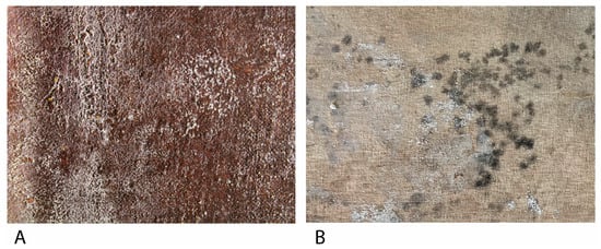
Cultural Heritage (CH) materials are susceptible to being complexly damaged physically, chemically, and aesthetically by the growing and metabolic activities of living beings, as investigators know as biodeterioration. Historical easel paintings have a mixture of materials and layers, increasing their conservation complexity. Materials such as cellulose on the support, rabbit skin glue, egg yolk, linseed oil, or varnishes on the polychromy are some organic materials that can compound an easel painting, although the painting layer also contains inorganic pigments. These organic materials can be degraded by fungi if the environmental conditions are favorable. Eliminating and controlling fungal biodeterioration is one of the most important challenges of easel painting conservation.
- biodeterioration
- fungi
- easel paintings
- wood
- textile fibers
1. Introduction
| Species | Genus | Order | Phylum | References |
|---|---|---|---|---|
| Aerobasidium pullulans | Aerobasidium | Dothideales | Ascomycota | [38,43,44,45,46,47,48][22][27][28][29][30][31][32] |
| Alternaria alternata | Alternaria | Pleosporales | Ascomycota | [38,2245,][2947,][3148,]49,50][[32][33][34] |
| Aspergillus clavatus | Aspergillus | Eurotiales | Ascomycota | [38][22] |
| Aspergillus flavus | Aspergillus | Eurotiales | Ascomycota | [45,46,51,52][29][30][35][36] |
| Aspergillus fumigatus | Aspergillus | Eurotiales | Ascomycota | [48][32] |
| Aspergillus niger | Apergillus | Eurotiales | Ascomycota | [43,44,47,48,49,50,51,53,54][27][28][31][32][33][34][35][37][38] |
| Aspergillus sp. | Apergillus | Eurotiales | Ascomycota | [41,55][25][39] |
| Aspergillus versicolor | Aspergillus | Eurotiales | Ascomycota | [43,45,48,50][27][29][32][34] |
| Bjerkandera adusta | Bjerkandera | Polyporales | Basidiomycota | [38][22] |
| Chaetonium globosum | Chaetonium | Sordariales | Ascomycota | [44,47,48,52,56][28][31][32][36][40] |
| Cladosporium cladosporioides | Cladosporium | Capnodiales | Ascomycota | [43,44,45,46,48,50][27][28][29][30][32][34] |
| Cladosporium sp. | Cladosporium | Capnodiales | Ascomycota | [41][25] |
| Filobasidium magnum | Filobasidium | Filobasidiales | Basidiomycota | [38,41,45][22][25][29] |
| Mucor spp. | Mucor | Mucorales | Mucoromycota | |
| Penicillium chrysogenum | Penicillium | Eurotiales | Ascomycota | [20,38,43,44,[45,50,22][52,27][53,54][20]28][29][34][36][37][38] |
| Penicillium sp. | Penicillium | Eurotiales | Ascomycota | [20,41][20][25] |
2. Fungal Biodeterioration of Wood on Easel Paintings
3. Fungal Biodeterioration of Textile Fibers on Easel Paintings
4. Fungal Biodeterioration of Oil Binders in Easel Paintings
References
- Russo, R.; Palla, F. Plant Essential Oils as Biocides in Sustainable Strategies for the Conservation of Cultural Heritage. Sustainability 2023, 15, 8522.
- Cennamo, P.; Luca, D.D. A Metabarcoding Approach for the Study of Biodeterioration of Ancient Wall Paintings in an Italian Cave. J. Phys. Conf. Ser. 2022, 2204, 012011.
- Zucconi, L.; Canini, F.; Isola, D.; Caneva, G. Fungi Affecting Wall Paintings of Historical Value: A Worldwide Meta-Analysis of Their Detected Diversity. Appl. Sci. 2022, 12, 2988.
- Fiorillo, F.; Fiorentino, S.; Montanari, M.; Roversi Monaco, C.; del Bianco, A.; Vandini, M. Learning from the Past, Intervening in the Present: The Role of Conservation Science in the Challenging Restoration of the Wall Painting Marriage at Cana by Luca Longhi (Ravenna, Italy). Herit. Sci. 2020, 8, 10.
- Suphaphimol, N.; Suwannarach, N.; Purahong, W.; Jaikang, C.; Pengpat, K.; Semakul, N.; Yimklan, S.; Jongjitngam, S.; Jindasu, S.; Thiangtham, S.; et al. Identification of Microorganisms Dwelling on the 19th Century Lanna Mural Paintings from Northern Thailand Using Culture-Dependent and-Independent Approaches. Biology 2022, 11, 228.
- Prieto, B.; Young, M.E.; Turmel, A.; Fuentes, E. Role of Masonry Fabric Subsurface Moisture on Biocolonisation. A Case Study. Build. Environ. 2022, 210, 108690.
- Schröer, L.; de Kock, T.; Godts, S.; Boon, N.; Cnudde, V. The Effects of Cyanobacterial Biofilms on Water Transport and Retention of Natural Building Stones. Earth Surf. Process. Landf. 2022, 47, 1921–1936.
- Soares, F.; Trovão, J.; Portugal, A. Phototrophic and Fungal Communities Inhabiting the Roman Cryptoporticus of the National Museum Machado de Castro (UNESCO Site, Coimbra, Portugal). World J. Microbiol. Biotechnol. 2022, 38, 157.
- Skipper, P.J.A.; Skipper, L.K.; Dixon, R.A. A Metagenomic Analysis of the Bacterial Microbiome of Limestone, and the Role of Associated Biofilms in the Biodeterioration of Heritage Stone Surfaces. Sci. Rep. 2022, 12, 4877.
- Dias, L.; Rosado, T.; Candeias, A.; Mirão, J.; Caldeira, A.T. A Change in Composition, a Change in Colour: The Case of Limestone Sculptures from the Portuguese National Museum of Ancient Art. J. Cult. Herit. 2020, 42, 255–262.
- Wang, Y.; Zhang, H.; Liu, X.; Liu, X.; Song, W. Fungal Communities in the Biofilms Colonizing the Basalt Sculptures of the Leizhou Stone Dogs and Assessment of a Conservation Measure. Herit. Sci. 2021, 9, 36.
- Isola, D.; Bartoli, F.; Meloni, P.; Caneva, G.; Zucconi, L. Black Fungi and Stone Heritage Conservation: Ecological and Metabolic Assays for Evaluating Colonization Potential and Responses to Traditional Biocides. Appl. Sci. 2022, 12, 2038.
- Silva, N.C.; Pintado, M.; Moreira, P.R. Sampling Methods for Outdoor Sculptures: Comparison of Swabs and Cryogels by Flow Cytometry as Novel Alternatives for Assessment and Quantification of Microbial Contamination. J. Cult. Herit. 2022, 54, 94–102.
- Silva, N.C.; Madureira, A.R.; Pintado, M.; Moreira, P.R. Biocontamination and Diversity of Epilithic Bacteria and Fungi Colonising Outdoor Stone and Mortar Sculptures. Appl. Microbiol. Biotechnol. 2022, 106, 3811–3828.
- Salvador, C.; Bordalo, R.; Silva, M.; Rosado, T.; Candeias, A.; Caldeira, A.T. On the Conservation of Easel Paintings: Evaluation of Microbial Contamination and Artists Materials. Appl. Phys. A Mater. Sci. Process. 2017, 123, 80.
- Caselli, E.; Pancaldi, S.; Baldisserotto, C.; Petrucci, F.; Impallaria, A.; Volpe, L.; D’Accolti, M.; Soffritti, I.; Coccagna, M.; Sassu, G.; et al. Characterization of Biodegradation in a 17 Th Century Easel Painting and Potential for a Biological Approach. PLoS ONE 2018, 13, e0207630.
- Salvador, C.; Sandu, I.; Sandbakken, E.; Candeias, A.; Caldeira, A.T. Biodeterioration in Art: A Case Study of Munch’s Paintings. Eur. Phys. J. Plus 2022, 137, 11.
- Kavkler, K.; Humar, M.; Kržišnik, D.; Turk, M.; Tavzes, Č.; Gostinčar, C.; Džeroski, S.; Popov, S.; Penko, A.; Gunde-Cimerman, N.; et al. A Multidisciplinary Study of Biodeteriorated Celje Ceiling, a Tempera Painting on Canvas. Int. Biodeterior. Biodegrad. 2022, 170, 105389.
- Poyatos, F.; Morales, F.; Nicholson, A.W.; Giordano, A. Physiology of Biodeterioration on Canvas Paintings. J. Cell. Physiol. 2018, 233, 2741–2751.
- López-Miras, M.; Piñar, G.; Romero-Noguera, J.; Bolívar-Galiano, F.C.; Ettenauer, J.; Sterflinger, K.; Martín-Sánchez, I. Microbial Communities Adhering to the Obverse and Reverse Sides of an Oil Painting on Canvas: Identification and Evaluation of Their Biodegradative Potential. Aerobiologia 2013, 29, 301–314.
- Colombini, M.P.; Degano, I.; Nevin, A. Analytical Approaches to the Analysis of Paintings: An Overview of Methods and Materials. In Cultural Heritage Science; Springer: Berlin/Heidelberg, Germany, 2022; pp. 95–111.
- Văcar, C.L.; Mircea, C.; Pârvu, M.; Podar, D. Diversity and Metabolic Activity of Fungi Causing Biodeterioration of Canvas Paintings. J. Fungi 2022, 8, 589.
- Jiang, L.; Pettitt, T.R.; Buenfeld, N.; Smith, S.R. A Critical Review of the Physiological, Ecological, Physical and Chemical Factors Influencing the Microbial Degradation of Concrete by Fungi. Build. Environ. 2022, 214, 108925.
- Cepero de García, M.C. Biologia De Hongos; Ediciones Unlandes: Bogotá, Colombia, 2012; ISBN 9586957942.
- Poyatos-Jiménez, F.; Morales, F.; Morales-Carrera, R.; Boffo, S.; Giordano, A.; Romero-Noguera, J. Fungal and Bacterial Biodeterioration of Outdoor Canvas Paintings: The Case of the Cloisters of Quito, Ecuador. Crit. Rev. Eukaryot. Gene Expr. 2021, 31, 45–63.
- Tortora, G.J.; Funke, B.R.; Case, C.L. Introducción a La Microbiología, 12th ed.; Editorial Médica Panamericana: Madrid, Spain, 2017; ISBN 9789500695404.
- Romero-Noguera, J.; Bolívar-Galiano, F.C.; Ramos-López, J.M.; Fernández-Vivas, M.A.; Martín-Sánchez, I. Study of Biodeterioration of Diterpenic Varnishes Used in Art Painting: Colophony and Venetian Turpentine. Int. Biodeterior. Biodegrad. 2008, 62, 427–433.
- Doménech-Carbó, M.T.; Bitossi, G.; de la Cruz-Cañizares, J.; Bolívar-Galiano, F.; López-Miras, M.d.M.; Romero-Noguera, J.; Martín-Sánchez, I. Microbial Deterioration of Mowilith DMC 2, Mowilith DM5 and Conrayt Poly(Vinyl Acetate) Emulsions Used as Binding Media of Paintings by Pyrolysis-Silylation-Gas Chromatography-Mass Spectrometry. J. Anal. Appl. Pyrolysis 2009, 85, 480–486.
- Sterflinger, K. Fungi: Their Role in Deterioration of Cultural Heritage. Fungal Biol. Rev. 2010, 24, 47–55.
- Kurowski, G.; Vogt, O.; Ogonowski, J. Paint-Degrading Microorganisms. Czas. Tech. 2017, 2017, 81–92.
- Minotti, D.; Vergari, L.; Proto, M.R.; Barbanti, L.; Garzoli, S.; Bugli, F.; Sanguinetti, M.; Sabatini, L.; Peduzzi, A.; Rosato, R.; et al. Il Silenzio: The First Renaissance Oil Painting on Canvas from the Uffizi Museum Restored with a Safe, Green Antimicrobial Emulsion Based on Citrus Aurantium Var. Amara Hydrolate and Cinnamomum Zeylanicum Essential Oil. J. Fungi 2022, 8, 140.
- Vukojevi, J.; Grbi, M.L. Moulds on Paintings in Serbian Fine Art Museums. Afr. J. Microbiol. Res. 2010, 4, 1453–1456.
- Elsayed, Y.; Shabana, Y. The Effect of Some Essential Oils on Aspergillus Niger and Alternaria Alternata Infestation in Archaeological Oil Paintings. Mediterr. Archaeol. Archaeom. 2018, 18, 71–87.
- Sabatini, L.; Sisti, M.; Campana, R. Evaluation of Fungal Community Involved in the Bioderioration Process of Wooden Artworks and Canvases in Montefeltro Area (Marche, Italy). Microbiol. Res. 2018, 207, 203–210.
- Ogbulie, J.; Obiajuru, I. Microbial Deterioration of Surface Paint Coatings. Glob. J. Pure Appl. Sci. 2004, 10, 485–490.
- Noohi, N.; Papizadeh, M. Study of Biodeterioration Potential of Microorganisms Isolated in the Paintings Storeroom of Mouze Makhsus Museum, Golestan Palace, Tehran. Stud. Conserv. 2022, 68, 720–730.
- Seves, A.M.; Sora, S.; Ciferri, O. The Microbial Colonization of Oil Paintings. A Laboratory Investigation. Int. Biodeterior. Biodegrad. 1996, 37, 215–224.
- Ciferri, O. Microbial Degradation of Paintings. Appl. Environ. Microbiol. 1999, 65, 879–885.
- Geweely, N.S.; Afifi, H.A.; Ibrahim, D.M.; Soliman, M.M. Efficacy of Essential Oils on Fungi Isolated from Archaeological Objects in Saqqara Excavation, Egypt. Geomicrobiol. J. 2019, 36, 148–168.
- Okpalanozie, O.E.; Adebusoye, S.A.; Troiano, F.; Polo, A.; Cappitelli, F.; Ilori, M.O. Evaluating the Microbiological Risk to a Contemporary Nigerian Painting: Molecular and Biodegradative Studies. Int. Biodeterior. Biodegrad. 2016, 114, 184–192.
- Sterflinger, K.; Piñar, G. Microbial Deterioration of Cultural Heritage and Works of Art—Tilting at Windmills? Appl. Microbiol. Biotechnol. 2013, 97, 9637–9646.
- Tiano, P. Biodegradation of Cultural Heritage: Decay Mechanisms and Control Methods. In Proceedings of the 9th ARIADNE Workshop “Historic Material and Their Diagnostic” ARCCHIP, Prague, Czech Republic, 22–28 April 2002; pp. 22–28.
- Kalra, R.; Conlan, X.A.; Goel, M. Fungi as a Potential Source of Pigments: Harnessing Filamentous Fungi. Front. Chem. 2020, 8, 369.
- Toma, M.A.; Nazir, K.H.M.N.H.; Mahmud, M.; Mishra, P.; Ali, K.; Kabir, A.; Shahid, A.H.; Siddique, M.P.; Alim, A. Isolation and Identification of Natural Colorant Producing Soil-Borne Aspergillus Niger from Bangladesh and Extraction of the Pigment. Foods 2021, 10, 1280.
- Wulandari, A.P.; Examinati, R.R.I.N.; Madihah, H.D.; Andayaningsih, P.; Andayaningsih, P. Citotoxicity of Metabolites Produced by Endophytic Fungus Cladosporium Sp. Isolated from Marine Macroalgae on Invitro MCF7, Hela, and DU-145 Cell Lines. Int. J. Pharm. Pharm. Sci. 2018, 10, 72.
- Kristensen, S.B.; Pedersen, T.B.; Nielsen, M.R.; Wimmer, R.; Muff, J.; Sørensen, J.L. Production and Selectivity of Key Fusarubins from Fusarium Solani Due to Media Composition. Toxins 2021, 13, 376.
- Miao, F.P.; Li, X.D.; Liu, X.H.; Cichewicz, R.H.; Ji, N.Y. Secondary Metabolites from an Algicolous Aspergillus Versicolor Strain. Mar. Drugs 2012, 10, 131–139.
- Dzurendova, S.; Losada, C.B.; Dupuy-Galet, B.X.; Fjær, K.; Shapaval, V. Mucoromycota Fungi as Powerful Cell Factories for Modern Biorefinery. Appl. Microbiol. Biotechnol. 2022, 106, 101–115.
- Fierascu, R.C.; Doni, M.; Fierascu, I. Selected Aspects Regarding the Restoration/Conservation of Traditional Wood and Masonry Building Materials: A Short Overview of the Last Decade Findings. Appl. Sci. 2020, 10, 1164.
- Pournou, A. Wood Deterioration by Terrestrial Microorganisms. In Biodeterioration of Wooden Cultural Heritage; Springer International Publishing: Cham, Switzerland, 2020; pp. 345–424.
- Goodell, B. Genetics and Biotechnology, 2nd ed.; Benz, J.P., Schipper, K., Eds.; Springer International Publishing: Cham, Switzerland, 2020; ISBN 978-3-030-49923-5.
- Branysova, T.; Demnerova, K.; Durovic, M.; Stiborova, H. Microbial Biodeterioration of Cultural Heritage and Identification of the Active Agents over the Last Two Decades. J. Cult. Herit. 2022, 55, 245–260.
- Afifi, H.A.M.; Mansour, M.M.A.; Hassan, A.G.A.I.; Salem, M.Z.M. Biodeterioration Effects of Three Aspergillus Species on Stucco Supported on a Wooden Panel Modeled from Sultan Al-Ashraf Qaytbay Mausoleum, Egypt. Sci. Rep. 2023, 13, 15241.
- Martín Rey, S.; Durand Alegría, J.S.; Martín Martínez, J.M. Adhesivos Tack-Melt Atóxicos Para Su Empleo En Tratamientos Restaurativos de Pintura Sobre Tela: Tipificación y Análisis. Ph.D. Thesis, Universidad Nacional de Educación a Distancia, Madrid, Spain, 2017.
- Caneva, G.; Nugari, M.P.; Salvadori, O. La Biología En La Restauración; Nerea: Hondarribia, Spain, 2000; ISBN 84-89569-48-7.
- Ranalli, G.; Zanardini, E.; Sorlini, C. Biodeterioration—Including Cultural Heritage. In Reference Module in Life Sciences; Elsevier: Amsterdam, The Netherlands, 2018.
- Szostak-Kotowa, J. Biodeterioration of Textiles. Int. Biodeterior. Biodegrad. 2004, 53, 165–170.
- Caneva, G.; Ceschin, S. Ecology of Biodeterioration. In Plant Biology for Cultural Heritage: Biodeterioration and Conservation; Caneva, G., Nugari, M.P., Salvadori, O., Eds.; The Getty Conservation Institute: Los Angeles, CA, USA, 2009; pp. 35–58.
- Kavkler, K.; Gunde-Cimerman, N.; Zalar, P.; Demšar, A. Fungal Contamination of Textile Objects Preserved in Slovene Museums and Religious Institutions. Int. Biodeterior. Biodegrad. 2015, 97, 51–59.
- Brzozowska, I.; Bogdanowicz, A.; Szczęsny, P.; Zielenkiewicz, U.; Laudy, A. Evaluation of Bacterial Diversity on Historical Silk Velvet Textiles from the Museum of King John III’s Palace at Wilanów, Poland. Int. Biodeterior. Biodegrad. 2018, 131, 78–87.
- Ravikumar, H.R. Biodegradation of Paints: A Current Status. Indian J. Sci. Technol. 2012, 5, 1–11.
- Breitbach, A.M.; Rocha, J.C.; Gaylarde, C.C. Influence of Pigment on Biodeterioration of Acrylic Paint Films in Southern Brazil. J. Coat. Technol. Res. 2011, 8, 619–628.
- López-Miras, M.D.M.; Martín-Sánchez, I.; Yebra-Rodríguez, Á.; Romero-Noguera, J.; Bolívar-Galiano, F.; Ettenauer, J.; Sterflinger, K.; Piñar, G. Contribution of the Microbial Communities Detected on an Oil Painting on Canvas to Its Biodeterioration. PLoS ONE 2013, 8, e80198.
- Mecklenburg, M.F. Micro Climates and Moisture Induced Damage to Paintings. In Proceedings of the Conference on Micro Climates in Museums, Copenhagen, Denmark, 19–23 November 2007; pp. 19–25.
- Zmeu, C.N.; Bosch-Roig, P. Risk Analysis of Biodeterioration in Contemporary Art Collections: The Poly-Material Challenge. J. Cult. Herit. 2022, 58, 33–48.
- Ortiz-Miranda, A.S.; Doménech-Carbó, A.; Doménech-Carbó, M.T.; Osete-Cortina, L.; Bolívar-Galiano, F.; Martín-Sánchez, I. Analyzing Chemical Changes in Verdigris Pictorial Specimens upon Bacteria and Fungi Biodeterioration Using Voltammetry of Microparticles. Herit. Sci. 2017, 5, 8.
- Santos, A.; Cerrada, A.; García, S.; Andrés, M.S.; Abrusci, C.; Marquina, D. Application of Molecular Techniques to the Elucidation of the Microbial Community Structure of Antique Paintings. Microb. Ecol. 2009, 58, 692–702.