Table 1. Most common fungal species on easel painting.
| Species |
Genus |
Order |
Phylum |
References |
| Aerobasidium pullulans |
Aerobasidium |
Dothideales |
Ascomycota |
[38,43,44,45,46,47,48] |
| Alternaria alternata |
Alternaria |
Pleosporales |
Ascomycota |
[38,45,47,48,49,50] |
| Aspergillus clavatus |
Aspergillus |
Eurotiales |
Ascomycota |
[38] |
| Aspergillus flavus |
Aspergillus |
Eurotiales |
Ascomycota |
[45,46,51,52] |
| Aspergillus fumigatus |
Aspergillus |
Eurotiales |
Ascomycota |
[48] |
| Aspergillus niger |
Apergillus |
Eurotiales |
Ascomycota |
[43,44,47,48,49,50,51,53,54] |
| Aspergillus sp. |
Apergillus |
Eurotiales |
Ascomycota |
[41,55] |
| Aspergillus versicolor |
Aspergillus |
Eurotiales |
Ascomycota |
[43,45,48,50] |
| Bjerkandera adusta |
Bjerkandera |
Polyporales |
Basidiomycota |
[38] |
| Chaetonium globosum |
Chaetonium |
Sordariales |
Ascomycota |
[44,47,48,52,56] |
| Cladosporium cladosporioides |
Cladosporium |
Capnodiales |
Ascomycota |
[43,44,45,46,48,50] |
| Cladosporium sp. |
Cladosporium |
Capnodiales |
Ascomycota |
[41] |
| Filobasidium magnum |
Filobasidium |
Filobasidiales |
Basidiomycota |
[38,41,45] |
| Mucor spp. |
Mucor |
Mucorales |
Mucoromycota |
| Penicillium chrysogenum |
Penicillium |
Eurotiales |
Ascomycota |
[20,38,43,44,45,50,52,53,54] |
| Penicillium sp. |
Penicillium |
Eurotiales |
Ascomycota |
[20,41] |
The development of fungal strains damages easel painting materials in different ways, causing a multitude of pathologies depending on the substrate and the strain [
57]. Generally, fungal growth produces acidification, chromatic changes, structural damage across chemical mechanisms—liberation of fatty acids and enzymatic action— and mycelium growth [
19] (
Figure 3). Microbial deterioration usually affects the painting support in the first instance, reaching and colonizing the pictorial layers in later phases [
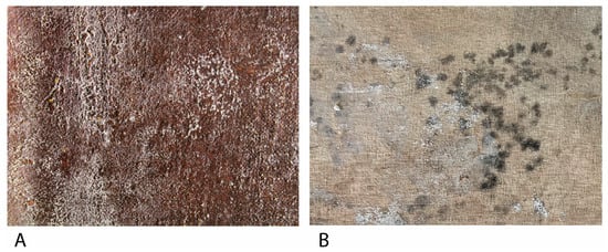
58]. The organic nature of these materials presents a large carbon source for microorganisms, which makes them susceptible to biological attack. In addition, the presence of glues or coating pastes applied as sizing treatments increases that susceptibility. Aesthetical damages are generally related to pigment production.
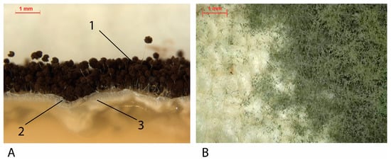
Ascomycota strains such as
A. niger produces azanigerones B and C—yellow [
59]—or other colors from pale yellow to brown [
60].
Cladosporium sp. and
Fusarium sp. produce anhydrofusarubin—red [
61,
62]—or
A. versicolor produces asperversin—yellow [
63]. Red-orange, β-carotene, is the most produced pigment by
Mucoromycota as a preventive measure for stress oxidative damages [
64].
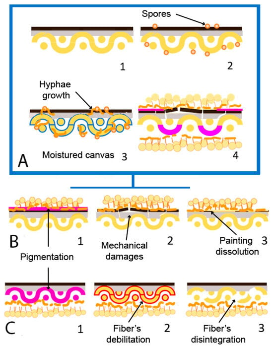
Figure 3. Fungal biodeterioration mechanism and damages on canvas painting (adapted from Poyatos et al., 2018 [
19]). (
A) (
1) Non-biodeteriorated oil on canvas painting; (
2) presence of fungal spores (orange) on the surface; (
3) moisture of canvas (blue) due to RH increase and fungal development. (
4) Biodeteriorated painting. (
B) Damages on painting strata; (
1) Pigmentation (pink); (
2) mechanical damage of painting film; (
3) dissolution of painting film. (
C) Damages on canvas support; (
1) pigmentation (pink); (
2) fiber’s debilitation (red); (
3) fiber’s disintegration.
2. Fungal Biodeterioration of Wood on Easel Paintings
Wood was the most important painting support until canvas popularization in the 16th century. It presents well-known biodeterioration problems due to its use in different industries or as a construction material [
65]. Wood fungal biodeterioration can be classified as white rot, brown rot (exclusively by
Basidiomycota), and soft rot (generally
Ascomycota), depending on the produced damages [
66,
67]. Brown rot fungi feed on cellulose and produce brown pigmentation and structural damage on wood, while white rot fungi also feed on lignin and produce discoloration [
68]. Soft rot fungi can produce lignin and cellulose, penetrating the surface at various centimeters, producing cavities and erosion [
67]. Recently, Afifi et al., 2023 [
69] detected biodeterioration damages produced by
Aspergillus sp., such as discoloration or hyphae penetration into the wooden support of mock-ups based on a historical stucco (Sultan al-Ashraf Qaytbay Mausoleum, Egypt).
3. Fungal Biodeterioration of Textile Fibers on Easel Paintings
Starting in the 17th century, textile fibers began to expand as the main artistic support due to their lower cost or technical characteristics, such as the possibility of producing larger and lighter formats, the contribution of texture, or the ease of transport and storage [
19,
70]. The historical textiles usually used are made of natural fiber fabrics, such as cotton, hemp, linen, or jute. These fabrics’ main component is cellulose, which comes from the plant fibers from which the materials are obtained [
58,
71]. For example, cotton comprises seed fibers, while flax or jute are made from phloem fibers [
58]. The main component of the cell walls of tissues of plant origin is cellulose. Cellulose is a polysaccharide formed by a crystalline region with great resistance to biodeterioration and an amorphous region easily attacked by microorganisms, in which the biological attack usually begins [
72]. The fungal deterioration of tissues happens mainly due to the production capacity of intracellular and extracellular hydrolytic enzymes and organic acids, using natural fibers as a carbon source for their growth and development. These enzymes are cellulases, breaking cellulose’s intramolecular bonds and obtaining glucose molecules [
71,
72]. That is, they decrease the degree of polymerization of the cellulose molecules [
73]. This can cause fiber breakage and thinning and, therefore, a decrease in the individual resistance of the fibers and the textile [
72].
In addition, organic acids produce minerals for some phototrophs and have a wide variety of pigments, being the first alteration of fungal biodeterioration and complex to eliminate. Therefore, the amount of cellulose in the tissues will be decisive regarding their vulnerability to being colonized by micro-organisms. Of the fabrics widely used as pictorial textile supports, cotton is the material that has the highest amount of cellulose (90%), followed by linen (80%), hemp (77%), and finally, jute (60%) [
58,
71]. Factors such as composition or fiber and the structural and chemical characteristics of the textile influence the risk of biodeterioration. Increasing the amount of cellulose—in addition to pectins or pentoses—on the textile composition will increase the biodeterioration susceptibility, too. However, some substances are present in textiles that few microorganisms cannot metabolize, such as lignin [
71,
74]. Structurally and chemically, the cellulose chains’ length, degree of polymerization and crystallinity, the fibers’ orientation or the threads’ thickness, and their chemical, photochemical, and mechanical degradation must be considered [
19,
71,
75,
76].
4. Fungal Biodeterioration of Oil Binders in Easel Paintings
On polychromies, fungal biodeterioration affects the chemical and physical stability: while fatty acids acidify the surface, esterases and lipases disaggregate the pictorial layer, damaging the oil medium and producing resistance loss, fracture, and detachments in extreme cases [
19,
77,
78]. Therefore, enzyme liberation supposes triacylglycerols hydrolyzation by lipases and the aqueous part hydrolyzation by esterases, favoring the mycelium growth, which produces microcracks [
54,
79]. As ideal conditions for fungal growth—high RH—are also prejudicial for easel paintings because of the different mechanical behavior between layers [
80], a synergy between biodeterioration agents and other types of alteration is caused, such as moisture, dust, or environmental contaminants [
81]. Contrary to support, pictorial films have more factors in the biodeterioration mechanisms, such as pigments. Paintings with Pb, Zn, Cr, or Cd are more resistant to fungal biodeterioration, while earth pigments are more susceptible [
82,
83]. Different investigations studied the effect of fungal strains isolated from easel paintings. Salvador et al., 2017 [
15] analyzed the biodeterioration of different Giorgio Marini (1836–1905) portraits conserved in museums or private collections in Évora, Portugal. Strains of
Aspergillus sp.,
Cladosporium sp.,
Penicillium sp., and
Mucor sp. were isolated, attributing their growth to the presence of proteinaceous binders and inadequate climate conditions. Even in outdoor environments, Poyatos-Jiménez et al., 2021 [
41] evaluated the biodeterioration of a collection of paintings placed on different Cloisters of Quito, Ecuador. In their study, researchers isolated strains of
Aspergillus sp.,
Mucor sp.,
Cladosporium sp., or
Penicillium sp. They attributed their biodeterioration pathologies to the semi-tropical environment where the paintings are exhibited.