Submitted Successfully!
To reward your contribution, here is a gift for you: A free trial for our video production service.
Thank you for your contribution! You can also upload a video entry or images related to this topic.
Version Summary Created by Modification Content Size Created at Operation
1 -- 2395 2024-02-06 14:58:42 |
2 format correct Meta information modification 2395 2024-02-07 07:55:21 |

Video Upload Options

Do you have a full video?

Confirm

Are you sure to Delete?
Cite
If you have any further questions, please contact Encyclopedia Editorial Office.
Bălăceanu-Gurău, B.; Apostol, E.; Caraivan, M.; Ion, A.; Tatar, R.; Mihai, M.M.; Popa, L.G.; Gurău, C.; Orzan, O.A. Infectious Tattoo-Related Side Effects. Encyclopedia. Available online: https://encyclopedia.pub/entry/54805 (accessed on 02 May 2024).
Bălăceanu-Gurău B, Apostol E, Caraivan M, Ion A, Tatar R, Mihai MM, et al. Infectious Tattoo-Related Side Effects. Encyclopedia. Available at: https://encyclopedia.pub/entry/54805. Accessed May 02, 2024.
Bălăceanu-Gurău, Beatrice, Eliza Apostol, Mădălina Caraivan, Ana Ion, Raluca Tatar, Mara Mădălina Mihai, Liliana Gabriela Popa, Cristian-Dorin Gurău, Olguța Anca Orzan. "Infectious Tattoo-Related Side Effects" Encyclopedia, https://encyclopedia.pub/entry/54805 (accessed May 02, 2024).
Bălăceanu-Gurău, B., Apostol, E., Caraivan, M., Ion, A., Tatar, R., Mihai, M.M., Popa, L.G., Gurău, C., & Orzan, O.A. (2024, February 06). Infectious Tattoo-Related Side Effects. In Encyclopedia. https://encyclopedia.pub/entry/54805
Bălăceanu-Gurău, Beatrice, et al. "Infectious Tattoo-Related Side Effects." Encyclopedia. Web. 06 February, 2024.
Infectious Tattoo-Related Side Effects
Edit

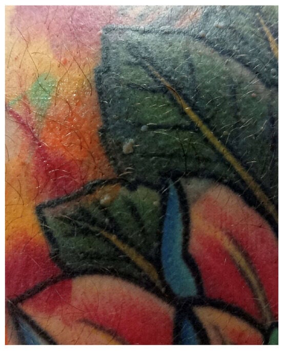
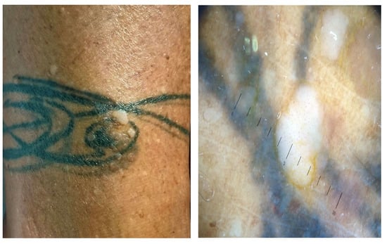
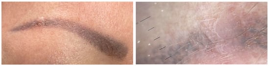
Tattooing is the procedure of implanting permanent pigment granules and additives into the dermal layer of the skin, serving various purposes such as decoration, medical identification, or accidental markings. There has been a significant rise in the popularity of decorative tattooing as a form of body art among both teenagers and young adults. Thus, the incidence of tattoos is increasing, with expanding applications such as permanent makeup, scar camouflage, nipple–areola, lips, and eyebrows tattooing, and utilization in oncological radiotherapy such as colon marking. However, there have been reported a broad range of adverse reactions linked to tattooing, encompassing allergic reactions, superficial and deep cutaneous infections, autoimmune disorders induced by the Koebner phenomenon, cutaneous tumors, and others.

tattoos side effects bacterial infections mycobacterial infections viral infections fungal infections

1. Introduction

The term “tattooing” is rooted in the Tahitian word “tattau”, meaning “to mark” [1]. It represents the procedure of implanting permanent pigment granules and additives into the dermal layer of the skin, serving various purposes such as cosmetic applications (decorative tattoos and permanent makeup) or therapeutic uses (medical tattoos) [1]. Medical therapeutic tattooing plays an important role in techniques such as camouflage for vitiligo, breast areola reconstruction after radical surgery, concealing permanent hair loss following craniofacial surgery, and addressing scars after plastic and reconstructive surgery [1]. Accidental occurrences, like traumatic tattoos resulting from abraded skin injuries, can also be encountered [1].
There has been a significant contemporary upswing in tattooing, particularly among teenagers and young adults, as a form of cosmetic and decorative body art [1]. Currently, there is a lack of stringent requirements, regulations, and legislative measures ensuring the safety of tattooing [1]. Consequently, the reported incidence of adverse reactions after tattooing has been increasing, although these are often observed by physicians but remain relatively unknown to the general public and tattoo artists [1]. The shift in tattoo-ink composition from inorganic pigments (heavy metals) to organic pigments (azo pigments) in recent decades and the subsequent use of postcare products adds another layer of complexity to understanding potential complications [2][3].
Common skin reactions documented in the medical literature encompass a transient acute inflammatory response resulting from skin trauma induced by needles, involving pain, development of blisters, crusts, and pinpoint hemorrhaging [2][3]. Moreover, there have been reported a wide range of emerging cutaneous manifestations. Skin conditions and issues following the process of tattooing can be classified into inflammatory disorders (allergic reactions, chronic inflammatory black tattoo reactions, autoimmune skin afflictions, foreign-body reactions, and pseudo lymphoma), infections (bacterial, mycobacterial, viral, fungal, and parasitic), neoplasms (benign and malignant tumors), miscellaneous complications (neuro-sensory issues, complications linked to magnetic resonance imaging, and photoinduced reactions) and cosmetic issues (misapplication, pigment fanning or migration, and scars) [1][2][3].
Delayed complications may include, in addition to scarring and cutaneous textural changes, pigmentary alterations associated with tattoo removal using Q-switched lasers, such as hypopigmentation and hyperpigmentation, and the occurrence of paradoxical darkening of the tattooed area or residual pigmentation [1].
The risk of infection is influenced by various factors, including the skin’s condition at the tattoo site, the proper sterilization of equipment, the use of contaminated tattoo ink, inadequate disinfection of the tattooed skin area, and inappropriate aftercare [3][4][5]. During the healing process of the injured tissue after tattooing, patients often experience pruritus and burning, which increase the risk of superinfection due to scratching and the subsequent introduction of microorganisms [3][4][5].
Infections on tattoos can manifest either as pyogenic or nonpyogenic. In contemporary times, due to standard hygiene practices and modern aseptic tattooing techniques, the majority of infections are typically superficial (acute superficial pyogenic infections, including folliculitis, impetigo, and ecthyma), of bacterial origin, and manifest within a few days post-tattooing [3][6][7]. One Danish study revealed that 10% of the unopened tattoo ink stock bottles were contaminated with a range of bacteria, including both pathogenic and nonpathogenic strains [7][8]. Examples of isolated strains include Pseudomonas species, Staphylococcus species, Streptococcus salivarius, Streptococcus sanguinis, Enterococcus faecium, and Acinetobacter species [7][8]. Additionally, 28% of the analyzed stock bottles were found to be inadequately sealed [7][8].
However, more severe systemic infections can also occur, such as cellulitis, furunculosis, necrotizing fasciitis, erysipelas, or bacterial endocarditis [9][10][11][12][13][14][15]. Historical records of gangrene, tetanus, amputations, and syphilis have also been documented [9][10][11][12][13][14][15].

2. Bacterial Infections

The most commonly encountered clinical infections related to tattoos include impetigo and folliculitis [3][15]. Staphylococcus aureus, Streptococcus pyogenes, Clostridium difficile, and Pseudomonas aeruginosa are the primary causative agents for these superficial infections [3][15] (Table 1).
Clinical manifestations of bacterial infections encompass local pain, erythema, and swelling, as well as fever and purulence [3][16]. It is crucial to differentiate cellulitis or erysipelas from temporary tattoo-induced edema, which is a transient reaction inherent to the tattooing process, particularly when applied to the lower extremities [3][16]. This reaction is inevitable and can occur in any individual [3][16].
Most bacterial infections are easily treatable, and their treatment generally aligns with standard bacterial infection management (Table 1). They can be verified through suitable cultures and subsequently treated accordingly. However, certain pathogens may pose greater challenges. For instance, an epidemic of cutaneous infections caused by methicillin-resistant Staphylococcus aureus was reported in the USA following tattooing [3][17][18].
Table 1. Bacterial and mycobacterial tattoo-related side effects and clinical measures.
Side
Effects
Bacterial Mycobacterial
Clinical measures Staphylococcus aureus/Streptococcus pyogenes/Clostridium difficile/Pseudomonas aeruginosa [3][15] Mycobacterium tuberculosis/Mycobacterium bovis [3][6][19][20][21] Mycobacterium chelonae/Mycobacterium abscessus/Mycobacterium fortuitum [22][23][24] Mycobacterium mageritense [3][25][26] Mycobacterium leprae [1][3][27]
Standard bacterial infection management Multidrug therapy administered in two phases [28]:
Isoniazid
Rifampicin
Pyrazinamide
Ethambutol or Streptomycin
Macrolide antibiotics [29]
(Clarithromycin)
4 months in mild cases
6–12 months in severe cases
Antibiotic therapy [29]:
Amikacin
Imipenem
Cefoxitin
Fluroquinolones
Sulfonamides
Paucibacillary disease [30]:
Dapsone + Rifampicin + Clofazimine for 6 months
Multibacillary disease [30]:
Dapsone + Rifampicin + Clofazimine for 12 months
Rifampicin resistance [30]:
24 months treatment broken down as 6 months of Clofazimine + Ofloxacin and Minocycline followed by 18 months of Clofazimine + Ofloxacin or Minocycline
Dapsone resistance [30]:
Clofazimine + Rifampicin for 6 months
In recent decades, one case of secondary syphilis occurring within a tattoo has been reported [3][31]. In the 19th century, syphilis was more frequently described in the context of tattoos [3][31]. During that period, tattoo artists often moistened needles with saliva or used nonsterile or previously used needles, potentially leading to the contamination of patients with Treponema pallidum [3][31].

3. Mycobacterial Infections

Tattoo-inoculated mycobacterial infections encompass tuberculosis, leprosy, and atypical mycobacteria such as Mycobacterium chelonae and Mycobacterium abscessus [22][23][24].
Tattooing can lead to the development of primary cutaneous tuberculosis [3][6][20][24]. This occurs when individuals lacking previous immunity are inoculated with Mycobacterium tuberculosis or Mycobacterium bovis [3][6][19][20][21]. Within 2–4 weeks, an erythematous papule or nodule emerges, eventually progressing to a superficial ulcer known as a tuberculous chancre [3][6][19][20][21]. Often, painless regional lymphadenopathy ensues within 3 to 8 weeks [3][6][19][20][21]. In cases where the patient’s immune system is compromised, there is a risk of progression to lupus vulgaris and tuberculosis cutis verrucose, or even hematogenous spread [3][19][20][21]. Differential diagnoses include foreign-body granuloma, sarcoidosis, inoculation leprosy, tertiary syphilis, and infections with atypical mycobacteria [6]. The histological examination typically reveals epithelioid histiocytes, Langhans giant cells, and tuberculoid granulomas, with or without central caseous necrosis [3][19][20][21]. A positive tuberculin test holds significant diagnostic value for primary tuberculosis [6][19][20][21].
The treatment approach for cutaneous tuberculosis is consistent with that of systemic tuberculosis and involves multidrug therapy [28]. Commonly used drugs include isoniazid, rifampicin, pyrazinamide, and ethambutol or streptomycin, administered in two phases: the intensive one (which aims to rapidly reduce the burden of Mycobacterium tuberculosis and typically spans about 8 weeks) and the continuation phase, designed to eradicate any remaining bacteria and extends for a duration of 9 to 12 months [28] (Table 1). Strict adherence to the treatment regimen is crucial for a successful cure [28].
Various factors influence the outcomes of treatment, including the patient’s immunity, overall health, disease stage, type of cutaneous lesions, treatment adherence, duration of therapy, and potential side effects [28].
Atypical mycobacterial infections, particularly with Mycobacterium chelonae, appear to be an emerging complication [3][32][33][34][35]. This occurrence is particularly associated with the preparation of grey ink, which is obtained by diluting black ink with water [3][33]. If the water used in this process is contaminated with Mycobacterium chelonae, a bacterium commonly found in nonsterile water, it can lead to infections [3][33]. Less commonly, skin infections can be caused by other mycobacterial species, such as Mycobacterium haemophilum, Mycobacterium abscessus, Mycobacterium immunogenum, Mycobacterium massiliense, Mycobacterium mageritense, and Mycobacterium fortuitum [3][25][26] (Table 1). Interestingly, mycobacterial infections tend to manifest more frequently in the grey or black areas of a tattoo [3][33]. Clinically, lesions present as chronic papules, pustules, lichenoid plaques, and plaques with scales, typically developing within 1 to 3 weeks after the procedure [3][33]. Ulcerated nodules primarily confined to the tattooed area have also been reported [3][33].
For skin and soft-tissue infections caused by nontuberculous mycobacteria, a prolonged treatment regimen involving combination therapy with at least two susceptible antimicrobials is recommended to minimize the risk of antibiotic resistance [29]. Typically, the recommended duration of therapy for mild cases is around 4 months, while severe cases may require treatment for 6–12 months [29]. Macrolide antibiotics, with clarithromycin commonly included, are considered standard treatment for nontuberculous mycobacteria infections, including those associated with tattoos and involving Mycobacterium chelonae, Mycobacterium abscessus, and Mycobacterium fortuitum [29]. However, it is important to note that Mycobacterium mageritense is known to be resistant to macrolides due to the presence of the erythromycin ribosomal methylase gene, which imparts resistance to macrolide antibiotics [29]. Mycobacterium mageritense generally exhibits susceptibility or intermediate susceptibility to amikacin, imipenem, cefoxitin, fluoroquinolones, and sulfonamides but is resistant to clarithromycin [29]. It is essential to guide antibiotic therapy based on susceptibility testing [29] (Table 1).
Instances of tattoo inoculation with Mycobacterium leprae are predominantly reported in regions where leprosy is endemic, and unhygienic tattooing practices are prevalent [1][3][27]. The onset of leprosy after tattooing can vary significantly, occurring between 10 to 20 years post-tattooing [1][3][27]. Outbreaks have been linked to the use of shared needles during unhygienic tattooing by roadside artists [3][6][27]. Manifestation of leprosy skin lesions may occur 10 to 20 years after the initial inoculation, and the clinical presentation is primarily influenced by the immunologic status of the host [3][6][27]. In cases where a mycobacterial infection is suspected, conducting a biopsy, tissue culture, and polymerase chain reaction for Mycobacterium species is recommended [3][6][27]. Histologically, these reactions are characterized by the formation of suppurative granulomas with the presence of polymorphonuclear leukocytes [3][6][27].
Treatment recommendations for leprosy in adults consist of long-term multidrug therapy: dapsone, rifampicin, and clofazimine for 6 months in paucibacillary disease and for 12 months in case of multibacillary disease [30]. In case of rifampicin resistance, clofazimine plus at least two of minocycline, clarithromycin, and quinolone for 6 months is recommended, followed by an additional 18 months of clofazimine plus one of the aforementioned drugs [30] (Table 1).

4. Viral Infections

The transmission of infections such as verrucae, molluscum contagiosum virus, human papillomavirus (HPV), herpes simplex virus (HSV), human immunodeficiency virus (HIV), and hepatitis B (HBV) and C viruses (HCV) has been documented [3][6] (Figure 1 and Table 2).
Figure 1. Multiple viral warts localized on the trunk.
Table 2. Viral, fungal, and parasitic tattoo-related side-effects and clinical measures.
Side Effects Viral Fungal Parasitic
Clinical measures Viral warts [3][12][36][37][38] Molluscum contagiosum [3][12][36][37][38] HPV, HSV, HIV, HBV and HCV [3][6] Dermatophytes/Aspergillus fumigatus/Sporotrichosis/Zygomycosis/Acremonium fungi/Candida [3][6][31][39][40] Leishmania species [3]
Firstline [41][42]:
Salicylic Acid
Cryotherapy
First-line [43][44][45]:
Cryotherapy
Curetage
Cantharidin
Podophyllotoxin
Multidisciplinary medical personnel (infectious disease specialist)
Antivirals as standard therapeutic approach
Topical antifungals:
Clotrimazole
Econazole
Miconazole
Ketoconazole
Nystatin Terbinafine
Cryotherapy
Photodynamic therapy
Imiquimod [46]
Refractory warts [42]:
Topical immunotherapy (contact allergens, intralesional Bleomycin, Fluorouracil)
Other [43][44][45][47][48][49][50][51]:
Imiquimod
Salicylic Acid
Topical retinoids
Systemic antifungals:
Amphotericin B
Itraconazole
Fluconazole
Voriconazole
Terbinafine
Griseofulvin
Intralesional or systemic antimonials [46]:
Sodium stibogluconate Meglumine antimoniate
Other [41][42][52][53][54][55][56][57]:
Cantharidin
Imiquimod
Trichloroacetic acid
Pulsed dye laser
Intralesional
immunotheraphy
Surgery
Other systemic therapies [46]:
AmphotericinB
Miltefosine
Pentamidin
Itraconazole
Fluconazole
Ketoconazole
Paromomycin
Zinc sulfate
Allopurinol
Viral warts and molluscum contagiosum lesions exhibit varying numbers and sizes, sometimes confined to a specific tattoo-ink color [3][12][36][37][38] (Figure 2). Onset may occur between 1 month and 10 years after tattooing [3][12][36][37][38] (Figure 3). The inoculation may be associated with contaminated instruments, alterations in local immunity related to the ink, or intense UV-light exposure [3][12][36][37][38]. However, the most plausible hypothesis remains the pre-existence of microscopic skin lesions disseminated through the tattoo drawing by a Koebner phenomenon [3][12][36][37][38]. When multiple viral lesions spontaneously appear within a tattoo, it may prompt testing for underlying immunodeficiencies [3][58].
Figure 2. Clinical and dermoscopic features of viral warts localized on the right leg.
Figure 3. Clinical and dermoscopic features of a viral wart in a microtattooed eyebrow.
First-line treatment approaches for viral warts are salicylic acid and cryotherapy [41][42]. Refractory warts could benefit from topical immunotherapy with contact allergens, intralesional bleomycin, and fluorouracil [42] (Table 2). A variety of other additional treatments include cantharidin, imiquimod, trichloroacetic acid, pulsed dye laser, intralesional immunotherapy, and surgery [41][42][52][53][54][55][56][57] (Table 2).
First-line therapies for molluscum contagiosum lesions include cryotherapy, curettage, cantharidin, and podophyllotoxin [43][44][45] (Table 2). Other treatment considerations involve imiquimod, salicylic acid, and topical retinoids [43][44][45][47][48][49][50][51] (Table 2).
Isolated cases of HPV and HSV within tattoos have been reported. HSV has been documented in people with cosmetically tattooed lips. These infections can either be transmitted during tattooing or reactivated from a previously dormant virus [3][6]. The incubation period typically spans weeks to months [3][6]. The triggering factor may be represented by a recent sunburn, suggesting that UV radiation could induce immunosuppression and activate HPV [3][6].
Severe viral infections, including HIV, HBV, and HCV have been reported in association with tattooing, the majority of these reports involving tattoos performed in nonprofessional settings [3][6]. With current hygiene regulations and tattoos administered by professional artists, the transmission of these viral infections is considered unlikely [3][22]. Additionally, many individuals with HIV, HBV, or HCV have other potential modes of transmission, such as injection drug use [3][6].
Antivirals represent the standard therapeutic approach, and the involvement of multidisciplinary medical personnel is advisable (Table 2).

5. Fungal Infections

Fungal infections following tattooing are infrequent. However, there have been rare cases of infections involving dermatophytes, Aspergillus fumigatus, sporotrichosis, zygomycosis, Acremonium fungi, or Candida [3][6][31][39][40]. The possibility of fungal infections should be taken into consideration when cutaneous complications worsen with the use of topical corticosteroids [3][6][31][39][40].
Antifungals, either systemic (amphotericin B, itraconazole, fluconazole, voriconazole, terbinafine, and griseofulvin) or topically applied (clotrimazole, econazole, miconazole, ketoconazole, nystatin, and terbinafine) represent the standard therapeutic approach (Table 2).

6. Parasitic Infections

Cases of cutaneous leishmaniasis emerging in tattoos are seldom documented, and all reported ones have been observed in individuals already diagnosed with visceral leishmaniasis or HIV, conditions associated with immunosuppression [3]. The reuse of needles may represent a potential mode of transmission [3].
Diagnosis of cutaneous leishmaniasis relies on a meticulous assessment of the patient’s medical history and a detailed examination of the lesion’s clinical characteristics [46]. In nonendemic areas, obtaining a comprehensive travel history is imperative, given the prolonged incubation period [46]. Confirmation of the diagnosis entails the identification of the parasite through procedures such as biopsy or split skin smear [46]. For a precise determination of the Leishmania species, especially in cases involving a risk of mucocutaneous leishmaniasis, culture and polymerase chain reaction (PCR) techniques are employed [46].
Therapy options include cryotherapy, photodynamic therapy, imiquimod, and intralesional or systemic antimonials (sodium stibogluconate, meglumine antimoniate) [46] (Table 2). Other systemic employed therapies involve amphotericin B, miltefosine, pentamidine, antifungal drugs (itraconazole, fluconazole, ketoconazole), paromomycin, zinc sulfate, and allopurinol [46].

References

  1. Khunger, N.; Molpariya, A.; Khunger, A. Complications of Tattoos and Tattoo Removal: Stop and Think Before you ink. J. Cutan. Aesthet. Surg. 2015, 8, 30–36.
  2. van der Bent, S.A.S.; Rauwerdink, D.; Oyen, E.M.M.; Maijer, K.I.; Rustemeyer, T.; Wolkerstorfer, A. Complications of tattoos and permanent makeup: Overview and analysis of 308 cases. J. Cosmet. Dermatol. 2021, 20, 3630–3641.
  3. Huisman, S.; van der Bent, S.A.S.; Maijer, K.I.; Tio, D.C.; Rustemeyer, T. Cutaneous non-allergic complications in tattoos: An overview of the literature. Presse Med. 2020, 49, 104049.
  4. Klügl, I.; Hiller, K.A.; Landthaler, M.; Bäumler, W. Incidence of health problems associated with tattooed skin: A nation-wide survey in German-speaking countries. Dermatology 2010, 221, 43–50.
  5. Wenzel, S.M.; Rittmann, I.; Landthaler, M.; Bäumler, W. Adverse reactions after tattooing: Review of the literature and comparison to results of a survey. Dermatology 2013, 226, 138–147.
  6. Kluger, N. Cutaneous complications related to permanent decorative tattooing. Expert Rev. Clin. Immunol. 2010, 6, 363–371.
  7. Kluger, N. Systemic diseases and infections, anecdotal complications and oddities associated with tattooing. Presse Med. 2020, 49, 104055.
  8. Høgsberg, T.; Saunte, D.M.; Frimodt-Møller, N.; Serup, J. Microbial status and product labelling of 58 original tattoo inks. J. Eur. Acad. Dermatol. Venereol. 2013, 27, 73–80.
  9. Beerman, H.; Lane, R.A. Tattoo; a survey of some of the literature concerning the medical complications of tattooing. Am. J. Med. Sci. 1954, 227, 444–464.
  10. Goldstein, N. Complications from tattoos. J. Dermatol. Surg. Oncol. 1979, 5, 869–878.
  11. Jacob, C.I. Tattoo-associated dermatoses: A case report and review of the literature. Dermatol. Surg. 2002, 28, 962–965.
  12. Long, G.E.; Rickman, L.S. Infectious complications of tattoos. Clin. Infect. Dis. 1994, 18, 610–619.
  13. Pentescu, A.; Orzan, M.C.; Stefanescu, C.D.; Orzan, O.A. Modelling Patient Satisfaction in Healthcare. J. Econ. Comput. Econ. Cybern. Stud. Res. 2014, 48, 153–166.
  14. Porter, C.J.; Simcock, J.W.; MacKinnon, C.A. Necrotising fasciitis and cellulitis after traditional Samoan tattooing: Case reports. J. Infect. 2005, 50, 149–152.
  15. Laux, P.; Tralau, T.; Tentschert, J.; Blume, A.; Dahouk, S.A.; Bäumler, W.; Bernstein, E.; Bocca, B.; Alimonti, A.; Colebrook, H.; et al. A medical-toxicological view of tattooing. Lancet 2016, 387, 395–402.
  16. Kluger, N. Acute complications of tattooing presenting in the ED. Am. J. Emerg. Med. 2012, 30, 2055–2063.
  17. Centers for Disease Control and Prevention. Methicillin-resistant Staphylococcus aureus skin infections among tattoo recipients. MMWR Morb. Mortal. Wkly. Rep. 2006, 55, 677–679.
  18. Cohen, P.R. Community-acquired methicillin-resistant Staphylococcus aureus skin infections: Implications for patients and practitioners. Am. J. Clin. Dermatol. 2007, 8, 259–270.
  19. Kazandjieva, J.; Tsankov, N. Tattoos: Dermatological complications. Clin. Dermatol. 2007, 25, 375–382.
  20. Wong, H.W.; Tay, Y.K.; Sim, C.S. Papular eruption on a tattoo: A case of primary inoculation tuberculosis. Australas. J. Dermatol. 2005, 46, 84–87.
  21. Horney, D.A.; Gaither, J.M.; Lauer, R.; Norins, A.L.; Mathur, P.N. Cutaneous inoculation tuberculosis secondary to ‘jailhouse tattooing’. Arch. Dermatol. 1985, 121, 648–650.
  22. Serup, J.; Sepehri, M.; Hutton Carlsen, K. Classification of Tattoo Complications in a Hospital Material of 493 Adverse Events. Dermatology 2016, 232, 668–678.
  23. Kennedy, B.S.; Bedard, B.; Younge, M.; Tuttle, D.; Ammerman, E.; Ricci, J.; Doniger, A.S.; Escuyer, V.E.; Mitchell, K.; Noble-Wang, J.A.; et al. Outbreak of Mycobacterium chelonae infection associated with tattoo ink. N. Engl. J. Med. 2012, 367, 1020–1024.
  24. Oyen, E.M.M.; Maijer, K.I.; van der Bent, S.A.S.; Prins, J.M.; Janssen, S.; Kuipers, S.; De Vries, H.J.C. Spontaneous resolution of multidrug-resistant Mycobacterium abscessus infection in tattoo. J. Eur. Acad. Dermatol. Venereol. 2021, 35, e328–e330.
  25. Conaglen, P.D.; Laurenson, I.F.; Sergeant, A.; Thorn, S.N.; Rayner, A.; Stevenson, J. Systematic review of tattoo-associated skin infection with rapidly growing mycobacteria and public health investigation of a cluster in Scotland, 2010. Eurosurveillance 2013, 18, 20553.
  26. Velez, L.; Harb, J.; Anuszewski, S.; Wesson, S. Mycobacterium massiliense infection from tattooing: A common yet under-reported and persistent epidemic hazard for dermatologists. BMJ Case Rep. 2018, 2018, bcr2017222762.
  27. Ghorpade, A. Inoculation (tattoo) leprosy: A report of 31 cases. J. Eur. Acad. Dermatol. Venereol. 2002, 16, 494–499.
  28. Charifa, A.; Mangat, R.; Oakley, A.M. Cutaneous Tuberculosis. In StatPearls; StatPearls Publishing: St. Petersburg, FL, USA, 2023.
  29. Lobo, Y.; Lun, K. Tattoo-Associated Cutaneous Mycobacterium mageritense Infection: A Case Report and Brief Review of the Literature. Case Rep. Dermatol. 2021, 13, 513–520.
  30. World Health Organization. WHO Model Prescribing Information: Drugs Used in Leprosy. 1998. Available online: http://apps.who.int/medicinedocs/en/d/Jh2988e/5.html (accessed on 8 January 2024).
  31. Simunovic, C.; Shinohara, M.M. Complications of decorative tattoos: Recognition and management. Am. J. Clin. Dermatol. 2014, 15, 525–536.
  32. Wolf, R.; Wolf, D. A tattooed butterfly as a vector of atypical Mycobacteria. J. Am. Acad. Dermatol. 2003, 48 (Suppl. 5), S73–S74.
  33. Kluger, N.; Muller, C.; Gral, N. Atypical mycobacteria infection following tattooing: Review of an outbreak in 8 patients in a French tattoo parlor. Arch. Dermatol. 2008, 144, 941–942.
  34. Preda, V.A.; Maley, M.; Sullivan, J.R. Mycobacterium chelonae infection in a tattoo site. Med. J. Aust. 2009, 190, 278–279.
  35. Drage, L.A.; Ecker, P.M.; Orenstein, R.; Phillips, P.K.; Edson, R.S. An outbreak of Mycobacterium chelonae infections in tattoos. J. Am. Acad. Dermatol. 2010, 62, 501–506.
  36. Kluger, N. Viral warts and seborrhoeic keratoses on tattoos: A review of nine cases. J. Eur. Acad. Dermatol. Venereol. 2017, 31, e340–e342.
  37. Sáez, M.; Rodríguez-Martín, M.; Sidro-Sarto, M.; Cabrera de Paz, R.; Rodríguez-García, F.; Fagundo-González, E.; Carnerero, A.; Guimerá, F.; García-Bustínduy, M.; Sánchez, R.; et al. Multiple verrucae vulgaris in a young woman’s tattoo. J. Eur. Acad. Dermatol. Venereol. 2006, 20, 356–357.
  38. Kluger, N.; Comte, C.; Guillot, B. Molluscum contagiosum sur tatouage . Ann. Dermatol. Venereol. 2007, 134 Pt 1, 506–507. (In French)
  39. Schwob, E.; Kluger, N. Tinea corporis within recent tattoos. Ann. Dermatol. Venereol. 2020, 147, 637–642. (In French)
  40. Kluger, N.; Saarinen, K. Aspergillus fumigatus infection on a home-made tattoo. Br. J. Dermatol. 2014, 170, 1373–1375.
  41. Sterling, J.C.; Gibbs, S.; Haque Hussain, S.S.; Mohd Mustapa, M.F.; Handfield-Jones, S.E. British Association of Dermatologists’ guidelines for the management of cutaneous warts. Br. J. Dermatol. 2014, 171, 696–712.
  42. Kwok, C.S.; Gibbs, S.; Bennett, C.; Holland, R.; Abbott, R. Topical treatments for cutaneous warts. Cochrane Database Syst. Rev. 2012, 2012, CD001781.
  43. Brown, J.; Janniger, C.K.; Schwartz, R.A.; Silverberg, N.B. Childhood molluscum contagiosum. Int. J. Dermatol. 2006, 45, 93–99.
  44. Hanna, D.; Hatami, A.; Powell, J.; Marcoux, D.; Maari, C.; Savard, P.; Thibeault, H.; McCuaig, C. A prospective randomized trial comparing the efficacy and adverse effects of four recognized treatments of molluscum contagiosum in children. Pediatr. Dermatol. 2006, 23, 574–579.
  45. Coloe, J.; Morrell, D.S. Cantharidin use among pediatric dermatologists in the treatment of molluscum contagiosum. Pediatr. Dermatol. 2009, 26, 405–408.
  46. Markle, W.H.; Makhoul, K. Cutaneous leishmaniasis: Recognition and treatment. Am. Fam. Physician 2004, 69, 1455–1460.
  47. Arican, O. Topical treatment of molluscum contagiosum with imiquimod 5% cream in Turkish children. Pediatr. Int. 2006, 48, 403–405.
  48. Short, K.A.; Fuller, L.C.; Higgins, E.M. Double-blind, randomized, placebo-controlled trial of the use of topical 10% potassium hydroxide solution in the treatment of molluscum contagiosum. Pediatr. Dermatol. 2006, 23, 279–281.
  49. Giner-Soriano, M.; Teixidó, C.; Marsal, J.R.; Díez, O.; Pera, H.; Vlacho, B.; Morros, R. Randomized placebo-controlled clinical trial on efficacy and safety of topical 10% Potassium hydroxide for molluscum contagiosumtreatment in children. J. Dermatol. Treat. 2019, 30, 750–756.
  50. Scheinfeld, N. Treatment of molluscum contagiosum: A brief review and discussion of a case successfully treated with adapelene. Dermatol. Online J. 2007, 13, 15.
  51. Lin, H.Y.; Linn, G.; Liu, C.B.; Chen, C.J.; Yu, K.J. An immunocompromised woman with severe molluscum contagiosum that responded well to topical imiquimod: A case report and literature review. J. Low. Genit. Tract. Dis. 2010, 14, 134–135.
  52. Pezeshkpoor, F.; Banihashemi, M.; Yazdanpanah, M.J.; Yousefzadeh, H.; Sharghi, M.; Hoseinzadeh, H. Comparative study of topical 80% trichloroacetic acid with 35% trichloroacetic acid in the treatment of the common wart. J. Drugs Dermatol. 2012, 11, e66–e69.
  53. Abdel Meguid, A.M.; Abdel Motaleb, A.A.; Abdel Sadek, A.M.I. Cryotherapy vs trichloroacetic acid 90% in treatment of common warts. J. Cosmet. Dermatol. 2019, 18, 608–613.
  54. Robson, K.J.; Cunningham, N.M.; Kruzan, K.L.; Patel, D.S.; Kreiter, C.D.; O’Donnell, M.J.; Arpey, C.J. ulsed-dye laser versus conventional therapy in the treatment of warts: A prospective randomized trial. J. Am. Acad. Dermatol. 2000, 43 Pt 1, 275–280.
  55. Park, H.S.; Choi, W.S. Pulsed dye laser treatment for viral warts: A study of 120 patients. J. Dermatol. 2008, 35, 491–498.
  56. Johnson, S.M.; Roberson, P.K.; Horn, T.D. Intralesional injection of mumps or Candida skin test antigens: A novel immunotherapy for warts. Arch. Dermatol. 2001, 137, 451–455.
  57. Clifton, M.M.; Johnson, S.M.; Roberson, P.K.; Kincannon, J.; Horn, T.D. Immunotherapy for recalcitrant warts in children using intralesional mumps or Candida antigens. Pediatr. Dermatol. 2003, 20, 268–271.
  58. Brajac, I.; Loncarek, K.; Stojnić-Sosa, L.; Gruber, F. Delayed onset of warts over tattoo mark provoked by sunburn. J. Eur. Acad. Dermatol. Venereol. 2005, 19, 247–248.
More
Information
Contributors MDPI registered users' name will be linked to their SciProfiles pages. To register with us, please refer to https://encyclopedia.pub/register : , , , , , , , ,
View Times: 99
Revisions: 2 times (View History)
Update Date: 07 Feb 2024
1000/1000