Submitted Successfully!
To reward your contribution, here is a gift for you: A free trial for our video production service.
Thank you for your contribution! You can also upload a video entry or images related to this topic.
Version Summary Created by Modification Content Size Created at Operation
1 -- 4677 2024-02-03 10:08:38 |
2 Reference format revised. + 2 word(s) 4679 2024-02-04 02:28:36 |

Video Upload Options

Do you have a full video?

Confirm

Are you sure to Delete?
Cite
If you have any further questions, please contact Encyclopedia Editorial Office.
Agostini, M.; Traldi, P.; Hamdan, M. Mass Spectrometry Investigation of ATP-Binding Cassette Proteins. Encyclopedia. Available online: https://encyclopedia.pub/entry/54728 (accessed on 30 June 2024).
Agostini M, Traldi P, Hamdan M. Mass Spectrometry Investigation of ATP-Binding Cassette Proteins. Encyclopedia. Available at: https://encyclopedia.pub/entry/54728. Accessed June 30, 2024.
Agostini, Marco, Pietro Traldi, Mahmoud Hamdan. "Mass Spectrometry Investigation of ATP-Binding Cassette Proteins" Encyclopedia, https://encyclopedia.pub/entry/54728 (accessed June 30, 2024).
Agostini, M., Traldi, P., & Hamdan, M. (2024, February 03). Mass Spectrometry Investigation of ATP-Binding Cassette Proteins. In Encyclopedia. https://encyclopedia.pub/entry/54728
Agostini, Marco, et al. "Mass Spectrometry Investigation of ATP-Binding Cassette Proteins." Encyclopedia. Web. 03 February, 2024.
Mass Spectrometry Investigation of ATP-Binding Cassette Proteins
Edit

Drug resistance remains one of the main causes of poor outcome in cancer therapy. It is also becoming evident that drug resistance to both chemotherapy and to antibiotics is driven by more than one mechanism. So far, there are at least eight recognized mechanisms behind such resistance. In normal tissues, ATP-binding cassette (ABC)  transporters protect the cells from the toxic effects of xenobiotics, whereas in tumor cells, they reduce the intracellular concentrations of anticancer drugs, which ultimately leads to the emergence of multidrug resistance (MDR). A deeper understanding of the structures and the biology of these proteins is central to current efforts to circumvent resistance to both chemotherapy, targeted therapy, and antibiotics. 

mass spectrometry multidrug efflux pumps (ABC) transporter proteins multidrug resistance ion mobility

1. Introduction

Resistance to chemotherapy and molecularly targeted therapy remains one of the major challenges in today’s oncology research. The current scientific literature indicates that resistance to a broad range of anticancer and antimicrobial drugs is related to the level of expression of one or more efflux pump proteins. These are membrane proteins that mediate various functions in their cellular environment, including the transport of noxious substances to the external environment. Membrane proteins, which this group belongs to, comprise about 30% of human proteins [1][2], yet almost 60% of the therapeutic targets belong to this class of proteins [3][4]. Despite their huge potential as therapeutic targets and their wide diffusion in nature, only ~1% of the structures in the Protein Data Bank are of transmembrane proteins. This evident discrepancy is commonly attributed to their physicochemical and biochemical characteristics as well as the additional problems related to their lipid environment, which limit the number of techniques suitable for their structural characterization [5].
Efflux pumps can not only expel a wide range of therapeutic compounds owing to their multi-substrate specificity, but also drive the acquisition of additional resistance mechanisms by lowering intracellular drug concentration and promoting mutation accumulation. The current literature classifies efflux pumps into five structural families, namely the resistance-nodulation-division (RND), the small multidrug resistance (SMR), the multi antimicrobial extrusion (MATE), the major facilitator superfamily (MFS), and the ATP-binding cassette (ABC) superfamilies [6]. It is relevant to point out that the basal level of expression of these proteins varies from one efflux pump to another. This level of expression can be influenced by the presence of compounds or conditions (effectors) [7]. Identification of effectors, which can trigger the expression of the genes that encode efflux pumps, is highly relevant to the understanding of what is known as transient reduction in the susceptibility to antibiotics [8]. Such information is not easily obtained using common susceptibility methods [9].
The ATP-binding cassette (ABC) transporter family of transmembrane proteins is capable of regulating the flux across the plasma membrane of structurally different chemotherapeutic agents. So far, there are 48 known members of this family [10]; however, only three members have been investigated in some detail, with a particular focus on their link to antineoplastic resistance, i.e., multidrug resistance (MDR). These three proteins are multidrug resistance protein 1 (MDR1; also known as P-glycoprotein and ABCB1), MDR-associated protein 1 (MRP1; also known as ABCC1), and breast cancer resistance protein (BCRP; also known as ABCG2). All three proteins have broad, overlapping substrate specificity and promote the elimination of various hydrophobic compounds, including major cancer chemotherapeutics [10]. The first ABC transporter to be identified was P-glycoprotein (P-gp). This membrane-bound glycoprotein is expressed at relatively low levels in most tissues; however, the same protein is found at much higher levels on the surface of epithelial cells with excretory roles, such as those lining the colon and the small intestine [11][12]. (P-gp) overexpression has been associated with chemotherapy failure in many cancers, including kidney, colon, and liver [13].
The insolubility of these proteins in water renders their purification and crystallization in preparation for their analyses highly demanding. Structural characterization of some members of the binding cassette (ABC) transporters is obtained using X-ray crystallography [5], cryo-electron microscopy [14][15], and, more recently, mass spectrometry (MS) [16][17]. In recent years, mass spectrometry (MS) has assumed a significant role in the characterization of membrane proteins. This emerging role can be attributed to a number of reasons, including the availability of MS-compatible detergents able to efficiently solubilize membrane proteins within the investigated samples, maintaining their stability in solution. These detergents have the advantage of being easily removed after the transfer of the precursor ions from solution into the gas phase in the mass spectrometer without significantly impacting on the structure/stoichiometry of the investigated proteins [18]. Additionally, advances in MS instrumentation and supporting bioinformatics have allowed for more sensitivity, higher mass resolution, and improved mass accuracy. Furthermore, the coupling of electrospray ionization (ESI) with ion mobility–mass spectrometry [19][20][21] and the use of isotope labelling (e.g., H/D exchange) [22] facilitated better structural and conformational characterization of these macromolecules. Mass spectrometry is unique in its capabilities, which can be applied to investigate proteins from different angles. This technique can be used to explore an amino acid’s composition, associated post-translational modifications (PTMs), protein–protein interaction, protein assemblies, protein–ligand interaction, and protein conformational changes both in the liquid and in the gas phase.

1.1. Developments Which Enhanced the Role of MS in Proteins Characterization

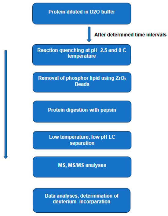
1.1.1. Hydrogen–Deuterium Exchange Mass Spectrometry(H/DX-MS)

In recent years, hydrogen–deuterium exchange combined with mass spectrometry (HDX-MS) [23] has emerged as a key player in studying the conformational dynamics and interaction of proteins in solution [16]. In this technique, a protein is diluted in a deuterated buffer, enabling H/D exchange of labile backbone amides. This process of isotopic exchange is strongly dependent on the protein secondary structure and solvent accessibility. In general, the exchange reaction can be quenched by dropping the pH and the temperature of the solution to 2.5 and 0 °C, respectively, conditions necessary to reduce the D-to-H back exchange. The quenched protein sample is then digested by an acid protease (e.g., pepsin), and the resulting peptides are separated by LC at low temperature and low pH, followed by MS and MS/MS analyses to determine the extent of deuterium incorporation (Figure 1).
Figure 1. The main steps in hydrogen–deuterium exchange combined with mass spectrometry HDX-MS to study protein conformations in solution. In this technique, a protein is diluted in a deuterated buffer, enabling H/D exchange of labile backbone amides. The process of isotopic exchange is strongly dependent on the protein secondary structure and solvent accessibility. The quenched protein sample is then digested by pepsin and the resulting peptides are separated by LC at low temperature and low pH. The extent of the deuterium incorporation is determined by MS and MS/MS analyses.
The lipid environment of membrane proteins is one of the main problems in HDX-MS analyses. This is because lipids hamper both the digestion process and the subsequent LC separation of the resulting peptides. Present day analyses of these proteins rely on the removal of the phospholipids prior to digestion, and this removal is normally carried out using ZrO2 coated beads [24], size exclusion chromatography [25], or trichloroacetic acid (TCA) precipitation [26].
Analyses of proteins containing certain post-translational modifications (PTMs), such as glycosylation and disulfide bonds can be challenging for HDX-MS. The presence of disulfide bonds can be responsible for poor digestion, which leads to poor sequence coverage. Furthermore, the MS/MS spectra generated in the presence of disulfide-linked peptides are not easy to interpret [27][28]. Although at room temperature and neutral pH disulfide bonds can be reduced by a variety of reagents, the low temperature and acidic pH used in HDX quenching narrows down the choice to a single reducing reagent, tris(2-carboxyethyl) phosphine (TCEP) [29]. Still, TCEP has its shortcomings related to the alkaline pH required for optimal disulfide reduction by this reagent. Various works have attempted to find alternative solutions to TCEP reduction. 
Glycosylation is another PTM, which requires more demanding HDX-MS and MS/MS analyses. This is because a single glycosylation site can be occupied by multiple, heterogeneous glycated structures, which inevitably results in different glycoforms of the same proteoform. This effect causes glycopeptide signal distribution and inevitable signal reduction in the individual structures [30]. Collision-induced dissociation of glycopeptides is also problematic because of the preferential cleavage of glycosidic bonds in carbohydrate moieties [31][32]. The same authors pointed out that the use of ETD instead of CID did not improve the signal [33].
In recent years there has been an increasing use of HDX-MS in the investigation of integral membrane proteins [34]. As mentioned earlier, this class of proteins is known to perform a range of diverse functions, and their dysfunctions are frequently linked to numerous diseases, which explains the huge interest of the pharmaceutical industry in these proteins as promising therapeutic targets. Over the last decade, HDX-MS has developed into a versatile tool for probing the protein dynamics of integral membrane proteins in solution. Earlier investigations included detergents’ extraction of the investigated proteins from their native environment, a step which is likely to interfere with the native structure. One of the approaches to simulate the native environment is the development of membrane mimetics [35].

1.1.2. Ion Mobility–Mass Spectrometry

Over the last 20 years the use of ion mobility in combination with mass spectrometry (IM-MS) to investigate macromolecules has furnished valuable information and deeper insight into the field of structural biology. This technique is considered an additional dimension to analyses performed by the LC-MS and MS/MS platform. The combination of ESI-MS/MS and IM-MS is a powerful tool for the resolution and identification of complex protein digests. Furthermore, such analytical platforms can provide much needed information on the confirmational states of a wide range of proteins. Before discussing the role of IM-MS in macromolecular analyses, there are two observations to consider. First, IM separates gas-phase macromolecular ions according to their mass, charge, size, and shape. While mass and charge can be determined by mass spectrometry (MS), it is the addition of ion mobility that enables the separation of isomeric and isobaric ions and the direct elucidation of conformation. Second, this technique has been around for over 50 years [36], and in the early years of its introduction it was mainly used to study gas-phase ion-molecule reactions to determine their rate constants [37]. The combination of this technique with commercially available mass spectrometers to investigate biomolecules appeared over a decade ago [38].
Under the general title “ion mobility”, there are different hardware configurations, including drift tube ion mobility, cyclic ion mobility, trapped ion mobility spectrometry, and differential ion mobility and field asymmetric waveform ion mobility spectrometry. All these ion mobility variants operate on the same basic principle, i.e., the separation of ions in a buffer gas (normally helium or nitrogen) under the influence of a relatively weak electric field. Under such conditions, the drift time of an ion is dependent on its mass, charge, size, and shape. Large, more structured ions experience more collisions with the buffer gas and, therefore, have longer drift times. The drift time of a given ion can be converted into a parameter known as the collision cross-section (CCS) [39], a parameter, which reflects the extent of interaction (collisions) between the drifting ion and the buffer gas. Experimentally determined CCS values can also be compared with those generated by computational modelling methods, such as structure prediction from sequence. 
Recent research demonstrated that IM-MS is capable of providing structural information, which for various reasons could not be provided by high-resolution techniques, such as X-ray crystallography and cryo-electron microscopy (cryo-EM). One of the interesting characteristics of IM-MS is that the transition of protein/peptide ions from the solution to gas phase does not seem to influence the secondary structure, compactness, or the quaternary structure of the investigated proteins [40][41].

1.1.3. More Efficient Activation Methods of Macromolecules

For over 40 years, tandem mass spectrometry (MS/MS) has been performed by accelerating a mass selected precursor ion into a collision cell containing a neutral gas (Ar, N2) maintained at pressure approximately 10−4 to 10−2 torr and a collision energy of 10–100 eV. Such collision conditions are commonly used in triple quadrupoles [42], ion traps, and hybrid time-of-flight (Q-TOF). Over the last twenty years, softer and more efficient fragmentation methods have been introduced as alternatives or as complementary techniques to collision-induced dissociation (CID). These methods include electron transfer dissociation (ETD), electron capture dissociation (ECD), and photodissociation.

1.1.4. Electron Capture Dissociation (ECD)

Over the last twenty years, electron capture (ECD) and electron transfer dissociation (ETD) have emerged as two of the most useful methods for peptide/protein fragmentation. The diffused use of both fragmentation methods coincided with unprecedented advances in mass spectrometry and liquid chromatography. In the ECD method, low-energy (less than 1 eV) electrons are captured by multiply charged protein/peptide positive ions generated by ESI [43]. This exothermic reaction induces dissociation through a H atom transfer to the backbone carbonyl group, resulting in a relatively soft cleavage of the Cα–N bond in a peptide backbone. Such low-energy interaction allows for much better detection and localization of labile PTMs as well as more sequencing information compared to classical CID. The predominant bonds cleaved in such a reaction are the backbone N–Cα, yielding c and z ions, with the exception of N-terminal proline cleavage, due to the proline’s cyclic nature [44][45]
Since its introduction, the application of ECD has been limited to Fourier transform ion cyclotron resonance instruments (FT-ICR) [46]. The main reason for such a limitation is that efficient ECD requires a dense population of thermal electrons to interact with the precursor ions for a given time window. Creating such conditions in instruments, which use radio frequency to trap the precursor ions (e.g., ion traps and hybrid Q-TOF), proved to be technically challenging.

1.1.5. Electron Transfer Dissociation (ETD)

Electron transfer dissociation (ETD) [47] is the direct result of interaction between multiply charged positive ions generated by electrospray ionization and a negatively charged radical reagent. The chemical properties of the radical reagent and the charge density of the precursor ion are the two parameters which influence the reaction. The two reaction pathways are deprotonation of the multiply charged ion and electron transfer from the radical reagent to the multiply charged ion (peptide or protein). The reactive radical anions are generated from polycyclic aromatic hydrocarbon molecules, such as anthracene or fluoranthene. The latter is considered as one of the more favorable ETD reagents. Electron transfer from the negatively charged reagent ions to multiply charged positive precursor ions induces dissociation of Cα–N bonds in a similar fashion to that observed in ECD. Both ETD and ECD are low-energy reactions, which explains their capability to detect and localize labile post-translational modifications, which are difficult to detect in collision-induced dissociation. That said, obtaining rich sequencing information by electron-based methods is closely related to the charge density of the precursor ion. Such influence manifests itself clearly in low charge density ions, where backbone cleavage takes place, but the resulting fragment ions are held together by non-covalent interactions present in the more compact structures, a process known as non-dissociative electron transfer [48]. One way to overcome such shortcoming is to chemically modify the investigated peptides [49][50] or to conduct combined ETD and CID, where the first method is used for high charge states, while the second is used for lower mass lower charge states [51][52].

1.1.6. Photodissociation Methods

Currently there are two main photon-based methods for the activation and subsequent fragmentation of macromolecular ions. The first method uses infrared multiphoton dissociation (IRMPD), which was first demonstrated almost 50 years ago [53]. The authors used the wavelength 10.6 μm from a tunable continuous wave CO2 laser to irradiate a proton-bound dimer of di-Et ether [(C2H5)2O]2H+. The measurements were conducted in Fourier transform ion cyclotron resonance (FTICR). For many years the use of IRMPD remained limited to fundamental studies of small molecules; however, more recently, it is increasingly being applied to the analyses of macromolecules. This increase in the use of IRMPD can be attributed to advances in the characteristics of IR lasers, particularly the IR optics. The extension of this photodissociation method to other mass spectrometers, including linear and quadrupole ion traps, orbitraps, and hybrid TOFs is another reason for the increasing use of this method in peptide/protein fragmentation This activation method combined with tandem mass spectrometry (MS/MS) can provide fairly precise information on the type and sites of various PTMs of individual amino acids as well as on amino acids within peptides and proteins. Some of the modifications examined by this method include phosphorylation, sulfation, acetylation, methylation, glycosylation, and disulfide bond formation [54][55]. The photodissociation of the precursor molecular ions containing one or more of these PTMs is induced by the presence of one or more reactant group(s) (e.g., -OH, -C-H, -S-H, -S-CH3) responsible for the absorption of the irradiating photons.
The second method uses ultraviolet photodissociation (UVPD) at different wavelengths. For instance, 193 nm was first applied in the 1980s [56][57]; however, such an application was limited to specific peptides performed on Fourier transform ion cyclotron resonance (FTICR) mass spectrometers. Over the last ten years and due to advances in UV lasers and in MS instrumentation, the technique has witnessed a resurgence, represented in its use in a wide range of instruments, including TOF [58], linear ion traps [59], and more recently in the orbitrap [60]. UVPD mainly uses two wave lengths, 157 and 193 nm; however, the wavelength 213 nm has been used to sequence intact proteoforms in top-down proteomics [61]. In UVPD, due to the absorption of a single photon having 5–7 eV energy by the precursor ion, fragmentation proceeds either through electronic dissociative excitation or through the interconversion of the electronic excitation to vibrational excitation, thus leading to bond rapture, which is the major fragmentation pathway [62][63]

2. Analysis of Some ATP-Binding Cassette (ABC) Transporters

The introduction of two soft ionization techniques, namely electrospray ionization (ESI) [64] and matrix-assisted laser desorption/ionization (MALDI) [65], was a major step towards a diffused use of mass spectrometry (MS) in biological and biochemical worlds. Over the last two decades, advances in MS instrumentation together with more refined labelling protocols, the combination of ion mobility with MS, and more frequent use of electron-based and photon-based fragmentation methods paved the way for what is known today as structural mass spectrometry. With these innovative developments, mass spectrometry can be applied for the structural characterization of a wide range of macromolecules and in some cases their respective assemblies. It is relevant to point out that continuous progress in sample preparations, including the use of a new class of detergents, extended the application of MS to membrane proteins, which are not water soluble.

2.1. Monitoring the Conformation of P-glycoprotein

P-glycoprotein (also known as ABCB1 and MDR1) is the first identified mammalian ABC transporter protein, discovered almost half a century ago [66]. It has a ~170 kDa molecular weight and is comprised of two homologous halves, each containing a nucleotide-binding domain (NBD) and a transmembrane domain (TMD) [67]. The transmembrane domain contains a hydrophobic cavity, accessed by portals in the membrane, that binds cytotoxic compounds as well as lipids. This protein is expressed in multiple key organs, such as the small intestine, blood–brain barrier, kidney, and liver. Therefore, P-glycoprotein mediated drug–drug interactions can occur in various organs and in tissues. The overexpression of P-glycoprotein has been linked to chemotherapy failure in various cancers, including kidney, colon, and liver. A hallmark characteristic of this transporter protein is its ability to bind and transport a wide range of structurally different molecules in the molecular mass range, 100 to 4000 Da, a range which covers most if not all anticancer and antimicrobial drugs currently in use [68]. The transportation of these molecules across the membrane was found to coincide with changes in the size and shape of a large multi-specific drug-binding pocket [67]. Although its physiological function(s) are yet to be fully understood, the well-recognized role of this protein in mediating multidrug resistance in many types of cancers has made it an attractive therapeutic target [69].
In recent years it has been demonstrated that hydrogen–deuterium exchange mass spectrometry (HDX-MS) is ideal for studying protein dynamics as they occur in solution. Proteins are known to assume a wide range of conformations in solution, which means that every amide proton will eventually exchange with deuterium. Measuring this rate of exchange as a function of time would result in a highly informative picture of the regional dynamics. Information provided by this technique are fundamental for the understanding of the functioning mechanism of ABC exporters, including P-gp, and complement the snapshots of the catalytic cycle provided by high-resolution 3D structures.
In a recent article, HDX-MS was used to investigate the conformational states as well as the dynamics and mechanism of transportation of P-glycoprotein. The authors detected three distinct conformational states and obtained information on transporter dynamics with a sequence coverage over 85% [70]. This high sequence coverage and the use of bio-layer interferometry to measure nucleotide affinity furnished further information on the states of conformation, dynamics, and mechanism of transportation by this protein.
Since its discovery almost 50 years ago, P-glycoprotein has been structurally characterized by X-ray, cryo-electron spectroscopy, and, more recently, by HDX-MS. This structural information, together with data furnished by analyses of clinical samples provided by patients undergoing chemotherapy and molecularly targeted therapy, gives a clearer picture of the role of this protein in MDR. The initial enthusiasm that such new information could lead to the discovery of P-glycoprotein inhibitors was quickly dampened. 

2.2. Breast Cancer Resistance Protein (ABCG2)

The human ATP-binding cassette transporter ABCG2, also known as breast cancer resistance protein (BCRP), is a key player in anticancer resistance and in physiological detoxification across tissue barriers [71]. Despite numerous investigations, the molecular mechanism of substrate transport by this protein remains to be fully clarified. The activities of this protein are known to affect the pharmacokinetics of commonly used drugs as well as interfering in the delivery of various therapeutics into tumor cells, thus contributing to multidrug resistance [72]. Unlike most of the other ABC transporters, which usually have two nucleotide-binding domains and two transmembrane domains, ABCG2 consists of only one nucleotide-binding domain followed by one transmembrane domain. Thus, ABCG2 has been thought to be a half-transporter that may function as a homodimer (molecular weight approximately 144 kDa).
The first high-resolution structure of human ABCG2 determined by cryo-electron microscopy was reported over five years ago [73]. The structure shows two cholesterol molecules bound in the multidrug-binding pocket that is located in a central, hydrophobic, inward-facing translocation pathway between the transmembrane domains. Within the same year, another research group used the X-ray crystal structure of ABCG5/G8 to generate a model of ABCG2 [73]. To validate structural and mechanistic predictions of their model, the authors used extensive molecular–genetic analyses. The ABCG2 structure contains two apparent cavities. The architecture of the central cavity includes the intracellular loop1, the elbow helix, and residues facing the cavity from transmembrane helices and the NBD dimer. The central cavity is part of the transmission interface, which is essential for ABCG2 drug transport. The smaller upper cavity is part of what is called the extracellular polar roof. These two cavities were found to be separated by two leucine residues, facing their equivalent residues in the core of the symmetric ABCG2 dimer [72][73][74][75].
The link between ABCG2 and resistance to anticancer drugs has been discussed in a number of studies conducted on cancer cell lines. In one of these studies from over 25 years ago, overexpression of ABCG2 in model cancer cell lines (MCF-7 breast cancer cells) was shown to cause resistance to a number of anticancer drugs, including doxorubicin and daunorubicin [76].
Over the last ten years, a new class of targeted anticancer drugs has emerged. One of these drugs, sonidegib (trade name odomzo), was approved by the FDA in 2015 for the treatment of adults with advanced basal cell carcinoma. This drug was tested as an inhibitor of the transport functions of both ABCB1 and ABCG2 [77]. The authors reported that in accumulation studies, the transport functions of both proteins were effectively inhibited by sonidegib.

3. Commenting on Inhibitors of P-glycoprotein

Four generations of potential inhibitors of P-Glycoprotein have been developed and tested over a period of 40 years [78]. During this period, many potential inhibitors were extensively investigated, but none of them obtained the approval of either the FDA (Food and Drug Administration) or the EMA (European Medicines Agency). Most experts in the field argue that the failure of these inhibitors to restore sensitivity to chemotherapy reside in their poor selectivity, low potency, inherent toxicity, and/or adverse pharmacokinetic interaction with anticancer drugs. However, it has to be said that despite the poor results of years of research, a number of studies during the same period indicated that under specific conditions the combination of potential inhibitors of MDR1 and ABCG2 with certain chemotherapeutics resulted in increased drug accumulation and drug resistance reversal.
- In vitro analyses: Cell lines are often used in research in place of primary cells. These in vitro analyses offer the advantages of low costs, ease of use, providing an unlimited supply of materials, and bypassing ethical concerns associated with the use of animal and human samples [79]. That said, the use of cell lines in cancer research has some drawbacks. A major drawback of in vitro models is their failure to capture the full reality of in vivo systems. For example, such models may not account for likely interactions between cells and other biological and biochemical processes. Cancer cell lines are often derived from a single subtype of tumor, which very often does not reflect the heterogeneity of tumors in patients. 
- The multifactorial nature of drug resistance is another factor to be considered as being partly responsible for the poor results in the search for specific inhibitors of P-gp. There is strong evidence that high expression of this protein remains one of the main reasons for the poor response to chemotherapy in many cancer types. That said, limiting the search for inhibitors to a single drug resistance mechanism is unlikely to restore chemotherapeutic sensitivity. As mentioned earlier, this protein is part of a superfamily containing at least 48 members. The existing literature shows that only three members have been studied in some detail.
- Ligand–protein interaction. Over the last 40 years, chemical, biochemical, and biological studies have provided a wealth of information on the mechanisms of interaction between a limited number of ABC transporter proteins and various ligands. The complexity of such interaction renders such information insufficient to identify the precise mechanism(s) of such interactions. Identification of the basic physical and chemical characteristics, which mediate ligand–protein interaction are highly relevant for inhibitor(s) selection.
- Level of expression. Accurate detection and quantification of ATP-binding cassette (ABC) transporters are highly relevant to the assessment of their role in chemoresistance. Such a parameter becomes more critical when expression is measured in vivo. Most studies conducted on cell lines indicate that the level of expression of one or more of these proteins varies from one tumor to another. The same literature shows that the detection and quantification of these levels have been mainly performed by reverse transcription–polymerase chain reaction (RT-PCR) for mRNA expression and by immunohistochemistry (IHC) for protein expression.
- The good and the bad. ATP-binding cassette (ABC) transporters are expressed in both healthy and cancerous cells. Within the healthy cells, some of these proteins play a crucial physiological role in protecting tissues from toxic xenobiotics and endogenous metabolites, and also affect the uptake and distribution of many clinically important drugs. The same proteins form a major component of the blood–brain barrier and restrict the uptake of drugs from the intestine. In cancerous cells, on the other hand, the overexpression of these proteins has been strongly linked to MDR. Although MDR is multifactorial in origin, it is strongly associated with the overexpression of ATP-binding cassette (ABC) transporters. 

4. Conclusions

Mass spectrometry is becoming an increasingly important tool in the field of structural biology. In recent years, the role of this technique has been enhanced by more frequent use of ion mobility-MS, HDX-MS, and electron/photon-based ion activation methods. These techniques, together with X-ray crystallography and cryo-electron microscopy, are furnishing much-needed information on the structure of membrane proteins, including ABC transporters. Information provided by these techniques together with those acquired through advanced molecular dynamics simulation, molecular docking studies, and protein–ligand investigations will no doubt contribute to the ongoing scientific research to fill the gaps, which for over 40 years have hindered the identification of specific and effective clinical inhibitors of ABC transporters.

References

  1. Almén, M.S.; Nordström, K.J.; Fredriksson, R.; Schiöth, H.B. Mapping the human membrane proteome: A majority of the human membrane proteins can be classified according to function and evolutionary origin. BMC Biol. 2009, 7, 50.
  2. Wallin, E.; von Heijne, G. Genome-wide analysis of integral membrane proteins from eubacterial, archaean, and eukaryotic organisms. Protein Sci. 1998, 7, 1029–1038.
  3. Arinaminpathy, Y.; Khurana, E.; Engelman, D.M.; Gerstein, M.B. Computational analysis of membrane proteins: The largest class of drug targets. Drug Discov. Today 2009, 14, 1130–1135.
  4. Overington, J.P.; Al-Lazikani, B.; Hopkins, A.L. Opinion—How many drug targets are there? Nat. Rev. Drug Discov. 2006, 5, 993–996.
  5. Billings, E.; Lundquist, K.; Overly, C.; Srinivasan, K.; Noinaj, N. Structure Determination of Membrane Proteins Using X-ray Crystallography. Methods Mol. Biol. 2021, 2302, 101–136.
  6. Hernando-Amado, S.; Blanco, P.; Alcalde-Rico, M.; Corona, F.; Reales-Calderón, J.A.; Sánchez, M.B.; Martínez, J.L. Multidrug efflux pumps as main players in intrinsic and acquired resistance to antimicrobials. Drug Resist. Update 2016, 28, 13–27.
  7. Sun, J.; Deng, Z.; Yan, A. Bacterial multidrug efflux pumps: Mechanisms, physiology and pharmacological exploitations. Biochem. Biophys. Res. Commun. 2014, 453, 254–267.
  8. Levin, B.R.; Rozen, D.E. Non-inherited antibiotic resistance. Nat. Rev. Microbiol. 2006, 4, 556–562.
  9. Corona, F.; Martinez, J.L. Phenotypic Resistance to Antibiotics. Antibiotics 2013, 2, 237–255.
  10. Gottesman, M.M.; Fojo, T.; Bates, S.E. Multidrug resistance in cancer: Role of ATP–dependent transporters. Nat. Rev. Cancer 2002, 2, 48–58.
  11. Ambudkar, S.V.; Dey, S.; Hrycyna, C.A.; Ramachandra, M.; Pastan, I.; Gottesman, M.M. Biochemical, cellular, and pharmacological aspects of the multidrug transporter. Annu. Rev. Pharmacol. Toxicol. 1999, 39, 361–398.
  12. Choi, C.-H. ABC transporters as multidrug resistance mechanisms and the development of chemosensitizers for their reversal. Cancer Cell Int. 2005, 5, 30.
  13. Thomas, H.; Coley, H.M. Overcoming multidrug resistance in cancer: An update on the clinical strategy of inhibiting p-glycoprotein. Cancer Control 2003, 10, 159–165.
  14. Kühlbrandt, W. The Resolution Revolution. Science 2014, 343, 1443–1444.
  15. Thompson, R.F.; Walker, M.; Siebert, C.A.; Muench, S.P.; Ranson, N.A. An introduction to sample preparation and imaging by cryo-electron microscopy for structural biology. Methods 2016, 100, 3–15.
  16. Konermann, L.; Pan, J.; Liu, Y.H. Hydrogen exchange mass spectrometry for studying protein structure and dynamics. Chem. Soc. Rev. 2011, 40, 1224–1234.
  17. Calabrese, A.N.; Radford, S.E. Mass spectrometry-enabled structural biology of membrane proteins. Methods 2018, 147, 187–205.
  18. Laganowsky, A.; Reading, E.; Hopper, J.T.S.; Robinson, C.V. Mass spectrometry of intact membrane protein complexes. Nat. Protoc. 2013, 8, 639–651.
  19. Lanucara, F.; Holman, S.W.; Gray, C.J.; Eyers, C.E. The power of ion mobility-mass spectrometry for structural characterization and the study of conformational dynamics. Nat. Chem. 2014, 6, 281–294.
  20. Konijnenberg, A.; Butterer, A.; Sobott, F. Native ion mobility-mass spectrometry and related methods in structural biology. BBA—Proteins Proteom. 2013, 1834, 1239–1256.
  21. Zhong, Y.; Hyung, S.-J.; Ruotolo, B.T. Ion mobility–mass spectrometry for structural proteomics. Expert Rev. Proteom. 2012, 9, 47–58.
  22. Engen, J.R.; Wales, T.E. Analytical Aspects of Hydrogen Exchange Mass Spectrometry. Annu. Rev. Anal. Chem. 2015, 8, 127–148.
  23. Katta, W.; Chait, B.T. Hydrogen/Deuterium Exchange Electrospray Ionization Mass Spectrometry: A Method for Probing Protein Conformational Changes in Solution. J. Am. Chem. Soc. 1993, 115, 6317–6321.
  24. Hammerschmid, D.; Calvaresi, V.; Bailey, C.; Russell, B.; Argyris Politis, L.; Morris, M.; Denbigh, L.; Anderson, M.; Reading, E. Chromatographic Phospholipid Trapping for Automated H/D Exchange Mass Spectrometry of Membrane Protein–Lipid assemblies. Anal. Chem. 2023, 95, 3002–3011.
  25. Calvaresi, V.; Redsted, A.; Norais, N.; Rand, K.D. Hydrogen–Deuterium exchange mass spectrometry with integrated size-exclusion chromatography for analysis of complex protein samples. Anal. Chem. 2021, 93, 11406–11414.
  26. Donnarumma, D.; Maestri, C.; Giammarinaro, P.I.; Capriotti, L.; Bartolini, E.; Veggi, D.; Petracca, R.; Scarselli, M.; Norais, N. Native state organization of outer membrane porins unraveled by HDx-MS. J. Proteome Res. 2018, 17, 1794–1800.
  27. Mysling, S.; Salbo, R.; Ploug, M.; Jørgensen, T.J.D. Electrochemical reduction of disulfide-containing proteins for hydrogen/deuterium exchange monitored by mass spectrometry. Anal. Chem. 2014, 86, 340–345.
  28. Bobst, C.E.; Kaltashov, I.A. Enhancing the Quality of H/D Exchange measurements with mass spectrometry detection in disulfide-rich proteins using electron capture dissociation. Anal. Chem. 2014, 86, 5225–5231.
  29. Burns, J.A.; Butler, J.C.; Moran, J.; Whitesides, G.M. Selective reduction of disulfides by tris(2-carboxyethyl)phosphine. J. Org. Chem. 1991, 56, 2648–2650.
  30. Larsen, M.R.; Højrup, P.; Roepstorff, P. Characterization of Gel-separated Glycoproteins Using Two-step Proteolytic Digestion Combined with Sequential Microcolumns and Mass Spectrometry. Mol. Cell. Proteom. 2005, 4, 107–119.
  31. Darula, Z.; Medzihradszky, K.F. Glycan side reaction may compromise ETD-based glycopeptide identification. J. Am. Soc. Mass Spectrom. 2014, 25, 977–987.
  32. Houel, S.; Hilliard, M.; Yu, Y.Q.; McLoughlin, N.; Martin, S.M.; Rudd, P.M.; Williams, J.P.; Chen, W. N- and O-glycosylation analysis of etanercept using liquid chromatography and quadrupole time-of-flight mass spectrometry equipped with electron-transfer dissociation functionality. Anal. Chem. 2014, 86, 576–584.
  33. Rand, K.D. Pinpointing changes in higher-order protein structure by hydrogen/deuterium exchange coupled to electron transfer dissociation mass spectrometry. Int. J. Mass Spectrom. 2013, 338, 2–10.
  34. Javed, W.; Griffiths, D.; Politis, A. Hydrogen/deuterium exchange-mass spectrometry of integral membrane proteins in native-like environments: Current scenario and the way forward. Essays Biochem. 2023, 67, 187–200.
  35. Majeed, S.; Ahmad, A.B.; Sehar, U.; Georgieva, E.R. Lipid Membrane Mimetics in Functional and Structural Studies of Integral Membrane Proteins. Membranes 2021, 11, 685.
  36. Karasek, F.W. Drift-mass spectrometer. Res. Dev. 1970, 21, 25–27.
  37. Alge, E.; Villinger, H.; Lindinger, W. Drift tube investigations on the reactions of O2 + with CH4 and of CO2 + with NO in various buffer gases. Plasma Chem. Plasma Process. 1981, 1, 65–71.
  38. Smith, D.P.; Giles, K.; Bateman, R.H.; Radford, S.E.; Ashcroft, A.E. Monitoring Copopulated Conformational States during Protein Folding Events Using Electrospray Ionization-Ion Mobility Spectrometry-Mass Spectrometry. J. Am. Soc. Mass Spectrom. 2007, 18, 2180–2190.
  39. May, J.C.; Morris, C.B.; McLean, J.A. Ion mobility collision cross section compendium. Anal. Chem. 2017, 89, 1032–1044.
  40. Wyttenbach, T.; Bowers, M.T. Structural stability from solution to the gas phase: Native solution structure of ubiquitin survives analysis in a solvent-free ion mobility–mass spectrometry environment. J. Phys. Chem. B 2011, 115, 12266–12275.
  41. Ruotolo, B.T.; Robinson, C.V. Aspects of native proteins are retained in vacuum. Curr. Opin. Chem. Biol. 2006, 10, 402–408.
  42. Yost, R.A.; Enke, C.G. Selected ion fragmentation with a tandem quadrupole mass spectrometer. J. Am. Chem. Soc. 1978, 100, 2274–2275.
  43. Zubarev, R.A.; Horn, D.M.; Fridriksson, E.K.; Kelleher, N.L.; Kruger, N.A.; Lewis, M.A.; Carpenter, B.K.; McLafferty, F.W. Electron capture dissociation for structural characterization of multiply charged protein cations. Anal. Chem. 2000, 72, 563–573.
  44. Syrstad, E.A.; Turecček, F. Toward a general mechanism of electron capture dissociation. J. Am. Soc. Mass Spectrom. 2005, 16, 208–224.
  45. Zubarev, R.A.; Kelleher, N.L.; McLafferty, F.W. Electron capture dissociation of multiply charged protein cations. A nonergodic process. J. Am. Chem. Soc. 1998, 120, 3265–3266.
  46. Qi, Y.; Volmer, D.A. Electron-based fragmentation methods in mass spectrometry: An overview. Mass Spectrom. Rev. 2017, 36, 4–15.
  47. Syka, J.E.P.; Coon, J.J.; Schroeder, M.J.; Shabanowitz, J.; Hunt, D.F. Peptide and Protein sequence analysis by electron transfer dissociation mass spectrometry. Proc. Natl. Acad. Sci. USA 2004, 101, 9528–9533.
  48. Pitteri, S.J.; Chrisman, P.A.; McLuckey, S.A. Electron-Transfer Ion/Ion Reactions of Doubly Protonated Peptides: Effect of Elevated Bath Gas Temperature. Anal. Chem. 2005, 77, 5662–5669.
  49. Ko, B.J.; Brodbelt, J.S. Enhanced electron transfer dissociation of peptides modified at C-terminus with fixed charges. J. Am. Soc. Mass Spectrum. 2012, 23, 1991–2000.
  50. Frey, B.L.; Ladror, D.T.; Sondalle, S.B.; Krusemark, C.J.; Jue, A.L.; Coon, J.J.; Smith, L.M. Chemical derivatization of peptide carboxyl groups for highly efficient electron transfer dissociation. J. Am. Soc. Mass Spectrom. 2013, 24, 1710–1721.
  51. Swaney, D.L.; McAlister, G.C.; Coon, J.J. Decision tree–driven tandem mass spectrometry for shotgun proteomics. Nat. Methods 2008, 5, 959–964.
  52. Frese, C.K.; Altelaar, A.F.M.; Hennrich, M.L.; Nolting, D.; Zeller, M.; Griep-Raming, J.; Heck, A.J.R.; Mohammed, S. Improved peptide identification by targeted fragmentation using CID, HCD and ETD on an LTQ-orbitrap velos. J. Proteome Res. 2011, 10, 2377–2388.
  53. Woodin, R.L.; Bomse, D.S.; Beauchamp, J.L. Multiphoton dissociation of molecules with low power continuous wave infrared laser radiation. J. Am. Chem. Soc. 1978, 100, 3248–3250.
  54. Maitre, P.; Scuderi, D.; Corinti, D.; Chiavarino, B.; Crestoni, M.E.; Fornarini, S. Applications of Infrared Multiple Photon Dissociation (IRMPD) to the Detection of Posttranslational Modifications. Chem. Rev. 2020, 120, 3261–3295.
  55. Greisch, J.-F.; van der Laarse, S.A.; Heck, A.J. Enhancing Top-Down Analysis Using Chromophore-Assisted Infrared Multiphoton Dissociation from (Phospho)peptides to Protein Assemblies. Anal. Chem. 2020, 92, 15506–15516.
  56. Bowers, W.D.; Delbert, S.S.; Hunter, R.L.; Mciver, R.T. Fragmentation of oligopeptide ions using ultraviolet laser radiation and fourier transform mass spectrometry. J. Am. Chem. Soc. 1984, 106, 7288–7289.
  57. Hunt, D.F.; Shabanowitz, J.; Yates, J.R. Peptide sequence analysis by laser photodissociation Fourier transform mass spectrometry. J. Chem. Soc. Chem. Commun. 1987, 548–550.
  58. Thompson, M.S.; Cui, W.; Reilly, J.P. Fragmentation of Singly Charged Peptide Ions by Photodissociation at λ = 157 nm. Angew. Chem. Int. Ed. 2004, 43, 4791–4794.
  59. Cannon, J.R.; Cammarata, M.B.; Robotham, S.A.; Cotham, V.C.; Shaw, J.B.; Fellers, R.T.; Early, B.P.; Thomas, P.M.; Kelleher, N.L.; Brodbelt, J.S. Ultraviolet photodissociation for characterization of whole proteins on a chromatographic time scale. Anal. Chem. 2014, 86, 2185–2192.
  60. Smyrnakis, A.; Levin, N.; Kosmopoulou, M.; Jha, A.; Fort, K.; Makarov, A.; Papanastasiou, D.; Mohammed, S. Characterization of an Omnitrap-Orbitrap Platform Equipped with Infrared Multiphoton Dissociation, Ultraviolet Photodissociation, and Electron Capture Dissociation for the Analysis of Peptides and Proteins. Anal. Chem. 2023, 95, 12039–12046.
  61. Fornelli, L.; Srzentić, K.; Toby, T.K.; Doubleday, P.F.; Huguet, R.; Mullen, C.; Melani, R.D.; dos Santos Seckler, H.; DeHart, C.J.; Weisbrod, C.R.; et al. Thorough Performance Evaluation of 213 nm Ultraviolet Photodissociation for Top-down Proteomics. Mol. Cell. Proteom. 2020, 19, 405–420.
  62. Julian, R.R.; Amster, I.J.; Kong, X.; Brodbelt, J.S.; Jørgensen, T.J.D.; Wysocki, V.H.; Hendrickson, C.L.; Santos, I.; Shaw, J.B.; Boyarkin, O.V.; et al. The mechanism behind top-down uvpd experiments: Making sense of apparent contradictions. J. Am. Soc. Mass Spectrom. 2017, 28, 1823–1826.
  63. Zabuga, A.V.; Kamrath, M.Z.; Boyarkin, O.V.; Rizzo, T.R. Fragmentation mechanism of UV-excited peptides in the gas phase. J. Chem. Phys. 2014, 141, 154309.
  64. Yamashita, M.; Fenn, J.B. Electrospray ion source. Another variation on the free-jet theme. J. Phys. Chem. 1984, 88, 4451–4459.
  65. Karas, M.; Bachmann, D.; Hillenkamp, F. Influence of the Wavelength in High-Irradiance Ultraviolet Laser Desorption Mass Spectrometry of Organic Molecules. Anal. Chem. 1985, 57, 2935–2939.
  66. Juliano, R.; Ling, V. A surface glycoprotein modulating drug permeability in Chinese hamster ovary cell mutants. BBA—Biomembr. 1976, 455, 152–162.
  67. Ward, A.B.; Szewczyk, P.; Grimard, V.; Lee, C.-W.; Martinez, L.; Doshi, R.; Caya, A.; Villaluz, M.; Pardon, E.; Cregger, C.; et al. Structures of P-glycoprotein reveal its conformational flexibility and an epitope on the nucleotide-binding domain. Proc. Natl. Acad. Sci. USA 2013, 110, 13386–13391.
  68. Schinkel, A.H.; Jonker, J.W. Mammalian drug efflux transporters of the ATP binding cassette (ABC) family: An overview. Adv. Drug Deliv. Rev. 2003, 55, 3–29.
  69. Marquez, B.; Van Bambeke, F. ABC multidrug transporters: Target for modulation of drug pharmacokinetics and drug-drug interactions. Curr. Drug Targets 2011, 12, 600–620.
  70. Kopcho, N.; Geoffrey Chang, G.; Komives, E.A. Dynamics of ABC transporter P-glycoprotein in three conformational states. Sci. Rep. 2019, 9, 15092.
  71. Van Herwaarden, A.E.; Schinkel, A.H. The function of breast cancer resistance protein in epithelial barriers, stem cells and milk secretion of drugs and xenotoxins. Trends Pharmacol. Sci. 2006, 27, 10–16.
  72. Taylor, N.M.I.; Manolaridis, I.; Jackson, S.M.; Kowal, J.; Stahlberg, H.; Locher, K.P. Structure of the human multidrug transporter ABCG2. Nature 2017, 546, 504–509.
  73. Khunweeraphong, N.; Stockner, T.; Kuchler, K. The structure of the human ABC transporter ABCG2 reveals a novel mechanism for drug extrusion. Sci. Rep. 2017, 7, 13767.
  74. Jackson, S.M.; Manolaridis, I.; Kowal, J.; Zechner, M.; Taylor, N.M.I.; Bause, M.; Bauer, S.; Bartholomaeus, R.; Bernhardt, G.; Koenig, B.; et al. Structural basis of small-molecule inhibition of human multidrug transporter ABCG2. Nat. Struct. Mol. Biol. 2018, 25, 333–340.
  75. Manolaridis, I.; Jackson, S.M.; Taylor, N.M.I.; Kowal, J.; Stahlberg, H.; Locher, K.P. Cryo-EM structures of a human ABCG2 mutant trapped in ATP-bound and substrate-bound states. Nature 2018, 563, 426–430.
  76. Doyle, L.A.; Yang, W.; Abruzzo, L.V.; Krogmann, T.; Gao, Y.; Rishi, A.K.; Ross, D.D. A multidrug resistance transporter from human MCF-7 breast cancer cells. Proc. Natl. Acad. Sci. 1998, 95, 15665–15670.
  77. Zhang, Y.; Vagiannis, D.; Budagaga, Y.; Sabet, Z.; Hanke, I.; Rozkoš, T.; Hofman, J. Sonidegib potentiates the cancer cells’ sensitivity to cytostatic agents by functional inhibition of ABCB1 and ABCG2 in vitro and ex vivo. Biochem. Pharmacol. 2022, 199, 115009.
  78. Modok, S.; Mellor, H.R.; Callaghan, R. Modulation of multidrug resistance efflux pump activity to overcome chemoresistance in cancer. Curr. Opin. Pharmacol. 2006, 6, 350–354.
  79. Crowley, E.; McDevitt, C.A.; Callaghan, R. Generating Inhibitors of P-Glycoprotein: Where to, now? Multi-Drug Resist. Cancer 2010, 596, 405–432.
More
Information
Subjects: Cell Biology
Contributors MDPI registered users' name will be linked to their SciProfiles pages. To register with us, please refer to https://encyclopedia.pub/register : , ,
View Times: 96
Revisions: 2 times (View History)
Update Date: 04 Feb 2024
1000/1000
Video Production Service