Submitted Successfully!
To reward your contribution, here is a gift for you: A free trial for our video production service.
Thank you for your contribution! You can also upload a video entry or images related to this topic.
Version Summary Created by Modification Content Size Created at Operation
1 -- 1641 2024-01-29 09:22:27 |
2 format correct Meta information modification 1641 2024-01-29 11:50:46 |

Video Upload Options

Do you have a full video?

Confirm

Are you sure to Delete?
Cite
If you have any further questions, please contact Encyclopedia Editorial Office.
Yazdani, E.; Geramifar, P.; Karamzade-Ziarati, N.; Sadeghi, M.; Amini, P.; Rahmim, A. Radiomics and Artificial Intelligence Workflow. Encyclopedia. Available online: https://encyclopedia.pub/entry/54467 (accessed on 06 July 2024).
Yazdani E, Geramifar P, Karamzade-Ziarati N, Sadeghi M, Amini P, Rahmim A. Radiomics and Artificial Intelligence Workflow. Encyclopedia. Available at: https://encyclopedia.pub/entry/54467. Accessed July 06, 2024.
Yazdani, Elmira, Parham Geramifar, Najme Karamzade-Ziarati, Mahdi Sadeghi, Payam Amini, Arman Rahmim. "Radiomics and Artificial Intelligence Workflow" Encyclopedia, https://encyclopedia.pub/entry/54467 (accessed July 06, 2024).
Yazdani, E., Geramifar, P., Karamzade-Ziarati, N., Sadeghi, M., Amini, P., & Rahmim, A. (2024, January 29). Radiomics and Artificial Intelligence Workflow. In Encyclopedia. https://encyclopedia.pub/entry/54467
Yazdani, Elmira, et al. "Radiomics and Artificial Intelligence Workflow." Encyclopedia. Web. 29 January, 2024.
Radiomics and Artificial Intelligence Workflow
Edit

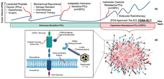
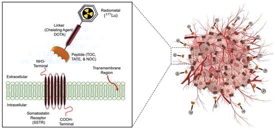
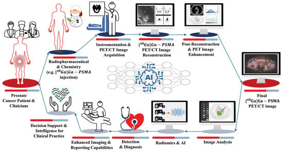
Radiotheranostics refers to the pairing of radioactive imaging biomarkers with radioactive therapeutic compounds that deliver ionizing radiation. Given the introduction of very promising radiopharmaceuticals, the radiotheranostics approach is creating a novel paradigm in personalized, targeted radionuclide therapies (TRTs), also known as radiopharmaceuticals (RPTs). Radiotherapeutic pairs targeting somatostatin receptors (SSTR) and prostate-specific membrane antigens (PSMA) are increasingly being used to diagnose and treat patients with metastatic neuroendocrine tumors (NETs) and prostate cancer. In parallel, radiomics and artificial intelligence (AI), as important areas in quantitative image analysis, are paving the way for significantly enhanced workflows in diagnostic and theranostic fields, from data and image processing to clinical decision support, improving patient selection, personalized treatment strategies, response prediction, and prognostication. Furthermore, AI has the potential for tremendous effectiveness in patient dosimetry which copes with complex and time-consuming tasks in the RPT workflow.

radiotheranostics radiomics artificial intelligence SSTR PSMA workflow

1. Introduction

Radiotheranostics represents a medical paradigm that uses radiopharmaceuticals for targeted radionuclide therapy (TRT), also known as radiopharmaceutical therapy (RPT). The approach involves the use of the same or different radiopharmaceuticals for both therapeutic and imaging purposes, enabling the matched targeting of specific disease sites [1][2][3]. The radiotheranostics paradigm enables the visualization of drug pharmacokinetics in the body, enabling personalized medicine frameworks [1]. Radiotheranostics makes it feasible to customize treatment planning based on individual variations by choosing “the right drug for the right patient at the right time” [4].
This study specifically concentrates on radiotheranostic ligand pairs that selectively bind to somatostatin receptors (SSTRs) and the prostate-specific membrane antigen (PSMA). In general, PSMA expression is higher in prostate cancer (PCa) cells than benign prostate cells, providing a comparatively specific target for patients with this tumor. Moreover, SSTRs are expressed much higher in neuroendocrine tumor (NET) cells or meningiomas than in normal tissues [5].
Prostate cancer is one of the three most common cancers in the world (7.1% of all cancers), with a high survival rate (a 5-year survival rate of >95%) and high recurrence rates [6][7][8][9]. Figure 1A illustrates a simplified disease course for PCa patients. In the first stage, patients are diagnosed through an abnormal serum prostate-specific antigen (PSA) level, a PCa tumor marker. Most of them will be treated (for example, with radiation therapy or surgery), and their PSA will nearly reach zero. However, some of them will have biochemical recurrences. A significant number of PCa patients will progress to metastatic castrate-resistant prostate cancer (mCRPC). Therefore, there is a growing need for alternative therapeutic strategies for these patients. In this regard, several molecules were tested to target PSMA expressed on the cell surface of mCRPC patients [10].
Figure 1. The principle of radiotheranostics in mCRPC patients. (A). The typical timeline of different therapies, including RPT (also known as RLT). (B). PSMA-binding domain, linker, and chelator labeled with Lu-177 deliver ionizing radiation to the tumor.
PSMA is a type II, 750-amino acid transmembrane protein anchored in the cell membrane of prostate epithelial cells [11]. Radiopharmaceuticals targeting PSMA for diagnostic imaging purposes include [68Ga]Ga-PSMA-11, [68Ga]Ga-PSMA-617, [68Ga]Ga-PSMA-I&T, [18F]DCFPyL, [18F]PSMA-1007 or [124I]MIP-1095, [64Cu]Cu-PSMA-617, and [44Sc]Sc-PSMA-617. For therapies, [90Y]Y-J591, [177Lu]Lu-PSMA-617, [177Lu]Lu-PSMA-I&T, [90Y]Y-PSMA-617, [225Ac]Ac-PSMA-I&T, and [225Ac]Ac-PSMA-617 have been used [12][13]. Currently, 68Ga or 18F labeled radioligand binding to PSMA are paramount players in PCa applications [14].
In March 2022, [177Lu]Lu-PSMA-617 received Food and Drug Administration (FDA) approval as a treatment option for adult patients with PSMA-positive mCRPC, marketed as Pluvicto® [15]. The use of radiolabeled PSMA-targeting ligands provides an important theranostic paradigm, with potential for treating mCRPC patients (Figure 1B) [16][17].
A similar scenario exists for neuroendocrine tumors (Figure 2). Although the epidemiologic importance of NETs is not as high as that of prostate cancer, NETs consist of 0.5% of all malignancies, and the incidence rate has increased six–fold over the past decades [18][19]. Patients with NETs showing high SSTR expression are appropriate candidates for [68Ga]Ga-/[177Lu]Lu-SSTR applications [20].
Figure 2. Schematic overview of radiotheranostics principle in NETs patients and the development of radiopharmaceutical. The chelator labeled with Lu-177 binds to SSTRs and delivers ionizing radiations to destroy tumor cells.
Several PET-labeled peptides, including [68Ga]Ga-DOTA-Tyr3 octreotide ([68Ga]Ga-DOTA-TATE), [68Ga]Ga-DOTA-Phe1 Tyr3 octreotide ([68Ga]Ga-DOTA-TOC), [68Ga]Ga-DOTA-1-NaI3 octreotide ([68Ga]Ga-DOTA-NOC), and [64Cu]Cu bound DOTA-TATE and DOTA-TOC are synthesized for diagnostic applications. Additionally compounds such as Lutetium-177 (177Lu), Yttrium-90 (90Y), and Actinium-225 (225Ac) bound DOTA-TATE and DOTA-TOC, are produced for therapeutic purposes. In January 2018, the FDA approved [177Lu]Lu-DOTA-TATE for the treatment of SSTR-positive gastroenteropancreatic neuroendocrine tumors (GEP-NETs) [21]. A complete list of clinically relevant radiotheranostic pairs targeting SSTR and PSMA is shown in Table 1 [22].
Table 1. Radiotheranostic pairs and targets in NETs and mCRPC diseases with an emphasis on clinical relevance.
Therapeutic
Radioisotopes
Diagnostic Radioisotopes-Pharmaceuticals
SSTRs Target/NET PSMA Target/mCRPC
177Lu [68Ga]Ga-DOTA-TATE PET [68Ga]Ga-PSMA-617 PET
[68Ga]Ga-DOTA-TOC PET [68Ga]Ga-PSMA-I&T PET
[68Ga]Ga-PSMA-11 PET
[64Cu]Cu-DOTA-TATE PET [64Cu]Cu-PSMA-617 PET
[64Cu]Cu-DOTA-TOC PET
No Clinical Match [18F]PSMA-617 PET
No Clinical Match [44Sc]Sc-PSMA-617 PET
225Ac [177Lu]Lu-DOTA-TATE SPECT [177Lu]Lu-PSMA-617 SPECT
[177Lu]Lu-DOTA-TOC SPECT
90Y [177Lu]Lu-DOTA-TATE SPECT [177Lu]Lu-PSMA-617 SPECT
[177Lu]Lu-DOTA-TOC SPECT [177Lu]Lu-J591 SPECT
[111In]In-DOTA-TATE SPECT [111In]In-J591 SPECT
[111In]In-DOTA-TOC SPECT
These clinical radiotheranostics pairs are listed for completeness, but they are not discussed further.
Artificial intelligence (AI)-based algorithms are increasingly being used to support, simplify, and facilitate dosimetry workflow. Moreover, AI has the potential to predict treatment outcomes and the absorbed dose. Compared to the visual/qualitative assessment of PET images and conventional PET parameters such as the standard uptake value (SUV), radiomics has additional value in diagnostics and prognostics [23].

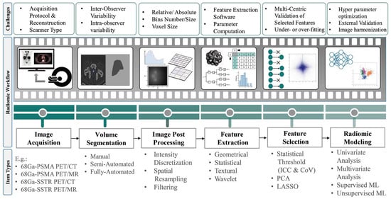
2. Radiomics and AI Workflow

In precision medicine, radiomics is currently underway in research based on feature extraction from medical images. Radiomics paves the way to map multimodal imaging into quantitative information on a large scale [24]. Figure 3 depicts the steps required to build a predictive model from medical images [25][26]. The radiomics workflow begins with image acquisition and segmentation. After image post-processing, hundreds of radiomic features (RFs) are measured from segmented regions to provide raw data for developing the final model. The major categories of features are as follows:
Figure 3. Radiomics and AI workflow from image acquisition to radiomics modeling.
  • Geometric or shape features: based on the segmented regions.
  • Statistical or intensity features: computed using intensity values in each image region.
  • Textural features (TFs): quantification of image intensity and regularity via mathematical functions.
  • Wavelet or high-order features: the image transformation process is essential to obtain these features.
Individual features are discarded for dimension reduction through feature selection in the next step. Options include intraclass correlation coefficients (ICC), principal component analysis (PCA), least absolute shrinkage and selection operator (LASSO), recursive feature elimination (RFE), and outputs from machine learning methods (ML).
Different approaches are applied in developing the model depending on the task: univariate or multivariate analysis and supervised or unsupervised ML methods. Supervised ML algorithms are classified into classification and regression algorithms if the variables are categorical or continuous, respectively. Classification algorithms are divided into linear and nonlinear models. Logistic regression and support vector machines (SVMs) are two methods of analyzing linear models.
In nonlinear models, k-nearest neighbors (KNNs), gradient boosting, decision tree, extra trees (ETs), and random forest are the most commonly used algorithms. The most widely used regression algorithms are linear, logistic, polynomial, support vector regression (SVR), decision regression, random forest regression (RFR), and ridge and lasso regressions.
In contrast to supervised learning methods, unsupervised learning approaches do not contain predefined response variables. Instead, the model finds hidden patterns and insights from the given data. In these procedures, similar data are grouped (clustering), or dimensionality is reduced. Some popular models in this category are K-means clustering, KNN, neural networks (NNs) or artificial neural networks (ANNs), PCA, and independent component analysis (ICA) [27].
Deep learning (DL) methods have been introduced as a more comprehensive part of ML methods with various techniques. Classic neural networks, convolutional neural networks (CNNs), recurrent neural networks, auto-encoders, generative adversarial networks (GANs), and gradient descent are examples of DL methods [28]. Challenges still need to be addressed to strengthen radiomics’ role in clinical practice. Most difficulties come from imaging feature variability among different devices and protocols, model robustness, and performance interpretation. In a multicenter context, addressing variability in acquisition and reconstruction protocols is crucial to ensure reproducibility [29]. Accordingly, harmonization procedures have been developed to provide a high reproducibility of RFs in multicenter studies. Reuze et al. [30] and Orlhac et al. [31] reviewed the radiomics workflow and its challenges.
The Society of Nuclear Medicine and Molecular Imaging (SNMMI) AI Task Force published a report on evaluating and validating AI algorithms [32]. This guideline applies extensively to radiomics studies involving AI. According to Figure 4, created based on this guideline, for a prostate cancer patient referred to PET/CT imaging, AI can be applied in a chain from radiochemistry to the physician’s report generation.
Figure 4. A broad range of AI applications in a chain from radiochemistry to a physician’s report generation for a prostate cancer patient who underwent [68Ga]Ga-PSMA-11 PET/CT scan based on SNMMI AI task-force guideline.
In the first step, AI could predict drug-target interactions, predict and optimize radiochemical reactions, carry out de novo drug design, and optimize radiopharmacy workflows. In the next step, ML-based methods may be well suited to difficult issues in image acquisition and instrumentation. For image reconstruction, AI may offer faster image reconstruction, a better signal-to-noise ratio, and fewer artifacts.
Image analysis can be automated using AI for different tasks, such as lesion detection, segmentation, and quantification for diagnosis and dosimetry. Moreover, AI has the potential to investigate patterns associated with patient results within large biological and imaging datasets. Additionally, AI can also detect and diagnose. By using ML methods, diagnostic images can be interpreted and translated into reports and clinical databases. Finally, clinicians can receive actionable advice after extracting, distilling, and integrating clinical information from various sources.

References

  1. Li, T.; Ao, E.C.; Lambert, B.; Brans, B.; Vandenberghe, S.; Mok, G.S. Quantitative imaging for targeted radionuclide therapy dosimetry-technical review. Theranostics 2017, 7, 4551.
  2. Teker, F.; Elboga, U. Is SUVmax a useful marker for progression-free survival 177 in patients with metastatic GEP-NET receiving Lu-DOTATATE therapy? Hell. J. Nucl. Med. 2021, 24, 122–131.
  3. Huizing, D.; Aalbersberg, E.A.; Versleijen, M.W.; Tesselaar, M.E.; Walraven, I.; Lahaye, M.J.; de Wit van der Veen, B.J.; Stokkel, M.P. Early response assessment and prediction of overall survival after peptide receptor radionuclide therapy. Cancer Imaging 2020, 20, 57.
  4. Jeelani, S.; Reddy, R.J.; Maheswaran, T.; Asokan, G.; Dany, A.; Anand, B. Theranostics: A treasured tailor for tomorrow. J. Pharm. Bioallied Sci. 2014, 6 (Suppl. S1), S6.
  5. Sjögreen Gleisner, K.; Chouin, N.; Gabina, P.M.; Cicone, F.; Gnesin, S.; Stokke, C.; Konijnenberg, M.; Cremonesi, M.; Verburg, F.A.; Bernhardt, P. EANM dosimetry committee recommendations for dosimetry of 177Lu-labelled somatostatin-receptor-and PSMA-targeting ligands. Eur. J. Nucl. Med. Mol. Imaging 2022, 49, 1778–1809.
  6. Khazaei, Z.; Jarrahi, A.M.; Momenabadi, V.; Ghorat, F.; Adineh, H.; Sohrabivafa, M.; Goodarzi, E. Global cancer statistics 2018: GLOBOCAN estimates of incidence and mortality worldwide stomach cancers and their relationship with the human development index (HDI). World Cancer Res. J. 2019, 6, e1257.
  7. Dy, G.W.; Gore, J.L.; Forouzanfar, M.H.; Naghavi, M.; Fitzmaurice, C. Global burden of urologic cancers, 1990–2013. Eur. Urol. 2017, 71, 437–446.
  8. Steele, C.B.; Li, J.; Huang, B.; Weir, H.K. Prostate cancer survival in the United States by race and stage (2001–2009): Findings from the CONCORD-2 study. Cancer 2017, 123, 5160–5177.
  9. Cooperberg, M.R.; Moul, J.W.; Carroll, P.R. The changing face of prostate cancer. J. Clin. Oncol. Off. J. Am. Soc. Clin. Oncol. 2005, 23, 8146–8151.
  10. Jones, W.; Griffiths, K.; Barata, P.C.; Paller, C.J. PSMA theranostics: Review of the current status of PSMA-targeted imaging and radioligand therapy. Cancers 2020, 12, 1367.
  11. Rahbar, K.; Afshar-Oromieh, A.; Jadvar, H.; Ahmadzadehfar, H. PSMA theranostics: Current status and future directions. Mol. Imaging 2018, 17, 1536012118776068.
  12. Ahmadzadehfar, H.; Aryana, K.; Pirayesh, E.; Farzanehfar, S.; Assadi, M.; Fallahi, B.; Shafiei, B.; Ayati, N.; Amoui, M. The Iranian Society of Nuclear Medicine practical guideline on radioligand therapy in metastatic castration-resistant prostate cancer using 177Lu-PSMA. Iran. J. Nucl. Med. 2018, 26, 2.
  13. Shakeri, S.; Askari, E.; Zarehparvar, S.; Farahmandfar, F.; Norouzbeigi, N.; Salek, R.; Aryana, K. Ga-PSMA-11 PET/CT for staging and patient management of high-risk prostate cancer: A single-center experience from Iran. Iran. J. Nucl. Med. 2022, 30, 33–39.
  14. Liberini, V.; Laudicella, R.; Balma, M.; Nicolotti, D.G.; Buschiazzo, A.; Grimaldi, S.; Lorenzon, L.; Bianchi, A.; Peano, S.; Bartolotta, T.V. Radiomics and artificial intelligence in prostate cancer: New tools for molecular hybrid imaging and theragnostics. Eur. Radiol. Exp. 2022, 6, 27.
  15. FDA. FDA Approves Pluvicto for Metastatic Castration-Resistant Prostate Cancer. 2022. Available online: https://www.fda.gov/drugs/resources-information-approved-drugs/fda-approves-pluvicto-metastatic-castration-resistant-prostate-cancer (accessed on 23 March 2022).
  16. Fallahi, B.; Khademi, N.; Karamzade-Ziarati, N.; Fard-Esfahani, A.; Emami-Ardekani, A.; Farzanefar, S.; Eftekhari, M.; Beiki, D. 99mTc-PSMA SPECT/CT versus 68Ga-PSMA PET/CT in the evaluation of metastatic prostate cancer. Clin. Nucl. Med. 2021, 46, e68–e74.
  17. Harsini, S.; Fallahi, B.; Ziarati, N.K.; Razi, A.; Amini, E.; Emami-Ardekani, A.; Fard-Esfahani, A.; Parizi, M.K.; Farzanehfar, S.; Beiki, D. A Prospective Study on -PSMA PET/CT Imaging in Newly Diagnosed Intermediate-and High-Risk Prostate Cancer. Asia Ocean. J. Nucl. Med. Biol. 2021, 9, 101.
  18. Das, S.; Dasari, A. Epidemiology, incidence, and prevalence of neuroendocrine neoplasms: Are there global differences? Curr. Oncol. Rep. 2021, 23, 43.
  19. Taal, B.; Visser, O. Epidemiology of neuroendocrine tumours. Neuroendocrinology 2004, 80 (Suppl. S1), 3–7.
  20. Virgolini, I.; Ambrosini, V.; Bomanji, J.B.; Baum, R.P.; Fanti, S.; Gabriel, M.; Papathanasiou, N.D.; Pepe, G.; Oyen, W.; De Cristoforo, C. Procedure guidelines for pet/ct tumour imaging with 68Ga-dota-conjugated peptides: 68Ga-dota-toc, 68Ga-dota-noc, 68Ga-dota-tate. Eur. J. Nucl. Med. Mol. Imaging 2010, 37, 2004–2010.
  21. FDA. FDA Approves Lutetium Lu, F. 177 Dotatate for Treatment of GEP-NETS. Available online: https://www.fda.gov/drugs/resources-information-approved-drugs/fda-approves-lutetium-lu-177-dotatate-treatment-gep-nets (accessed on 26 January 2018).
  22. Miller, C.; Rousseau, J.; Ramogida, C.F.; Celler, A.; Rahmim, A.; Uribe, C.F. Implications of physics, chemistry and biology for dosimetry calculations using theranostic pairs. Theranostics 2022, 12, 232.
  23. Yousefirizi, F.; Decasez, P.; Amyar, A.; Ruan, S.; Saboury, B.; Rahmim, A. Artificial Intelligence-Based Detection, Classification and Prediction/Prognosis in PET Imaging: Towards Radiophenomics. arXiv 2021, arXiv:2110.10332.
  24. Gillies, R.J.; Kinahan, P.E.; Hricak, H. Radiomics: Images are more than pictures, they are data. Radiology 2016, 278, 563.
  25. Mayerhoefer, M.E.; Materka, A.; Langs, G.; Häggström, I.; Szczypiński, P.; Gibbs, P.; Cook, G. Introduction to radiomics. J. Nucl. Med. 2020, 61, 488–495.
  26. Lambin, P.; Leijenaar, R.T.; Deist, T.M.; Peerlings, J.; De Jong, E.E.; Van Timmeren, J.; Sanduleanu, S.; Larue, R.T.; Even, A.J.; Jochems, A. Radiomics: The bridge between medical imaging and personalized medicine. Nat. Rev. Clin. Oncol. 2017, 14, 749–762.
  27. Ray, S. A quick review of machine learning algorithms. In Proceedings of the 2019 International Conference on Machine Learning, Big Data, Cloud and Parallel Computing (COMITCon), Faridabad, India, 14–16 February 2019; IEEE: New York, NY, USA, 2019; pp. 35–39.
  28. Alzubaidi, L.; Zhang, J.; Humaidi, A.J.; Al-Dujaili, A.; Duan, Y.; Al-Shamma, O.; Santamaría, J.; Fadhel, M.A.; Al-Amidie, M.; Farhan, L. Review of deep learning: Concepts, CNN architectures, challenges, applications, future directions. J. Big Data 2021, 8, 1–74.
  29. Kumar, V.; Gu, Y.; Basu, S.; Berglund, A.; Eschrich, S.A.; Schabath, M.B.; Forster, K.; Aerts, H.J.; Dekker, A.; Fenstermacher, D. Radiomics: The process and the challenges. Magn. Reson. Imaging 2012, 30, 1234–1248.
  30. Reuzé, S.; Schernberg, A.; Orlhac, F.; Sun, R.; Chargari, C.; Dercle, L.; Deutsch, E.; Buvat, I.; Robert, C. Radiomics in nuclear medicine applied to radiation therapy: Methods, pitfalls, and challenges. Int. J. Radiat. Oncol. Biol. Phys. 2018, 102, 1117–1142.
  31. Orlhac, F.; Nioche, C.; Klyuzhin, I.; Rahmim, A.; Buvat, I. Radiomics in PET imaging: A practical guide for newcomers. PET Clin. 2021, 16, 597–612.
  32. Bradshaw, T.J.; Boellaard, R.; Dutta, J.; Jha, A.K.; Jacobs, P.; Li, Q.; Liu, C.; Sitek, A.; Saboury, B.; Scott, P.J. Nuclear medicine and artificial intelligence: Best practices for algorithm development. J. Nucl. Med. 2022, 63, 500–510.
More
Information
Contributors MDPI registered users' name will be linked to their SciProfiles pages. To register with us, please refer to https://encyclopedia.pub/register : , , , , ,
View Times: 84
Revisions: 2 times (View History)
Update Date: 29 Jan 2024
1000/1000
Video Production Service