Submitted Successfully!
To reward your contribution, here is a gift for you: A free trial for our video production service.
Thank you for your contribution! You can also upload a video entry or images related to this topic.
Version Summary Created by Modification Content Size Created at Operation
1 -- 3610 2024-01-25 11:43:49 |
2 references update and layout Meta information modification 3610 2024-01-30 06:41:40 |

Video Upload Options

We provide professional Video Production Services to translate complex research into visually appealing presentations. Would you like to try it?

Confirm

Are you sure to Delete?
Cite
If you have any further questions, please contact Encyclopedia Editorial Office.
Begagić, E.; Bečulić, H.; Đuzić, N.; Džidić-Krivić, A.; Pugonja, R.; Muharemović, A.; Jaganjac, B.; Salković, N.; Sefo, H.; Pojskić, M. CRISPR/Cas9 Gene Editing. Encyclopedia. Available online: https://encyclopedia.pub/entry/54341 (accessed on 19 November 2024).
Begagić E, Bečulić H, Đuzić N, Džidić-Krivić A, Pugonja R, Muharemović A, et al. CRISPR/Cas9 Gene Editing. Encyclopedia. Available at: https://encyclopedia.pub/entry/54341. Accessed November 19, 2024.
Begagić, Emir, Hakija Bečulić, Nermin Đuzić, Amina Džidić-Krivić, Ragib Pugonja, Asja Muharemović, Belma Jaganjac, Naida Salković, Haso Sefo, Mirza Pojskić. "CRISPR/Cas9 Gene Editing" Encyclopedia, https://encyclopedia.pub/entry/54341 (accessed November 19, 2024).
Begagić, E., Bečulić, H., Đuzić, N., Džidić-Krivić, A., Pugonja, R., Muharemović, A., Jaganjac, B., Salković, N., Sefo, H., & Pojskić, M. (2024, January 25). CRISPR/Cas9 Gene Editing. In Encyclopedia. https://encyclopedia.pub/entry/54341
Begagić, Emir, et al. "CRISPR/Cas9 Gene Editing." Encyclopedia. Web. 25 January, 2024.
CRISPR/Cas9 Gene Editing
Edit

The complexity of CRISPR-Cas9 applications in GBM research is highlighted, providing unique insights into apoptosis, cell proliferation, and immune responses within the tumor microenvironment. The studies challenge conventional perspectives on specific genes, emphasizing the potential therapeutic implications of manipulating key molecular players in cell cycle dynamics. Exploring CRISPR/Cas9 gene therapy in GBMs yields significant insights into the regulation of cellular processes, spanning cell interphase, renewal, and migration. Researchers, by precisely targeting specific genes, uncover the molecular orchestration governing cell proliferation, growth, and differentiation during critical phases of the cell cycle. The findings underscore the potential of CRISPR/Cas9 technology in unraveling the complex dynamics of the GBM microenvironment, offering promising avenues for targeted therapies to curb GBM growth. 

glioblastoma (GBM) gene therapy CRISPR Cas9

1. Introduction

Glioblastoma (GBM) stands out as the predominant and highly aggressive primary brain tumor among adults, constituting approximately 45.2% of all tumors affecting the brain and central nervous system (CNS) [1]. This neoplasm is categorized as a grade IV diffuse astrocytic glioma, distinguished by its elevated cellular density, nuclear abnormalities, microvascular proliferation, necrosis, and invasive characteristics. The molecular features defining GBM encompass mutations in the telomerase reverse transcriptase (TERT) promoter, amplification of the epidermal growth factor receptor (EGFR) gene, and variations in chromosome copy numbers (+7/−10) [2]. Notably, GBM exhibits marked heterogeneity, both in its histological and molecular aspects, contributing significantly to its resistance to therapeutic interventions and dismal prognosis [3].
Primary and secondary GBMs represent discrete disease entities distinguished by disparate genetic pathways, patient demographics, and prognostic outcomes. Primary GBMs arise de novo in older individuals, marked by the upregulation of the epidermal growth factor receptor (EGFR) gene, mutations in the phosphatase and tensin homolog (PTEN), deletions in the cyclin-dependent kinase inhibitor 2A (CDKN2A) gene, and, occasionally, amplification of the mouse double minute 2 homolog (MDM2) gene [1]. On the other hand, secondary GBMs emerge from low-grade or anaplastic astrocytomas, typically occurring in younger patients. They are notably characterized by mutations in the TP53 gene, serving as the earliest detectable genetic alteration. In summary, primary and secondary GBMs are distinct subtypes within the spectrum of GBM, exhibiting contrasting genetic and clinical characteristics [4].
The prevailing standard of care for newly diagnosed GBM involves extensive surgical resection, followed by concurrent chemoradiation employing temozolomide, and subsequent adjuvant temozolomide chemotherapy [5]. Despite the aggressiveness of this therapeutic regimen, its impact on improving survival outcomes is only marginal [1]. Consequently, there exists a compelling demand for novel and efficacious therapeutic approaches for GBM. The past two decades have witnessed a heightened interest in the exploration of targeted agents and immunotherapies for GBM, as reported by Begagić et al. [1]. Regrettably, these endeavors have not yielded a substantive influence on patient survival. GBM remains an incurable affliction, and its management continues to pose one of the most formidable challenges in the realm of neuro-oncology [6]. Furthermore, there is an imperative to formulate therapeutic strategies that are both more potent and less toxic, specifically tailored to address the unique biological characteristics of GBM [7].
As previously mentioned, the presence of genetic mutations, including chromosomal changes such as the loss of chromosomes 10 and 9p, and the gain of chromosomes 7 and 19, suggests the potential utility of gene-oriented therapy as an option in the treatment of GBM [8][9]. One such approach that has garnered significant interest in the last decade across various medical conditions is the Clustered Regularly Interspaced Short Palindromic Repeat CRISPR-associated (Cas) nuclease 9 (CRISPR-Cas9) system [10], which facilitates gene editing technology [11]. CRISPR is recognized as the fastest, cheapest, most versatile, and most reliable gene editing tool available, extensively employed for uncovering genetic alterations, oncogenic targets, and epigenetic regulation. CRISPR-Cas9 stands out as the preferred choice for editing genes or genomes in various cancers, including GBM [12][13][14][15]. Considering the current trend in medical research towards more accessible treatments for diverse pathologies [16], with an aim for broader applicability and treatment options in low- and middle-income countries [17], the potential for gene editing using this method has emerged, even in cases of GBM. There are evident studies employing this method for treating GBM, although the data are dispersed across individual studies.

2. CRISPR/Cas9-Mediated GBM Therapy

Distinctive genetic polymorphisms, ionizing radiation exposure, and the impact of chemical carcinogens on brain cells are among the key pathogenic factors driving the development of GBM [18]. Current research is honing in on the promising potential of CRISPR/Cas9 as a cutting-edge gene-editing technology in the realm of immunotherapy for GBM. This innovation is gaining traction in various studies and holds the promise of evolving into a pivotal tool for advancing gene research and engineering strategies in glioma therapy [19][20].

2.1. Targeting Specific Genetic Mutations in GBM

Previous research has not provided a clear classification of precise gene therapy for GBM. Based on the study by Begagić et al. [1], it is observed that the main focuses in GBM therapy are the protein kinase pathway, cell-cycle-related mechanisms, and microenvironmental and immunomodulatory targets. In the realm of CRISPR/Cas9 gene editing for GBM, researchers categorizes specific gene targets into distinct groups, namely: cell cycle regulation, regulation related to the microenvironment, regulation during cell interphase, and targets related to therapy resistance reduction.

2.1.1. Cell Cycle Regulation

The cell cycle, a meticulously regulated and intricately orchestrated biological process, stands as a fundamental mechanism governing the growth, development, and maintenance of living organisms [1]. Comprising a series of precisely coordinated events leading to cell division, the cell cycle ensures the accurate transmission of genetic information from one generation of cells to the next. In instances where genetic mutations precede this transmission, the altered information is passed on to progeny cells through the process of cell division. This paradigm is particularly relevant to GBM, as disruptions in the cell cycle regulation, stemming from genetic alterations, contribute to the excessive division and proliferation of neoplastic GBM tissue.
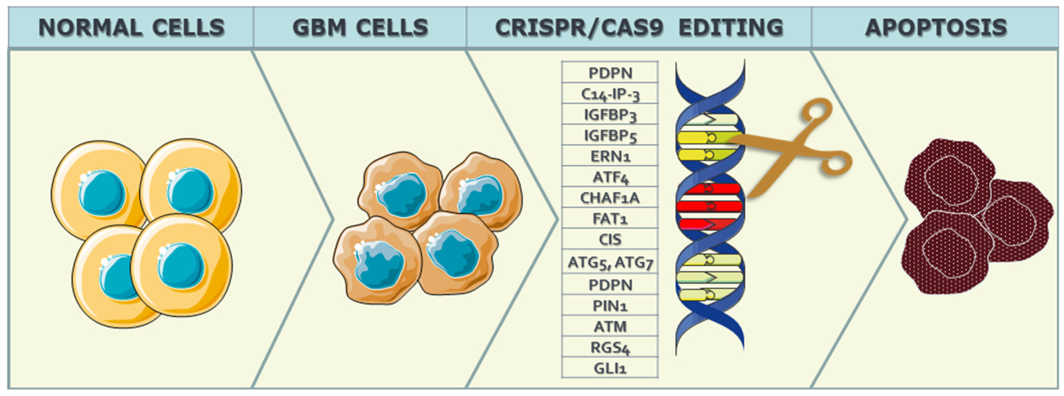
Given the strict control exerted by genes over the cell cycle, alterations in these genes lead to dysregulation of cell cycle control mechanisms, fostering uncontrolled division and proliferation of neoplastic GBM cells. Several mutations associated with GBM malignancy have been identified, including those affecting genes such as Epidermal Growth Factor Receptor (EGFR), Erb-B2 Receptor Tyrosine Kinase 2 (ERBB2), Isocitrate Dehydrogenase 1 (IDH1), Neurofibromin 1 (NF1), Phosphatidylinositol-4,5-Bisphosphate 3-Kinase Catalytic Subunit Alpha (PIK3CA), Phosphoinositide-3-Kinase Regulatory Subunit 1 (PIK3R1), and Phosphatase and Tensin Homolog (PTEN), among others. The application of CRISPR/Cas9 technology seeks to intervene in the cell cycle of neoplastic cells, aiming to induce apoptosis or autophagy in these aberrant cells, as can be seen in Figure 1.
Figure 1. Representation of the CRISPR/Cas9 precise gene editing process. Normal cells refer to cells before neoplastic changes in GBM, followed by gene editing of the target gene to halt the proliferation of malignant cells, slow down the expression of the target gene (knockdown), or completely halt it (knockout).
The promotion of apoptosis and autophagy through CRISPR-Cas9 technology has been substantiated through research conducted on GBM cellular lines and in vitro models. Various target genes have been investigated in this context, including Podoplanin (PDPN), C14orf166 (C14-IP-3), Insulin-Like Growth Factor Binding Protein 3 and 5 (IGFBP3 and IGFBP5), Endoplasmic Reticulum To Nucleus Signaling 1 (ERN1), Activating Transcription Factor 4 (ATF4), Autophagy Related 5 (ATG5), Chromatin Assembly Factor 1 Subunit A (CHAF1A), FAT Atypical Cadherin 1 (FAT1), Cytokine-Inducible SH2-Containing Protein (CIS), Autophagy Related 7 (ATG7), and PIN1 (Peptidylprolyl Isomerase NIMA-Interacting 1).
In the context of CRISPR-Cas9 technology, it is essential to distinguish between knockout, knockdown, and knock-in strategies. While knockout involves the complete elimination of a targeted gene, exemplified by the knockout of the FAT1 gene in GBM cells to enhance susceptibility to apoptosis, knockdown selectively reduces the expression of a gene, as demonstrated in the PDPN gene knockdown study, aiming to modulate apoptosis and cell proliferation.
Wang et al. [21] employed a knockdown strategy targeting the PDPN gene to intricately modulate apoptosis and cell proliferation. This approach specifically focused on manipulating PDPN surface-membrane cell molecules. The presence of mutations in PDPN aligns with the malignancy, aggressiveness, and invasiveness of GBM. The identified association between PDPN overexpression and the facilitation of macrophage M2 polarization and neutrophil degranulation underscores the immunomodulatory impact of PDPN within the tumor microenvironment. The observed shift towards M2 polarization of macrophages and the induction of neutrophil degranulation collectively indicate a coordinated effort by PDPN to establish an environment conducive to immune evasion and tumor progression [21]. This nuanced insight challenges conventional perspectives on PDPN, positioning it as a pivotal orchestrator in shaping an immunosuppressive milieu specifically within IDH wildtype gliomas. These findings highlight the complex and multifaceted role of PDPN in influencing not only the cellular aspects of apoptosis and proliferation but also the intricate immunological dynamics within the tumor microenvironment. Employing a strategy that initiated apoptosis through the Unfolded Protein Response (UPR) mechanism, IGFBP3 and IGFBP5 were targeted for knockout, shedding light on their influence on cell cycle dynamics [22]. Further insights were contributed by investigating ATG5 knockout, revealing its dual impact on apoptosis and autophagy and providing understanding within the framework of cell cycle processes associated with GBM [23]. Also, CHAF1A was subjected to CRISPR-Cas9 knockout, dissecting its consequences on the AKT/FOXO3a/Bim pathway and influencing proliferation and DNA repair mechanisms integral to cell cycle regulation [24]. Targeting the FAT1 gene for knockout revealed its active involvement in apoptosis through the Death-Inducing Signaling Complex (DISC), contributing valuable insights into the understanding of key molecular players that impact the cell cycle during GBM progression [25]. Shifting the focus to the CIS gene, investigations aimed to unravel its role in NK-cell activation and apoptosis, establishing a link between immune responses and the intricate molecular mechanisms that influence the cell cycle [26]. Furthermore, examinations delved into the consequences of knockout for ATG5 and ATG7 on the autophagosome membrane [27]. This shed light on the potential significance of autophagy in regulating the cell cycle in the context of GBM. PIN1, targeted for knockout, offered a comprehensive exploration of its multifaceted role in influencing the cell cycle within GBM development, encompassing aspects of apoptosis, migration, and cell cycle progression. The knockout study involving ATM, PTEN, p85α, and XIAP genes uncovered their roles as tumor suppressors, advancing researchers' comprehension of the intricate regulatory mechanisms that govern the cell cycle in GBM [28]. Further investigations focused on RGS4 knockout explored its impact on apoptosis through G protein signaling, expanding the repertoire of molecular targets with potential therapeutic implications in the context of cell cycle regulation. Lastly, the exploration of GLI1’s involvement in apoptosis through the PI3K/Akt pathway contributed to the elucidation of critical signaling pathways that influence the cell cycle in the pathophysiology of GBM [29].
In summary, the intricate landscape of CRISPR-Cas9 applications in GBM research unveils a spectrum of gene manipulation strategies, each offering unique insights into the regulation of apoptosis, cell proliferation, and immune responses within the tumor microenvironment. From knockout endeavors targeting genes such as FAT1, ATM, PTEN, and GLI1 to deciphering the roles of PDPN, IGFBP3, and CHAF1A through knockdown and knock-in approaches, the studies presented here collectively broaden researchers' understanding of the complex molecular orchestration shaping GBM progression. These findings not only challenge conventional perspectives on specific genes but also underscore the potential therapeutic implications of manipulating key molecular players in the intricate web of cell cycle dynamics. As the field continues to unravel the complexities of CRISPR-Cas9 technology in the context of GBM, these insights hold promise for advancing targeted therapeutic interventions and refining researchers' approach to combating this formidable malignancy.

2.1.2. Cell-Interphase-Related Targets

Exploring the realm of CRISPR/Cas9 gene therapy in the context of GBMs, researchers have delved into a diverse array of cell-interphase-related targets to unravel the intricacies of gene regulation during critical phases of the cell cycle. The focus on cell interphase, the period between cell divisions encompassing G1, S, and G2 phases, is crucial in understanding the dynamics of GBM progression and identifying potential therapeutic avenues [30].
In recent studies, various genes have been targeted using CRISPR-Cas9 gene editing technology to elucidate their roles in cell proliferation and related functions. Fierro et al. [31] focused on PD-L1, employing a knockout strategy to investigate its impact on proliferation, invasion, and macrophage polarization. Lumibao et al. [32] targeted CHCHD2, aiming for knockout to understand its influence on mitochondrial respiration, glutathione status, and cell growth inhibition, particularly in the context of EGFRvIII. Toledano et al. [33] explored Plexin-A2 through knockout, shedding light on its involvement in cytoskeletal organization, cell flattening, and cell cycle arrest, with a focus on β-galactosidase, MAPK, and FARP2. Gallo et al. [13] delved into the knockout of 14-3-3β, unraveling its effects on proliferation, spheroid formation, and interactions with Bad, FBI1, Raf-1, and Cdc25b. Additionally, Meng et al. [34] investigated CDK7, employing a knockout strategy to understand its role in cellular growth. Guda et al. [35] targeted RGS4 for knockout, exploring its influence on MMP2 and proliferation. Zhang et al. [36] utilized a knockdown strategy for Nanos3, examining its effects on CD133, Oct4, and its implications for proliferation, migration, and chemoresistance. Godoy et al. [37] employed knockdown of NRF2, investigating its role in self-renewal and cell proliferation, particularly in relation to SOD. Zhang et al. [38] focused on Dazl, utilizing knockout to study its involvement in the CD133/Oct4/Nanog/Sox2 regulatory axis and its impact on proliferation. Lastly, Liu et al. [39] explored ERβ through knockout, elucidating its effects on proliferation and apoptosis by targeting ERβ1, ERβ2, ERβ3, ERβ4, ERβ5 (exon 8), mTOR, and STAT-3.
Cell renewal studies employing CRISPR/Cas9 technology have investigated specific genes and their roles in regulating crucial aspects of this process. In the work by Bulstrode et al. [40], the focus was on Foxo3, utilizing a knockdown approach to assess its impact on differentiation. Specifically, the study targeted FOXG1, SOX2, EGFR, and EGFRvIII in order to delineate their involvement in cell renewal pathways. Saenz-Antonanzas et al. [41] explored the role of SRR2 through deletion, aiming to understand its influence on self-renewal capacity with a particular emphasis on SOX2. Additionally, Song et al. (2019) investigated SRSF3 using knockout techniques, unraveling its significance in glioma-associated alternative splicing processes involving SR proteins.
Studies targeting cell migration through CRISPR/Cas9 technology have provided valuable insights into the molecular underpinnings of this crucial cellular process. Ogawa et al. [42] focused on TP53, employing recombination techniques to explore its influence on migration. Smolkin et al. [43] investigated NRP2, Plexin-A4, Plexin-D1, and Semaphorin-3C through knockout strategies, shedding light on their roles in regulating migration processes. Prolo et al. [44] delved into MAP4K4 using knockout, elucidating its impact on both migration and invasion. Wang et al. [45] explored the knockout of BRG1, revealing its involvement in migration, proliferation, and resistance to TMZ. Shao et al. [46] targeted PIK3CD along with PAK3 and PLEK2 for knockout, unraveling their roles in migration and invasion. Chen et al. [47] investigated THBS1 and TNF through knockout, shedding light on their contributions to proliferation and migration. Ozyerli-Gokna et al. [48] focused on ASH2L and SET1/MLL, employing knockout to understand their roles in both proliferation and migration. Nieland et al. [49] targeted miR21 and SOX2 through knockout, providing insights into their contributions to migration, invasion, and proliferation. Lastly, Uceda-Castro et al. [50] investigated GFAP, GFAPα, and GFAPδ through knockout, revealing their involvement in invasion processes.
In summary, the comprehensive exploration of CRISPR/Cas9 gene therapy in the intricate landscape of GBMs has yielded significant insights into the regulation of crucial cellular processes, spanning cell interphase, renewal, and migration. Through precise targeting of specific genes, researchers have unraveled the complex molecular orchestration governing cell proliferation, growth, and differentiation during critical phases of the cell cycle. The investigations into cell renewal shed light on the roles of Foxo3, SRR2, and SRSF3 in influencing self-renewal capacity and alternative splicing processes. Furthermore, studies elucidating the molecular underpinnings of cell migration, targeting genes such as TP53, NRP2, MAP4K4, BRG1, PIK3CD, THBS1, PD-L1, ASH2L, SET1/MLL, miR21, SOX2, and GFAP, have provided valuable insights into the regulation of migration, invasion, and proliferation in the context of GBMs.

2.1.3. Microenvironmental CRISPR/Cas9 Targets in GBM Cells
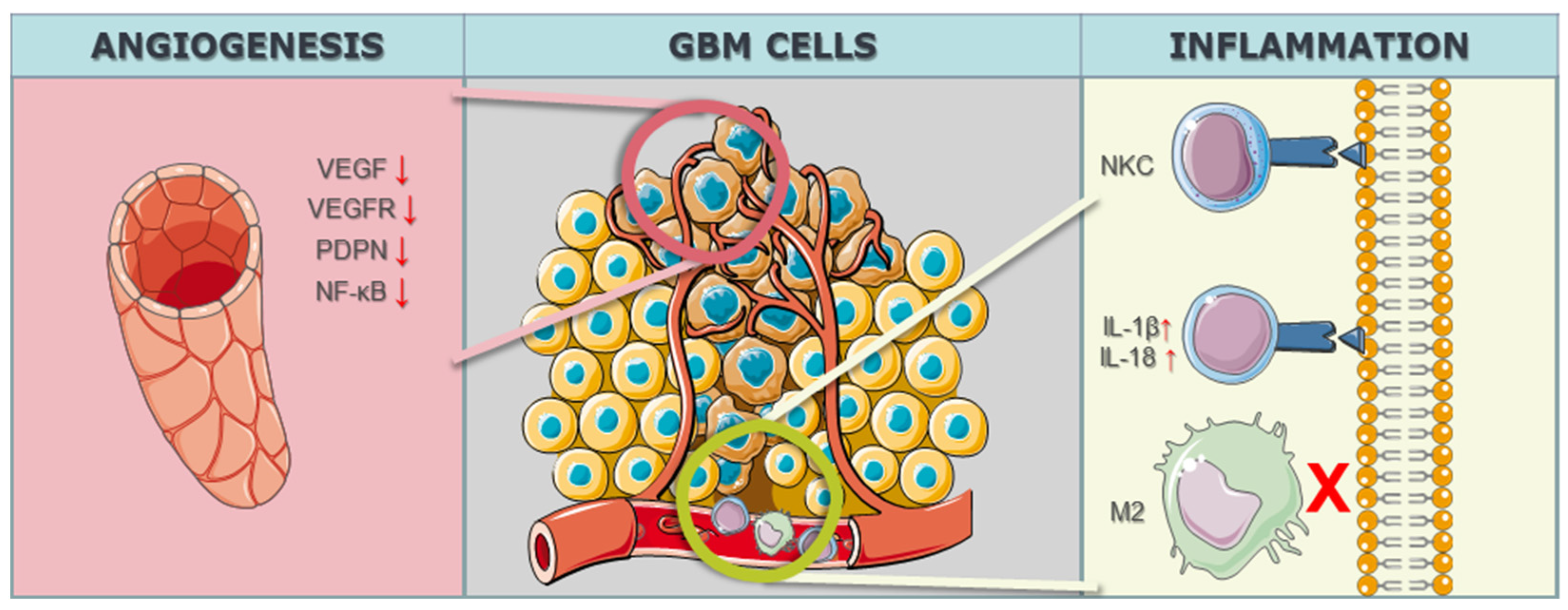
In the realm of GBM research, the intricate modulation of the tumor microenvironment, particularly in the context of angiogenesis, has become a focal point for therapeutic interventions. The study by Han et al. [51] targeted the Notch1 gene, employing a knockdown strategy to address hypoxia, angiogenesis, and tumor growth. Notch1 is known for its involvement in diverse cellular processes, and its modulation in the study aimed at disrupting key pathways associated with angiogenesis, a hallmark feature of GBM progression. By utilizing CRISPR/Cas9 technology to downregulate Notch1 expression, the study sought to unravel the intricate interplay between hypoxia, angiogenesis, and the overall growth dynamics of GBM malignant cells. Eisemann et al. [52] delved into the role of PDPN, employing a knockout strategy to investigate its influence on the maturation and integrity of the developing vasculature in the murine brain. PDPN, when interacting with C-type lectin-like receptor 2 on platelets, has been implicated in mediating vascular development. By utilizing CRISPR/Cas9 to knockout PDPN, the study aimed to disrupt the finely tuned mechanisms governing vasculature maturation, potentially impeding the vascular support crucial for GBM growth and progression. The targeted gene PDPN serves as a molecular focal point, shedding light on its intricate involvement in orchestrating the vascular microenvironment within the context of GBM. Szymura et al. [53] explored the role of DDX39B in regulating the extracellular extracellular matrix (ECM) and promoting angiogenesis through the NF-κB pathway. By employing a knockdown strategy, the study aimed to decipher the contributions of DDX39B in modulating the complex network of signals involved in angiogenesis and ECM regulation. The NF-κB pathway, known for its involvement in various cellular processes, including inflammation and angiogenesis, was specifically targeted to understand its role in the GBM microenvironment. The study adds depth to our understanding of how specific genes can be manipulated to influence the intricate balance of proangiogenic factors in the context of GBM. Continuing the exploration of angiogenesis-related genes, Lu et al. [54] investigated the genes BIG1 and BIG2, targeting VEGF through a knockdown approach in 2019. VEGF is a key player in angiogenesis, promoting the formation of new blood vessels to sustain tumor growth. By employing CRISPR/Cas9 to knock down BIG1 and BIG2 and subsequently reduce VEGF levels, the study aimed to disrupt the angiogenic signals that contribute to the vascularization of GBM tumors. The modulation of these specific genes provides insights into the intricate regulatory mechanisms underlying angiogenesis and presents potential avenues for therapeutic interventions aimed at curbing the growth and progression of GBM through microenvironmental control (Figure 2).
Figure 2. CRISPR/Cas9-related targets within the microenvironment. Angiogenesis is attenuated or entirely halted through the reduction (marked as ↓) in VEGF (Vascular Endothelial Growth Factor) and its receptor (VEGFR), as well as PDPN and NF-kB. CRISPR/Cas9 technology has effectively directed an inflammatory response against GBM cells in in vitro models, activating Natural Killer Cells (NKC) and increasing IL-1 and IL-18 (marked as ↑), inducing pyroptosis in the cells. Inhibiting (marked as X) the actions of M2 macrophages has proven effective in the immune regulation of GBM.
Nakazawa et al. [26] investigated the CIS gene, employing CRISPR/Cas9 knockout to enhance the effects of Natural Killer Cells (NKCs) and bolster the inflammatory response against GBM. Similarly, Wei et al. [55] targeted the OPN gene for knockout, resulting in the reduction in M2 macrophages and a concurrent elevation in T-lymphocyte effector activity, thereby influencing the inflammatory landscape within the tumor microenvironment. Additionally, Chen et al. [56] utilized CRISPR/Cas9 knockdown to modulate the AIM2 gene, leading to a downregulation of interleukins IL-1β and IL-18 and inducing pyroptosis, an inflammatory programmed cell death.
In summary, the collective findings from these studies underscore the potential role of CRISPR/Cas9 technology in unraveling the complex dynamics of the GBM microenvironment. Through precise manipulation of key genes involved in angiogenesis, such as Notch1, PDPN, DDX39B, and VEGF-related genes (BIG1 and BIG2), researchers have gained insights into the molecular intricacies governing vascularization, providing potential targets for therapeutic intervention. By targeting genes like CIS, OPN, and AIM2, these studies leverage CRISPR/Cas9 to enhance Natural Killer Cell effects, modulate M2 macrophages, and induce pyroptosis, collectively contributing to the immune regulation of GBM. The versatility of CRISPR/Cas9 gene editing in manipulating these microenvironment-related targets presents promising avenues for developing targeted therapies to curb the growth and progression of GBM, showcasing its potential as a transformative tool in the quest for effective GBM treatments.

2.2. Contribution of CRISPR/Cas9 Technology in Alleviating Therapy Resistance of GBM

In the study by Wu et al. [57], the ALDH1A3 gene was targeted for knockdown, focusing on ALDHs to mitigate resistance to temozolomide (TMZ), a common chemotherapy agent. Similarly, Han et al. [58] in 2023 employed knockdown targeting the MGMT gene, specifically addressing TMZ resistance. Tong et al. [59], also in 2023, utilized knockdown techniques targeting the MUC1 gene, associated with EGFRvIII, to overcome TMZ resistance. Liu et al. [60] focused on the GSS gene, employing knockout to address radiotherapy resistance, particularly in the context of Angiopep-2. Rocha et al. [61], in 2020, targeted multiple genes, including MSH2, PTCH2, CLCA2, FZD6, CTNNB1, and NRF2, focusing on transmembrane proteins to counter TMZ resistance through CRISPR/Cas9 knockout. Lastly, Yin et al. [62] targeted the HPRT1 gene for knockout, with a focus on AMPK, aiming to alleviate TMZ resistance.
CRISPR/Cas9 gene editing has proven successful in addressing treatment resistance in GBM. Wu et al. [57] targeted ALDH1A3, achieving a significant impact on TMZ resistance, particularly at dosages ≤300 μM. Han et al. [58] successfully used knockdown of MGMT to sensitize GBM cells to TMZ treatment. Additionally, Tong et al. [59] demonstrated success by targeting MUC1 with knockdown, revealing its role in DNA damage repair during chemotherapy and radiation.
However, not all interventions yielded successful outcomes. Rocha et al. [61] targeted MSH2, PTCH2, CLCA2, FZD6, CTNNB1, and NRF2 for TMZ resistance through knockout, but silencing the top three genes did not sensitize GBM cells to TMZ.

References

  1. Begagić, E.; Pugonja, R.; Bečulić, H.; Čeliković, A.; Tandir Lihić, L.; Kadić Vukas, S.; Čejvan, L.; Skomorac, R.; Selimović, E.; Jaganjac, B.; et al. Molecular Targeted Therapies in Glioblastoma Multiforme: A Systematic Overview of Global Trends and Findings. Brain Sci. 2023, 13, 1602.
  2. Stoyanov, G.S.; Lyutfi, E.; Georgieva, R.; Georgiev, R.; Dzhenkov, D.L.; Petkova, L.; Ivanov, B.D.; Kaprelyan, A.; Ghenev, P. Reclassification of Glioblastoma Multiforme According to the 2021 World Health Organization Classification of Central Nervous System Tumors: A Single Institution Report and Practical Significance. Cureus 2022, 14, e21822.
  3. Jain, K.K. A Critical Overview of Targeted Therapies for Glioblastoma. Front. Oncol. 2018, 8, 419.
  4. Tatebayashi, K.; Nakayama, N.; Sakamoto, D.; Iida, T.; Ono, S.; Matsuda, I.; Enomoto, Y.; Tanaka, M.; Fujita, M.; Hirota, S.; et al. Clinical Significance of Early Venous Filling Detected via Preoperative Angiography in Glioblastoma. Cancers 2023, 15, 3800.
  5. Angom, R.S.; Nakka, N.M.R.; Bhattacharya, S. Advances in Glioblastoma Therapy: An Update on Current Approaches. Brain Sci. 2023, 13, 1536.
  6. Agosti, E.; Zeppieri, M.; De Maria, L.; Tedeschi, C.; Fontanella, M.M.; Panciani, P.P.; Ius, T. Glioblastoma Immunotherapy: A Systematic Review of the Present Strategies and Prospects for Advancements. Int. J. Mol. Sci. 2023, 24, 15037.
  7. Riemenschneider, M.J.; Jeuken, J.W.; Wesseling, P.; Reifenberger, G. Molecular diagnostics of gliomas: State of the art. Acta Neuropathol. 2010, 120, 567–584.
  8. Isachesku, E.; Braicu, C.; Pirlog, R.; Kocijancic, A.; Busuioc, C.; Pruteanu, L.-L.; Pandey, D.P.; Berindan-Neagoe, I. The Role of Non-Coding RNAs in Epigenetic Dysregulation in Glioblastoma Development. Int. J. Mol. Sci. 2023, 24, 16320.
  9. Senhaji, N.; Squalli Houssaini, A.; Lamrabet, S.; Louati, S.; Bennis, S. Molecular and Circulating Biomarkers in Patients with Glioblastoma. Int. J. Mol. Sci. 2022, 23, 7474.
  10. Khlidj, Y. What did CRISPR-Cas9 accomplish in its first 10 years? Biochem. Med. 2023, 33, 030601.
  11. Peixoto, J.; Príncipe, C.; Pestana, A.; Osório, H.; Pinto, M.T.; Prazeres, H.; Soares, P.; Lima, R.T. Using a Dual CRISPR/Cas9 Approach to Gain Insight into the Role of LRP1B in Glioblastoma. Int. J. Mol. Sci. 2023, 24, 11285.
  12. Brennan, C.W.; Verhaak, R.G.; McKenna, A.; Campos, B.; Noushmehr, H.; Salama, S.R.; Zheng, S.; Chakravarty, D.; Sanborn, J.Z.; Berman, S.H.; et al. The somatic genomic landscape of glioblastoma. Cell 2013, 155, 462–477.
  13. Gallo, K.; Srinageshwar, B.; Ward, A.; Diola, C.; Dunbar, G.; Rossignol, J.; Bakke, J. Inducible Knockout of 14-3-3β Attenuates Proliferation and Spheroid Formation in a Human Glioblastoma Cell Line U87MG. Brain Sci. 2023, 13, 868.
  14. Motoche-Monar, C.; Ordoñez, J.E.; Chang, O.; Gonzales-Zubiate, F.A. gRNA Design: How Its Evolution Impacted on CRISPR/Cas9 Systems Refinement. Biomolecules 2023, 13, 1698.
  15. Ding, S.; Liu, J.; Han, X.; Tang, M. CRISPR/Cas9-Mediated Genome Editing in Cancer Therapy. Int. J. Mol. Sci. 2023, 24, 16325.
  16. Begagić, E.; Pugonja, R.; Bečulić, H.; Selimović, E.; Skomorac, R.; Saß, B.; Pojskić, M. The new era of spinal surgery: Exploring the utilization of exoscopes as a viable alternative to operative microscopes—A systematic review and meta-analysis. World Neurosurg. 2023, in press.
  17. Begagić, E.; Bečulić, H.; Skomorac, R.; Pojskić, M. Accessible Spinal Surgery: Transformation Through the Implementation of Exoscopes As Substitutes for Conventional Microsurgery in Low- and Middle-Income Settings. Cureus 2023, 15, e45350.
  18. Kang, X.; Wang, Y.; Liu, P.; Huang, B.; Zhou, B.; Lu, S.; Geng, W.; Tang, H. Progresses, Challenges, and Prospects of CRISPR/Cas9 Gene-Editing in Glioma Studies. Cancers 2023, 15, 396.
  19. Qazi, M.A.; Vora, P.; Venugopal, C.; Sidhu, S.S.; Moffat, J.; Swanton, C.; Singh, S.K. Intratumoral heterogeneity: Pathways to treatment resistance and relapse in human glioblastoma. Ann. Oncol. 2017, 28, 1448–1456.
  20. Lake, J.A.; Donson, A.M.; Prince, E.; Davies, K.D.; Nellan, A.; Green, A.L.; Mulcahy Levy, J.; Dorris, K.; Vibhakar, R.; Hankinson, T.C.; et al. Targeted fusion analysis can aid in the classification and treatment of pediatric glioma, ependymoma, and glioneuronal tumors. Pediatr. Blood Cancer 2020, 67, e28028.
  21. Wang, X.; Wang, X.; Li, J.; Liang, J.; Ren, X.; Yun, D.; Liu, J.; Fan, J.; Zhang, Y.; Zhang, J.; et al. PDPN contributes to constructing immunosuppressive microenvironment in IDH wildtype glioma. Cancer Gene Therapy 2023, 30, 345–357.
  22. Rodvold, J.J.; Xian, S.; Nussbacher, J.; Tsui, B.; Cameron Waller, T.; Searles, S.C.; Lew, A.; Jiang, P.; Babic, I.; Nomura, N.; et al. IRE1α and IGF signaling predict resistance to an endoplasmic reticulum stress-inducing drug in glioblastoma cells. Sci. Rep. 2020, 10, 8348.
  23. Vu, H.T.; Kobayashi, M.; Hegazy, A.M.; Tadokoro, Y.; Ueno, M.; Kasahara, A.; Takase, Y.; Nomura, N.; Peng, H.; Ito, C.; et al. Autophagy inhibition synergizes with calcium mobilization to achieve efficient therapy of malignant gliomas. Cancer Sci. 2018, 109, 2497–2508.
  24. Peng, H.; Du, B.; Jiang, H.; Gao, J. Over-expression of CHAF1A promotes cell proliferation and apoptosis resistance in glioblastoma cells via AKT/FOXO3a/Bim pathway. Biochem. Biophys. Res. Commun. 2016, 469, 1111–1116.
  25. Kranz, D.; Boutros, M. A synthetic lethal screen identifies FAT1 as an antagonist of caspase-8 in extrinsic apoptosis. Embo J. 2014, 33, 181–197.
  26. Nakazawa, T.; Morimoto, T.; Maeoka, R.; Matsuda, R.; Nakamura, M.; Nishimura, F.; Ouji, N.; Yamada, S.; Nakagawa, I.; Park, Y.S.; et al. CIS deletion by CRISPR/Cas9 enhances human primary natural killer cell functions against allogeneic glioblastoma. J. Exp. Clin. Cancer Res. 2023, 42, 205.
  27. Zielke, S.; Meyer, N.; Mari, M.; Abou-El-Ardat, K.; Reggiori, F.; van Wijk, S.J.L.; Kögel, D.; Fulda, S. Loperamide, pimozide, and STF-62247 trigger autophagy-dependent cell death in glioblastoma cells. Cell. Death Dis. 2018, 9, 994.
  28. Ali, R.; Alabdullah, M.; Miligy, I.; Normatova, M.; Babaei-Jadidi, R.; Nateri, A.S.; Rakha, E.A.; Madhusudan, S. ATM Regulated PTEN Degradation Is XIAP E3 Ubiquitin Ligase Mediated in p85α Deficient Cancer Cells and Influence Platinum Sensitivity. Cells 2019, 8, 1271.
  29. Ranjan, A.; Srivastava, S.K. Penfluridol suppresses glioblastoma tumor growth by Akt-mediated inhibition of GLI1. Oncotarget 2017, 8, 32960–32976.
  30. Esemen, Y.; Awan, M.; Parwez, R.; Baig, A.; Rahman, S.; Masala, I.; Franchini, S.; Giakoumettis, D. Molecular Pathogenesis of Glioblastoma in Adults and Future Perspectives: A Systematic Review. Int. J. Mol. Sci. 2022, 23, 2607.
  31. Fierro, J., Jr.; DiPasquale, J.; Perez, J.; Chin, B.; Chokpapone, Y.; Tran, A.M.; Holden, A.; Factoriza, C.; Sivagnanakumar, N.; Aguilar, R.; et al. Dual-sgRNA CRISPR/Cas9 knockout of PD-L1 in human U87 glioblastoma tumor cells inhibits proliferation, invasion, and tumor-associated macrophage polarization. Sci. Rep. 2022, 12, 2417.
  32. Lumibao, J.C.; Haak, P.L.; Kolossov, V.L.; Chen, J.E.; Stutchman, J.; Ruiz, A.; Sivaguru, M.; Sarkaria, J.N.; Harley, B.A.C.; Steelman, A.J.; et al. CHCHD2 mediates glioblastoma cell proliferation, mitochondrial metabolism, hypoxia-induced invasion and therapeutic resistance. Int. J. Oncol. 2023, 63, 117.
  33. Toledano, S.; Sabag, A.D.; Ilan, N.; Liburkin-Dan, T.; Kessler, O.; Neufeld, G. Plexin-A2 enables the proliferation and the development of tumors from glioblastoma derived cells. Cell Death Dis. 2023, 14, 41.
  34. Harutyunyan, A.S.; Krug, B.; Chen, H.; Papillon-Cavanagh, S.; Zeinieh, M.; De Jay, N.; Deshmukh, S.; Chen, C.C.L.; Belle, J.; Mikael, L.G.; et al. H3K27M induces defective chromatin spread of PRC2-mediated repressive H3K27me2/me3 and is essential for glioma tumorigenesis. Nat. Commun. 2019, 10, 1262.
  35. Guda, M.R.; Velpula, K.K.; Asuthkar, S.; Cain, C.P.; Tsung, A.J. Targeting RGS4 Ablates Glioblastoma Proliferation. Int. J. Mol. Sci. 2020, 21, 3300.
  36. Zhang, F.; Liu, R.; Liu, C.; Zhang, H.; Lu, Y. Nanos3, a cancer-germline gene, promotes cell proliferation, migration, chemoresistance, and invasion of human glioblastoma. Cancer Cell. Int. 2020, 20, 197.
  37. Godoy, P.R.D.V.; Pour Khavari, A.; Rizzo, M.; Sakamoto-Hojo, E.T.; Haghdoost, S. Targeting NRF2, Regulator of Antioxidant System, to Sensitize Glioblastoma Neurosphere Cells to Radiation-Induced Oxidative Stress. Oxidative Med. Cell. Longev. 2020, 2020, 2534643.
  38. Zhang, F.; Liu, R.; Zhang, H.; Liu, C.; Liu, C.; Lu, Y. Suppressing Dazl modulates tumorigenicity and stemness in human glioblastoma cells. BMC Cancer 2020, 20, 673.
  39. Liu, J.; Sareddy, G.R.; Zhou, M.; Viswanadhapalli, S.; Li, X.; Lai, Z.; Tekmal, R.R.; Brenner, A.; Vadlamudi, R.K. Differential Effects of Estrogen Receptor β Isoforms on Glioblastoma Progression. Cancer Res. 2018, 78, 3176–3189.
  40. Bulstrode, H.; Johnstone, E.; Marques-Torrejon, M.A.; Ferguson, K.M.; Bressan, R.B.; Blin, C.; Grant, V.; Gogolok, S.; Gangoso, E.; Gagrica, S.; et al. Elevated FOXG1 and SOX2 in glioblastoma enforces neural stem cell identity through transcriptional control of cell cycle and epigenetic regulators. Genes. Dev. 2017, 31, 757–773.
  41. Saenz-Antoñanzas, A.; Moncho-Amor, V.; Auzmendi-Iriarte, J.; Elua-Pinin, A.; Rizzoti, K.; Lovell-Badge, R.; Matheu, A. CRISPR/Cas9 Deletion of SOX2 Regulatory Region 2 (SRR2) Decreases SOX2 Malignant Activity in Glioblastoma. Cancers 2021, 13, 1574.
  42. Ogawa, J.; Pao, G.M.; Shokhirev, M.N.; Verma, I.M. Glioblastoma Model Using Human Cerebral Organoids. Cell. Rep. 2018, 23, 1220–1229.
  43. Smolkin, T.; Nir-Zvi, I.; Duvshani, N.; Mumblat, Y.; Kessler, O.; Neufeld, G. Complexes of plexin-A4 and plexin-D1 convey semaphorin-3C signals to induce cytoskeletal collapse in the absence of neuropilins. J. Cell. Sci. 2018, 131, jcs208298.
  44. Prolo, L.M.; Li, A.; Owen, S.F.; Parker, J.J.; Foshay, K.; Nitta, R.T.; Morgens, D.W.; Bolin, S.; Wilson, C.M.; Vega, L.J.; et al. Targeted genomic CRISPR-Cas9 screen identifies MAP4K4 as essential for glioblastoma invasion. Sci. Rep. 2019, 9, 14020.
  45. Wang, Y.; Yang, C.H.; Schultz, A.P.; Sims, M.M.; Miller, D.D.; Pfeffer, L.M. Brahma-Related Gene-1 (BRG1) promotes the malignant phenotype of glioblastoma cells. J. Cell. Mol. Med. 2021, 25, 2956–2966.
  46. Shao, W.; Azam, Z.; Guo, J.; To, S.S.T. Oncogenic potential of PIK3CD in glioblastoma is exerted through cytoskeletal proteins PAK3 and PLEK2. Lab. Investig. 2022, 102, 1314–1322.
  47. Chen, L.; Fang, W.; Chen, W.; Wei, Y.; Ding, J.; Li, J.; Lin, J.; Wu, Q. Deciphering the molecular mechanism of the THBS1 gene in the TNF signaling axis in glioma stem cells. Cell. Signal. 2023, 106, 110656.
  48. Ezgi, O.-G.; Ezgi Yagmur, K.; Ali Cenk, A.; Ipek, B.; Ahmet, C.; Sheikh, N.; Martin, B.; Fidan, S.-P.; Tunc, M.; Can, A.; et al. Epigenetic-focused CRISPR/Cas9 screen identifies ASH2L as a regulator of glioblastoma cell survival. bioRxiv 2022. peer review.
  49. Nieland, L.; van Solinge, T.S.; Cheah, P.S.; Morsett, L.M.; El Khoury, J.; Rissman, J.I.; Kleinstiver, B.P.; Broekman, M.L.D.; Breakefield, X.O.; Abels, E.R. CRISPR-Cas knockout of miR21 reduces glioma growth. Mol. Ther. Oncolytics 2022, 25, 121–136.
  50. Uceda-Castro, R.; van Asperen, J.V.; Vennin, C.; Sluijs, J.A.; van Bodegraven, E.J.; Margarido, A.S.; Robe, P.A.J.; van Rheenen, J.; Hol, E.M. GFAP splice variants fine-tune glioma cell invasion and tumour dynamics by modulating migration persistence. Sci. Rep. 2022, 12, 424.
  51. Han, N.; Hu, G.; Shi, L.; Long, G.; Yang, L.; Xi, Q.; Guo, Q.; Wang, J.; Dong, Z.; Zhang, M. Notch1 ablation radiosensitizes glioblastoma cells. Oncotarget 2017, 8, 88059–88068.
  52. Eisemann, T.; Costa, B.; Harter, P.N.; Wick, W.; Mittelbronn, M.; Angel, P.; Peterziel, H. Podoplanin expression is a prognostic biomarker but may be dispensable for the malignancy of glioblastoma. Neuro-Oncol. 2019, 21, 326–336.
  53. Szymura, S.J.; Bernal, G.M.; Wu, L.; Zhang, Z.; Crawley, C.D.; Voce, D.J.; Campbell, P.A.; Ranoa, D.E.; Weichselbaum, R.R.; Yamini, B. DDX39B interacts with the pattern recognition receptor pathway to inhibit NF-κB and sensitize to alkylating chemotherapy. BMC Biol. 2020, 18, 32.
  54. Lu, F.I.; Wang, Y.T.; Wang, Y.S.; Wu, C.Y.; Li, C.C. Involvement of BIG1 and BIG2 in regulating VEGF expression and angiogenesis. FASEB J. 2019, 33, 9959–9973.
  55. Wei, J.; Marisetty, A.; Schrand, B.; Gabrusiewicz, K.; Hashimoto, Y.; Ott, M.; Grami, Z.; Kong, L.Y.; Ling, X.; Caruso, H.; et al. Osteopontin mediates glioblastoma-associated macrophage infiltration and is a potential therapeutic target. J. Clin. Investig. 2019, 129, 137–149.
  56. Chen, P.A.; Shrivastava, G.; Balcom, E.F.; McKenzie, B.A.; Fernandes, J.; Branton, W.G.; Wheatley, B.M.; Petruk, K.; van Landeghem, F.K.H.; Power, C. Absent in melanoma 2 regulates tumor cell proliferation in glioblastoma multiforme. J. Neurooncol. 2019, 144, 265–273.
  57. Wu, W.; Wu, Y.; Mayer, K.; von Rosenstiel, C.; Schecker, J.; Baur, S.; Würstle, S.; Liesche-Starnecker, F.; Gempt, J.; Schlegel, J. Lipid Peroxidation Plays an Important Role in Chemotherapeutic Effects of Temozolomide and the Development of Therapy Resistance in Human Glioblastoma. Transl. Oncol. 2020, 13, 100748.
  58. Han, X.; Abdallah, M.O.E.; Breuer, P.; Stahl, F.; Bakhit, Y.; Potthoff, A.L.; Pregler, B.E.F.; Schneider, M.; Waha, A.; Wüllner, U.; et al. Downregulation of MGMT expression by targeted editing of DNA methylation enhances temozolomide sensitivity in glioblastoma. Neoplasia 2023, 44, 100929.
  59. Tong, F.; Zhao, J.X.; Fang, Z.Y.; Cui, X.T.; Su, D.Y.; Liu, X.; Zhou, J.H.; Wang, G.X.; Qiu, Z.J.; Liu, S.Z.; et al. MUC1 promotes glioblastoma progression and TMZ resistance by stabilizing EGFRvIII. Pharmacol. Res. 2023, 187, 106606.
  60. Liu, X.; Cao, Z.; Wang, W.; Zou, C.; Wang, Y.; Pan, L.; Jia, B.; Zhang, K.; Zhang, W.; Li, W.; et al. Engineered Extracellular Vesicle-Delivered CRISPR/Cas9 for Radiotherapy Sensitization of Glioblastoma. ACS Nano 2023, 17, 16432–16447.
  61. Rocha, C.R.R.; Rocha, A.R.; Silva, M.M.; Gomes, L.R.; Latancia, M.T.; Andrade-Tomaz, M.; de Souza, I.; Monteiro, L.K.S.; Menck, C.F.M. Revealing Temozolomide Resistance Mechanisms via Genome-Wide CRISPR Libraries. Cells 2020, 9, 2573.
  62. Yin, J.; Wang, X.; Ge, X.; Ding, F.; Shi, Z.; Ge, Z.; Huang, G.; Zhao, N.; Chen, D.; Zhang, J.; et al. Hypoxanthine phosphoribosyl transferase 1 metabolizes temozolomide to activate AMPK for driving chemoresistance of glioblastomas. Nat. Commun. 2023, 14, 5913.
More
Information
Subjects: Neurosciences; Biology
Contributors MDPI registered users' name will be linked to their SciProfiles pages. To register with us, please refer to https://encyclopedia.pub/register : , , , , , , , , ,
View Times: 180
Revisions: 2 times (View History)
Update Date: 30 Jan 2024
1000/1000
ScholarVision Creations