Submitted Successfully!
To reward your contribution, here is a gift for you: A free trial for our video production service.
Thank you for your contribution! You can also upload a video entry or images related to this topic.
Version Summary Created by Modification Content Size Created at Operation
1 -- 2722 2024-01-24 14:19:36 |
2 Reference format revised. -1 word(s) 2721 2024-01-25 01:38:15 |

Video Upload Options

Do you have a full video?

Confirm

Are you sure to Delete?
Cite
If you have any further questions, please contact Encyclopedia Editorial Office.
Christou, C.; Stylianou, A.; Gkretsi, V. Role of Midkine in Hepatocellular Carcinoma. Encyclopedia. Available online: https://encyclopedia.pub/entry/54295 (accessed on 01 July 2024).
Christou C, Stylianou A, Gkretsi V. Role of Midkine in Hepatocellular Carcinoma. Encyclopedia. Available at: https://encyclopedia.pub/entry/54295. Accessed July 01, 2024.
Christou, Christiana, Andreas Stylianou, Vasiliki Gkretsi. "Role of Midkine in Hepatocellular Carcinoma" Encyclopedia, https://encyclopedia.pub/entry/54295 (accessed July 01, 2024).
Christou, C., Stylianou, A., & Gkretsi, V. (2024, January 24). Role of Midkine in Hepatocellular Carcinoma. In Encyclopedia. https://encyclopedia.pub/entry/54295
Christou, Christiana, et al. "Role of Midkine in Hepatocellular Carcinoma." Encyclopedia. Web. 24 January, 2024.
Role of Midkine in Hepatocellular Carcinoma
Edit

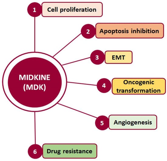
Midkine (MDK) is a multifunctional secreted protein that can act as a cytokine or growth factor regulating multiple signaling pathways and being implicated in fundamental cellular processes, such as survival, proliferation, and migration. Although its expression in normal adult tissues is barely detectable, MDK serum levels are found to be elevated in several types of cancer, including hepatocellular carcinoma (HCC). 

metastasis EMT AFP liver cancer NEGF-2

1. Hepatocellular Carcinoma

Cancer, characterized by uncontrolled cell division and proliferation [1], is one of the most lethal diseases and comprises an important global health challenge. Hepatocellular carcinoma (HCC), in particular, is one of the two main types of primary liver cancer, accounting for 70% of primary liver cancer cases, and in 2020 was ranked sixth among the most frequently diagnosed cancer types and was the third leading cause of cancer-related mortality worldwide [2][3]. In fact, recent data from the United States of America show that, in 2023, 42,210 individuals were estimated to be diagnosed with HCC, and that HCC was estimated to account for 4% of cancer-related mortality in females and 6% in males [4]. Globally, liver cancer was among the top three causes of cancer-related mortality in 46 countries and among the top five causes of cancer-related death in 90 countries. Interestingly, the number of new cases of per year is predicted to increase by 55.0% between 2020 and 2040 [3].

2. MDK-Mediated Signaling and Cellular Function

Midkine (MDK) is a multifunctional secreted protein that has emerged as a promising biomarker with the potential to aid in early diagnosis and intervention [5]. MDK, also known as neurite growth-promoting factor-2 (NEGF-2), amphiregulin-associated protein (ARAP), mid-gestation and kidney protein, and retinoic acid inducible factor, is a 13 kDa cysteine-rich protein encoded by the MDK gene [6], which is located on chromosome 11 [7]. It was originally discovered as a highly expressed gene during mouse embryogenesis [8] and, to date, seven MDK mRNA isoforms have been reported due to alternative splicing and differences in the transcription initiation site [9]. Classified as a heparin-binding protein, MDK can act as a cytokine or growth factor [6][8][10]. It is composed of two domains, each including three antiparallel β-strands and various heparin-binding consensus sites [6], which enable its binding to heparan sulfate and chondroitin sulfate and the formation of a molecular complex with proteoglycans [10]. Importantly, MDK binding to sulfated glycosaminoglycans (GAGs) leads to interactions with several key receptors, including protein-tyrosine phosphatase-ζ (PTP-ζ), syndecans, low-density lipoprotein receptor-related protein (LRP), α4β1 and α6β1 integrins, and neurogenic locus notch homolog protein 2 (Notch-2), while it has also been reported to phosphorylate the cell-matrix adhesion proteins paxillin and focal adhesion kinase (FAK) [11] (Figure 1).
Figure 1. Diagrammatic representation of the receptors interacting with MDK, and the respective signaling pathways that are activated.
Such interactions prompt, in turn, the activation of major pro-survival signaling pathways, including Src family kinase, phosphoinositide 3 kinase (PI3K), mitogen-activated protein kinase (MAPK), and nuclear factor kappa B (NF-κΒ), which in turn promote various cellular functions, such as cell proliferation, survival, adhesion, migration, and angiogenesis (Figure 2) [12][13]. Moreover, MDK binding to receptors such as the Notch2, LRP1/integrin, and transforming growth factor beta (TGF-β) receptors activates Janus kinase/signal transducer and activator of transcription 1 (JAK/STAT) and STAT3 [14] as well as MAPK pathways, resulting in epithelial to mesenchymal transition (EMT), a process that defines cancer progression and leads to metastasis [10][12]. It is worth noting that MDK can also induce EMT through interactions with β-catenin via Wnt signaling and the estrogen receptor (ER) [8]. Additionally, its interactions with LRP1, anaplastic lymphoma kinase (ALK), and integrins along with the downstream pathway MAPK/PI3K/Akt lead to increased proliferation and angiogenesis [12][15]. MDK also has the ability to induce cell proliferation by inhibiting apoptosis through the inhibition of caspase-3 and anoikis [15]. Finally, several studies in various cancers highlight MDK as an emerging player in drug resistance [6] (Figure 2).
Figure 2. Cellular processes in which MDK is crucially involved and which are related to cancer development and progression.

3. MDK Localization, Expression, and Detection

While MDK is considered to be a secreted protein, ultrastructural analysis with immunoelectronic microscopy has also identified the presence of MDK in the nucleolus. Specifically, MDK was found to be localized in the granular component, the dense fibrillar component, and the border between that and the fibrillar center. Moreover, exogenous MDK inhibits apoptosis in the HepG2 HCC cell line and plays an important role in rRNA transcription, ribosome biogenesis, and cell proliferation, confirming a functional role related to its localization [16].
Interestingly, MDK expression in normal adult tissues is undetectable or weak [7]. In fact, the highest expression of MDK is detected in infants, while plasma MDK levels progressively decline with age in healthy children [17]. Interestingly enough, though, it is found to be overexpressed in several types of cancer, including HCC, especially during tumor progression to more advanced stages. Specifically, apart from HCC, MDK expression is found to be elevated in lung cancer [18][19], glioblastoma [20], childhood lymphoblastic leukemia [21], gastric cardia adenocarcinoma [22], prostate cancer [23], head and neck squamous cell carcinoma [24], pancreatic cancer [25], endometrial cancer [26], and bladder cancer [27], and, in most cases, it is also associated with a more metastatic phenotype and a poor prognosis.
This, in conjunction with the fact that MDK is a secreted protein that can be easily detected in the blood, provides an extra advantage, as its levels can be determined through non-invasive blood and urine analyses, and it can therefore be utilized as a biomarker [8].

4. MDK’s Potential as a Biomarker for HCC Diagnosis and Progression

MDK was therefore proposed as a potential biomarker over a decade ago, as its levels were observed to be elevated in the serum of HCC patients. Indeed, several studies have shown that MDK is upregulated in HCC patients compared with healthy individuals
More specifically, Omran et al. [28] assessed MDK serum levels by ELISA, initially in 104 HCC patients and 92 individuals with non-malignant liver disease, and subsequently in 80 HCC patients and 42 patients with liver cirrhosis. They found elevated MDK levels in HCC patients compared with cirrhotic patients, suggesting MDK’s potential as a distinctive marker in HCC diagnosis and its differentiation from cirrhosis. 
Notably, a recent comprehensive study in HCC using bioinformatics analysis approaches identified 10 hub genes from protein–protein interaction networks, among which MDK was further verified by polymerase chain reaction (PCR) and immunohistochemistry (IHC) and found to be highly expressed in HCC [29].
Interestingly, Sun et al. [15] showed elevated MDK levels in the serum of 341 HCC patients who also had curative partial hepatectomy compared with healthy individuals. More importantly, these were positively correlated with increased recurrence rate compared with normal MDK levels. Moreover, they analyzed MDK mRNA expression in nine HCC cell lines (PLC/PRF5/F, Huh7, Hep3B, HepG2, SMMC7721, MHCC97L, and MHCC97H) and two normal liver cell lines (WRL68 and Chang liver) and showed that although MDK mRNA expression varied significantly among the HCC cell lines tested, in all cases it was much higher than that in the normal cell lines used. Moreover, they continued with a series of in vitro and in vivo experiments and reported a negative correlation of differential MDK expression in the cell lines tested with resistance to suspension-induced cell death, known as anoikis. Moreover, PLC/PRF/5 cells lacking MDK via small interfering RNA (siRNA)-mediated silencing exhibited reduced in vitro expression of pro-apoptotic caspase-3 and Bax and a concurrent upregulation of anti-apoptotic B-cell lymphoma 2 (Bcl2) and tropomyosin receptor kinase B (TrkB), a known and potent suppressor of anoikis.

5. MDK in Comparison with AFP

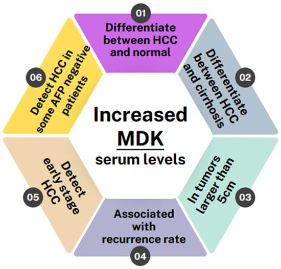
Few studies showed no correlation with AFP levels. Specifically, Saad et al. [30] determined MDK mRNA expression using real-time PCR in 29 patients with HCC and compared it with that of 7 healthy individuals, 27 patients who had chronic HCV, and 18 patients who had liver cirrhosis. They found that MDK mRNA expression was higher in HCC patients compared with the other groups. However, no significant correlation was observed between MDK and tumor characteristics (number of nodules, lesion size, and extrahepatic metastases) or AFP levels. In agreement with the above, Shaheen et al. [7], evaluated MDK protein levels as a biomarker in patients with newly diagnosed HCC. Among 100 participants, including 40 HCC patients, 30 liver cirrhosis patients, and 30 controls, MDK was found to be significantly upregulated in HCC patients compared with both liver cirrhosis patients and controls but showed no association with tumor diameter, number of nodules, AFP levels, or Barcelona Clinic liver cancer (BCLC) staging, which is widely used for staging primary liver cancer. However, it is worth noting that MDK showed greater sensitivity compared with AFP in HCC diagnosis, especially in the early stages, highlighting its potential as a novel marker, especially in differentiating HCC from liver cirrhosis.

The majority of studies, however, showed that MDK provides some additional advantages as a biomarker compared with AFP. Specifically, in a recent study [31], serum MDK levels were assessed in 238 individuals, including 78 HCC patients with HCV-related HCC, 40 with HCV-related liver cirrhosis, 40 with chronic HCV without liver cirrhosis, and 80 healthy controls. The findings consistently confirmed elevated MDK levels in HCC patients compared with other groups. However, although no association with tumor size was found, and while there was no significant difference in MDK protein expression between the other groups, the authors highlighted that MDK diagnostic accuracy for HCC diagnosis was high. 

Interestingly, in a recent phase II validation study, Zhu et al. [32] evaluated MDK as a diagnostic biomarker in early-stage HCC for those with negative AFP. Their study also included in vitro work in nine HCC cell lines (Bel-7402, Huh-7, HCCLM3, MHCC97H, PLC, HepG2, and Hep3B, with L-O2 and Chang liver cells serving as controls), as well as evaluation of 88 HCC samples, their corresponding adjacent tissues, and serum samples from 388 HCC cases and 545 controls. MDK expression was evaluated using IHC in tissue microarrays (TMA), ELISA in serum samples, and Western blotting in cell lines. They found elevated MDK protein expression in all HCC cell lines compared with controls, while IHC analysis in the TMA exhibited high MDK expression in the form of diffused cytoplasmic staining in 72% of HCCs compared with normal adjacent liver tissue or cancer-free cirrhotic samples. Although serum MDK levels did not exhibit correlations with tumor aggressiveness indicators, such as poor differentiation, microvascular invasion, larger tumor size, advanced tumor stage, survival, and tumor recurrence, they did correlate with MDK expression in tumor tissues. No association was found between MDK and BCLC staging. Compared with AFP, in AFP-positive patients, MDK and AFP had similar specificities; however, MDK showed superior sensitivity to AFP (86.9% and 51.9%, respectively). On the other hand, in patients with early-stage HCC and negative AFP, MDK had better performance compared with AFP for distinguishing early-stage HCC and small-sized tumors from non-HCC cases, including cirrhosis. 

In another study, El-Shayeb et al. [33] tested MDK serum levels in 89 patients with liver cirrhosis without HCC, 86 patients with cirrhotic HCV-induced HCC, and 69 healthy controls. They found serum MDK levels to be increased in HCC patients compared with the other two control groups. Interestingly, however, MDK exhibited higher levels in patients with multiple focal lesions, lesions exceeding 5 cm, and those with portal vein thrombosis, compared with those with single focal lesions, lesions smaller than 5 cm, and those without portal vein thrombosis. Finally, MDK was proven to have superior performance compared with AFP in differentiating HCC patients from individuals with liver cirrhosis. 

Furthermore, in a study by Hodeib et al. [34], the diagnostic utility of serum MDK compared with AFP for the diagnosis of HCC in HCV-related liver cirrhosis was evaluated. The study involved a total of 140 participants, encompassing 35 HCV patients without cirrhosis, 35 with HCV and liver cirrhosis, 35 with HCC on top of liver cirrhosis, and 35 healthy controls, and serum MDK levels were evaluated by ELISA. Their main findings showed that serum MDK levels were elevated in HCC patients compared with the other groups and did not correlate with AFP levels, but that MDK had better sensitivity in diagnosing HCC compared with AFP (98.4% vs. 97%). However, the combination of MDK and AFP had a diagnostic value of 98% for HCC diagnosis.
Corroborating further the idea that a combination of MDK and AFP increases diagnostic capacity for HCC, Mashaly et al. [35] also evaluated serum MDK levels by ELISA in 44 HCC patients, 31 with liver cirrhosis, and 15 healthy controls. They also found increased MDK levels in HCC patients compared with cirrhotic patients and controls. More importantly, 21 out of the 44 HCC patients were AFP-negative, but 17 of these AFP-negative patients were MDK-positive, while no correlation was revealed between MDK and AFP, suggesting the independent nature of MDK’s increase in relation to AFP.
Similarly, Hung et al. [36] included in their study four groups: 18 samples from HCC tissues with adjacent non-cancerous liver tissues, 85 archival HCC patient samples, 165 samples from patients with active HCC, and 285 samples from individuals in remission after HCC treatment or at high risk for HCC development. MDK serum levels were assessed by ELISA, while gene expression in the tissue samples was evaluated with microarrays and real-time PCR. MDK mRNA expression was shown to be elevated in all HCC samples compared with the respective adjacent tissue samples. When measuring serum MDK and AFP levels in the 85 archival patients, they found that 25% had elevated levels of both MDK and AFP, 16% had elevated MDK levels only, 33% had elevated AFP levels only, and 26% had neither elevated MDK nor AFP, suggesting that the combination of MDK and AFP can increase the sensitivity of HCC detection. In order to assess the specificity of serum MDK in HCC detection, they also measured serum MDK levels in 72 patients with newly diagnosed HCC from the follow-up group. Of these, 60% had elevated MDK levels. 
Finally, Daif et al. [37] evaluated the gene expression of MDK in HCC patients compared with serum AFP levels in 40 HCC patients, 30 HCV patients with no evidence of HCC, and 30 healthy controls. Gene expression levels were assessed using qRT-PCR, and MDK mRNA expression was elevated in the HCC and HCV groups compared with controls. Contrary to the other studies described above, MDK alone showed lower sensitivity (50%) and specificity (68.2%) in detecting HCC compared with AFP (78.6% and 72.7% respectively). However, the combination of MDK and AFP reached a sensitivity of 71.4% and a specificity of 81.8%.
Although most studies clearly demonstrated a strong, indisputable correlation between MDK levels and HCC disease progression as well as certain benefits in terms of early detection, sensitivity, or specificity compared with AFP, there was one study by Vongsuvanh et al. [38] showing elevated MDK levels in HCC compared with other groups (86 HCC, 86 liver cirrhosis, 86 non-cirrhotic chronic liver disease, and 86 controls), but, when compared, AFP was proven to be superior to MDK in distinguishing HCC from non-HCC cases and HCV or HBV-associated HCC from liver cirrhosis. However, MDK could differentiate NASH-related HCC from cirrhosis. In a later phase, however, they recruited 112 individuals, including 28 with HCC, 28 with HBV cirrhosis, 28 with non-cirrhotic chronic HBV, and 28 with HCV cirrhosis. MDK was elevated in 54% of the HCC patients at the time of diagnosis and, remarkably, MDK was elevated 6 months prior to diagnosis in 67% of patients, suggesting MDK’s role in pre-clinical HCC diagnosis.
Last but not least, a comparison of MDK’s specificity and sensitivity with the published specificity and sensitivity of in vitro diagnostic assays such as the GAAD or GALAD score-based models shows that MDK is comparable to both GAAD and GALAD scores. More specifically, the average MDK specificity based on the 11 studies described above is 80.34%, while the GAAD score’s specificity is 90% [39] and the GALAD score’s average specificity is 87.71% [40][41][42]. With regard to sensitivity, the average MDK sensitivity value from the 11 studies described above was 83.77%, compared with 71.8% (for GAAD) [39] and 85.82% (the average for GALAD) [40][41][42]. This further demonstrates that MDK could be used in the clinic in combination with AFP, GAAD, and GALAD scores for more effective HCC diagnosis.

6. Conclusions

Overall, all of the studies conducted so far indicate that MDK levels are elevated in HCC patients and can, in fact, differentiate not only between HCC patients and normal individuals but also between HCC patients and patients with other liver pathologies, such as liver cirrhosis, regardless of whether it is HCV/HBV-induced or not [16][17][18][22][24][26]. Furthermore, two studies have shown MDK levels to be correlated with a tumor size greater than 5 cm [19][24] and it is also associated with high recurrence rates [12]. MDK was also shown to monitor disease progression and response to therapy, as it remains elevated in patients with incompletely treated or recurrent HCC while it drops after curative surgery [43]. Most importantly, though, MDK levels can be used to diagnose early-stage HCC and can even diagnose HCC in patients who have tested negative for AFP [30][32][33][44][45] (Figure 3).
Figure 3. Schematic showing the benefits of MDK as a biomarker in HCC detection.

References

  1. Dehghani, S.H.H.a.M. Review of cancer from perspective of molecular. J. Cancer Res. Pract. 2017, 4, 127–129.
  2. Calderon-Martinez, E.; Landazuri-Navas, S.; Vilchez, E.; Cantu-Hernandez, R.; Mosquera-Moscoso, J.; Encalada, S.; Al Lami, Z.; Zevallos-Delgado, C.; Cinicola, J. Prognostic Scores and Survival Rates by Etiology of Hepatocellular Carcinoma: A Review. J. Clin. Med. Res. 2023, 15, 200–207.
  3. Rumgay, H.; Arnold, M.; Ferlay, J.; Lesi, O.; Cabasag, C.J.; Vignat, J.; Laversanne, M.; McGlynn, K.A.; Soerjomataram, I. Global burden of primary liver cancer in 2020 and predictions to 2040. J. Hepatol. 2022, 77, 1598–1606.
  4. Siegel, R.L.; Miller, K.D.; Wagle, N.S.; Jemal, A. Cancer statistics, 2023. CA Cancer J. Clin. 2023, 73, 17–48.
  5. Tsuchiya, N.; Sawada, Y.; Endo, I.; Saito, K.; Uemura, Y.; Nakatsura, T. Biomarkers for the early diagnosis of hepatocellular carcinoma. World J. Gastroenterol. 2015, 21, 10573–10583.
  6. Saikia, M.; Cheung, N.; Singh, A.K.; Kapoor, V. Role of Midkine in Cancer Drug Resistance: Regulators of Its Expression and Its Molecular Targeting. Int. J. Mol. Sci. 2023, 24, 8739.
  7. Shaheen, K.Y.; Abdel-Mageed, A.I.; Safwat, E.; AlBreedy, A.M. The value of serum midkine level in diagnosis of hepatocellular carcinoma. Int. J. Hepatol. 2015, 2015, 146389.
  8. Filippou, P.S.; Karagiannis, G.S.; Constantinidou, A. Midkine (MDK) growth factor: A key player in cancer progression and a promising therapeutic target. Oncogene 2020, 39, 2040–2054.
  9. Muramatsu, T. Structure and function of midkine as the basis of its pharmacological effects. Br. J. Pharmacol. 2014, 171, 814–826.
  10. Muramatsu, T. Midkine, a heparin-binding cytokine with multiple roles in development, repair and diseases. Proc. Jpn. Acad. Ser. B Phys. Biol. Sci. 2010, 86, 410–425.
  11. Majaj, M.; Weckbach, L.T. Midkine-A novel player in cardiovascular diseases. Front. Cardiovasc. Med. 2022, 9, 1003104.
  12. Gowhari Shabgah, A.; Ezzatifar, F.; Aravindhan, S.; Olegovna Zekiy, A.; Ahmadi, M.; Gheibihayat, S.M.; Gholizadeh Navashenaq, J. Shedding more light on the role of Midkine in hepatocellular carcinoma: New perspectives on diagnosis and therapy. IUBMB Life 2021, 73, 659–669.
  13. Polykratis, A.; Katsoris, P.; Courty, J.; Papadimitriou, E. Characterization of heparin affin regulatory peptide signaling in human endothelial cells. J. Biol. Chem. 2005, 280, 22454–22461.
  14. Bie, C.; Chen, Y.; Tang, H.; Li, Q.; Zhong, L.; Peng, X.; Shi, Y.; Lin, J.; Lai, J.; Wu, S.; et al. Insulin-Like Growth Factor 1 Receptor Drives Hepatocellular Carcinoma Growth and Invasion by Activating Stat3-Midkine-Stat3 Loop. Dig. Dis. Sci. 2022, 67, 569–584.
  15. Sun, B.; Hu, C.; Yang, Z.; Zhang, X.; Zhao, L.; Xiong, J.; Ma, J.; Chen, L.; Qian, H.; Luo, X.; et al. Midkine promotes hepatocellular carcinoma metastasis by elevating anoikis resistance of circulating tumor cells. Oncotarget 2017, 8, 32523–32535.
  16. Dai, L.C.; Shao, J.Z.; Min, L.S.; Xiao, Y.T.; Xiang, L.X.; Ma, Z.H. Midkine accumulated in nucleolus of HepG2 cells involved in rRNA transcription. World J. Gastroenterol. 2008, 14, 6249–6253.
  17. Wu, L.; Chen, H.; Fu, C.; Xing, M.; Fang, H.; Yang, F.; Yang, Q.; Zhang, Y.; Li, W.; Chen, Z. Midkine mediates dysfunction of liver sinusoidal endothelial cells through integrin alpha4 and alpha6. Vasc. Pharmacol. 2022, 147, 107113.
  18. Li, J.; Li, J.; Hao, H.; Lu, F.; Wang, J.; Ma, M.; Jia, B.; Zhuo, M.; Wang, J.; Chi, Y.; et al. Secreted proteins MDK, WFDC2, and CXCL14 as candidate biomarkers for early diagnosis of lung adenocarcinoma. BMC Cancer 2023, 23, 110.
  19. Yuan, K.; Chen, Z.; Li, W.; Gao, C.E.; Li, G.; Guo, G.; Yang, Y.; Ai, Y.; Wu, L.; Zhang, M. MDK Protein Overexpression Correlates with the Malignant Status and Prognosis of Non-small Cell Lung Cancer. Arch. Med. Res. 2015, 46, 635–641.
  20. Hu, B.; Qin, C.; Li, L.; Wei, L.; Mo, X.; Fan, H.; Lei, Y.; Wei, F.; Zou, D. Midkine promotes glioblastoma progression via PI3K-Akt signaling. Cancer Cell Int. 2021, 21, 509.
  21. Jia, M.; Zhao, H.Z.; Cheng, Y.P.; Luo, Z.B.; Zhang, J.Y.; Li, S.S.; Xu, X.J.; Tang, Y.M. High expression of Midkine (MK) indicates poor prognosis in childhood acute lymphoblastic leukemia. Hematology 2016, 21, 69–77.
  22. Hu, X.F.; Yao, J.; Gao, S.G.; Yang, Y.T.; Peng, X.Q.; Feng, X.S. Midkine and syndecan-1 levels correlate with the progression of malignant gastric cardiac adenocarcinoma. Mol. Med. Rep. 2014, 10, 1409–1415.
  23. Zhou, Q.; Yang, C.; Mou, Z.; Wu, S.; Dai, X.; Chen, X.; Ou, Y.; Zhang, L.; Sha, J.; Jiang, H. Identification and validation of a poor clinical outcome subtype of primary prostate cancer with Midkine abundance. Cancer Sci. 2022, 113, 3698–3709.
  24. Chiu, T.J.; Chen, C.H.; Chen, Y.J.; Wee, Y.; Wang, C.S.; Luo, S.D. Prognosis of Midkine and AT1R expression in resectable head and neck squamous cell carcinoma. Cancer Cell Int. 2023, 23, 212.
  25. Yao, J.; Li, W.Y.; Li, S.G.; Feng, X.S.; Gao, S.G. Midkine promotes perineural invasion in human pancreatic cancer. World J. Gastroenterol. 2014, 20, 3018–3024.
  26. Tanabe, K.; Matsumoto, M.; Ikematsu, S.; Nagase, S.; Hatakeyama, A.; Takano, T.; Niikura, H.; Ito, K.; Kadomatsu, K.; Hayashi, S.; et al. Midkine and its clinical significance in endometrial carcinoma. Cancer Sci. 2008, 99, 1125–1130.
  27. Lin, H.; Zhou, Q.; Wu, W.; Ma, Y. Midkine Is a Potential Urinary Biomarker for Non-Invasive Detection of Bladder Cancer with Microscopic Hematuria. Onco. Targets Ther. 2019, 12, 11765–11775.
  28. Omran, M.M.; Farid, K.; Omar, M.A.; Emran, T.M.; El-Taweel, F.M.; Tabll, A.A. A combination of alpha-fetoprotein, midkine, thioredoxin and a metabolite for predicting hepatocellular carcinoma. Ann. Hepatol. 2020, 19, 179–185.
  29. Shang, B.; Wang, R.; Qiao, H.; Zhao, X.; Wang, L.; Sui, S. Multi-omics analysis of pyroptosis regulation patterns and characterization of tumor microenvironment in patients with hepatocellular carcinoma. PeerJ 2023, 11, e15340.
  30. Saad, Y.; El-Serafy, M.; Eldin, M.S.; Abdellatif, Z.; Khatab, H.; Elbaz, T.; Elgarem, H. New genetic markers for diagnosis of hepatitis C related hepatocellular carcinoma in Egyptian patients. J. Gastrointest. Liver Dis. 2013, 22, 419–425.
  31. Zekri, A.-R.N.; El Kassas, M.; Salam, E.S.E.; Hassan, R.M.; Mohanad, M.; Gabr, R.M.; Lotfy, M.M.; Abdel-Zaher, R.A.T.; Bahnassy, A.A.; Ahmed, O.S. The possible role of Dickkopf-1, Golgi protein- 73 and Midkine as predictors of hepatocarcinogenesis: A review and an Egyptian study. Sci. Rep. 2020, 10, 5156.
  32. Zhu, W.W.; Guo, J.J.; Guo, L.; Jia, H.L.; Zhu, M.; Zhang, J.B.; Loffredo, C.A.; Forgues, M.; Huang, H.; Xing, X.J.; et al. Evaluation of midkine as a diagnostic serum biomarker in hepatocellular carcinoma. Clin. Cancer Res. 2013, 19, 3944–3954.
  33. El-Shayeb, A.F.; El-Habachi, N.M.; Mansour, A.R.; Zaghloul, M.S. Serum midkine is a more sensitive predictor for hepatocellular carcinoma than Dickkopf-1 and alpha-L-fucosidase in cirrhotic HCV patients. Medicine 2021, 100, e25112.
  34. Hodeib, H.; ELshora, O.; Selim, A.; Sabry, N.M.; El-Ashry, H.M. Serum Midkine and Osteopontin Levels as Diagnostic Biomarkers of Hepatocellular Carcinoma. Electron. Physician 2017, 9, 3492–3498.
  35. Mashaly, A.H.; Anwar, R.; Ebrahim, M.A.; Eissa, L.A.; El Shishtawy, M.M. Diagnostic and Prognostic Value of Talin-1 and Midkine as Tumor Markers in Hepatocellular Carcinoma in Egyptian Patients. Asian Pac. J. Cancer Prev. 2018, 19, 1503–1508.
  36. Hung, Y.J.; Lin, Z.H.; Cheng, T.I.; Liang, C.T.; Kuo, T.M.; Kao, K.J. Serum midkine as a prognostic biomarker for patients with hepatocellular carcinoma. Am. J. Clin. Pathol. 2011, 136, 594–603.
  37. Daif, A.; Al-Azzawi, M.A.; Sakr, M.A.; Ismail, H.A.; Gadallah, M. Noninvasive identifcation of molecular biomarkers of hepatocellular carcinoma in HCV-Egyptian patients. J. Egypt. Nat. Cancer Instit. 2023, 35, 11.
  38. Vongsuvanh, R.; van der Poorten, D.; Iseli, T.; Strasser, S.I.; McCaughan, G.W.; George, J. Midkine Increases Diagnostic Yield in AFP Negative and NASH-Related Hepatocellular Carcinoma. PLoS ONE 2016, 11, e0155800.
  39. Piratvisuth, T.; Hou, J.; Tanwandee, T.; Berg, T.; Vogel, A.; Trojan, J.; De Toni, E.N.; Kudo, M.; Eiblmaier, A.; Klein, H.G.; et al. Development and clinical validation of a novel algorithmic score (GAAD) for detecting HCC in prospective cohort studies. Hepatol. Commun. 2023, 7, e0317.
  40. Eletreby, R.; Elsharkawy, M.; Taha, A.A.; Hassany, M.; Abdelazeem, A.; El-Kassas, M.; Soliman, A. Evaluation of GALAD Score in Diagnosis and Follow-up of Hepatocellular Carcinoma after Local Ablative Therapy. J. Clin. Transl. Hepatol. 2023, 11, 334–340.
  41. Guan, M.C.; Zhang, S.Y.; Ding, Q.; Li, N.; Fu, T.T.; Zhang, G.X.; He, Q.Q.; Shen, F.; Yang, T.; Zhu, H. The Performance of GALAD Score for Diagnosing Hepatocellular Carcinoma in Patients with Chronic Liver Diseases: A Systematic Review and Meta-Analysis. J. Clin. Med. 2023, 12, 949.
  42. Yang, J.D.; Addissie, B.D.; Mara, K.C.; Harmsen, W.S.; Dai, J.; Zhang, N.; Wongjarupong, N.; Ali, H.M.; Ali, H.A.; Hassan, F.A.; et al. GALAD Score for Hepatocellular Carcinoma Detection in Comparison with Liver Ultrasound and Proposal of GALADUS Score. Cancer Epidemiol. Biomark. Prev. 2019, 28, 531–538.
  43. Parikh, N.D.; Mehta, A.S.; Singal, A.G.; Block, T.; Marrero, J.A.; Lok, A.S. Biomarkers for the Early Detection of Hepatocellular Carcinoma. Cancer Epidemiol. Biomark. Prev. 2020, 29, 2495–2503.
  44. Pinero, F.; Dirchwolf, M.; Pessoa, M.G. Biomarkers in Hepatocellular Carcinoma: Diagnosis, Prognosis and Treatment Response Assessment. Cells 2020, 9, 1370.
  45. Zheng, L.; Li, H.; Huang, J.; Shin, J.; Luo, S.; Guo, C.; Zhao, Y.; Li, F. Serum midkine levels for the diagnosis and assessment of response to interventional therapy in patients with hepatocellular carcinoma. J. Interv. Med. 2021, 4, 39–45.
More
Information
Subjects: Cell Biology; Oncology
Contributors MDPI registered users' name will be linked to their SciProfiles pages. To register with us, please refer to https://encyclopedia.pub/register : , ,
View Times: 100
Revisions: 2 times (View History)
Update Date: 25 Jan 2024
1000/1000
Video Production Service