Submitted Successfully!
To reward your contribution, here is a gift for you: A free trial for our video production service.
Thank you for your contribution! You can also upload a video entry or images related to this topic.
Version Summary Created by Modification Content Size Created at Operation
1 -- 5391 2024-01-09 12:12:13 |
2 format correct -38 word(s) 5353 2024-01-10 09:20:02 |

Video Upload Options

Do you have a full video?

Confirm

Are you sure to Delete?
Cite
If you have any further questions, please contact Encyclopedia Editorial Office.
Saurav, S.; Sharma, P.; Kumar, A.; Tabassum, Z.; Girdhar, M.; Mamidi, N.; Mohan, A. Nano-Scaffolds in Bone Tissue Engineering. Encyclopedia. Available online: https://encyclopedia.pub/entry/53604 (accessed on 18 May 2024).
Saurav S, Sharma P, Kumar A, Tabassum Z, Girdhar M, Mamidi N, et al. Nano-Scaffolds in Bone Tissue Engineering. Encyclopedia. Available at: https://encyclopedia.pub/entry/53604. Accessed May 18, 2024.
Saurav, Sushmita, Prashish Sharma, Anil Kumar, Zeba Tabassum, Madhuri Girdhar, Narsimha Mamidi, Anand Mohan. "Nano-Scaffolds in Bone Tissue Engineering" Encyclopedia, https://encyclopedia.pub/entry/53604 (accessed May 18, 2024).
Saurav, S., Sharma, P., Kumar, A., Tabassum, Z., Girdhar, M., Mamidi, N., & Mohan, A. (2024, January 09). Nano-Scaffolds in Bone Tissue Engineering. In Encyclopedia. https://encyclopedia.pub/entry/53604
Saurav, Sushmita, et al. "Nano-Scaffolds in Bone Tissue Engineering." Encyclopedia. Web. 09 January, 2024.
Nano-Scaffolds in Bone Tissue Engineering
Edit

Numerous surgeries are carried out to replace tissues that have been harmed by an illness or an accident. Due to various surgical interventions and the requirement of bone substitutes, the emerging field of bone tissue engineering attempts to repair damaged tissues with the help of scaffolds. These scaffolds act as template for bone regeneration by controlling the development of new cells. For the creation of functional tissues and organs, there are three elements of bone tissue engineering that play very crucial role: cells, signals and scaffolds. For the achievement of these aims, various types of natural polymers, like chitosan, chitin, cellulose, albumin and silk fibroin, have been used for the preparation of scaffolds. Scaffolds produced from natural polymers have many advantages: they are less immunogenic as well as being biodegradable, biocompatible, non-toxic and cost effective. The hierarchal structure of bone, from microscale to nanoscale, is mostly made up of organic and inorganic components like nanohydroxyapatite and collagen components. 

tissue engineering nano-scaffolds biopolymers nanostructure bone regeneration

1. Natural Polymers

Natural polymer-based nanomaterials are used for bone tissue engineering scaffolding materials due to their biodegradation and ability to stimulate regeneration of bone and tissues at both the starting and end of the process [1].
There are diverse kinds of polymeric scaffolds employed for bone tissue engineering. The natural polymers are proteins (such as soy, fibrin, silk, collagen, gelatin, actin, keratin, etc.) [2], polysaccharides (such as cellulose, starch, amylose, alginate, dextran, chitin/chitosan, hyaluronic acid, etc.) and polynucleotides (i.e., DNA and RNA). They typically exhibit enhanced biocompatibility, great cell adherence and growth promotion because of their resemblance to the elements in the extracellular matrix [3]. Hyaluronic acid, elastin, alginate, collagen/gelatin, silk fibroin, chitosan, GAGs (glycosaminoglycans), peptides and others are the natural polymers of origin for bone tissue engineering and are most frequently researched [4].

2. Chitosan and Chitin

Chitin is the main constituent of the exoskeleton of crustaceans and the major provenance of the natural polysaccharide known as chitosan. Chitosan is synthesized by partial deacetylation of chitin by biological, chemical or combined approaches [5]. Chitosan is not precisely defined, but generally, the degree of deacetylation of chitin of 70% or more is regarded as chitosan. By pro viding nitrogen atmosphere or by adding sodium borohydride to the solution of NaOH to prevent any form of unfavorable reactions, the process of deacetylation of chitin is performed. Chitosan has an approximate molecular weight of 1.2 × 10⁵ g/mol. Chitosan is tough, because it has an H-bond and can be easily modified into films of good mechanical strength. Chitosan is different from chitin due to the presence of amino groups in chitosan, which provide several unique qualities [6]. Chitin has very few applications owing to its weak solubility. The chitosan d-glucosamine amino group can be protonated and soluble in a weak acidic aqueous solution with pH < 6. Chitosan is also employed in the creation of multilayered films via layer-by-layer deposition as a polyelectrolyte. In mineral acid like hydrochloric acid, chitosan dissolves in sulfuric acid, and it forms insoluble chitosan sulfate [7].
Since the only naturally occurring polysaccharide is chitosan is positively charged, it can form complexes with synthetic polymers that are negatively charged, such as poly acrylic acid; it can also form films on negatively charged surfaces, such as those found on proteins, fats, macromolecules and cholesterol [8]. In chitosan chains, hydroxyl and amino functional groups allow them to create strong covalent connections with other functional groups. Some general reactions, including etherification and esterification, can take place in the hydroxyl groups [9]. In molecular structure, both hydrophobic acetyl groups and hydrophilic amino groups are present; chitosan shows amphiphilic properties that affect the physical properties in solid and liquid states. In addition to its capacity to bind fat, chitosan also has antibacterial, antifungal, hemostatic, analgesic, wound-healing and mucoadhesive properties in both humans and animals. Chitosan can be biodegraded into a substance that is non-toxic and shows biocompatibility to some extent [10]. Because of this, chitosan can be employed in an array of medical procedures, such as topical ocular treatment and implantation. Chitosan is an excellent candidate for tissue engineering application due to all these distinctive qualities [11].

2.1. Applications of Chitin/Chitosan in Bone Tissue Engineering

Due to its versatility in being employed as fibers, sponges, and hydrogels, chitosan is employed for tissue engineering purposes. Its implementation in various fields, like drug delivery, gene therapy and as an antimicrobial agent, is shown in Figure 1. Chitosan is similar to the glycosaminoglycans (GAGs) present in the ECM (extracellular matrix) and has significant additional characteristics that make it a perfect natural polymer to use in bone tissue engineering as a scaffold material [12].
Figure 1. Applications of chitosan in various fields.
The significant features of glycosaminoglycans (GAGs) include various electrostatic interactions with cytokines, receptors, and cell adhesion molecules. Chitosan interacts with negatively charged GAGs and promotes cartilage chondrogenesis. Chitosan has the ability to stimulate the synthesis of special cartilage GAGs, so chitosan-based composite scaffolds are popular for the restoration of articular cartilage. Biopolymers like collagen, silk fibroin and alginate in combination with chitosan have generated interest in cartilage tissue engineering due to the non-toxicity and biocompatibility of biopolymers. Gelatin has the potential to uptake high water content, which is useful to impart alimentation to scaffolds like hydrogels and also an important element of the cartilage extracellular matrix [7].

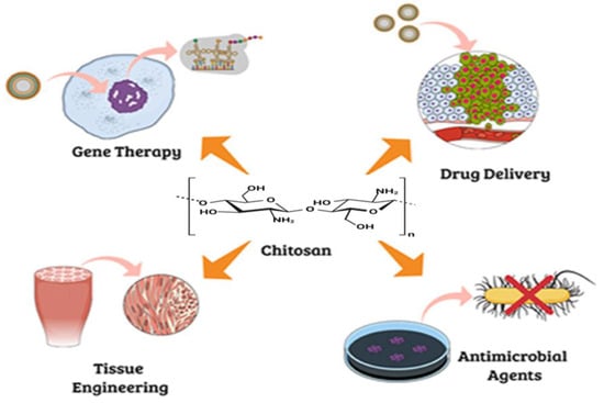
1.2. Application of Chitin/Chitosan in Bone Implants and Healing of Osteoarthritis Defects

A chitosan-poly(3-hydroxybutyrate) scaffold using β-tricalcium phosphate (β-TCP) as a reinforcement material was prepared through a novel and intriguing electrospinning technique [13]. The electrospun scaffold had higher mechanical protection, a large surface area and high porosity. Additionally, it had crucial characteristics like biocompatibility and a rate of deterioration comparable to situations of osteoarthritis defect healing [14]. With 82% porosity, this nanocomposite scaffold demonstrated a strong tensile strength of 9 MPa. The addition of β-TCP rendered the scaffold hydrophilic, which worked as a stimulant in the growth and proliferation of chondrocytes. As compared to the normal bone, the mechanical properties of the chitosan scaffold are inferior [15]; they are incapable of supporting the load-bearing demands of bone implants. As chitosan lacks osteoconductive qualities, these scaffolds are unable to mimic the characteristics of real bones. To improve the mechanical strength and structural integrity of chitosan biocomposites for bone tissue engineering applications, biopolymers including silk fibers, alginate, chitin, polycaprolactone and polylactic acid as well as bioactive nanoceramics such as Hap/TiO2, ZrO2 and SiO2 etc. have been created [13].
Numerous studies have found that Hydroxyapatite (Ca10(PO4)6(OH)2) can boost the mechanical strength and osteoconductivity of implants. It is one of the more stable varieties of calcium phosphate and makes up 60–65 percent of bone. HAp also interacts with the living system and promotes the production of new bone without resorption [15]. Additionally, nanostructured HAp has greater bioactivity and a greater surface area. As a result, the chitosan and HAp composite may replicate both the inorganic and organic components of the real bone and is currently the subject of additional research [16]. Collagen, polyethylene glycol, chitosan and HAp were combined and freeze=dried using a de-hydrothermal cross-linking process to create a porous 3D scaffold. This research investigated how HAp affected chitosan-based materials. Hyaluronic acid, collagen, silk fibroin, gelatin and alginate are the other popular natural polymers that have been extensively used in this field. Moreira et al. created an intriguing in situ forming hydrogel by using gelatin, chitosan and bioactive glass. This injectable hydrogel undergoes in situ gelation in response to body temperature stimulation. This hydrogel is a novel, thermo-sensitive, minimally invasive device that may be delivered as fluid using a syringe and needle. The hydrogel’s ability to be injected has added the benefit of filling small irregular holes and transporting cells and medicinal substances [17].

1.3. Application of Chitosan/Chitin in Periodontal Regeneration

The periodontium, which consists of the gingiva, alveolar bone, periodontal ligament and cement, is severely damaged by periodontitis. It is a chronic inflammatory condition brought on by bacterial infection. This illness, which accounts for more than 50 percent of cases of tooth loss in the U.S. population, is widespread around the world [18]. The formations of the periodontal pocket, and in more severe instances, infrabony (below the crest of bone) abnormalities are its defining features. Such lesions constitute a challenge to doctors in terms of treatment [19]. To combat inflammation and infection and to encourage tissue regeneration, it has been proposed to administer local delivery of active medications or substances [20]. The chitosan-based delivery method has drawn particular attention over the past ten years [21]. Many chitosan-based technologies, including micro/nanoparticles, fibers, membranes and gels, have been developed and tested under certain conditions [22]. Some interesting physical characteristics of chitosan gels may be influenced by chitosan concentration. In fact, it has been demonstrated that these chitosan-based gels (1–4%) have an intriguing viscosity that allows them to be injected within periodontal pockets. Most significantly, they can be trusted to deliver active medications to the location of the aliment. While a continuous release may be beneficial, it is crucial to remember that the kinetics of release is a characteristic behavior that is also determined by the proportion of chitosan [23]. It has also been shown that chitosan might potentially enhance the antibacterial effects of chlorhexidine, demonstrating chitosan’s inherent qualities [24].

2. Cellulose

Cellulose is a versatile material with adjustable properties and can be used in systems with a wide range of biochemical and biophysical conditions. In comparison to traditional synthetic materials, cellulose-based biomaterials have several significant benefits, and they hold considerable promise for expanding the frontiers of scientific understanding [25]. Cellulose has tunable chemical, physical and mechanical properties, which is why it is an ideal candidate for biomaterial manufacturing [26].
One of the most significant and prevalent biopolymers in nature is cellulose. Cellulose is a linear syndiotactic homopolymer of d-anhydroglucopyranose that is linked by β-(1-4) glycosidic bonds. In general, cellulose is fibrillar and semi-crystalline [27]. Rapid advancement in nanotechnology offers more opportunities for cellulose. Because of features like biodegradability, renewability, sustainability, chemical stability and economy, cellulose has become popular in the form of cellulose nanofibers (CNFs), which may be used as advanced and novel biomaterials. Cellulose nanofibers are cost-effective, with generated agricultural residues or wastes providing nanosized reinforcements in composite materials [28]. The cellulose nanofibers that are segregated from lingo-cellulosic plants typically show distinctiveness primarily through a high modulus and high surface area. They exhibit enormous potential for use as reinforcement in biopolymer environments. Due to their similarity to numerous biomaterials, polymeric materials and metal oxides, cellulose nanofibers have a high crystalline content, making them ideal fillers. The leading edge of nanotechnology, material science and biological science is represented by the creative production of hybrid nanostructured materials known as cellulose nanomaterials. The most common method to prepare cellulose nanocrystals is acid hydrolysis [29]. Cellulose synthase proteins, which catalyze glucan chain polymerization, are present in all cellulose-synthesizing species, including bacteria, algae, tunicates and higher plants. The vast differences in the lifestyle of the organism and the structure of the cellulose it produces suggests that regulatory proteins and the underlying mechanisms of cellulose synthesis may have evolved independently, even though the catalytic domains of cellulose synthase are preserved in all cellulose-synthesizing organisms [30]. Cellulose obtained from plants can be converted into cellulose microfibrils. Plant cellulose and bacterial cellulose are the two main classes of cellulose, and both have good applications in tissue engineering, as depicted in Figure 2. Bacteria produce bacterial cellulose, which is a natural biomaterial. It has outstanding mechanical properties, water-holding capacity and suspension ability. Additionally, it possesses great biocompatibility and biodegradability as well as a high degree of crystallinity and high purity. As a result, bacterial cellulose has gained considerable interest from both academia and industry. Plant cellulose frequently contains impurities, including lignin, pectin, hemicellulose and other substances, whereas bacterial cellulose is nearly pure, has much higher water content and exhibits noticeably increased tensile strength because of its longer chain length [31].
Figure 2. Sources of plant and bacterial cellulose and its application in tissue engineering.
A mixture of polycaprolactone (PCL), gelatin (GEL) and bacterial cellulose (BC) reinforced with various quantities of hydroxyapatite (HA) nanoparticles was used to create a unique porous scaffold for 3D printing [32]. Four distinct composites were created, with infill rates ranging from fifty to eighty percent, to obtain ideal pore size and a homogenous blending ratio. At the 80% infill rate, the optimal pore size for a bone tissue imitating ECM was attained, producing a uniformity ratio of more than 90%. In comparison to composites devoid of bacterial cellulose, bacterial cellulose-containing composites displayed a lower tensile strength and a higher cell survival rate.
Additionally, compared to the other composites, adding 0.25% HA to the blends improved cell adherence and vitality. Comparative research on these printed scaffolds showed that they might be used as bone implants [33].

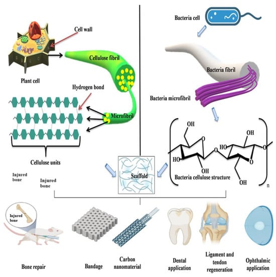
2.1. Application of Plant Cellulose in Bone Tissue Engineering

Cellulose derivatives are now being used in biomedical applications. In the field of biomaterials, the use of cellulose for membranes in biomedical application is very important. For the preparation of membranes for osseointegration, cellulose acetate is proven to be an appropriate biomaterial due to its excellent biocompatibility [34]. When cellulose acetate breaks down, it produces fragments that are not cytotoxic at the site of implantation and an acetyl group that slightly alters the local pH. These cellulose acetate membranes were employed to cover magnesium alloy implants, and they produced positive results for both in vitro tests and after being implanted into the mice’s intermedullary canal [35]. In the osseointegration process, cellulose acetate (CA) prepared with sericin or resveratrol was successfully applied [36]. In MC3T3 osteoblast precursor cells, the proliferation ability implicitly improved the osseointegration of implants covered with cellulose acetate membranes. To avoid any post-operative infections, especially in dental areas, controlled antibiotics were applied at the implantation site. For such uses, dual role membranes that promote osseointegration and permit the release of the drug doxycycline were developed [37]. For hemodialysis, composites of cellulose acetate (CA) were created using fillers like silica nanowires or hydroxyapatite or by combining carbon nanotubes and graphene. All of them had a non-cytotoxic character, high bovine serum (BSA) binding ability and high flow rates. Cellulose acetate has some other applications, like in the preparation of membranes having antibacterial properties or antibiotics from aqueous solutions [38].

2.2. Applications of Bacterial Cellulose for Bone Regeneration

A wide range of research has been conducted using cellulose from bacteria (BC) in guided bone regeneration on several animals. Noncritical bone defects were evaluated in vivo in rat tibiae on a bacterial cellulose membrane, and these defects were entirely filled by fresh bone tissue after four weeks [39]. Studies on the impact of BC membranes on bone regeneration on rat skulls produced results that were equivalent, showing that after 8 weeks of implantation, new bone had formed on both the periphery and the core of the bone defect [40]. The study of bacterial cellulose membranes as a barrier membrane for directed bone regeneration on rat calvarial defects did not cause an inflammatory reaction and preserved appropriate spaces for bone regeneration [41], and no signs of a foreign body reaction were noticed when bacterial cellulose grafts were utilized to treat rabbit nasal dorsum, which fragmented in six months, a favorable sign of bacterial cellulose integration [42]. At day 14 after implant, mineralized formation of bones was seen on the exterior and inner surface of the femoral cortical bone of dogs [43].
To treat maxillary canine periodontal abnormalities in beagle dogs, a bacterial cellulose composite membrane was utilized as a guided tissue regeneration membrane. The results reveal that the membrane encouraged periodontal tissue regeneration by generating new bone [44].

3. Albumin

One of the most important and fundamental study fields in nanomedicine is the use of peptides and proteins. Experts from different fields, like nanobiotechnology, pharmacy, toxicology, nanomedicine, immunology and other medical sciences, are currently examining the many facets of these essential biomolecules, including understanding how the resulting nanostructures interact with the body, and are using them for therapeutic and diagnostic purposes. The animal protein albumin has several therapeutic applications [45]. Because albumin contains several functional groups, it can interact with a large number of drugs. The ability to fully recognize albumin’s structure and amino acid sequence as well as its multiple charged groups enables the binding of different drugs to albumin nanoparticles by a variety of mechanisms, including electrostatic attraction with negatively charged drugs and positively charged drugs like oligonucleotides and dual compounds. The albumin-based nanoparticulate structure is used in the administration of medicines in the categories of anticancer agents, vaccines, hormones, anti-asthmatics and anti-inflammatory agents [46]. The most prevalent human serum albumin has been reported to influence cell attachment to different scaffold materials in a way that is similar and superior to collagen and fibronectin following particular treatments. It may act as a bridge between the scaffold and the cells, facilitating the fusion of these two elements. With little variation in the sequence of amino acid content, it is present in the white portions of eggs, milk, cow blood, pig blood serum, sheep blood serum, human blood serum, human liver and many other animal and plant tissues [47]. Bovine serum albumin (BSA), human serum albumin (HSA), porcine serum albumin (PSA) and ovalbumin are the major types of albumins most often used in tissue engineering scaffolds. The most prevalent protein in plasma is serum albumin; it has tremendous importance because it regulates a variety of vital processes, including blood pH and osmotic pressure. This globular protein albumin has several binding sites and is non-toxic, highly soluble in water and stable from pH 4 to 9 as well as in organic solvents. Albumins detoxify blood plasma, lowering the amount of dangerous chemicals such as heavy metals, reactive oxygen species (ROS) and ions. It also works as a solubilizing agent for fatty acids [48].
Yamaguchi et al. produced a significant body of research on the use of albumin in bone regeneration trials. Firstly, they demonstrated that local albumin production increases following bone fracture. Additionally, they examined the effects of supplementing a medium with bovine serum albumin on bone explants in vitro. They discovered that albumin enhances the calcium and DNA content of the fragments of bone [49]. Sources of albumin, like human serum albumin (HSA), bovine serum albumin (BSA) and ovalbumin (OVA), can be used to prepare scaffolds in in vitro as well as in vivo analysis for bone healing by blending it with different polymers and adding some growth factors, as shown in Figure 3. A variety of sources, including eggs (ovalbumin), bovine serum albumin, human serum albumin, rat serum, soy, milk and grains all are used to obtain albumin [50]. They are biocompatible and have high stability, low temperature and pH sensitivity. For biophysical and biochemical research, BSA, HSA and OVA (ovalbumin) are most frequently used, and all three albumins are commercially accessible. The presence of sulfhydryl groups and disulfide bonds in albumins permit interaction with both organic and inorganic ligands [46]. OVA, HSA and BSA have similar characteristics, and all are primarily composed of α-helix, with a globular structure. They have both hydrophilic and hydrophobic sites and exhibit an acidic nature.
Figure 3. Sources and preparation of albumin scaffold for bone regeneration.
Ovalbumin (OVA) is a sequence of a single 385 amino acid polypeptide chain with a spring-like structure and a helical reactive loop configuration. Ovalbumin can form gel and foam, which makes it a common ingredient in the food industry [51]. Ovalbumin is one of the first proteins identified and accounts for 60–65% of the protein in avian white eggs. Its structure and properties show that it is a member of the serpin superfamily of proteins. Despite sharing a 30% sequence identity with antitrypsin and other inhibitor proteins of the serpin family, ovalbumin does not exhibit any protease inhibitory function [52].
Bovine serum albumin (BSA) is a protein made up of three homologous domains and two subdomains. It has 583 amino acid residues and has 67% α-helical structure. It is a 66 kDa globular protein and is procured from the blood of cows. BSA is a vital part of tissue media used for culture, and a scaffold based on albumin will continue to be an excellent substrate, since it provides structural support for cells and tissue engineering [53]. The three corresponding domains, I, II and III, which make up the three-dimensional structure of BSA are selective for fatty acids and metals. Each domain is the result of two sub-domains, with most of them helical. Sulfide bridges are used to connect these sub-domains widely [54]. Due to its accessibility, binding affinity in the development of ligand protein complexes, intrinsic fluorescence caused by its two tryptophan residues (Trp 134 in the first domain and Trp 212 in the second domain) and structural and functional resemblances to HSA, BSA is intriguing. Human serum albumin (HSA) has 585 amino acids in a globular conformation, the same as BSA, and three homologous domains that are shaped like an α-helical ellipsoid [55]. The amino acids in HSA are preferentially absorbed by tumors and inflammatory tissue, and they can be dissimilated and utilized as a source of sustenance for peripheral tissues [56]. Additionally, HSA has a variety of binding sites, which enables it to transport ions, medicines, hormones and fatty acids throughout the body. These qualities make it a strong candidate to be used as a drug delivery system and in pharmaceutical formulations [57]. In terms of structure, characteristics and conformation, BSA and HSA are similar. The tryptophan residue position is the primary distinction between BSA and HSA, which share 76% sequence similarity. HSA only contains one Trp at position 214, but BSA contains two tryptophans at two different positions, at Trp-212 and Trp-134 in solvents. The differences between ovalbumin and HSA/BSA in terms of weight, size structure, S-S bond bridges and SH groups are remarkable [58]. BSA and HSA are protein carriers utilized in various delivery methods, because they are less immunogenic than other albumins and are thought to be better tolerated by humans. Currently, BSA and HSA are used in drug delivery systems in which drugs are conjugated to the binding sites of proteins, improving drug water solubility, therapeutic efficacy, bioavailability, biocompatibility and biodistribution as well as lowering adverse drug reactions, including anti-inflammatory, chemotherapeutic and hypoglycemic drugs [59]. The amino acid of HSA is made up of one polypeptide strand, which combines to create three different secondary structures. Thirty-five cysteine roots make up albumin, and they play a crucial part in this protein by generating 17 disulfide linkages. Additionally, a large number of charged amino acids, including lysine, arginine, glutamic acid and aspartic acid, are present in albumins and fabricate its structure, which is foremost for the proteins in many biological functions as well as for the formation of nanoparticles and the binding of various substances [56]. By using X-ray crystallography, the three-dimensional structure of human albumin has been identified. The albumin has a heart-shaped morphology, with dimensions of 80 by 30 angstrom. Around 80% of the plasma osmolarity pressure is generated only by albumin, which also has a significant buffering effect on blood pH. The usual half-life of this protein in human blood serum is 19 days, and it is produced in the liver, along with many other plasma proteins, at a daily rate production of 10–15 grams. Fatty acids, eicosanoids, bile acids, steroid hormones, vitamins C and D, foliate and magnesium, as well as numerous drugs, like penicillin, sulfonamides and benzodiazepine compounds, work as carriers of many molecules through albumin [60]. The protective function of albumin is to bind with toxic substances that are of external origin, like benzene, other carcinogenic compounds and various others. Human serum albumin is applied as a therapeutic agent to treat a variety of ailments, including shock, burns, albumin deficiencies, trauma, cardiac surgery, acute respiratory issues and blood dialysis [61].

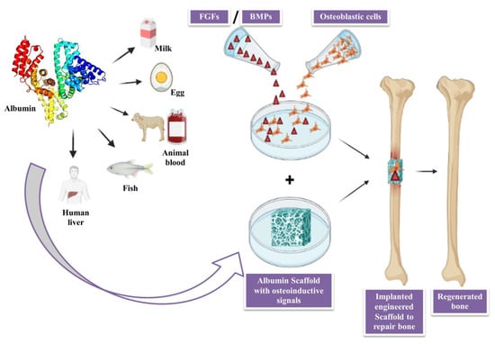
3.1. Applications of Albumin in Bone Tissue Engineering

The cross-linked albumin scaffolds have high wettability, which promotes craniofacial regeneration. When combined with collagen I, they encourage the development of mesenchymal stem cells into osteoblasts, has a very porous structure, resilience, moderate mechanical strength and good compatibility. The mechanical strength of albumin scaffolds produced by heat aggregation has been reported to be 8.5, with pH 12 showing the highest biodegradation rate and with pH 4.8 showing the lowest biodegradation rate [62]. Another technique involved electrospinning albumin to create a scaffold, which was found to be nontoxic, biodegradable and supportive of endothelial muscle cell adhesion in vivo. The functional group on the electron fiber facilitates protein conjugation, which can promote physiological processes that aid in the growth of tissue, like the lungs. For protein in general and albumin in particular, cross-linking, freeze-drying, heat-aggregation and electrospinning techniques have been shown to be effective in manufacturing the scaffold.
The use of albumin scaffolds in bone tissue engineering has been effectively demonstrated and utilized. They have high seeding efficacy, are biocompatible, non-immunogenic and affordable, and have regulated decomposition [63]. Successful fabrication of albumin fiber scaffolds with mechanical characteristics like heart tissue has been accomplished. When compared to PCL fibers, these fibers perform better and act as scaffolds for the fabrication of functioning cardiac tissues. Cell adhesion is encouraged by this cardiac scaffold’s capacity to bind serum proteins, and the functional group on the albumin scaffold facilitates protein conjugation, increasing physiological processes and promoting tissue growth [64]. However, further research needs to be conducted on serum protein binding, the release of therapeutic biomolecules that will enhance tissue function, and the capacity of the tailored patches to enhance heart functions following infraction [65].

3.2. Role of Albumin as Biomaterial in Regenerative Medicine

Gold, steel, platinum, titanium and other metal-based biomaterials are ideal because of their inertness and structural capabilities; nevertheless, their surfaces lack bioactivity [66]. Despite its lack of flexibility, bio glasses and ceramics, with great biocompatibility and strong mechanical qualities, find use in dentistry and bone regeneration [67]. Some synthetic polymers like PLA, PCL and PGA are used for the preparation of scaffolds, medical devices and implants. They have good physical characteristics, moderate rates of degradation and customizable design, but they do not encourage cell adhesion and spreading [66]. Utilization of natural components, blood-derived products, decellularized organs, primary cells and stem cells is an innovative technique for improving biological compatibility. Albumin is a blood-derived protein substance having the potential for autologous or allogeneic tissue engineering and has been successfully used in the production of scaffolds, hydrogels and coatings [57].
Further in vivo testing of albumin coating on bone allografts involved creating bone lesions in the parietal bones of old female rats. The bone defects were filled in with bone albumin and a non-coated demineralized bone matrix, and mechanical tests, CT (computed topography) and micro-CT were performed to access the graft’s integration with the surrounding tissue. The bone development was more rapid, more durable and had two times greater fracturing force compared to the uncoated bone grafts, according to in vivo computed topography and ex vivo micro-computed topography tests. The grafts with albumin coating attracted around twice as many cells as the grafts without albumin coating when MSCs were incubated in vitro [68].

3.3. Role of Albumin as a Nano-Scaffolds
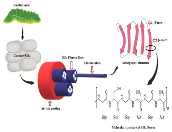
When used in biomedicine, albumin has various benefits; nevertheless, for some reasons, albumin works best when coupled with other biomaterials [69]. Due to their resemblance to the inorganic components of bone tissue, bioactivity, osteoconductivity and mold ability, calcium phosphate-based biomaterials are commonly employed in biomedicine, particularly for bone regeneration [70]. The production of (ACP-PLA) ACP-poly (l,d-lactic acid) nanofibers with the addition of BSA (bovine serum albumin), reported by Fu et al., utilized amorphous calcium phosphate (ACP) nanoparticles. The ACP-PLA solution containing BSA was easily electrospun into nanofibers with a fibrous structure. The PLA, PLA-ACP and BSA containing ACP-PLA nano-fibrous round mats were submerged in a simulated body fluid with ion concentrations that were approximately identical to those in human blood plasma to promote mineralization. Except for PLA, the shape of the nanofibers had already changed after just one day of immersion due to the deposition of tiny nanoparticles on their surfaces. Increased inorganic matter deposition and the development of a nanosheet network were produced when the mineralization time was extended. The total hydrophilicity of the surfaces of the BSA containing ACP-PLA and the ACP-PLA alone was presumably brought on by the enhanced water adsorption inside the recently constructed porous nanosheet network. After a week of cultivating MG-63 cells (human osteosarcoma cell lines) on the scaffolds, a steady rise in cell metabolic activity on all the examined fibers was observed. Water-soluble drug-delivery methods for tissue engineering may be able to use the hybrid material with BSA and ACP-PLA, since it quickly mineralized and had great biocompatibility [71]. Patel et al. and Haag et al. studied the intricate relationships between BSA and calcium. When albumin is exposed to calcium, it undergoes several structural changes that increase its bioactivity and have several positive impacts on bone tissue regeneration, according to their modeling research [72].

4. Silk Fibroin

Silk fibroin is a kind of protein that is derived from silkworms. It has great biodegradibility, bioresorbability and biocompatibility, low immunogenicity and tunable mechanical properties. Due to its unique properties, silk fibroin is used as a potential biopolymer for bone tissue engineering [73]. The integration of silk fibroin with other biopolymers to create silk fibroin composite scaffolds can encourage cellular behaviors like cell differetiation, cell proliferation and cell adhesion. Additionally, silk fibroin-based biomaterials can be made into a variety of materials formats, including hydrogels, sponges, 3D structures, films and nanoparticles. It is a natural protein made up of amino acids in which 90% of the amino acids are glycine, alanine and serine, which form crystalline β-pleated sheets in silk fibers [74].
There are numerous taxonomic families that produce silk, including those of silkworms, glowworms, lacewings, mites and spiders; some of these species have the ability to spin silk into threads for cocoon regeneration [75]. According to recent research by Yoshika et al., the psychidae family, popularly called Bagworm moths, is expected to create the hardest type of moth silk, which is used currently [76]. The most often used types of silk for biological applications are from spiders and silkworms. In spider silk, when the silk is spun and when it comes in contact with the air, it hardens, which makes it difficult to generate a large quantity of silk fibers. Compared to spiders, the yield of fibers obtained from one silkworm cocoon is around 10 times as much [77]. The two families of silkworms on either the mulberry tree (Bombycidae) or alternative food sources known as non-mulberry (Saturniidae) silks are recognised to play the most significant roles in the study of silk produced from silkworms. Bombyx mori is a mullbery-feeding silkworm; it produces a higher quality of silk than saturniidae. It is the most widely used silkworm, producing large amounts of silk with better quality [78]. The cocoons of Bombyx mori contain impurities called sericin (16.7–25%) and a protein known as silk fibroin, as shown in Figure 4 (75–83.3%). Silk fibroin is a protein with a semi-crystalline structure that is mostly used for its ability to support weight. On the other hand, sericin is an unstructured protein polymer that acts as a gumming substance [79]. Sericin-free fibroin has been discovered to have superior mechanical qualities over fibroin wrapped in sericin, which has a 50 percent increase in tear strength, a 15–17 GPa modulus and a stress break reaching up to 19%. To produce pure silk fibers of a higher quality, scientists and many researchers are working to improve the degumming procedure, which normally calls for reagents and organic solvents. The traditional Marseilles soap degumming method has been supplanted by the sodium carbonate degumming method, which is presently the most popular method due to its speed and affordability [80].
Figure 4. Sources and structure of silk fiber.
The structure of silk fibroin extracted from the cocoons of Bombyx mori is shown in Figure 4. Light (L) 26 (kDa) and Heavy (H) chain (390 kDa) are the two major chains that make up silk fibroin, and they are joined by a disulphide bond to create the H-L complex. The three polypeptides that make up the cocoons of Bombyx mori are the L-chain, H-chain and P25, which are each visible at a molar ratio 6:6:1. Glycine 45.9%, Serine 5.3%, Alanine 30.3%, Valine 1.8%, and 15 other amino acid types 4.5% make up the H-chain amino acid sequence [81]. The H-chain is composed of 60–75% repeats of the Gly-X dipeptide motifs. Dipeptide repeat hydrophobic residues can form stable antiparallel β-pleated sheets. The two hexapeptides whose peptide sequences are Gly-Ala-Gly-Ala-Gly-Ser and Gly-Ala-Gly-Ala-Gly-Tyr make up 70% of the GX dipeptide motif region. Crystalline and amorphous structures can be found in the secondary structure made from solutions of regenerated silk fibroin (RSF) [82]. When silk is crystalline, it has β-turns and an insoluble structure made of folded β-sheets, whereas when it is amorphous, it contains helix turns and random coil structures [83].

References

  1. Hasnain, M.S.; Ahmad, S.A.; Chaudhary, N.; Hoda, M.N.; Nayak, A.K. Biodegradable polymer matrix nanocomposites for bone tissue engineering. In Applications of Nanocomposite Materials in Orthopedics; Elsevier: Amsterdam, The Netherlands, 2019; pp. 1–37.
  2. Nagarajan, S.; Radhakrishnan, S.; Kalkura, S.N.; Balme, S.; Miele, P.; Bechelany, M. Overview of protein-based biopolymers for biomedical application. Macromol. Chem. Phys. 2019, 220, 1900126.
  3. Reddy, M.S.; Ponnamma, D.; Choudhary, R.; Sadasivuni, K.K. A comparative review of natural and synthetic biopolymer composite scaffolds. Polymers 2021, 13, 1105.
  4. Sivakumar, P.M.; Yetisgin, A.A.; Sahin, S.B.; Demir, E.; Cetinel, S. Bone tissue engineering: Anionic polysaccharides as promising scaffolds. Carbohydr. Polym. 2022, 283, 119142.
  5. Iber, B.T.; Kasan, N.A.; Torsabo, D.; Omuwa, J.W. A review of various sources of chitin and chitosan in nature. J. Renew. Mater. 2022, 10, 1097.
  6. Dey, S.C.; Al-Amin, M.; Rashid, T.U.; Ashaduzzaman, M.; Shamsuddin, S.M. pH induced fabrication of kaolinite-chitosan biocomposite. Int. Lett. Chem. Phys. Astron 2016, 68, 1–9.
  7. Ressler, A. Chitosan-Based Biomaterials for Bone Tissue Engineering Applications: A Short Review. Polymers 2022, 14, 3430.
  8. Vander Straeten, A.; Lefèvre, D.; Demoustier-Champagne, S.; Dupont-Gillain, C. Protein-based polyelectrolyte multilayers. Adv. Colloid Interface Sci. 2020, 280, 102161.
  9. Jaworska, M.M.; Antos, D.; Górak, A. Review on the application of chitin and chitosan in chromatography. React. Funct. Polym. 2020, 152, 104606.
  10. Craciun, A.-M.; Mititelu-Tartau, L.; Gavril, G.; Marin, L. Chitosan crosslinking with pyridoxal 5-phosphate vitamer toward biocompatible hydrogels for in vivo applications. Int. J. Biol. Macromol. 2021, 193, 1734–1743.
  11. Kaviani, A.; Zebarjad, S.M.; Javadpour, S.; Ayatollahi, M.; Bazargan-Lari, R. Fabrication and characterization of low-cost freeze-gelated chitosan/collagen/hydroxyapatite hydrogel nanocomposite scaffold. Int. J. Polym. Anal. Charact. 2019, 24, 191–203.
  12. Tamimi, M.; Rajabi, S.; Pezeshki-Modaress, M. Cardiac ECM/chitosan/alginate ternary scaffolds for cardiac tissue engineering application. Int. J. Biol. Macromol. 2020, 164, 389–402.
  13. Saravanan, S.; Leena, R.; Selvamurugan, N. Chitosan based biocomposite scaffolds for bone tissue engineering. Int. J. Biol. Macromol. 2016, 93, 1354–1365.
  14. Karbasi, S.; Alizadeh, Z.M. Effects of multi-wall carbon nanotubes on structural and mechanical properties of poly (3-hydroxybutyrate)/chitosan electrospun scaffolds for cartilage tissue engineering. Bull. Mater. Sci. 2017, 40, 1247–1253.
  15. Jodati, H.; Yılmaz, B.; Evis, Z. A review of bioceramic porous scaffolds for hard tissue applications: Effects of structural features. Ceram. Int. 2020, 46, 15725–15739.
  16. Islam, S.; Bhuiyan, M.R.; Islam, M. Chitin and chitosan: Structure, properties and applications in biomedical engineering. J. Polym. Environ. 2017, 25, 854–866.
  17. Moreira, C.D.; Carvalho, S.M.; Sousa, R.G.; Mansur, H.S.; Pereira, M.M. Nanostructured chitosan/gelatin/bioactive glass in situ forming hydrogel composites as a potential injectable matrix for bone tissue engineering. Mater. Chem. Phys. 2018, 218, 304–316.
  18. Kinane, D.F.; Stathopoulou, P.G.; Papapanou, P.N. Periodontal diseases. Nat. Rev. Dis. Primers 2017, 3, 1–14.
  19. Cortellini, P.; Tonetti, M.S. Clinical concepts for regenerative therapy in intrabony defects. Periodontology 2000 2015, 68, 282–307.
  20. Petit, C.; Batool, F.; Bugueno, I.M.; Schwinté, P.; Benkirane-Jessel, N.; Huck, O. Contribution of statins towards periodontal treatment: A review. Mediat. Inflamm. 2019, 2019, 6367402.
  21. Sah, A.K.; Dewangan, M.; Suresh, P.K. Potential of chitosan-based carrier for periodontal drug delivery. Colloids Surf. B Biointerfaces 2019, 178, 185–198.
  22. Kamal, T.; Khan, S.B.; Haider, S.; Alghamdi, Y.G.; Asiri, A.M. Thin layer chitosan-coated cellulose filter paper as substrate for immobilization of catalytic cobalt nanoparticles. Int. J. Biol. Macromol. 2017, 104, 56–62.
  23. Ganguly, A.; Ian, C.K.; Sheshala, R.; Sahu, P.S.; Al-Waeli, H.; Meka, V.S. Application of diverse natural polymers in the design of oral gels for the treatment of periodontal diseases. J. Mater. Sci. Mater. Med. 2017, 28, 39.
  24. Barreras, U.S.; Méndez, F.T.; Martínez, R.E.M.; Valencia, C.S.; Rodríguez, P.R.M.; Rodríguez, J.P.L. Chitosan nanoparticles enhance the antibacterial activity of chlorhexidine in collagen membranes used for periapical guided tissue regeneration. Mater. Sci. Eng. C 2016, 58, 1182–1187.
  25. Gregory, D.A.; Tripathi, L.; Fricker, A.T.; Asare, E.; Orlando, I.; Raghavendran, V.; Roy, I. Bacterial cellulose: A smart biomaterial with diverse applications. Mater. Sci. Eng. R Rep. 2021, 145, 100623.
  26. Courtenay, J.C.; Deneke, C.; Lanzoni, E.M.; Costa, C.A.; Bae, Y.; Scott, J.L.; Sharma, R.I. Modulating cell response on cellulose surfaces; tunable attachment and scaffold mechanics. Cellulose 2018, 25, 925–940.
  27. Lamm, M.E.; Li, K.; Qian, J.; Wang, L.; Lavoine, N.; Newman, R.; Gardner, D.J.; Li, T.; Hu, L.; Ragauskas, A.J. Recent advances in functional materials through cellulose nanofiber templating. Adv. Mater. 2021, 33, 2005538.
  28. Mohajer, S.; Rezaei, M.; Hosseini, S.F. Physico-chemical and microstructural properties of fish gelatin/agar bio-based blend films. Carbohydr. Polym. 2017, 157, 784–793.
  29. Aghazadeh, M.R.; Delfanian, S.; Aghakhani, P.; Homaeigohar, S.; Alipour, A.; Shahsavarani, H. Recent advances in development of natural cellulosic non-woven scaffolds for tissue engineering. Polymers 2022, 14, 1531.
  30. Allen, H.; Wei, D.; Gu, Y.; Li, S. A historical perspective on the regulation of cellulose biosynthesis. Carbohydr. Polym. 2021, 252, 117022.
  31. Zhong, C. Industrial-scale production and applications of bacterial cellulose. Front. Bioeng. Biotechnol. 2020, 8, 605374.
  32. Choi, S.M.; Rao, K.M.; Zo, S.M.; Shin, E.J.; Han, S.S. Bacterial cellulose and its applications. Polymers 2022, 14, 1080.
  33. Cakmak, A.M.; Unal, S.; Sahin, A.; Oktar, F.N.; Sengor, M.; Ekren, N.; Gunduz, O.; Kalaskar, D.M. 3D printed polycaprolactone/gelatin/bacterial cellulose/hydroxyapatite composite scaffold for bone tissue engineering. Polymers 2020, 12, 1962.
  34. Oprea, M.; Pandele, A.M.; Nicoara, A.I.; Nicolescu, A.; Deleanu, C.; Voicu, S.I. Crown ether-functionalized cellulose acetate membranes with potential applications in osseointegration. Int. J. Biol. Macromol. 2023, 230, 123162.
  35. Neacsu, P.; Staras, A.I.; Voicu, S.I.; Ionascu, I.; Soare, T.; Uzun, S.; Cojocaru, V.D.; Pandele, A.M.; Croitoru, S.M.; Miculescu, F. Characterization and in vitro and in vivo assessment of a novel cellulose acetate-coated Mg-based alloy for orthopedic applications. Materials 2017, 10, 686.
  36. Voicu, S.I.; Condruz, R.M.; Mitran, V.; Cimpean, A.; Miculescu, F.; Andronescu, C.; Miculescu, M.; Thakur, V.K. Sericin covalent immobilization onto cellulose acetate membrane for biomedical applications. ACS Sustain. Chem. Eng. 2016, 4, 1765–1774.
  37. Corobea, M.S.; Albu, M.G.; Ion, R.; Cimpean, A.; Miculescu, F.; Antoniac, I.V.; Raditoiu, V.; Sirbu, I.; Stoenescu, M.; Voicu, S.I. Modification of titanium surface with collagen and doxycycline as a new approach in dental implants. J. Adhes. Sci. Technol. 2015, 29, 2537–2550.
  38. Pandele, A.; Comanici, F.; Carp, C.; Miculescu, F.; Voicu, S.; Thakur, V.; Serban, B. Synthesis and characterization of cellulose acetate-hydroxyapatite micro and nano composites membranes for water purification and biomedical applications. Vacuum 2017, 146, 599–605.
  39. Saska, S.; Barud, H.; Gaspar, A.; Marchetto, R.; Ribeiro, S.J.L.; Messaddeq, Y. Bacterial cellulose-hydroxyapatite nanocomposites for bone regeneration. Int. J. Biomater. 2011, 2011, 175362.
  40. Lee, Y.-J.; An, S.-J.; Bae, E.-B.; Gwon, H.-J.; Park, J.-S.; Jeong, S.I.; Jeon, Y.-C.; Lee, S.-H.; Lim, Y.-M.; Huh, J.-B. The effect of thickness of resorbable bacterial cellulose membrane on guided bone regeneration. Materials 2017, 10, 320.
  41. Lee, S.-H.; Lim, Y.-M.; Jeong, S.I.; An, S.-J.; Kang, S.-S.; Jeong, C.-M.; Huh, J.-B. The effect of bacterial cellulose membrane compared with collagen membrane on guided bone regeneration. J. Adv. Prosthodont. 2015, 7, 484–495.
  42. Amorim, W.L.; Costa, H.O.; Souza, F.C.d.; Castro, M.G.d.; Silva, L.d. Experimental study of the tissue reaction caused by the presence of cellulose produced by Acetobacter xylinum in the nasal dorsum of rabbits. Rev. Bras. De Otorrinolaringol. 2009, 75, 200–207.
  43. Yuan, H.; Van Blitterswijk, C.; De Groot, K.; de Bruijn, J.D. A comparison of bone formation in biphasic calcium phosphate (BCP) and hydroxyapatite (HA) implanted in muscle and bone of dogs at different time periods. J. Biomed. Mater. Res. Part A: Off. J. Soc. Biomater. Jpn. Soc. Biomater. Aust. Soc. Biomater. Korean Soc. Biomater. 2006, 78, 139–147.
  44. Zhang, H.; Wang, J.; Wang, K.; Xu, L. A bilayered PLGA/multiwall carbon nanotubes/bacterial cellulose composite membrane for tissue regeneration of maxillary canine periodontal bone defects. Mater. Lett. 2018, 212, 118–121.
  45. Karimi, M.; Bahrami, S.; Ravari, S.B.; Zangabad, P.S.; Mirshekari, H.; Bozorgomid, M.; Shahreza, S.; Sori, M.; Hamblin, M.R. Albumin nanostructures as advanced drug delivery systems. Expert Opin. Drug Deliv. 2016, 13, 1609–1623.
  46. Bolaños, K.; Kogan, M.J.; Araya, E. Capping gold nanoparticles with albumin to improve their biomedical properties. Int. J. Nanomed. 2019, ume 14, 6387–6406.
  47. Jiang, X.; Mu, H.; Hsieh, Y.-H.P.; Rao, Q. Isolation and Characterization of Chicken Serum Albumin (Hen Egg Alpha-Livetin, Gal d 5). Foods 2022, 11, 1637.
  48. De Simone, G.; di Masi, A.; Ascenzi, P. Serum albumin: A multifaced enzyme. Int. J. Mol. Sci. 2021, 22, 10086.
  49. Horvathy, D.B.; Vacz, G.; Szabo, T.; Szigyarto, I.C.; Toro, I.; Vamos, B.; Hornyak, I.; Renner, K.; Klara, T.; Szabo, B.T. Serum albumin coating of demineralized bone matrix results in stronger new bone formation. J. Biomed. Mater. Res. Part B Appl. Biomater. 2016, 104, 126–132.
  50. Kuten Pella, O.; Hornyák, I.; Horváthy, D.; Fodor, E.; Nehrer, S.; Lacza, Z. Albumin as a Biomaterial and Therapeutic Agent in Regenerative Medicine. Int. J. Mol. Sci. 2022, 23, 10557.
  51. Aiyelabegan, H.T.; Zaidi, S.S.; Fanuel, S.; Eatemadi, A.; Ebadi, M.T.; Sadroddiny, E. Albumin-based biomaterial for lung tissue engineering applications. Int. J. Polym. Mater. Polym. Biomater. 2016, 65, 853–861.
  52. Nosrati, H.; Salehiabar, M.; Manjili, H.K.; Danafar, H.; Davaran, S. Preparation of magnetic albumin nanoparticles via a simple and one-pot desolvation and co-precipitation method for medical and pharmaceutical applications. Int. J. Biol. Macromol. 2018, 108, 909–915.
  53. Yuan, H.; Zheng, X.; Liu, W.; Zhang, H.; Shao, J.; Yao, J.; Mao, C.; Hui, J.; Fan, D. A novel bovine serum albumin and sodium alginate hydrogel scaffold doped with hydroxyapatite nanowires for cartilage defects repair. Colloids Surf. B Biointerfaces 2020, 192, 111041.
  54. Prasopdee, T.; Sinthuvanich, C.; Chollakup, R.; Uttayarat, P.; Smitthipong, W. The albumin/starch scaffold and its biocompatibility with living cells. Mater. Today Commun. 2021, 27, 102164.
  55. Lv, L.; Chi, Y.; Chen, C.; Xu, W. Structural and functional properties of ovalbumin glycated by dry-heating in the presence of maltodextrin. Int. J. Food Prop. 2015, 18, 1326–1333.
  56. Swiercz, R.; Mo, M.; Khare, P.; Schneider, Z.; Ober, R.J.; Ward, E.S. Loss of expression of the recycling receptor, FcRn, promotes tumor cell growth by increasing albumin consumption. Oncotarget 2017, 8, 3528.
  57. Li, P.-S.; -Liang Lee, I.; Yu, W.-L.; Sun, J.-S.; Jane, W.-N.; Shen, H.-H. A novel albumin-based tissue scaffold for autogenic tissue engineering applications. Sci. Rep. 2014, 4, 5600.
  58. Spada, A.; Emami, J.; Tuszynski, J.A.; Lavasanifar, A. The uniqueness of albumin as a carrier in nanodrug delivery. Mol. Pharm. 2021, 18, 1862–1894.
  59. Kianfar, E. Protein nanoparticles in drug delivery: Animal protein, plant proteins and protein cages, albumin nanoparticles. J. Nanobiotechnol. 2021, 19, 159.
  60. Visentini, F.F.; Perez, A.A.; Santiago, L.G. Bioactive compounds: Application of albumin nanocarriers as delivery systems. Crit. Rev. Food Sci. Nutr. 2023, 63, 7238–7268.
  61. Iravani, S.; Varma, R.S. Plants and plant-based polymers as scaffolds for tissue engineering. Green Chem. 2019, 21, 4839–4867.
  62. Ma, C.; Wang, H.; Chi, Y.; Wang, Y.; Jiang, L.; Xu, N.; Wu, Q.; Feng, Q.; Sun, X. Preparation of oriented collagen fiber scaffolds and its application in bone tissue engineering. Appl. Mater. Today 2021, 22, 100902.
  63. Kong, F.; Mehwish, N.; Lee, B.H. Emerging albumin hydrogels as personalized biomaterials. Acta Biomater. 2023, 157, 67–90.
  64. Biru, E.I.; Necolau, M.I.; Zainea, A.; Iovu, H. Graphene Oxide–Protein-Based Scaffolds for Tissue Engineering: Recent Advances and Applications. Polymers 2022, 14, 1032.
  65. Vasu, S.; Zhou, J.; Chen, J.; Johnston, P.V.; Kim, D.-H. Biomaterials-based approaches for cardiac regeneration. Korean Circ. J. 2021, 51, 943–960.
  66. Raut, H.K.; Das, R.; Liu, Z.; Liu, X.; Ramakrishna, S. Biocompatibility of biomaterials for tissue regeneration or replacement. Biotechnol. J. 2020, 15, 2000160.
  67. Punj, S.; Singh, J.; Singh, K. Ceramic biomaterials: Properties, state of the art and future prospectives. Ceram. Int. 2021, 47, 28059–28074.
  68. Horváthy, D.B.; Schandl, K.; Schwarz, C.M.; Renner, K.; Hornyák, I.; Szabó, B.T.; Niculescu-Morzsa, E.; Nehrer, S.; Dobó-Nagy, C.; Doros, A. Serum albumin-coated bone allograft (BoneAlbumin) results in faster bone formation and mechanically stronger bone in aging rats. J. Tissue Eng. Regen. Med. 2019, 13, 416–422.
  69. An, F.-F.; Zhang, X.-H. Strategies for preparing albumin-based nanoparticles for multifunctional bioimaging and drug delivery. Theranostics 2017, 7, 3667.
  70. Xu, H.H.; Wang, P.; Wang, L.; Bao, C.; Chen, Q.; Weir, M.D.; Chow, L.C.; Zhao, L.; Zhou, X.; Reynolds, M.A. Calcium phosphate cements for bone engineering and their biological properties. Bone Res. 2017, 5, 17056.
  71. Fu, Q.-W.; Zi, Y.-P.; Xu, W.; Zhou, R.; Cai, Z.-Y.; Zheng, W.-J.; Chen, F.; Qian, Q.-R. Electrospinning of calcium phosphate-poly (D, L-lactic acid) nanofibers for sustained release of water-soluble drug and fast mineralization. Int. J. Nanomed. 2016, ume 11, 5087–5097.
  72. Patel, D.; Haag, S.L.; Patel, J.S.; Ytreberg, F.M.; Bernards, M.T. Paired Simulations and Experimental Investigations into the Calcium-Dependent Conformation of Albumin. J. Chem. Inf. Model. 2022, 62, 1282–1293.
  73. Zhang, C.; Zhang, Y.; Shao, H.; Hu, X. Hybrid silk fibers dry-spun from regenerated silk fibroin/graphene oxide aqueous solutions. ACS Appl. Mater. Interfaces 2016, 8, 3349–3358.
  74. Gore, P.M.; Naebe, M.; Wang, X.; Kandasubramanian, B. Progress in silk materials for integrated water treatments: Fabrication, modification and applications. Chem. Eng. J. 2019, 374, 437–470.
  75. von Byern, J.; Chandler, P.; Merritt, D.; Adlassnig, W.; Stringer, I.; Meyer-Rochow, V.B.; Kovalev, A.; Dorrer, V.; Dimartino, S.; Marchetti-Deschmann, M. Biomechanical properties of fishing lines of the glowworm Arachnocampa luminosa (Diptera; Keroplatidae). Sci. Rep. 2019, 9, 3082.
  76. Klein, B.A. Wax, wings, and swarms: Insects and their products as art media. Annu. Rev. Entomol. 2022, 67, 281–303.
  77. Narimani, M.; Teimouri, A.; Shahbazarab, Z. Synthesis, characterization and biocompatible properties of novel silk fibroin/graphene oxide nanocomposite scaffolds for bone tissue engineering application. Polym. Bull. 2019, 76, 725–745.
  78. Fang, G.; Sapru, S.; Behera, S.; Yao, J.; Shao, Z.; Kundu, S.C.; Chen, X. Exploration of the tight structural–mechanical relationship in mulberry and non-mulberry silkworm silks. J. Mater. Chem. B 2016, 4, 4337–4347.
  79. Malay, A.D.; Sato, R.; Yazawa, K.; Watanabe, H.; Ifuku, N.; Masunaga, H.; Hikima, T.; Guan, J.; Mandal, B.B.; Damrongsakkul, S. Relationships between physical properties and sequence in silkworm silks. Sci. Rep. 2016, 6, 27573.
  80. Dou, H.; Zuo, B. Effect of sodium carbonate concentrations on the degumming and regeneration process of silk fibroin. J. Text. Inst. 2015, 106, 311–319.
  81. Reizabal, A.; Costa, C.M.; Pérez-Álvarez, L.; Vilas-Vilela, J.L.; Lanceros-Méndez, S. Silk fibroin as sustainable advanced material: Material properties and characteristics, processing, and applications. Adv. Funct. Mater. 2023, 33, 2210764.
  82. Qiu, W.; Liu, X.Y. Recent progress of applying mesoscopic functionalization engineering principles to spin advanced regenerated silk fibroin fibers. Adv. Fiber Mater. 2022, 4, 390–403.
  83. Sun, W.; Gregory, D.A.; Tomeh, M.A.; Zhao, X. Silk fibroin as a functional biomaterial for tissue engineering. Int. J. Mol. Sci. 2021, 22, 1499.
More
Information
Contributors MDPI registered users' name will be linked to their SciProfiles pages. To register with us, please refer to https://encyclopedia.pub/register : , , , , , ,
View Times: 76
Revisions: 2 times (View History)
Update Date: 10 Jan 2024
1000/1000