You're using an outdated browser. Please upgrade to a modern browser for the best experience.
Submitted Successfully!
Thank you for your contribution! You can also upload a video entry or images related to this topic. For video creation, please contact our Academic Video Service.
Version Summary Created by Modification Content Size Created at Operation
1 -- 3459 2023-12-13 11:54:40 |
2 Reference format revised. -3 word(s) 3456 2023-12-19 04:08:52 | |
3 revise for increase clarity -6 word(s) 3409 2024-08-05 12:42:03 |

Video Upload Options

We provide professional Academic Video Service to translate complex research into visually appealing presentations. Would you like to try it?

Confirm

Are you sure to Delete?
Yes No
Cite
If you have any further questions, please contact Encyclopedia Editorial Office.
Błaszkiewicz, M.; Walulik, A.; Florek, K.; Górecki, I.; Sławatyniec, O.; Gomułka, K.; Sarmoko, S. Molecular Basis of Diabetic Retinopathy. Encyclopedia. Available online: https://encyclopedia.pub/entry/52685 (accessed on 19 July 2025).
Błaszkiewicz M, Walulik A, Florek K, Górecki I, Sławatyniec O, Gomułka K, et al. Molecular Basis of Diabetic Retinopathy. Encyclopedia. Available at: https://encyclopedia.pub/entry/52685. Accessed July 19, 2025.
Błaszkiewicz, Michał, Agata Walulik, Kamila Florek, Ignacy Górecki, Olga Sławatyniec, Krzysztof Gomułka, Sarmoko Sarmoko. "Molecular Basis of Diabetic Retinopathy" Encyclopedia, https://encyclopedia.pub/entry/52685 (accessed July 19, 2025).
Błaszkiewicz, M., Walulik, A., Florek, K., Górecki, I., Sławatyniec, O., Gomułka, K., & Sarmoko, S. (2023, December 13). Molecular Basis of Diabetic Retinopathy. In Encyclopedia. https://encyclopedia.pub/entry/52685
Błaszkiewicz, Michał, et al. "Molecular Basis of Diabetic Retinopathy." Encyclopedia. Web. 13 December, 2023.
Molecular Basis of Diabetic Retinopathy
Edit

Diabetes mellitus (DM) is a growing problem nowadays, and diabetic retinopathy (DR) is its predominant complication. 

diabetes proliferative diabetic retinopathy retina vascular endothelial growth factor asymmetric dimethylarginine microRNAs

1. Introduction

Diabetic retinopathy (DR) remains a major ocular complication of diabetes mellitus (DM) and is the leading cause of irreversible yet preventable vision loss among the working-age adult population, particularly in low- and middle-income areas [1]. Among the 537 million adults (20–79 years) with diabetes mellitus, approximately one-third have diabetic retinopathy signs, and one-third of this group may go on to develop severe retinopathy or macular edema [2][3]. Beyond its ocular effects, the presence of diabetic retinopathy also signifies an evaluated future risk of myocardial infarction, heart failure, and cerebrovascular accidents [4].
As the worldwide prevalence of diabetes mellitus has significantly increased over the past twenty years, the persistently high incidence of diabetic retinopathy among individuals with diabetes makes screening crucial. This screening is necessary for the early detection of individuals displaying signs of visual impairment due to chronic hyperglycemia, who require a comprehensive ophthalmic examination and appropriate treatment [5].

2. Risk Factors

2.1 Duration of Diabetes and Hyperglycemia

The most relevant risk factors for the development of diabetic retinopathy include the duration of diabetes, greater uncontrolled hyperglycemia as indicated by high HbA1c levels, and the presence of hypertension [6]. Research showed that maintaining proper blood glucose control has a notably stronger effect on diabetic retinopathy prevention compared to controlling blood pressure [7][8]. The risk of diabetic retinopathy gradually increases over time, making regular eye examinations essential for individuals with diabetes identified for more than a decade.

2.2 Nephropathy and High BMI

Other well-known risk factors for diabetic retinopathy are nephropathy and high body mass index (BMI) [6][9]. Although there are no definite associations between traditional lipid markers and diabetic retinopathy, several studies over the years have suggested that lipid-lowering therapy might be an effective adjunctive agent for diabetic retinopathy and may reduce the risk of its development [10][11][12][13][14]. Both diabetic retinopathy and nephropathy are complications of DM resulting from microvascular damage through, i.e., inflammation and oxidative stress attributed to uncontrolled blood glucose levels. These mechanisms can result in the simultaneous occurrence of nephropathy and DM, consequently, it is important to regularly screen patients with severe nephropathy for eventual diabetic retinopathy development [3][15].

2.3 Smoking

Smoking is an additional risk factor for diabetic retinopathy. A study by Xiaoling et al., which identified and compared 73 studies involving type 1 and type 2 diabetes patients, established a clear association between smoking and diabetic retinopathy. In type 1 diabetes, the risk of diabetic retinopathy significantly increased among smokers compared to non-smokers. Surprisingly, in type 2 diabetes, the risk of diabetic retinopathy was found to be lower in smokers than in non-smokers [16]. However, this result should not change the importance of smoking cessation for overall health benefits.

2.4 Pregnancy

For women with diabetes, pregnancy can pose an additional risk factor for developing or worsening already existing diabetic retinopathy. The prevalence of diabetic retinopathy in women with type 1 diabetes is higher than in type 2, and it tends to worsen in type 1 diabetic women compared to type 2 diabetes [15][17]. Consequently, it is crucial for pregnant women with diabetes to closely monitor blood glucose levels and manage the condition effectively.
Diabetic retinopathy is a complex condition that requires diligent management to prevent or slow down its progression. By understanding the risk factors associated with diabetic retinopathy, individuals with diabetes can take proactive measures to protect their vision. Consistently managing blood sugar levels, blood pressure, and cholesterol and making healthy lifestyle choices, such as quitting smoking, are crucial steps in reducing the risk and severity of diabetic retinopathy.

3. Pathophysiology

Diabetic retinopathy is primarily associated with microvascular abnormalities and retinal neurodegeneration [18]. The neurovascular unit comprises endothelial cells and pericytes, basement membrane, glial cells (including astrocytes and Müller cells), microglia, and neurons. The degeneration of this unit is considered a primary indicator of diabetic retinopathy [19].
Hyperglycemia induces non-enzymatic advanced glycation end products creation, increases oxidative stress, and promotes the growth in proinflammatory cytokines, leukocyte migration, and adhesion, which may lead to leukostasis (microcapillaries blockade with leukocytes), moreover, it influences epigenetic modifications [20][21]. Hyperglycemia, chronic inflammation, and microthrombi induce hypoxia and via hypoxia-inducible factor (HIF-1α) upregulates growth factors, mainly VEGF (vascular endothelial growth factor) [22]. The VEGF isoforms promote endothelial cell proliferation during early angiogenesis, and some of its isoforms take part in pathological neovascularization.
Hypertension and local retinal vasoconstriction also play a role in diabetic retinopathy development and are associated with increased VEGF production [23].
The clinical classification divides diabetic retinopathy into non-proliferative diabetic retinopathy (NPDR) and proliferative diabetic retinopathy (PDR) [24]. In the pathogenesis of NPDR, there is a loss of pericytes, a decrease in their protective role, damage to endothelial cells, and excessive thickening of the basement membrane. These changes lead to vascular leakage and cellular damage [22][25]. The clinical manifestation of NPDR in the fundoscopic examination typically reveals microaneurysms, which may rupture and cause hemorrhages.

4. Molecular Biomarkers

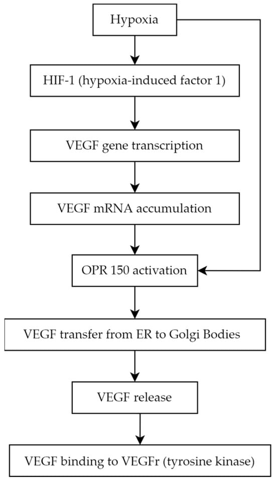
4.1. Vascular Endothelial Growth Factor

Angiogenesis is a complex biological process that underlies the development of proliferative diabetic retinopathy, representing the advanced stage of diabetic retinopathy [26]. Among the many pro-angiogenic factors, vascular endothelial growth factor (VEGF) is notably significant.
VEGF is a homodimer glycoprotein with a molecular weight of 46 kDa, connected by three disulphide bonds in cystine-knot form. The VEGF family consists of the following members:
  • VEGF-A (also called VEGF or vascular permeability factor, the first discovered molecule of the whole family in 1983)
  • VEGF-B
  • VEGF-C (essential for the formation of lymphatic vessels) [27]
  • VEGF-D (known as c-Fos-induced growth factor, FIGF)
  • VEGF-E (connected with parapoxvirus Orf, which causes pustular dermatitis) [28]
  • placenta growth factor (PGF) [29][30].
VEGF production is stimulated by ischemia and hypoxia. Low pO2 induces the production of the crucial mediator of hypoxic responses—DNA-binding protein called hypoxia-induced factor 1 (HIF-1). HIF-1 binds to specific enhancer elements, stimulating the transcription of the VEGF gene, which leads to increased VEGF mRNA production and decreased mRNA degradation. Accumulated intracellular VEGF is transported from endoplasmic reticulum to Golgi bodies by a chaperone protein known as ORP 150 (oxygen-regulated protein 150), whose secretion is augmented in a hypoxic environment [31][32] (Figure 1).
Figure 1. VEGF production and secretion pathway.
VEGF has been shown to play a role in physiological processes such as vasculogenesis—de novo formation of blood vessels during embryogenesis, angiogenesis—vessels formation from already existing vasculature, and pathological processes like tumor growth, tissue remodeling, and metastasis. VEGF mediates its effects by binding to the tyrosine kinase receptors (VEGFRs). The family of VEGFRs consists of the following members: VEGFR1 and VEGFR2 (mainly located on blood vessels endothelial cells) and VEGFR3 (expressed in lymphatic endothelium). The molecular structure of each receptor is similar [33].
VEGFR1 can be secreted in the soluble form (sFlt1—expressed in the placenta during gestation) to prevent endothelial overproliferation due to VEGF accumulation. VEGFR1 is a crucial factor in providing a proper level of VEGF-A, and it plays an important role in the negative regulation of vascular modification. VEGFR1 is also necessary for the process of monocyte migration. PGF bound to VEGFR1 is responsible for initiating inflammatory-related angiogenesis, which is vital in the pathogenesis of various diseases. VEGF-B characteristically binds to VEGFR1 in tissues with high metabolic activity such as myocardial cells.
VEGFR2, the most comprehensively studied among the VEGF receptors, binds with VEGF-A, VEGF-C, and VEGF-D. The activation of VEGFR2 kinase through VEGF-induced VEGFR2 homodimerization is responsible for the majority of the known VEGF-related processes, such as mitosis stimulation, migration, and survival of endothelial cells, ultimately leading to the formation of new blood vessels [35]. The crucial functions of VEGFR2 in endothelial biology are evident from the various processes involved in its tight regulation such as internalization to early endosomes for the activation of specific pathways and the involvement of phosphatases like vascular endothelial protein tyrosine phosphatase.
VEGFR3 has an affinity to VEGF-C and VEGF-D. Initially identified as a regulator of lymphatic endothelial development and biology, VEGFR3 is also present in blood vascular endothelial cells. Endothelial cells involved in angiogenesis express both VEGFR2 and VEGFR3. VEGFR3 activation is not solely dependent on VEGF-C/VEGF-D binding, as it can also be activated through integrin-mediated mechanisms, it leads to lymphatic vessel expansion and the absorption of interstitial fluid [36].
There are known VEGF coreceptors, including the neuropilins, neuropilin-1 and neuropilin-2, whose main role is to enhance the binding of VEGF to the VEGFR2 and stimulate the migration of endothelial cells [34]. Heparan sulfate and integrins also modulate the signal from VEGFRs. 
VEGF is critical for ensuring the proper morphology and function of vascular structures [35]. Notably, mutations resulting in the loss of the VEGF gene allele are lethal [37]. The development of proliferative diabetic retinopathy (PDR) is linked to relative retinal ischemia, creating a hypoxic environment favors HIF-1 activation and VEGF production. Pathological revascularization in the retina has been attributed to VEGF-A165, a specific splice variant of VEGF-A [38].

4.2. Asymmetric Dimethylarginine

Arginine is an amino acid essential for normal growth and development. Endogenous synthesis is adequate in healthy people but might be deficient in many pathological states [39]. The earliest sign of vascular complications is endothelial dysfunction [40]. Nitric oxide (NO) is an important vasodilator that is crucial in maintaining the health of the vascular endothelium. Studies demonstrate that endothelial dysfunction plays a critical role in the development of diabetes-associated microvascular complications and often precedes advanced diabetic retinopathy (DR) [41][42][43].
Nitric oxide (NO) is synthesized from the guanidine group of arginine by the enzyme family NO synthases (NOSs), which consist of three isoforms [44][45]. Asymmetric dimethylarginine (ADMA) is an active endogenous methylated amino acid, a structural analogue of L-arginine, which inhibits the activity of all isoforms of NOS, inhibiting the formation of nitric oxide in tissues and blood plasma [46][47].
ADMA is synthesized by the protein arginine N-methyltransferase 1 (PRMT1), mainly metabolized by the dimethylarginine dimethylaminohydrolases (DDAHs) pathway, and eliminated from the body by kidneys [48][49]. ADMA enters cells through cationic amino-acid transporters (CATs) [50]. Plasma levels of ADMA in healthy people vary between 0.3 and 0.5 μmol/L [51], but in pathological states, it may increase even tenfold [52].
ADMA has a negative effect on cells, contributing to oxidative stress, shortening telomeres, inhibiting the release of NO, and increasing the secretion of interleukin-8 and monocyte chemotaxis factor 1 [51]. Under normal conditions, endothelial NOS is inhibited by 10%, but in pathological situations, even by 30–70% [52]. When the plasma ADMA level increases, the NO synthesis in the environment decreases, vascular homeostasis degrades due to vasoconstriction, and endothelial dysfunction begins [45].
Endothelial dysfunction and impaired ocular hemodynamics prime diabetic retinopathy development are associated with decreased NOS activity and NO bioavailability, thus resulting in increased reactive oxygen species (ROS) and vasoconstriction [52][53]. Oxidative stress is closely related to DDAH activity, which further affects ADMA concentrations in patients with diabetes [54][55]. Increased oxidative stress contributes to elevated ADMA, and by the upregulation of circulating markers of oxidative stress, increased serum ADMA concentration is associated with increased vascular oxidative stress [56][57][58].
ADMA accumulation was first reported in patients characterized by endothelial dysfunction including hyperglycemia, hypercholesterolemia, and hypertension [59][60]. Impaired liver or renal function could also have an impact on the plasma concentration of ADMA. The significance of ADMA in the inhibition of vascular endothelial growth factor-mediated angiogenesis has been demonstrated in numerous studies. Some evidence suggests that diabetes mellitus with microvascular complications has increased serum levels of ADMA [61][62][63][64]. Elevated ADMA was detected in aqueous humor in diabetic patients, especially those with severe retinopathy [65]. The plasma ADMA level is elevated in patients with diabetic microangiopathy such as DR [42][62][66][67][68][69].
Lowering ADMA levels may delay the progression of diabetic retinopathy by reducing the formation of neovascularization, providing protective advantages for the blood–retinal barrier [68].

4.3. MicroRNAs

MicroRNAs (miRNAs) are single-strengthened, non-coding RNA, which affect gene expression regulation. Their suppressor interaction with mRNA usually is associated with 3′ untranslated regions (3′ UTRs), although data claim as well its interaction potential according to different sequences such as gene promoters. Moreover, they also have a regulatory role in transcription and translation processes [70]. The creation process of those micromolecules goes from DNA transcription to primary miRNA (pri-mRNA) through precursor miRNA (pre-miRNA) leading to mature miRNA formation [71]. The role of miRNA in signalization pathways is studied nowadays excessively because of those particles’ multiplicity.
Molecular bases of miRNA mechanisms of action are distinct for different miRNAs, and it is possible to distinguish which particles affect which pathway leading to diabetic retinopathy, such as affecting cell proliferation, angiogenesis, apoptosis, or basement membrane thickening [72]. It has been proven that directly or indirectly particles such as miRNA-9, miRNA-152, miRNA-15b, miRNA-29b-3p, miRNA-199a-3p, miRNA-203a-3p, miRNA-200b-3p, and miRNA-30a-3p downregulate VEGF expression, which lowers the range of active cell-cycle-related proteins and by that protects RMECs (retinal microvascular endothelial cells) from abnormal proliferation [73]. In addition, from previously mentioned biomolecules, the alternative pathway to downregulate VEGF is SIRT1 (nicotinamide adenosine dinucleotide (NAD+)-dependent deacetylase) upregulation, which is possible by miRNA-29b-3p and miRNA-34a inhibition, moreover, causing an increase in proinflammatory cytokines [73]. MiRNA-34a was evaluated to be an interesting therapeutic target, as in rats with induced diabetic retinopathy, its silencing was observed as an apoptosis regulation [74]
MiRNA-20a and miRNA-20b were revealed to downregulate VEGF as well but in different mechanisms—first act by Yes-associated protein (YAP)/hypoxia-inducible factor 1α (HIF1α)/VEGF axis, and second was revealed in the study on rats to be correlated with downregulation of AKT3, lowering VEGF expression [75][76]. Moreover, it was assessed that resolvin D1 modulates the intracellular VEGF-related miRNAs—miRNA-20a-3p, miRNA-20a-5p, miRNA-106a-5p, and miRNA-20b—expression of retinal photoreceptors challenged with high glucose [77].
The role of the miRNA as biomarkers for diabetic retinopathy was investigated using various sample types and study designs, comparing different groups based on diabetes type (T1DM or T2DM), patients with diabetes,and healthy individuals, as well studies examining the progression of diabetic retinopathy. In blood serum samples in T1DM patients with and without diabetic retinopathy, miRNA-211 was the most significant. Additionally, miRNA-18b and miRNA-19b were found to be upregulated, along with miRNA-29a, miRNA-148a, miRNA-181a, and miRNA-200a, which also showed notable impact [78][79].
In T2DM patients, a study identified differences in the following particles: hsa-let-7a-5p, hsa-miRNA-novel-chr5_15976, hsa-miRNA-28-3p, hsa-miRNA-151a-5p, and hsa-miRNA-148a-3p, which were upregulated compared to DM group without retinopathy. Notably, a panel of the first three miRNA (hsa-let-7a-5p, hsa-miRNA-novel-chr5_15976, and hsa-miRNA-28-3p) showed the highest diagnostic potential with sensitivity and specificity of 0.92 and 0.94, respectively [80]. Another study showed that in T2DM patients, diabetic retinopathy was associated with increased circulating levels of miRNA-25-3p and miRNA-320b, and decreased levels of miRNA-495-3p [81]
Plasma results among T2DM patients revealed lower levels of miRNA-29b in those with diabetic retinopathy, and miRNA-21 was significantly associated with proliferative diabetic retinopathy (PDR). Other parameters increased in T2DM patients with diabetic retinopathy included miRNA-93 via SIRT1, miRNA-21, and miRNA-152 [82][83]. Conversely, miRNA-15a, miRNA-20b, miRNA-21, miRNA-24, miRNA-320, miRNA-486, and miRNA-150, miRNA-126, miRNA-191, miRNA-197 were downregulated in the plasma samples of these patients [84].
Importantly, miRNA-150 is observed in the circulation of both T1DM and T2DM patients circulation and in the neutral retina. miRNA-150 upregulates Elk1, stimulating proinflammatory, pro-angiogenic, and apoptotic influences. A lower range of miRNA-150 in serum impacts Elk1 and Myb overexpression, leading to similar pathway resulting in microvascular complications and neovascularization, culminating in diabetic retinopathy. Therefore, miRNA-150 is not only a diagnostic biomarker but also significantly involved in the diabetic retinopathy pathogenesis [85].

4.4. Endothelin-1

Endothelin-1 (ET-1) in its active form is a 21-amino acid hormone that helps to maintain basal vascular tone and metabolic function in healthy individuals [86]. ET-1, an endothelium-derived factor, exhibits proliferative, profibrotic, and proinflammatory properties [87]. It is the most abundantly expressed member of the endothelin family, which includes ET-1, ET-2, and ET-3. Immature ET-1 undergoes extensive post-transcriptional processing, culminating in cleavage by endothelin converting enzymes (ECEs) and the release of mature ET-1 primarily into the interstitial space, with a smaller proportion entering the circulation [86].
ET-1 exerts its biological effect through two receptor subtypes: ETA and ETB [88]. ETA receptors are predominantly localized on vascular smooth muscle cells (VSMCs) of blood vessels, where they mediate contractile and proliferative response to ET-1. In contrast, ETB receptors have a more complex role in vascular regulation; they can cause vasodilation by releasing relaxing factors when present on endothelial cells or vasoconstriction when located on VSMCs in certain vascular beds [87]. Therefore, the overall effect of ET-1 on different tissues largely depend on the expression and relative densities of these receptor subtypes. ET-1 is a crucial marker of endothelial dysfunction, a condition characterized by an imbalance between vasoconstrictors and vasodilators [89].
Due to its vasoconstrictive properties, ET-1 has been widely studied for its role in hypertension and has proven clinically significant, as evidenced by the use of endothelin receptor antagonists in treating pulmonary arterial hypertension (PAH) [90]. The vasoconstrictive and in turn hypertensive properties of ET-1 can explain a possible link between elevated plasma ET-1 level and retinopathy under ischemia, a finding relevant to diabetic retinopathy, which is thought to be the consequence of retinal ischemia. Animal models have shown that administering ET-1 into the posterior vitreous body or optic nerve leads to ischemic-related physiological and cellular damage, including obstruction of retinal blood flow, elevated scotopic b-wave in electroretinogram, and apoptosis of cells in the ganglion cell layer of the retina [91].

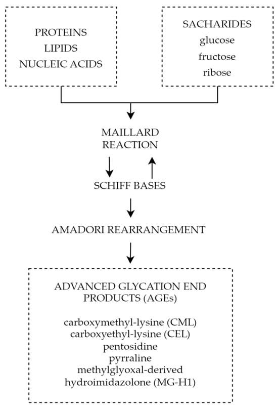
4.5. Advanced Glycation End Products

One of the mechanisms connecting chronic hyperglycemia with diabetic retinopathy is the formation and accumulation of advanced glycation end products (AGEs). Advanced glycation end products are heterogeneous groups of molecules formed from post-translational non-enzymatic modifications of proteins, lipids, or nucleic acids by saccharides including glucose, fructose, and pentose through the Maillard reaction represented by Figure 2 [92][93]. There are over 20 AGEs identified in human tissues, but some of the most common ones are carboxymethyl-lysine (CML), carboxyethyl-lysine (CEL), pentosidine, pyrraline, and methylglyoxal-derived hydroimidazolone (MG-H1) [94]. The characteristic factor of AGEs that distinguishes them from early glycation products, such as glycohemoglobin A1c (HbA1c), is the lack of spontaneous reversion ability, which once derived results in the accumulation in tissues over time [95]. Even though the discovery of AGEs dates to the early 20th century, not until the 1980s, the role of AGEs in aging and chronic diseases was recognized [96]. The first mention of AGEs and their accumulation in human tissues and their potential role in diabetic complications appeared in 1988 in a scientific article published by Helen Vlassara et al. [97]. Since then, AGEs and their involvement in pathophysiological processes have been the subject of extensive research.
Figure 2. Forming of AGEs through Maillard reaction.

5. Summary

ADMA inhibits the activity of NOS, which results in decreased levels of NO and leads to vasoconstriction and endothelial dysfunction. Increased ADMA levels may be considered an early prognostic factor of diabetes complications such as PDR. The use of ADMA as a biomarker may help in early diagnosis, monitoring, and effective therapeutic management of the disease. Reducing ADMA levels in patients with diabetes may be a new therapeutic target to prevent the development of diabetic retinopathy. Endothelin-1 is another factor with an undoubted relationship to diabetic retinopathy. Increased serum and aqueous humor levels are observed in patients with ET-1 elevation dependent on the severity of the progression of the disease. This, juxtaposed with promising results of ET-1 receptor antagonist animal studies, showcases the potential of ET-1 as a possible target for future therapy. It is important to note that miRNAs are not only supposed to be an innovative predictive biomarker and progression indicator in DR but also a potential therapeutic target. Different miRNAs can be found in T1DM and T2DM as well depending on sample type, moreover, some of them differ depending on DR type. The variety of miRNAs and frequently high amounts of particles involved in several pathogenesis pathways can be at the same time the advantage and disadvantage of that prospective novel biomarkers group; hence, miRNAs panels are more adequate than a single biomarker rating. Finally, advanced glycation end products play a significant role in the pathophysiology of diabetic retinopathy causing impairment of the neurovascular units through reactive oxygen species, inflammatory reactions, and cell death pathways. All the above mechanisms play a significant role not only in diabetic retinal disorders, but also other chronic oxidative-based diseases; therefore, a thorough understanding of their properties and mechanisms will allow advances in the diagnosis and treatment of chronic diseases and most importantly diabetic retinopathy. The above factors and signaling pathways can help to create multimodal and highly specified therapies for patients suffering from DR. It is crucial to investigate molecular agents participating in DR pathogenesis. Hopefully, it will provide the ability to inhibit this progressive disease at its early stage.

References

  1. Tan, T.E.; Wong, T.Y. Diabetic retinopathy: Looking forward to 2030. Front. Endocrinol. 2023, 13, 1077669.
  2. International Diabetes Federation. International Diabetes Federation—Facts & Figures. Idf.org. Published 12 September 2021. Available online: https://www.idf.org/aboutdiabetes/what-is-diabetes/facts-figures.html (accessed on 15 May 2023).
  3. Cheung, N.; Mitchell, P.; Wong, T.Y. Diabetic retinopathy. Lancet 2010, 376, 124–136.
  4. Modjtahedi, B.S.; Wu, J.; Luong, T.Q.; Gandhi, N.K.; Fong, D.S.; Chen, W. Severity of Diabetic Retinopathy and the Risk of Future Cerebrovascular Disease, Cardiovascular Disease, and All-Cause Mortality. Ophthalmology 2021, 128, 1169–1179.
  5. Teo, Z.L.; Tham, Y.C.; Yu, M.; Chee, M.L.; Rim, T.H.; Cheung, N.; Bikbov, M.M.; Wang, Y.X.; Tang, Y.; Lu, Y.; et al. Global Prevalence of Diabetic Retinopathy and Projection of Burden through 2045: Systematic Review and Meta-analysis. Ophthalmology 2021, 128, 1580–1591.
  6. Lin, K.Y.; Hsih, W.H.; Lin, Y.B.; Wen, C.Y.; Chang, T.J. Update in the epidemiology, risk factors, screening, and treatment of diabetic retinopathy. J. Diabetes Investig. 2021, 12, 1322–1325.
  7. Chew, E.Y.; Davis, M.D.; Danis, R.P.; Locato, J.F. The effects of medical management on the progression of diabetic retinopathy in persons with type 2 diabetes: The Action to Control Cardiovascular Risk in Diabetes (ACCORD) Eye Study. Ophthalmology. 2014, 121, 2443–2451.
  8. Action to Control Cardiovascular Risk in Diabetes Follow-On (ACCORDION) Eye Study Group; The Action to Control Cardiovascular Risk in Diabetes Follow-On (ACCORDION) Study Group. Persistent effects of intensive glycemic control on retinopathy in type 2 diabetes in the Action to Control Cardiovascular Risk in Diabetes (ACCORD) follow-on study. Diabetes Care 2016, 39, 1089–1100.
  9. Kaštelan, S.; Tomić, M.; Gverović Antunica, A.; Ljubić, S.; Salopek Rabatić, J.; Karabatić, M. Body mass index: A risk factor for retinopathy in type 2 diabetic patients. Mediat. Inflamm. 2013, 2013, 436329.
  10. Chang, Y.C.; Wu, W.C. Dyslipidemia and diabetic retinopathy. Rev. Diabet. Stud. 2013, 10, 121–132.
  11. Kohner, E.M.; Aldington, S.J.; Stratton, I.M.; Manley, S.E.; Holman, R.R.; Matthews, D.R.; Turner, R.C. United Kingdom Prospective Diabetes Study 30. Diabetic retinopathy at diagnosis of non-insulin-dependent diabetes mellitus and associated risk factors. Arch. Ophthalmol. 1998, 116, 297–303.
  12. Klein, R.; Sharrett, A.R.; Klein, B.E.; Moss, S.E.; Folsom, A.R.; Wong, T.Y.; Brancati, F.L.; Hubbard, L.D.; Couper, D.; ARIC Group. The association of atherosclerosis, vascular risk factors, and retinopathy in adults with diabetes: The Atherosclerosis Risk in Communities study. Ophthalmology 2002, 109, 1225–1234.
  13. Klein, R.; Marino, E.K.; Kuller, L.H.; Polak, J.F.; Tracy, R.P.; Gottdiener, J.S.; Burke, G.L.; Hubbard, L.D.; Boineau, R. The relation of atherosclerotic cardiovascular disease to retinopathy in people with diabetes in the Cardiovascular Health Study. Br. J. Ophthalmol. 2002, 86, 84–90.
  14. van Leiden, H.A.; Dekker, J.M.; Moll, A.C.; Nijpels, G.; Heine, R.J.; Bouter, L.M.; Stehouwer, C.D.; Polak, B.C. Blood pressure, lipids, and obesity are associated with retinopathy: The Hoorn study. Diabetes Care 2002, 25, 1320–1325.
  15. Vujosevic, S.; Aldington, S.J.; Silva, P.; Peto, P. Screening for diabetic retinopathy: New perspectives and challenges. Lancet Diabetes Endocrinol. 2020, 8, 337–347.
  16. Cai, X.; Chen, Y.; Yang, W.; Gao, X.; Han, X.; Ji, L. The association of smoking and risk of diabetic retinopathy in patients with type 1 and type 2 diabetes: A meta-analysis. Endocrine 2018, 62, 299–306.
  17. Rasmussen, K.L.; Laugesen, C.S.; Ringholm, L.; Vestgaard, M.; Damm, P.; Mathiesen, E.R. Progression of diabetic retinopathy during pregnancy in women with type 2 diabetes. Diabetologia 2010, 53, 1076–1083.
  18. Rubsam, A.; Parikh, S.; Fort, P.E. Role of Inflammation in Diabetic Retinopathy. Int. J. Mol. Sci. 2018, 19, 942.
  19. Simo, R.; Stitt, A.W.; Gardner, T.W. Neurodegeneration in diabetic retinopathy: Does it really matter? Diabetologia 2018, 61, 1902–1912.
  20. Ansari, P.; Tabasumma, N.; Snigdha, N.N.; Siam, N.H.; Panduru, R.V.N.R.S.; Azam, S.; Hannan, J.M.A.; Abdel-Wahab, Y.H.A. Diabetic Retinopathy: An Overview on Mechanisms, Pathophysiology and Pharmacotherapy. Diabetology 2022, 3, 159–175.
  21. Kowluru, R.A.; Santos, J.M.; Mishra, M. Epigenetic modifications and diabetic retinopathy. Biomed. Res. Int. 2013, 2013, 635284.
  22. Kollias, A.N.; Ulbig, M.W. Diabetic retinopathy: Early diagnosis and effective treatment. Dtsch. Arztebl. Int. 2010, 107, 75–83.
  23. Williams, B.; Baker, A.Q.; Gallacher, B.; Lodwick, D. Angiotensin II increases vascular permeability factor gene expression by human vascular smooth muscle cells. Hypertension 1995, 25, 913–917.
  24. Viswanath, K.; McGavin, D.D. Diabetic retinopathy: Clinical findings and management. Community Eye Health 2003, 16, 21–24.
  25. Roy, S.; Kim, D. Retinal capillary basement membrane thickening: Role in the pathogenesis of diabetic retinopathy. Prog. Retin. Eye Res. 2021, 82, 100903.
  26. Nawaz, I.M.; Rezzola, S.; Cancarini, A.; Russo, A.; Costagliola, C.; Semeraro, F.; Presta, M. Human vitreous in proliferative diabetic retinopathy: Characterization and translational implications. Prog. Retin. Eye Res. 2019, 72, 100756.
  27. Homsi, J.; Daud, A.I. Spectrum of activity and mechanism of action of VEGF/PDGF inhibitors. Cancer Control 2007, 14, 285–294.
  28. Shibuya, M. Vascular endothelial growth factor receptor-2: Its unique signaling and specific ligand, VEGF-E. Cancer Sci. 2005, 94, 751–756.
  29. Arrigo, A.; Aragona, E.; Bandello, F. VEGF-targeting drugs for the treatment of retinal neovascularization in diabetic retinopathy. Ann. Med. 2022, 54, 1089–1111.
  30. Holmes, D.I.; Zachary, I. The vascular endothelial growth factor (VEGF) family: Angiogenic factors in health and disease. Genome Biol. 2005, 6, 209.
  31. Gupta, N.; Mansoor, S.; Sharma, A.; Sapkal, A.; Sheth, J.; Falatoonzadeh, P.; Kuppermann, B.; Kenney, M. Diabetic retinopathy and VEGF. Open Ophthalmol. J. 2013, 7, 4–10.
  32. Ferrara, N. Vascular Endothelial Growth Factor: Basic Science and Clinical Progress. Endocr. Rev. 2004, 25, 581–611.
  33. Stuttfeld, E.; Ballmer-Hofer, K. Structure and function of VEGF receptors. IUBMB Life 2009, 61, 915–922.
  34. Clauss, M. Molecular Biology of the VEGF and the VEGF Receptor Family. Semin. Thromb. Hemost. 2000, 26, 561–570.
  35. Wang, X.; Bove, A.M.; Simone, G.; Ma, B. Molecular Bases of VEGFR-2-Mediated Physiological Function and Pathological Role. Front. Cell Dev. Biol. 2020, 8, 599281.
  36. Claesson-Welsh, L. VEGF receptor signal transduction—A brief update. Vasc. Pharmacol. 2016, 86, 14–17.
  37. Carmeliet, P.; Ferreira, V.; Breier, G.; Harpal, K. Abnormal blood vessel development and lethality in embryos lacking a single VEGF allele. Nature 1996, 380, 435–439.
  38. Bucolo, C.; Barbieri, A.; Vigano, I.; Band, F. Short-and Long-Term Expression of Vegf: A Temporal Regulation of a Key Factor in Diabetic Retinopathy. Front. Pharmacol. 2021, 12, 707909.
  39. Grillo, M.A.; Colombatto, S. Arginine revisited: Minireview article. Amino Acids 2004, 26, 345–351.
  40. Endemann, D.H.; Schiffrin, E.L. Endothelial dysfunction. J. Am. Soc. Nephrol. 2004, 15, 1983–1992.
  41. Yonem, A.; Duran, C.; Unal, M.; Ipcioglu, O.M.; Ozcan, O. Plasma apelin and asymmetric dimethylarginine levels in type 2 diabetic patients with diabetic retinopathy. Diabetes Res. Clin. Pract. 2009, 84, 219–223.
  42. Narayanan, P.S.; Rojas, M.; Suwanpradid, J.; Toque, H.A.; Caldwell, W.R.; Caldwell, R.B. Arginase in retinopathy. Prog. Retin. Eye Res. 2013, 36, 260–280.
  43. Sena, C.M.; Pereira, A.M.; Seica, R. Endothelial dysfunction—A major mediator of diabetic vascular disease. Biochim. Biophys. Acta 2013, 1832, 2216–2231.
  44. Forstermann, U.; Closs, E.I.; Pollock, J.S.; Nakane, M.; Schwarz, P.; Gath, I.; Kleinert, H. Nitric oxide synthase isozymes. Characterization, purification, molecular cloning, and functions. Hypertension 1994, 23, 1121–1131.
  45. Toutouzas, K.; Riga, M.; Stefanadi, E.; Stefanadis, C. Asymmetric dimethylarginine (ADMA) and other endogenous nitric oxide synthase (NOS) inhibitors as an important cause of vascular insulin resistance. Horm. Metab. Res. 2008, 40, 655–659.
  46. Bode-Boger, S.M.; Scalera, F.; Martens-Lobenhoffer, J. Asymmetric dimethylarginine (ADMA) accelerates cell senescence. Vasc. Med. 2005, 10, 65–71.
  47. Sirman, Y.V.; Savytskyi, I.V. Study of endothelial dysfunction and asymmetric dimethylarginine levels. J. Educ. Health Sport 2019, 9, 395–412.
  48. Leiper, J.M.; Vallance, P. The synthesis and metabolism of asymmetric dimethylarginine (ADMA). Eur. J. Clin. Pharmacol. 2006, 62, 33–38.
  49. Morris, S.M. Arginine metabolism revisited. J. Nutr. 2016, 146, 2579–2586.
  50. Trocha, M.; Merwid-Lad, A.; Szuba, A.; Sozanski, T.; Magdalan, J.; Szelag, A. Asymmetric dimethylarginine synthesis and degradation under physiological and pathological conditions. Adv. Clin. Exp. Med. 2010, 19, 233–243.
  51. Vallance, P.; Leone, A.; Calver, A.; Collier, J.; Moncada, S. Accumulation of an endogenous inhibitor of nitric oxide synthesis in chronic renal failure. Lancet 1992, 339, 572–575.
  52. Sydow, K.; Munzel, T. ADMA and oxidative stress. Atheroscler. Suppl. 2003, 4, 41–51.
  53. Cardounel, A.J.; Cui, H.; Samouilov, A.; Johnson, W.; Kearns, P.; Tsai, A.-L.; Berka, V.; Zweier, J.L. Evidence for the pathophysiological role of endogenous methylarginines in regulation of endothelial NO production and vascular function. J. Biol. Chem. 2007, 282, 879–887.
  54. Jian, Q.; Wu, Y.; Zhang, F. Metabolomics in diabetic retinopathy: From potential biomarkers to molecular basis of oxidative stress. Cells 2022, 11, 3005.
  55. Peters, K.S.; Rivera, E.; Warden, C.; Harlow, P.A.; Mitchell, S.L.; Calcutt, M.W.; Samuels, D.C.; Brantley, M.A. Plasma arginine and citrulline are elevated in diabetic retinopathy. Am. J. Ophthalmol. 2022, 235, 154–162.
  56. Sumarriva, K.; Uppal, K.; Ma, C.; Herren, D.J.; Wang, Y.; Chocron, I.M.; Warden, C.; Mitchell, S.L.; Burgess, G.L.; Goodale, M.P.; et al. Arginine and carnitine metabolites are altered in diabetic retinopathy. Investig. Ophthalmol. Vis. Sci. 2019, 60, 3119–3126.
  57. Dag, U.; Caglayan, M.; Alakus, M.F.; Oncul, H. The relationship between reduced choroidal thickness due to high plasma asymmetrical dimethylarginine level and increased severity of diabetic retinopathy. Arq. Bras. Oftalmol. 2023, 86, 27–32.
  58. Lamprou, S.; Koletsos, N.; Mintziori, G.; Anyfanti, P.; Trakatelli, C.; Kotsis, V.; Gkaliagkousi, E.; Triantafyllou, A. Microvascular and endothelial dysfunction in prediabetes. Life 2023, 13, 644.
  59. Krasnicki, P.; Proniewska-Skretek, E.; Dmuchowska, D.A.; Dobrzycki, S.; Mariak, Z. Asymmetric dimethylarginine (ADMA) as a marker of blood flow disturbances in ocular circulation in patients with type 2 diabetes and coronary artery disease. Mag. Lek. Okulisty 2009, 3, 325–331.
  60. Tousoulis, D.; Kampoli, A.-M.; Stefanadis, C. Diabetes mellitus and vascular endothelial dysfunction: Current perspectives. Curr. Vasc. Pharmacol. 2012, 10, 19–32.
  61. Stepien, E.; Szuscik, I.; Tokarz, A.; Enguita, F.J.; Solnica, B.; Zurakowski, A.; Malecki, M. The role of microparticles in pathomechanisms of diabetic retinopathy—Analysis of intercellular communication mechanisms in endothelial aging. Case control study in patients with metabolic syndrome, diabetes type 1 and type 2. J. Med. Sci. 2014, 83, 322–327.
  62. Huang, C.-Y.; Zhou, T.; Li, G.; Li, M.-Y.; Xiong, X.-M.; Wu, M.-T.; Jiang, J.-L. Asymmetric dimethylarginine aggravates blood-retinal barrier breakdown of diabetic retinopathy via inhibition of intercellular communication in retinal pericytes. Amino Acids 2019, 51, 1515–1526.
  63. Liu, J.; Li, C.; Chen, W.; He, K.; Ma, H.; Ma, B.; Zhao, P.; Tian, L. Relationship between serum asymmetric dimethylarginine level and microvascular. Bio. Med. Res. Int. 2019, 2019, 2941861.
  64. Alpay, A.; Ozcan, O.; Ugurbas, S.C.; Ugurbas, S.H. Investigated of vitreous and serum asymmetric dimethylarginine levels in diabetic. Res. Sq. 2019, 2019.
  65. Sugai, M.; Ohta, A.; Ogata, Y.; Nakanishi, M.; Ueno, S.; Kawata, T.; Saito, N.; Tanaka, Y. Asymmetric dimethylarginine (ADMA) in the aqueous humor of diabetic patients. Endocr. J. 2007, 54, 303–309.
  66. Abhary, S.; Kasmeridis, N.; Burdon, K.P.; Kuot, A.; Whiting, M.J.; Yew, W.P.; Petrovsky, N.; Craig, J.E. Diabetic retinopathy is associated with elevated serum asymmetric and symmetric dimethylarginines. Diabetes Care 2009, 32, 2084–2086.
  67. Eliana, F.; Suwondo, P.; Makmun, L.H.; Harbuwono, D.S. ADMA as a marker of endothelial dysfunction in prediabetic women. Acta Medica Indones. 2011, 43, 92–98.
  68. Du, M.-R.; Yan, L.; Li, N.-S.; Wang, Y.-J.; Zhou, T.; Jiang, J.-L. Asymmetric dimethylarginine contributes to retinal neovascularization of diabetic retinopathy through EphrinB2 pathway. Vasc. Pharmacol. 2018, 108, 46–56.
  69. Yun, J.H.; Kim, J.-M.; Jeon, H.J.; Oh, T.; Choi, H.J.; Kim, B.-J. Metabolomics profiles associated with diabetic retinopathy in type 2 diabetes patients. PLoS ONE 2020, 15, e241365.
  70. O’Brien, J.; Hayder, H.; Zayed, Y.; Peng, C. Overview of MicroRNA Biogenesis, Mechanisms of Actions, and Circulation. Front. Endocrinol. 2018, 9, 402.
  71. Ha, M.; Kim, V. Regulation of microRNA biogenesis. Nat. Rev. Mol. Cell Biol. 2014, 15, 509–524.
  72. Karbasforooshan, H.; Karimi, G. The role of SIRT1 in diabetic retinopathy. Biomed. Pharmacother. 2018, 97, 190–194.
  73. Chang, X.; Zhu, G.; Cai, Z.; Wang, Y.; Lian, R.; Tang, X.; Ma, C.; Fu, S. miRNA, lncRNA and circRNA: Targeted Molecules Full of Therapeutic Prospects in the Development of Diabetic Retinopathy. Front. Endocrinol. 2021, 12, 771552.
  74. Ji, Q.; Han, J.; Wang, L.; Liu, J.; Dong, Y.; Zhu, K.; Shi, L. MicroRNA-34a promotes apoptosis of retinal vascular endothelial cells by targeting SIRT1 in rats with diabetic retinopathy. Cell Cycle 2020, 19, 2886–2896.
  75. Pan, Q.; Gao, Z.; Zhu, C.; Peng, Z.; Song, M.; Li, L. Overexpression of histone deacetylase SIRT1 exerts an antiangiogenic role in diabetic retinopathy via miR-20a elevation and YAP/HIF1α/VEGFA depletion. Am. J. Physiol. Endocrinol. Metab. 2020, 319, 932–943.
  76. Qin, B.; Liu, J.; Liu, S.; Li, B.; Ren, J. MiR-20b targets AKT3 and modulates vascular endothelial growth factor-mediated changes in diabetic retinopathy. Acta Biochim. Biophys. Sin. 2016, 48, 732–740.
  77. Maisto, R.; Trotta, M.C.; Petrillo, F.; Izzo, S.; Cuomo, G.; Alfano, R.; Hermenean, A.; Barcia, J.M.; Galdiero, M.; Platania, C.B.M.; et al. Resolvin D1 Modulates the Intracellular VEGF-Related miRNAs of Retinal Photoreceptors Challenged With High Glucose. Front. Pharmacol. 2020, 11, 235.
  78. Liu, H.N.; Cao, N.J.; Li, X.; Qian, W.; Chen, X.L. Serum microRNA-211 as a biomarker for diabetic retinopathy via modulating Sirtuin 1. Biochem. Biophys. Res. Commun. 2018, 505, 1236–1243.
  79. Miao, C.; Chang, J.; Zhang, G.; Fang, Y. MicroRNAs in type 1 diabetes: New research progress and potential directions. Biochem. Cell Biol. 2018, 96, 498–506.
  80. Liang, Z.; Gao, K.P.; Wang, Y.X.; Liu, Z.C.; Tian, L.; Yang, X.Z.; Ding, J.Y.; Wu, W.T.; Yang, W.H.; Li, Y.L.; et al. RNA sequencing identified specific circulating miRNA biomarkers for early detection of diabetes retinopathy. Am. J. Physiol. Endocrinol. Metab. 2018, 315, 374–385.
  81. Santovito, D.; Toto, L.; De Nardis, V.; Ces, D. Plasma microRNA signature associated with retinopathy in patients with type 2 diabetes. Sci Rep. 2021, 11, 4136.
  82. Guo, J.; Zhou, P.; Liu, Z.; Dai, F.; Pan, M.; An, G.; Han, J.; Du, L.; Jin, X. The Aflibercept-Induced MicroRNA Profile in the Vitreous of Proliferative Diabetic Retinopathy Patients Detected by Next-Generation Sequencing. Front. Pharmacol. 2021, 12, 781276.
  83. Saleh, A.A.; El-Hefnawy, S.M.; Kasemy, Z.A.; Alhagaa, A.A.; Nooh, M.Z.; Arafat, E.S. Mi-RNA-93 and Mi-RNA-152 in the Diagnosis of Type 2 Diabetes and Diabetic Retinopathy. Br. J. Biomed. Sci. 2022, 79, 10192.
  84. Zampetaki, A.; Kiechl, S.; Drozdov, I.; Willeit, P.; Mayr, U.; Prokopi, M.; Mayr, A.; Weger, S.; Oberhollenzer, F.; Bonora, E.; et al. Plasma microRNA profiling reveals loss of endothelial miR-126 and other microRNAs in type 2 diabetes. Circ. Res. 2010, 107, 810–817.
  85. Ko, G.Y.; Yu, F.; Bayless, K.J.; Ko, M.L. MicroRNA-150 (miR-150) and Diabetic Retinopathy: Is miR-150 Only a Biomarker or Does It Contribute to Disease Progression? Int. J. Mol. Sci. 2022, 23, 99.
  86. Jenkins, H.N.; Rivera-Gonzalez, O.; Gibert, Y.; Speed, J.S. Endothelin-1 in the pathophysiology of obesity and insulin resistance. Obes. Rev. 2020, 21, e13086.
  87. Ergul, A. Endothelin-1 and diabetic complications: Focus on the vasculature. Pharmacol. Res. 2011, 63, 477–482.
  88. Stow, L.R.; Jacobs, M.E.; Wingo, C.S.; Cain, B.D. Endothelin-1 gene regulation. FASEB J. 2010, 25, 16–28.
  89. Kostov, K. The causal relationship between endothelin-1 and hypertension: Focusing on endothelial dysfunction, arterial stiffness, vascular remodeling, and Blood Pressure Regulation. Life 2021, 11, 986.
  90. Abman, S.H. Role of endothelin receptor antagonists in the treatment of pulmonary arterial hypertension. Annu. Rev. Med. 2009, 60, 13–23.
  91. Cheung, S.S.; Leung, J.W.; Lam, A.K.; Acy, L. Selective over-expression of endothelin-1 in endothelial cells exacerbates inner retinal edema and neuronal death in ischemic retina. PLoS ONE 2011, 6, e26184.
  92. Shen, C.Y.; Lu, C.H.; Wu, C.H.; Li, K.J. The Development of Maillard Reaction, and Advanced Glycation End Product (AGE)-Receptor for AGE (RAGE) Signaling Inhibitors as Novel Therapeutic Strategies for Patients with AGE-Related Diseases. Molecules 2020, 25, 5591.
  93. Ruiz, H.H.; Ramasamy, R.; Schmidt, A.M. Advanced Glycation End Products: Building on the Concept of the “Common Soil” in Metabolic Disease. Endocrinology 2020, 161, bqz006.
  94. Reddy, V.P.; Aryal, P.; Darkwah, E.K. Advanced Glycation End Products in Health and Disease. Microorganisms 2022, 10, 1848.
  95. Khalid, M.; Petroianu, G.; Adem, A. Advanced Glycation End Products and Diabetes Mellitus: Mechanisms and Perspectives. Biomolecules 2022, 12, 542.
  96. Mao, L.; Yin, R.; Yang, L.; Zhao, D. Role of advanced glycation end products on vascular smooth muscle cells under diabetic atherosclerosis. Front. Endocrinol. 2022, 13, 983723.
  97. Vlassara, H.; Bucala, R.; Striker, L. Pathogenic effects of advanced glycosylation: Biochemical, biologic, and clinical implications for diabetes and aging. Lab. Investig. 1988, 58, 317–326.
More
Upload a video for this entry
Information
Subjects: Ophthalmology
Contributors MDPI registered users' name will be linked to their SciProfiles pages. To register with us, please refer to https://encyclopedia.pub/register : , , , , , ,
View Times: 345
Revisions: 3 times (View History)
Update Date: 05 Aug 2024
1000/1000
Hot Most Recent
Academic Video Service