Submitted Successfully!
To reward your contribution, here is a gift for you: A free trial for our video production service.
Thank you for your contribution! You can also upload a video entry or images related to this topic.
Version Summary Created by Modification Content Size Created at Operation
1 -- 3772 2023-11-13 14:47:42 |
2 Reference format revised. + 3 word(s) 3775 2023-11-14 01:37:55 |

Video Upload Options

Do you have a full video?

Confirm

Are you sure to Delete?
Cite
If you have any further questions, please contact Encyclopedia Editorial Office.
Jain, K.G.; Xi, N.M.; Zhao, R.; Ahmad, W.; Ali, G.; Ji, H. Alveolar Type 2 Epithelial Cell Organoid Culture Methods. Encyclopedia. Available online: https://encyclopedia.pub/entry/51485 (accessed on 03 May 2024).
Jain KG, Xi NM, Zhao R, Ahmad W, Ali G, Ji H. Alveolar Type 2 Epithelial Cell Organoid Culture Methods. Encyclopedia. Available at: https://encyclopedia.pub/entry/51485. Accessed May 03, 2024.
Jain, Krishan Gopal, Nan Miles Xi, Runzhen Zhao, Waqas Ahmad, Gibran Ali, Hong-Long Ji. "Alveolar Type 2 Epithelial Cell Organoid Culture Methods" Encyclopedia, https://encyclopedia.pub/entry/51485 (accessed May 03, 2024).
Jain, K.G., Xi, N.M., Zhao, R., Ahmad, W., Ali, G., & Ji, H. (2023, November 13). Alveolar Type 2 Epithelial Cell Organoid Culture Methods. In Encyclopedia. https://encyclopedia.pub/entry/51485
Jain, Krishan Gopal, et al. "Alveolar Type 2 Epithelial Cell Organoid Culture Methods." Encyclopedia. Web. 13 November, 2023.
Alveolar Type 2 Epithelial Cell Organoid Culture Methods
Edit

Lung diseases rank third in terms of mortality and represent a significant economic burden globally. Scientists have been conducting research to better understand respiratory diseases and find treatments for them. An ideal in vitro model must mimic the in vivo organ structure, physiology, and pathology. Organoids are self-organizing, three-dimensional (3D) structures originating from adult stem cells, embryonic lung bud progenitors, embryonic stem cells (ESCs), and induced pluripotent stem cells (iPSCs). These 3D organoid cultures may provide a platform for exploring tissue development, the regulatory mechanisms related to the repair of lung epithelia, pathophysiological and immunomodulatory responses to different respiratory conditions, and screening compounds for new drugs. To create 3D lung organoids in vitro, both co-culture and feeder-free methods have been used. However, there exists substantial heterogeneity in the organoid culture methods, including the sources of type 2 alveolar cells (AT2) cells, media composition, and feeder cell origins. 

organoids alveolar type 2 cells stem cells 3D cultures pulmonary diseases

1. Introduction

Alveoli, located at the distal end of the lung, are tiny air sacs whose epithelial layer consists of two types of epithelial cells [1]. Type 1 epithelial (AT1) cells cover approximately 95% of the alveolar surface area and are responsible for gaseous exchange. Type 2 alveolar cells (AT2) form the remaining 5% of the alveolar surface area and are responsible for surfactant secretion, immune defense against infections, and the regulation of fluid volume. AT2 cells serve as the facultative adult stem cells of the distal lung, and they are responsible for regenerating the alveoli through self-renewal and differentiation into AT1 cells [2][3][4]. Distal lung diseases, such as COPD/emphysema, and infections including severe acute respiratory syndrome (SARS) and COVID-19, can cause damage to the alveolar epithelium and are among the leading causes of global mortality [5][6][7][8].
Studies on primary and secondary respiratory diseases have been limited by the scarcity of models for the human alveolar epithelium that faithfully mimic in vivo physiology and pathophysiology [9]. For decades, the 2D air–liquid interface model of AT2 cells has been the most extensively studied tool for modeling epithelial barrier function and lung diseases [10][11]. While this 2D model has provided valuable insights into the alveolar structure and function of distal lungs, enriching our knowledge of alveolar diseases, it has often failed to maintain cell proliferation and the expression of tissue-specific functions [12]. Typically, 2D AT2 cell models require primary AT2 cells isolated from humans and animals, with only a few research groups successfully cultivating polarized mouse AT2 monolayers [13][14][15][16][17]. In contrast, 3D model culture systems are far superior in mimicking in vivo lung conditions [18]. Over the past decade, 3D organoid models have been extensively used to study lung diseases. Organoids are self-organized, three-dimensional cultures derived from stem cells in vitro, closely recapitulating the natural structural organization and physiological conditions of organs [19].
The term “organoid” was initially used in 1946 to describe the histological features of tumors [20]. Later, this term came to refer to tissues or structures that resemble an organ in vitro (Figure 1). These structures are often called miniature organs due to their ability to recapitulate the complex structure and function of the corresponding organs. After six decades, in 2014, Lancaster et al. defined organoids as a 3D self-organizing structure [21]. The first 3D organoids were “enteroids” developed from mouse intestinal stem cells [22]. Advancements in comprehending the techniques and identifying the necessary growth factors for organoid culture have led to the establishment of organoids from a range of tissues, such as the salivary gland, midbrain, colon, pancreas, retina, liver, prostate, and lung, in multiple laboratory settings [23][24][25][26][27][28][29]. The initial lung organoids were airway organoids pioneered by Rock et al. [30]. Shortly thereafter, McQuarter et al. developed a mixed culture of airway and alveolar organoids [31]. In 2013, Barkauskas et al. made a significant advancement by successfully developing alveolar organoids from both human and mouse AT2 cells [4]. Concurrently, the progress in stem cell research sparked interest among scientists in utilizing embryonic stem cells (ESCs) and induced pluripotent stem cells (iPSCs) as a consistent cell source for cultivating organoids [32]. Nikolic et al. harnessed multipotent human embryonic lung bud tip epithelial progenitor cells to grow lung organoids [33]. Initially, organoid cultures relied on feeder cells, but later, chemically defined media replaced the need for feeder cells in 3D organoid cultures [3][34][35]. Feeder-free alveolar organoids offer an ideal platform for the in vitro expansion of AT2 cells, a task that was previously challenging in 2D cultures due to their tendency to spontaneously differentiate into AT1 cells. These 3D organoid models can be employed for the proliferation and expansion of AT2 cells for months. Recently, these feeder-free organoid-derived AT2 cells have been used in mimicking the lung-on-chip model [36]. These organoids mimic both the structure and functions of the alveoli. Recently, Lim et al. [37] showed that NKX2.1 regulates alveolar differentiation and functional maturation like the presence of lamellar bodies and the production and secretion of surfactants in human fetal lung AT2-derived organoids. Organoid technology is increasingly employed to model animal and human organ development, as well as various human pathologies in a dish.
Figure 1. The milestones in 3D alveolar organoid development [4][20][21][22][24][29][31][32][33][34][36].
Mouse lungs are not ideal models for investigating human lung diseases due to the significant differences between them and human lungs in various aspects. These distinctions include size, lung structure, cellular composition, the absence of cartilaginous rings, respiratory bronchioles, and cytokeratin 5+ basal stem cells in mouse lungs [38]. Furthermore, the developmental stages of mouse lungs differ from those of human lungs in terms of molecular pathways, metabolic processes, and the absence of the ACE2 receptor [39][40]. Therefore, it is of utmost importance to identify a model that closely mimics the human lung in vitro. Three-dimensional organoids derived from human AT2 cells play a crucial role in bridging the gaps between human and mouse lungs, offering a robust platform for studying human lung pathologies, lung development, and the development of new therapeutic approaches [41].
Given their miniature organ properties, organoids offer a valuable alternative to reduce, refine, and replace (3R) animal experiments, addressing ethical issues. Moreover, data generated from transformed or cancer cell lines and animal models may not be effectively translated into clinical applications due to genetic abnormalities and differences in genetic makeup [42][43]. Human-tissue-derived organoids currently represent the best model for faithfully mimicking cell composition, tissue organization, and physiological functions akin to natural organs within the body. Findings from human tissue models can be directly applied in clinical contexts.

2. Sources of AT2 Cells

Adult lungs consist of more than 40 types of cells, with AT2 cells comprising about 90% of the alveolar epithelial cell population in the distal lung [12][44]. Various strategies have been employed to isolate these cells for alveolosphere cultures. The distal portion, representing the alveolar region, has been collected and subjected to enzymatic digestion. This process allowed for the collection of a cell mixture comprising various cell types, including stromal cells, the capillary endothelium, pericytes, and macrophages. AT2 cells have been harvested with a purity of 90–95% using the “panning method”, in which IgG-coated plates are used to attach AT2 cells [45]. Others have also reported using the density gradient centrifugation method to isolate AT2 cells with 90% purity [46]. Other sophisticated methods include genetic lineage labeling (Sftpc-CreERT2; Rosa-tdTomato) in vivo or surface marker labeling in vitro (EpCAM, HTII280). Subsequently, lineage-positive or surface-marker-positive cells have been purified using MACS (magnetic cell sorting), FACS (fluorescent cell sorting), or a combination of both methods [4][31][47][48]. Furthermore, research studies have noted the presence of distinct subsets of AT2 cells within the alveoli that display variations in their proliferative potential and exhibit unique responses during both homeostasis and injury [1]. In an interesting study, Hasegawa et al. revealed that relying solely on the EpCAM+ marker is insufficient for the purification of AT2 cells from a mixed lung cell population [49][50]. By introducing major histocompatibility complex class II (MHCII) as a secondary marker, they demonstrated that the FACS-sorted EpCAM+ epithelial cells comprised three distinct subpopulations. Subpopulation 1 consisted of EpCAMmed MHCII+ cells enriched in proSP-C+ expressing cells. Subpopulation 2, characterized by EpCAMhi MHCII expression, primarily consisted of ciliated cells. Subpopulation 3, marked by EpCAMlow MHCII expression, comprised AT1 cells. Notably, the expression levels of EpCAM and MHCII remained largely unchanged across different mouse strains and ages or in response to lipopolysaccharide (LPS)-induced lung injury [49]. Embryonic bipotent alveolar epithelial progenitors, specifically at the e16.5 stage, have been employed as a valuable source of 3D organoid cultures as well [2]. To enhance the yield of bipotent alveolar epithelial progenitors, multiple embryos have been combined from the same litter, typically five to seven embryos. onetheless, the isolation of primary AT2 cells from both adult and embryonic lungs is a labor-intensive and intricate procedure. More recently, researchers have turned to pluripotent stem cells (PSCs), including both iPSC-derived and ESC-derived AT2 (iAT2) cells, as an alternative approach to cultivating alveolar organoids. This method offers advantages in terms of efficiency and reproducibility [19][51][52]. Thanks to their unlimited proliferation potential in vitro, PSCs could serve as a continuous source of iAT2 cells and contribute to animal welfare. To derive iAT2, pluripotent stem cells have been directed to differentiate into distal lung SFTPC+ alveolar epithelial cells using a multistep protocol.

3. AT2 Organoid Culture Methods

Lung AT2 organoids serve as a robust model for studying lung diseases, drug screening, investigating lung development, and designing new therapies (Figure 2). Over past decades, various methods have been employed to cultivate distal lung organoids, depending on the cell sources, growth supplements, and supporting cells. The most utilized approaches include co-culture and feeder-free (AT2-cell-only) procedures.
Figure 2. Overview of AT2 organoid cultures. AT2 cells can be derived from primary lung tissues, ESCs, embryonic lung buds, or iPSC lines. EPCAM+ rodent or HTII-280+ human AT2 cells are sorted via MACS or FACS for growing organoids. AT2 cells suspended in Matrigel with or without feeder cells are planted in transwells or as drops into six well plates or petri dishes. Organoids can be analyzed for colony-forming efficiency, morphology and structure, cell lineages, omics, and the development of disease models. In addition, organoids have been used for lung development studies, drug screenings, and transplantation investigations. iAT2: induced AT2 cells; EAT2: embryonic lung derived AT2 cells; DIC: differential interference contrast.

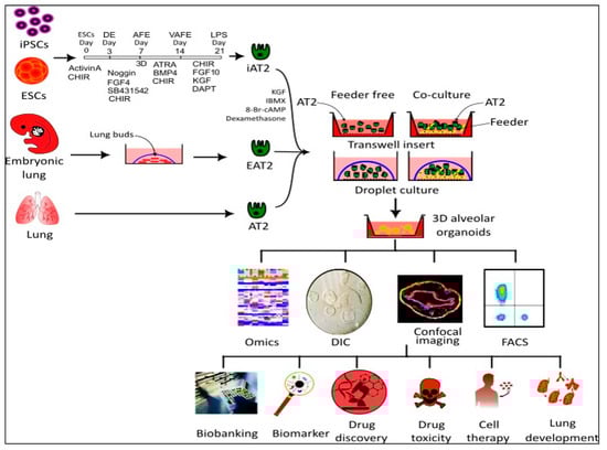
3.1. Co-Culture Method

Three-dimensional in vitro organoid cultures rely on the components present in the microenvironment of cells in vivo. Fibroblast niches regulate AT2 cells’ self-renewal or trans-differentiation into AT1 via Wnt signaling both in vitro and in vivo [53]. These paracrine signals from fibroblast niches are crucial in determining the destiny of AT2 cells. To grow AT2 organoids and replicate their function in vitro, feeder cells serve as a source of niche signals when co-cultured with AT2 cells. Various supporting cell types, including cell lines [54], primary lung fibroblasts [55], PDGFRa+ fibroblasts or endothelial cells [56], MSCs [31], or PSC-derived lung mesenchymal cells, have been used with AT2 cells to support organoid growth [57].
To determine the growth factor requirements for AT2 organoid colony formation, co-cultures are supplemented with a medium consisting of various cytokines known to be involved in lung development. McQualter et al. demonstrated that epithelial growth factors like fibroblast growth factor (FGF)-10 and/or hepatocyte growth factor (HGF) significantly increased organoid colony counts, but FGF-7 had no effect. In contrast, the addition of mesenchymal growth factors such as bone morphogenic protein 4 (BMP-4), TGF-β1, or platelet-derived growth factor (PDGF) either significantly reduced or completely inhibited organoid colony formation [31]

3.1.1. EpCAM+ AT2 Co-Cultured with Fibroblast Cells

Earlier attempts to grow organoids in Matrigel from EpCAM+ AT2 cells alone failed, but complex epithelial cell colonies were generated when co-cultured with Sca-1+ mesenchymal cells [31]. This study showed that αSMA+ mesenchymal cells tightly surrounded epithelial colonies, suggesting a dependency of lung epithelial cells on feeder (fibroblast)-cell-released FGF-10 and HGF to form colonies in vitro. This observation aligns with in vivo studies demonstrating that FGF-10 and HGF regulate lung development [44][58][59]. Later, McQualter et al. [60] showed that CD166 and CD166+ lung stromal cells exhibited different epithelial-supportive capacities. They reported that the in vitro expansion of lung stromal cells resulted in the downregulation of FGF10 expression, reducing their ability to support epithelial colony formation.

3.1.2. AT2 Co-Cultured with PDGFRA+ Cells

Barkauskas et al. reported that AT2 cells function as stem cells in the distal lung and form alveolospheres in vitro [4]. They genetically labeled AT2 cells and purified Sftpc-CreER:Rosa-Tm lineage-positive cells using FACS. To validate that PDGFRA+ mesenchyme cells were an integral part of AT2 niches, these AT2 populations were co-cultured with primary PDGFRA+ mesenchyme cells (in a 1:20 ratio) or with neonatal mouse lung fibroblasts. They found that AT2 cells co-cultured with PDGFRA+ mesenchyme cells developed larger and more numerous colonies compared to those co-cultured with immortalized fibroblast cell lines. Furthermore, the same group reproduced their murine alveolar model in humans by growing HTII-280+ AT2 cells co-cultured with the MRC5 cell line to form alveolospheres. Subsequently, several studies adapted this protocol with variations in culture media composition, the AT2-to-fibroblast ratio, and the types of feeder cells used.

3.1.3. Human iPSC-Derived AT2

Induced pluripotent stem cells are adult cells genetically reprogrammed to resemble embryonic-like stem cells [61]. Patient-specific iPSCs can be a valuable tool for directly studying human diseases and developing personalized medicine using human-derived in vitro models. Several studies have reported the differentiation of iPSCs into NKX2-1+ and SFTPC-expressing AT2 cells [62][63]. Recently, Jacob et al. reported a modified, robust protocol for the differentiation of iPSC into mature, functional AT2 comparable to primary adult AT2 cells. They found that Wnt signaling, in addition to FGF signaling, together with corticosteroids and cyclic AMP, promotes the maturation of SFTPC+ AT2 from NKX2-1+ precursors in vitro. The feeder-free alveolosphere derived from these AT2 cells displayed classical functional features of mature AT2 cells, including innate immune responsiveness and the processing of surfactant lamellar bodies. This iAT2-derived organoid model could be passaged for up to one year without differentiating into AT1 cells and serving as a source of pure AT2 population [64].
In an interesting study, patient-derived iPSCs were differentiated into NKX2-1+ lung epithelial progenitor cells through a 21-day differentiation method. The NKX2-1+ progenitor cells were sorted using Carboxypeptidase M (CPM) as their cell surface marker and cultured alone to grow lung bud organoids. Alternatively, these cells were co-cultured with primary lung fibroblasts to grow alveolar organoids to model HPS-associated interstitial pneumonia (HPSIP) [55]. However, this study could not show any significant differences in organoid morphology and size between patient-specific and gene-corrected organoids, likely due to the immaturity of iPSC-derived AT2 cells.

3.1.4. Mouse and Human ESC-Derived AT2 Cells

Embryonic stem cells are pluripotent cells with the potential to differentiate into multiple tissue types. Recently, bone-marrow-derived “very small embryonic-like stem cells” (VSELs) have been shown to have the potential to differentiate in vivo into SPC-producing AT2 cells. In a remarkable experiment, VSELs were isolated from SPC-H2B-GFP BAC transgenic mice and administered to a bleomycin-injured lung injury mouse model. After three weeks, VSELs differentiated into GFP+ AT2 cells and regenerated the epithelium in vivo. These VSELs-derived GFP+ AT2 cells were FACS-sorted from mice lungs and used to cultivate organoids in vitro to study their proliferation and differentiation potential. After 21 days of co-culture with MLG cells, AT2 cells from VSEL-transplanted mice developed GFP+ organoids, whereas no GFP+ organoids were observed in the control groups. The lineage of VSEL cells was confirmed via the colocalization of the TTF1 (transcription termination factor 1) marker with GFP+ AT2 cells [65].
Nikolić et al. utilized human embryonic lung bud tips progenitor cells to cultivate long-term self-renewing, branching organoids for studying lung development. The embryonic tips, embedded in Matrigel with EGF, FGF7, FGF10, Noggin, RSPO1, CHIR99021, and SB431542, formed spheres within 12 h with a 100% colony-forming efficiency. They were able to passage these organoids for nine passages without changes in morphology, SOX2 and SOX9 expression, or karyotype alterations. This study also showed that the growth conditions for human lung tip organoids do not support the long-term self-renewal of mouse lung tip cells, suggesting species-specific differences during lung development. Interestingly, they observed that mesenchymal cells disappeared after the second passage of organoid cultures, indicating that organoids can be maintained without mesenchymal cells. However, co-culture with canalicular-stage mesenchyme improved alveolar differentiation, leading to the attainment of a bipotent progenitor stage (pro-SFTPC+, HTII-280+, HOPX+ and PDPN+ co-expression, and NKX2-1+) [33][66].

3.2. Feeder-Free Organoid Culture Systems

Although AT2 organoid co-cultures have been used to model lung diseases, they have several drawbacks that render them an undesirable system. One major drawback is the separation of AT2 cells from feeder cells for subsequent transplantation, as transplanting fibroblast cells can induce fibrosis following injury [67][68]. The variations in the type of feeder cells and the composition and concentration of growth factors secreted by the lung mesenchyme make co-culture disadvantageous for modeling cellular therapies. The co-culture system’s niche induces the differentiation of AT2 to AT1 cells. Recent studies have revealed that, in feeder-free organoid cultures, both AT2 cells and iAT2 cells do not spontaneously differentiate into AT1 cells. Instead, their differentiation into AT1 cells necessitates the addition of specific AT1 differentiation induction factors [69]. The feeder-free organoid cultures of AT2 cells in defined medium conditions (AT2 maintenance medium) maintain AT2 cells in a proliferative state and can be scaled up and passaged multiple times to yield a large number of AT2 cells. Feeder-free, serum-free cultures maintain the clonal expansion of EpCAM+ LysoTracker+ AT2 cells for up to 180 days [70]. These AT2 cells can be stored in liquid nitrogen for extended periods, and the same passage can be used for multiple experiments. This approach increases the reproducibility of results and reduces the number of animals used in the research for isolating AT2 cells, as the cells isolated from one mouse can be used for multiple experiments or studies.

3.3. D Matrix Alternatives to Matrigel for Organoid Cultures

Organoid growth not only depends on growth factors and cell–cell interactions but also on signals from the extracellular matrix (ECM), which is an intriguing aspect of stem cell niches. Accumulated evidence has suggested that both physiological and mechanical cues from the extracellular matrix (ECM) may contribute to the maturation and differentiation of type 2 alveolar epithelial cells (AT2) [69]. In co-culture experiments involving iAT2 and fibroblasts, it was observed that fibroblasts express ECM genes and growth factors when AT2 cells are undergoing a transition or transdifferentiation into AT1 cells [52]. To date, both co-cultures and feeder-free 3D cultures of AT2 organoids have primarily relied on Matrigel [4][9][52]. Matrigel is the most commonly used medium to support 3D organoid growth. It is an ECM derived from cancerous mouse tissue and is commonly utilized for stem and cancer cell proliferation. While organoids grown in Matrigel mimic tissue structure, organ physiology, and function, it is derived from animal sources and presents a significant challenge in defining culture conditions due to lot-to-lot variability. The Matrigel system also produces heterogeneous organoids in terms of shape, size, and composition [71]. A chemically defined matrix system would provide more reliable data that could be clinically translated. Hoffman et al. conducted a study on a hydrogel derived from the extracellular matrix (ECM) of human alveolar cells (referred to as aECM hydrogel) for 3D organoid culture [52]. This hydrogel was obtained by processing alveolar-enriched fractions of decellularized human lungs. Their proteomics analysis revealed, that while the hydrogel did not encompass the complete spectrum of native ECM proteins, it exhibited enrichment in key proteins like COL1, COL3, and FBN1. The study demonstrated that the aECM hydrogel not only supported the proliferation of iAT2 (type 2 alveolar epithelial) cells and the formation of alveolospheres but also played a role in promoting the morphological differentiation of a subset of iAT2 cells into structures resembling human AT1-like cells. Additionally, the researchers investigated the influence of matrix stiffness by using different concentrations of aECM. Intriguingly, the higher stiffness of the aECM hydrogel led to a decrease in the SFTPC expression in iAT2 cells compared to hydrogels with less stiffness. Moreover, the bulk RNA sequencing of iAT2 cells cultured in the aECM hydrogel revealed changes in the expression of genes associated with iAT2 maturation, transitional cell states, and AT1-associated markers. These findings suggest that ECM stiffness may play a significant role in directing cellular differentiation processes [52].

4. Applications

Lung cancer is the leading cause of cancer-related deaths globally. The absence of appropriate ex vivo models of the human alveolar epithelium has hindered our understanding of lung cancer pathogenesis and related therapy development. Early-stage diagnosis and treatment are essential for preventing cancer relapse and saving lives. Recently, Dost et al. [72] used both mouse AT2 and human iPSCAT2 organoid models to uncover the early consequences of oncogenic KRAS expression in vivo. Their work has provided novel tools for extensive data collection and studying the transcriptional and proteomic changes that distinguish normal epithelial progenitor cells from early-stage lung cancer. Their study revealed that reductions in AT2 lineage marker gene expression are an early consequence of oncogenic KRAS. Multiomics studies demonstrated that SPC-high cells in Kras activation and p53 loss (KP) lung tumor organoids exhibit higher tumorigenic capacity in the lung microenvironment compared to Hmga2-high cells [73]

HESC-derived organoids have been used to model human lung development. These organoids contained early-stage proximal and distal airway epithelial cells, including early-staged alveolar type 2 (AT2) cells (SPC+/SOX9+) and immature alveolar type 1 (AT1) cells (HOPX+/SOX9+) in vitro. However, when transplanted in vivo for the short term, these organoids differentiated into only a few distal progenitor epithelial cells (NKX2.1+, SOX9+, and P63+). In contrast, the long-term transplantation of these organoids resulted in the differentiation of lung distal bipotent progenitor cells (PDPN+/SPC+/SOX9+), AT2 cells (SPC+ SPB+), and immature AT1 cells (PDPN+, AQP5). These long-term transplanted organoids also contained other cell types present in lung tissues, such as mesenchymal cells, vasculature, neuroendocrine-like cells, and nerve fiber structures [74].

In a recent study, mouse primary AT2 and human iPSCs-derived AT2 organoids were used to investigate the early stages of lung adenocarcinoma (LUAD) driven by KRAS mutation. The data from the alveolar organoids model may be useful in screening novel drug targets and developing new drug molecules to prevent lung cancer growth at the early stage. 

Feeder-free organoid-derived AT2 cells have shown great potential for cellular therapy in lung regeneration. In a recent study, mesenchyme-free AT2 organoids were transplanted into the lungs of mice injured by influenza. The transplanted organoids retained their AT2 fate; however, in some cases, they adopted a dysplastic fate. These dysplastic organoids did not appear to improve the oxygen-exchange capability of the injured lungs in recipient mice. Further investigations have been requested to understand the molecular changes that occur in AT2 organoids after transplantation in the influenza-injured microenvironment in order to optimize organoid transplants [34].

5. Conclusions

Three-dimensional organoid technology has opened new avenues for regenerative medicine and provided a platform for novel drug screening, as well as for developing diagnostics in combination with editing technology for gene therapy and tissue engineering. In addition, patient-derived organoids have given scientists a new tool to predict drug responses in a personalized fashion.

References

  1. Chong, L.; Ahmadvand, N.; Noori, A.; Lv, Y.; Chen, C.; Bellusci, S.; Zhang, J.S. Injury activated alveolar progenitors (IAAPs): The underdog of lung repair. Cell Mol. Life Sci. 2023, 80, 145.
  2. Brownfield, D.G.; de Arce, A.D.; Ghelfi, E.; Gillich, A.; Desai, T.J.; Krasnow, M.A. Alveolar cell fate selection and lifelong maintenance of AT2 cells by FGF signaling. Nat. Commun. 2022, 13, 7137.
  3. Konishi, S.; Tata, A.; Tata, P.R. Defined conditions for long-term expansion of murine and human alveolar epithelial stem cells in three-dimensional cultures. STAR Protoc. 2022, 3, 101447.
  4. Barkauskas, C.E.; Cronce, M.J.; Rackley, C.R.; Bowie, E.J.; Keene, D.R.; Stripp, B.R.; Randell, S.H.; Noble, P.W.; Hogan, B.L. Type 2 alveolar cells are stem cells in adult lung. J. Clin. Investig. 2013, 123, 3025–3036.
  5. Martinez, F.J.; Collard, H.R.; Pardo, A.; Raghu, G.; Richeldi, L.; Selman, M.; Swigris, J.J.; Taniguchi, H.; Wells, A.U. Idiopathic pulmonary fibrosis. Nat. Rev. Dis. Primers 2017, 3, 17074.
  6. Barnes, P.J.; Burney, P.G.; Silverman, E.K.; Celli, B.R.; Vestbo, J.; Wedzicha, J.A.; Wouters, E.F. Chronic obstructive pulmonary disease. Nat. Rev. Dis. Primers 2015, 1, 15076.
  7. Lamers, M.M.; Beumer, J.; van der Vaart, J.; Knoops, K.; Puschhof, J.; Breugem, T.I.; Ravelli, R.B.G.; Paul van Schayck, J.; Mykytyn, A.Z.; Duimel, H.Q.; et al. SARS-CoV-2 productively infects human gut enterocytes. Science 2020, 369, 50–54.
  8. Franks, T.J.; Chong, P.Y.; Chui, P.; Galvin, J.R.; Lourens, R.M.; Reid, A.H.; Selbs, E.; McEvoy, C.P.; Hayden, C.D.; Fukuoka, J.; et al. Lung pathology of severe acute respiratory syndrome (SARS): A study of 8 autopsy cases from Singapore. Hum. Pathol. 2003, 34, 743–748.
  9. Katsura, H.; Sontake, V.; Tata, A.; Kobayashi, Y.; Edwards, C.E.; Heaton, B.E.; Konkimalla, A.; Asakura, T.; Mikami, Y.; Fritch, E.J.; et al. Human Lung Stem Cell-Based Alveolospheres Provide Insights into SARS-CoV-2-Mediated Interferon Responses and Pneumocyte Dysfunction. Cell Stem Cell 2020, 27, 890–904.e898.
  10. Baldassi, D.; Gabold, B.; Merkel, O.M. Air-Liquid Interface Cultures of the Healthy and Diseased Human Respiratory Tract: Promises, Challenges, and Future Directions. Adv. NanoBiomed Res. 2021, 1, 2000111.
  11. Miller, A.J.; Spence, J.R. In Vitro Models to Study Human Lung Development, Disease and Homeostasis. Physiology 2017, 32, 246–260.
  12. Yamamoto, Y.; Korogi, Y.; Hirai, T.; Gotoh, S. A method of generating alveolar organoids using human pluripotent stem cells. Methods Cell Biol. 2020, 159, 115–141.
  13. Fang, X.; Song, Y.; Hirsch, J.; Galietta, L.J.; Pedemonte, N.; Zemans, R.L.; Dolganov, G.; Verkman, A.S.; Matthay, M.A. Contribution of CFTR to apical-basolateral fluid transport in cultured human alveolar epithelial type II cells. Am. J. Physiol. Lung Cell Mol. Physiol. 2006, 290, L242–L249.
  14. Cheek, J.M.; Kim, K.J.; Crandall, E.D. Tight monolayers of rat alveolar epithelial cells: Bioelectric properties and active sodium transport. Am. J. Physiol. 1989, 256, C688–C693.
  15. Demaio, L.; Tseng, W.; Balverde, Z.; Alvarez, J.R.; Kim, K.J.; Kelley, D.G.; Senior, R.M.; Crandall, E.D.; Borok, Z. Characterization of mouse alveolar epithelial cell monolayers. Am. J. Physiol. Lung Cell Mol. Physiol. 2009, 296, L1051–L1058.
  16. Ali, G.; Zhang, M.; Zhao, R.; Jain, K.G.; Chang, J.; Komatsu, S.; Zhou, B.; Liang, J.; Matthay, M.A.; Ji, H.L. Fibrinolytic niche is required for alveolar type 2 cell-mediated alveologenesis via a uPA-A6-CD44(+)-ENaC signal cascade. Signal Transduct. Target. Ther. 2021, 6, 97.
  17. Ali, G.; Zhang, M.; Chang, J.; Zhao, R.; Jin, Y.; Zhang, J.; Ji, H.-L. PAI-1 regulates AT2-mediated re-alveolarization and ion permeability. Stem Cell Res. Ther. 2023, 14, 185.
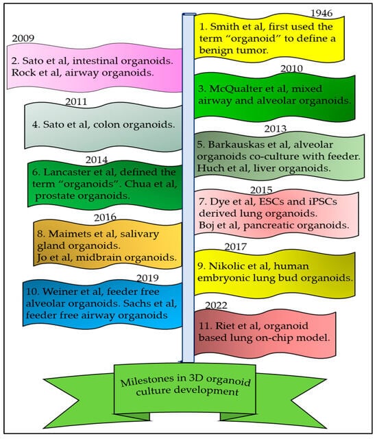
  18. Zhao, Z.; Chen, X.; Dowbaj, A.M.; Sljukic, A.; Bratlie, K.; Lin, L.; Fong, E.L.S.; Balachander, G.M.; Chen, Z.; Soragni, A.; et al. Organoids. Nat. Rev. Methods Primers 2022, 2, 94.
  19. Pei, R.; Feng, J.; Zhang, Y.; Sun, H.; Li, L.; Yang, X.; He, J.; Xiao, S.; Xiong, J.; Lin, Y.; et al. Host metabolism dysregulation and cell tropism identification in human airway and alveolar organoids upon SARS-CoV-2 infection. Protein Cell 2021, 12, 717–733.
  20. Smith, E.; Cochrane, W.J. CYSTIC ORGANOID TERATOMA: (Report of a Case). Can. Med. Assoc. J. 1946, 55, 151–152.
  21. Lancaster, M.A.; Knoblich, J.A. Organogenesis in a dish: Modeling development and disease using organoid technologies. Science 2014, 345, 1247125.
  22. Sato, T.; Vries, R.G.; Snippert, H.J.; van de Wetering, M.; Barker, N.; Stange, D.E.; van Es, J.H.; Abo, A.; Kujala, P.; Peters, P.J.; et al. Single Lgr5 stem cells build crypt-villus structures in vitro without a mesenchymal niche. Nature 2009, 459, 262–265.
  23. Clevers, H. Modeling Development and Disease with Organoids. Cell 2016, 165, 1586–1597.
  24. Sato, T.; Stange, D.E.; Ferrante, M.; Vries, R.G.; Van Es, J.H.; Van den Brink, S.; Van Houdt, W.J.; Pronk, A.; Van Gorp, J.; Siersema, P.D.; et al. Long-term expansion of epithelial organoids from human colon, adenoma, adenocarcinoma, and Barrett’s epithelium. Gastroenterology 2011, 141, 1762–1772.
  25. Huch, M.; Dorrell, C.; Boj, S.F.; van Es, J.H.; Li, V.S.; van de Wetering, M.; Sato, T.; Hamer, K.; Sasaki, N.; Finegold, M.J.; et al. In vitro expansion of single Lgr5+ liver stem cells induced by Wnt-driven regeneration. Nature 2013, 494, 247–250.
  26. Chua, C.W.; Shibata, M.; Lei, M.; Toivanen, R.; Barlow, L.J.; Bergren, S.K.; Badani, K.K.; McKiernan, J.M.; Benson, M.C.; Hibshoosh, H.; et al. Single luminal epithelial progenitors can generate prostate organoids in culture. Nat. Cell Biol. 2014, 16, 951–961.
  27. Boj, S.F.; Hwang, C.I.; Baker, L.A.; Chio, I.I.; Engle, D.D.; Corbo, V.; Jager, M.; Ponz-Sarvise, M.; Tiriac, H.; Spector, M.S.; et al. Organoid models of human and mouse ductal pancreatic cancer. Cell 2015, 160, 324–338.
  28. Jo, J.; Xiao, Y.; Sun, A.X.; Cukuroglu, E.; Tran, H.D.; Göke, J.; Tan, Z.Y.; Saw, T.Y.; Tan, C.P.; Lokman, H.; et al. Midbrain-like Organoids from Human Pluripotent Stem Cells Contain Functional Dopaminergic and Neuromelanin-Producing Neurons. Cell Stem Cell 2016, 19, 248–257.
  29. Maimets, M.; Rocchi, C.; Bron, R.; Pringle, S.; Kuipers, J.; Giepmans, B.N.; Vries, R.G.; Clevers, H.; de Haan, G.; van Os, R.; et al. Long-Term In Vitro Expansion of Salivary Gland Stem Cells Driven by Wnt Signals. Stem Cell Rep. 2016, 6, 150–162.
  30. Rock, J.R.; Onaitis, M.W.; Rawlins, E.L.; Lu, Y.; Clark, C.P.; Xue, Y.; Randell, S.H.; Hogan, B.L. Basal cells as stem cells of the mouse trachea and human airway epithelium. Proc. Natl. Acad. Sci. USA 2009, 106, 12771–12775.
  31. McQualter, J.L.; Yuen, K.; Williams, B.; Bertoncello, I. Evidence of an epithelial stem/progenitor cell hierarchy in the adult mouse lung. Proc. Natl. Acad. Sci. USA 2010, 107, 1414–1419.
  32. Dye, B.R.; Hill, D.R.; Ferguson, M.A.H.; Tsai, Y.-H.; Nagy, M.S.; Dyal, R.; Wells, J.M.; Mayhew, C.N.; Nattiv, R.; Klein, O.D.; et al. In vitro generation of human pluripotent stem cell derived lung organoids. eLife 2015, 4, e05098.
  33. Nikolić, M.Z.; Caritg, O.; Jeng, Q.; Johnson, J.-A.; Sun, D.; Howell, K.J.; Brady, J.L.; Laresgoiti, U.; Allen, G.; Butler, R.; et al. Human embryonic lung epithelial tips are multipotent progenitors that can be expanded in vitro as long-term self-renewing organoids. eLife 2017, 6, e26575.
  34. Weiner, A.I.; Jackson, S.R.; Zhao, G.; Quansah, K.K.; Farshchian, J.N.; Neupauer, K.M.; Littauer, E.Q.; Paris, A.J.; Liberti, D.C.; Scott Worthen, G.; et al. Mesenchyme-free expansion and transplantation of adult alveolar progenitor cells: Steps toward cell-based regenerative therapies. NPJ Regen. Med. 2019, 4, 17.
  35. Sachs, N.; Papaspyropoulos, A.; Zomer-van Ommen, D.D.; Heo, I.; Böttinger, L.; Klay, D.; Weeber, F.; Huelsz-Prince, G.; Iakobachvili, N.; Amatngalim, G.D.; et al. Long-term expanding human airway organoids for disease modeling. Embo J. 2019, 38, e100300.
  36. van Riet, S.; van Schadewijk, A.; Khedoe, P.; Limpens, R.; Bárcena, M.; Stolk, J.; Hiemstra, P.S.; van der Does, A.M. Organoid-based expansion of patient-derived primary alveolar type 2 cells for establishment of alveolus epithelial Lung-Chip cultures. Am. J. Physiol. Lung Cell Mol. Physiol. 2022, 322, L526–L538.
  37. Lim, K.; Donovan, A.P.A.; Tang, W.; Sun, D.; He, P.; Pett, J.P.; Teichmann, S.A.; Marioni, J.C.; Meyer, K.B.; Brand, A.H.; et al. Organoid modeling of human fetal lung alveolar development reveals mechanisms of cell fate patterning and neonatal respiratory disease. Cell Stem Cell 2023, 30, 20–37.e29.
  38. Nadkarni, R.R.; Abed, S.; Draper, J.S. Stem Cells in Pulmonary Disease and Regeneration. Chest 2018, 153, 994–1003.
  39. Aichler, M.; Kunzke, T.; Buck, A.; Sun, N.; Ackermann, M.; Jonigk, D.; Gaumann, A.; Walch, A. Molecular similarities and differences from human pulmonary fibrosis and corresponding mouse model: MALDI imaging mass spectrometry in comparative medicine. Lab. Investig. 2018, 98, 141–149.
  40. Pan, H.; Deutsch, G.H.; Wert, S.E. Comprehensive anatomic ontologies for lung development: A comparison of alveolar formation and maturation within mouse and human lung. J. Biomed. Semant. 2019, 10, 18.
  41. Huang, X.; Fan, W.; Sun, J.; Yang, J.; Zhang, Y.; Wang, Q.; Li, P.; Zhang, Y.; Zhang, S.; Li, H.; et al. SARS-CoV-2 induces cardiomyocyte apoptosis and inflammation but can be ameliorated by ACE inhibitor Captopril. Antivir. Res. 2023, 215, 105636.
  42. Warren, H.S.; Tompkins, R.G.; Moldawer, L.L.; Seok, J.; Xu, W.; Mindrinos, M.N.; Maier, R.V.; Xiao, W.; Davis, R.W. Mice are not men. Proc. Natl. Acad. Sci. USA 2015, 112, E345.
  43. Martić-Kehl, M.I.; Schibli, R.; Schubiger, P.A. Can animal data predict human outcome? Problems and pitfalls of translational animal research. Eur. J. Nucl. Med. Mol. Imaging 2012, 39, 1492–1496.
  44. Yuan, T.; Volckaert, T.; Chanda, D.; Thannickal, V.J.; De Langhe, S.P. Fgf10 Signaling in Lung Development, Homeostasis, Disease, and Repair After Injury. Front. Genet. 2018, 9, 418.
  45. Abraham, V.; Chou, M.L.; DeBolt, K.M.; Koval, M. Phenotypic control of gap junctional communication by cultured alveolar epithelial cells. Am. J. Physiol. 1999, 276, L825–L834.
  46. Weller, N.K.; Karnovsky, M.J. Improved isolation of rat lung alveolar type II cells. More representative recovery and retention of cell polarity. Am. J. Pathol. 1986, 122, 92–100.
  47. Choi, J.; Park, J.E.; Tsagkogeorga, G.; Yanagita, M.; Koo, B.K.; Han, N.; Lee, J.H. Inflammatory Signals Induce AT2 Cell-Derived Damage-Associated Transient Progenitors that Mediate Alveolar Regeneration. Cell Stem Cell 2020, 27, 366–382.e367.
  48. Gonzalez, R.F.; Allen, L.; Gonzales, L.; Ballard, P.L.; Dobbs, L.G. HTII-280, a Biomarker Specific to the Apical Plasma Membrane of Human Lung Alveolar Type II Cells. J. Histochem. Cytochem. 2010, 58, 891–901.
  49. Hasegawa, K.; Sato, A.; Tanimura, K.; Uemasu, K.; Hamakawa, Y.; Fuseya, Y.; Sato, S.; Muro, S.; Hirai, T. Fraction of MHCII and EpCAM expression characterizes distal lung epithelial cells for alveolar type 2 cell isolation. Respir. Res. 2017, 18, 150.
  50. Toulmin, S.A.; Bhadiadra, C.; Paris, A.J.; Lin, J.H.; Katzen, J.; Basil, M.C.; Morrisey, E.E.; Worthen, G.S.; Eisenlohr, L.C. Type II alveolar cell MHCII improves respiratory viral disease outcomes while exhibiting limited antigen presentation. Nat. Commun. 2021, 12, 3993.
  51. Zhao, S.; Wu, X.; Tan, Z.; Ren, Y.; Li, L.; Ou, J.; Lin, Y.; Song, H.; Feng, L.; Seto, D.; et al. Generation of Human Embryonic Stem Cell-Derived Lung Organoids for Modeling Infection and Replication Differences between Human Adenovirus Types 3 and 55 and Evaluating Potential Antiviral Drugs. J. Virol. 2023, 97, e0020923.
  52. Hoffman, E.T.; Uriarte, J.J.; Uhl, F.E.; Eckstrom, K.; Tanneberger, A.E.; Becker, C.; Moulin, C.; Asarian, L.; Ikonomou, L.; Kotton, D.N.; et al. Human alveolar hydrogels promote morphological and transcriptional differentiation in iPSC-derived alveolar type 2 epithelial cells. Sci. Rep. 2023, 13, 12057.
  53. Nabhan, A.N.; Brownfield, D.G.; Harbury, P.B.; Krasnow, M.A.; Desai, T.J. Single-cell Wnt signaling niches maintain stemness of alveolar type 2 cells. Science 2018, 359, 1118–1123.
  54. Jain, K.G.; Zhao, R.; Liu, Y.; Guo, X.; Yi, G.; Ji, H.L. Wnt5a/β-catenin axis is involved in the downregulation of AT2 lineage by PAI-1. Am. J. Physiol. Lung Cell Mol. Physiol. 2022, 323, L515–L524.
  55. Suezawa, T.; Kanagaki, S.; Korogi, Y.; Nakao, K.; Hirai, T.; Murakami, K.; Hagiwara, M.; Gotoh, S. Modeling of lung phenotype of Hermansky-Pudlak syndrome type I using patient-specific iPSCs. Respir. Res. 2021, 22, 284.
  56. Lee, J.H.; Bhang, D.H.; Beede, A.; Huang, T.L.; Stripp, B.R.; Bloch, K.D.; Wagers, A.J.; Tseng, Y.H.; Ryeom, S.; Kim, C.F. Lung stem cell differentiation in mice directed by endothelial cells via a BMP4-NFATc1-thrombospondin-1 axis. Cell 2014, 156, 440–455.
  57. Tamai, K.; Sakai, K.; Yamaki, H.; Moriguchi, K.; Igura, K.; Maehana, S.; Suezawa, T.; Takehara, K.; Hagiwara, M.; Hirai, T.; et al. iPSC-derived mesenchymal cells that support alveolar organoid development. Cell Rep. Methods 2022, 2, 100314.
  58. Nyeng, P.; Norgaard, G.A.; Kobberup, S.; Jensen, J. FGF10 maintains distal lung bud epithelium and excessive signaling leads to progenitor state arrest, distalization, and goblet cell metaplasia. BMC Dev. Biol. 2008, 8, 2.
  59. Ohmichi, H.; Koshimizu, U.; Matsumoto, K.; Nakamura, T. Hepatocyte growth factor (HGF) acts as a mesenchyme-derived morphogenic factor during fetal lung development. Development 1998, 125, 1315–1324.
  60. McQualter, J.L.; McCarty, R.C.; Van der Velden, J.; O’Donoghue, R.J.J.; Asselin-Labat, M.-L.; Bozinovski, S.; Bertoncello, I. TGF-β signaling in stromal cells acts upstream of FGF-10 to regulate epithelial stem cell growth in the adult lung. Stem Cell Res. 2013, 11, 1222–1233.
  61. Takahashi, K.; Tanabe, K.; Ohnuki, M.; Narita, M.; Ichisaka, T.; Tomoda, K.; Yamanaka, S. Induction of pluripotent stem cells from adult human fibroblasts by defined factors. Cell 2007, 131, 861–872.
  62. Gotoh, S.; Ito, I.; Nagasaki, T.; Yamamoto, Y.; Konishi, S.; Korogi, Y.; Matsumoto, H.; Muro, S.; Hirai, T.; Funato, M.; et al. Generation of alveolar epithelial spheroids via isolated progenitor cells from human pluripotent stem cells. Stem Cell Rep. 2014, 3, 394–403.
  63. Hawkins, F.; Kramer, P.; Jacob, A.; Driver, I.; Thomas, D.C.; McCauley, K.B.; Skvir, N.; Crane, A.M.; Kurmann, A.A.; Hollenberg, A.N.; et al. Prospective isolation of NKX2-1-expressing human lung progenitors derived from pluripotent stem cells. J. Clin. Investig. 2017, 127, 2277–2294.
  64. Jacob, A.; Vedaie, M.; Roberts, D.A.; Thomas, D.C.; Villacorta-Martin, C.; Alysandratos, K.D.; Hawkins, F.; Kotton, D.N. Derivation of self-renewing lung alveolar epithelial type II cells from human pluripotent stem cells. Nat. Protoc. 2019, 14, 3303–3332.
  65. Ciechanowicz, A.K.; Sielatycka, K.; Cymer, M.; Skoda, M.; Suszyńska, M.; Bujko, K.; Ratajczak, M.Z.; Krause, D.S.; Kucia, M. Bone Marrow-Derived VSELs Engraft as Lung Epithelial Progenitor Cells after Bleomycin-Induced Lung Injury. Cells 2021, 10, 1570.
  66. Chen, Y.-W.; Huang, S.X.; de Carvalho, A.L.R.T.; Ho, S.-H.; Islam, M.N.; Volpi, S.; Notarangelo, L.D.; Ciancanelli, M.; Casanova, J.-L.; Bhattacharya, J.; et al. A three-dimensional model of human lung development and disease from pluripotent stem cells. Nat. Cell Biol. 2017, 19, 542–549.
  67. Rock, J.R.; Barkauskas, C.E.; Cronce, M.J.; Xue, Y.; Harris, J.R.; Liang, J.; Noble, P.W.; Hogan, B.L.M. Multiple stromal populations contribute to pulmonary fibrosis without evidence for epithelial to mesenchymal transition. Proc. Natl. Acad. Sci. USA 2011, 108, E1475–E1483.
  68. Tanjore, H.; Xu, X.C.; Polosukhin, V.V.; Degryse, A.L.; Li, B.; Han, W.; Sherrill, T.P.; Plieth, D.; Neilson, E.G.; Blackwell, T.S.; et al. Contribution of epithelial-derived fibroblasts to bleomycin-induced lung fibrosis. Am. J. Respir. Crit. Care Med. 2009, 180, 657–665.
  69. Alysandratos, K.-D.; Garcia-de-Alba, C.; Yao, C.; Pessina, P.; Huang, J.; Villacorta-Martin, C.; Hix, O.T.; Minakin, K.; Burgess, C.L.; Bawa, P.; et al. Culture impact on the transcriptomic programs of primary and iPSC-derived human alveolar type 2 cells. JCI Insight 2023, 8, e158937.
  70. Yamamoto, Y.; Gotoh, S.; Korogi, Y.; Seki, M.; Konishi, S.; Ikeo, S.; Sone, N.; Nagasaki, T.; Matsumoto, H.; Muro, S.; et al. Long-term expansion of alveolar stem cells derived from human iPS cells in organoids. Nat. Methods 2017, 14, 1097–1106.
  71. Loebel, C.; Weiner, A.I.; Eiken, M.K.; Katzen, J.B.; Morley, M.P.; Bala, V.; Cardenas-Diaz, F.L.; Davidson, M.D.; Shiraishi, K.; Basil, M.C.; et al. Microstructured Hydrogels to Guide Self-Assembly and Function of Lung Alveolospheres. Adv. Mater. 2022, 34, e2202992.
  72. Dost, A.F.M.; Moye, A.L.; Vedaie, M.; Tran, L.M.; Fung, E.; Heinze, D.; Villacorta-Martin, C.; Huang, J.; Hekman, R.; Kwan, J.H.; et al. Organoids Model Transcriptional Hallmarks of Oncogenic KRAS Activation in Lung Epithelial Progenitor Cells. Cell Stem Cell 2020, 27, 663–678.e668.
  73. Kim, C.; Li, J.; Dang, S.; Schurmann, P.; Dost, A.; Moye, A.; Paschini, M.; Bhetariya, P.; Bronson, R.; Sui, S.H. Organoid modeling reveals the tumorigenic potential of the alveolar progenitor cell state. Res. Sq. 2023; preprint.
  74. Chen, Y.; Feng, J.; Zhao, S.; Han, L.; Yang, H.; Lin, Y.; Rong, Z. Long-Term Engraftment Promotes Differentiation of Alveolar Epithelial Cells from Human Embryonic Stem Cell Derived Lung Organoids. Stem Cells Dev. 2018, 27, 1339–1349.
More
Information
Contributors MDPI registered users' name will be linked to their SciProfiles pages. To register with us, please refer to https://encyclopedia.pub/register : , , , , ,
View Times: 97
Revisions: 2 times (View History)
Update Date: 14 Nov 2023
1000/1000