Submitted Successfully!
To reward your contribution, here is a gift for you: A free trial for our video production service.
Thank you for your contribution! You can also upload a video entry or images related to this topic.
Version Summary Created by Modification Content Size Created at Operation
1 -- 2014 2023-11-09 10:34:18 |
2 format change Meta information modification 2014 2023-11-10 03:00:01 |

Video Upload Options

Do you have a full video?

Confirm

Are you sure to Delete?
Cite
If you have any further questions, please contact Encyclopedia Editorial Office.
Miranda, M.; Gianfreda, F.; Carlotta, D.; Armati, S.; Barlattani, A.; Bollero, P. Oral Manifestations of Graft vs. Host Disease. Encyclopedia. Available online: https://encyclopedia.pub/entry/51346 (accessed on 03 May 2024).
Miranda M, Gianfreda F, Carlotta D, Armati S, Barlattani A, Bollero P. Oral Manifestations of Graft vs. Host Disease. Encyclopedia. Available at: https://encyclopedia.pub/entry/51346. Accessed May 03, 2024.
Miranda, Michele, Francesco Gianfreda, Danesi Carlotta, Sofia Armati, Alberta Barlattani, Patrizio Bollero. "Oral Manifestations of Graft vs. Host Disease" Encyclopedia, https://encyclopedia.pub/entry/51346 (accessed May 03, 2024).
Miranda, M., Gianfreda, F., Carlotta, D., Armati, S., Barlattani, A., & Bollero, P. (2023, November 09). Oral Manifestations of Graft vs. Host Disease. In Encyclopedia. https://encyclopedia.pub/entry/51346
Miranda, Michele, et al. "Oral Manifestations of Graft vs. Host Disease." Encyclopedia. Web. 09 November, 2023.
Oral Manifestations of Graft vs. Host Disease
Edit

Graft-versus-host disease (GVHD) is a complication of hematopoietic stem cell transplantation (HSCT). GVHD may also develop following solid transplants or blood transfusions if white blood cells are transferred. GVHD affects multiple organs, including the oral tissues. 

graft-versus-host disease oral manifestations dental treatment

1. Introduction

GvHD is known as the worst complication of allogeneic hematopoietic stem cell (allo-HSCT) transplants, making it the leading cause of morbidity and mortality associated with this transplant [1]. The immunological mechanism underlying GvHD can be considered as “opposite” to rejection: while in the latter case, the host’s immune cells recognize the cells of the transplanted tissue as “non-self”, in the graft-versus-host disease it is the transplanted immune cells that recognize the recipient’s organs as foreign and trigger an immune attack against them [2][3].
In 1960, Billingham established the criteria for the onset of GvHD:
  • Graft containing supply of immunocompetent cells [4]
  • Immunological dissimilarity between host and donor
  • Immunosuppressed host.

1.1. How It Works: Allo-HSCT

The Billingham criteria can be found in allo-HSCT, a transplant that consists of the infusion of hematopoietic stem cells from a compatible donor into a recipient who has been appropriately conditioned with high-dose chemo- and radiotherapy [2]. The goals of chemo-radiotherapy treatment and the infusion of allogeneic hematopoietic stem cells are:
  • Resolution of the disease
  • Allogeneic stem cell engraftment (production of the necessary space for the infusion)
  • Bone marrow restoration by the infused cells after a period of aplasia (which takes about 100 days)
  • Elimination of remaining diseased cells thanks to the graft-versus-tumor effect (GvT): allogeneically transplanted human hematopoietic cells can eliminate residual tumor cells in the recipient by an immune-mediated mechanism [3].
The main indication for this transplant is acute myeloid leukemia, but it is not the only one [5].
The compatibility of HLA genes must be the same (autotransplant) or very similar since the reduction in compatibility is associated with an increasing risk of developing GvHD [4][6]. Technological advancement and a better understanding of the characteristics of the epitope structure of HLA antigens have allowed the development of a test that identifies the compatibility between patient and possible donor, which is achieved through a tool, the HLA-MatchMaker, a computerized algorithm that analyzes a simple venous blood draw from both the donor and the recipient [7][8]. The probability of finding a compatible donor in the family environment, given the huge polymorphism of the HLA system, is around 25%, while the probability of finding it in the National and International Registers of bone marrow donors is around 40% [9].
The sources of stem cells used can be:
  • Bone marrow (BM).
  • Peripheral blood stem cells (PBSC), which is the most innovative and most used technique to date.
  • Umbilical cord blood, which, however, does not provide a quantity of stem cells sufficient to treat an adult.

1.2. Pre-HSCT Dentistry Videat

In patients who must undergo HSCT, the consensus is that all low-invasive dental care is necessary to eliminate any traumatic factors and infectious foci, postponing any elective treatments (such as extended surgical treatments) after the post-transplant immune reconstitution [10]. The trend is to end the necessary dental therapies about 10 days before the transplant, to give the patient time to undergo conditioning therapy [11]. The current protocol, based on the most frequently encountered pathologies, is summarized in Table 1 [4].
Table 1. Billingham’s criteria.
Traumatic factors (overflowing restorations, fractured teeth, root debris) could be etiological factors in the development of oral ulcers or contribute to the development of oral mucositis in HSCT transplant patients who develop GvHD. Therefore, these should be eliminated [12]. For the same reason, prostheses should be cleaned and relined to perfection. No dental therapy should ever be performed in the 6–12-month post-transplant period, including dental hygiene maneuvers that could generate aerosols with accidental aspiration of bacteria or debris [13].

Epidemiology

Approximately 35–50% of patients undergoing HSCT develop acute GvHD. In addition, it has been estimated that about 50% of patients with the acute form also subsequently develop the chronic form [14].
Regarding prevalence, a 2018 study by the U.S. Research and Markets statistical center calculated that in the seven largest pharmaceutical markets, diagnosed cases of chronic and acute GvHD stood at around 18,408, set to rise above 22,000 in 2028.

1.3. Acute and Chronic GVHD

In the past, GvHD was divided into two main forms: acute and chronic. This designation was based exclusively on a temporal criterion, the so-called “10-day dividing line”. The 2005 NIH Consensus Conference abolished this classification criterion and redefined chronic and acute GvHD solely based on different clinical manifestations [15]. Acute GvHD (aGvHD) is thus subdivided into classic and late forms, based on whether symptoms occur before or after 100 days post-transplant. The late form is further subdivided into the following forms:
  • Persistent, if characterized by the persistence of signs and symptoms of classic aGvHD initiated before day 100 post-transplant;
  • Recurrent, if it arises as a classic form that resolves but recurs after day 100 post-transplant;
  • De novo, if it occurs after day 100 post-transplant without any signs or symptoms in the previous time frame.
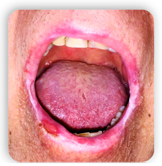
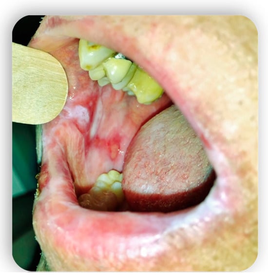
As far as the chronic form (cGvHD) is concerned, this can arise following the evolution of the acute form (PTO, progressive type of onset), after the resolution of a previous acute form (quiescent), or “de novo”. Also, cGvHD is divided into classic and superimposed forms, if it occurs with the typical acute form manifestations on a pre-existing chronic form [16].

1.4. Oral GVHD

The oral manifestations of GvHD can be divided into three groups: the manifestations derived from the conditioning regimen and those of the acute and chronic form of GvHD (Figure 1 and Figure 2) [17].
Figure 1. Oral mucositis manifestations from GvHD.
Figure 2. Oral mucositis manifestations from GvHD.

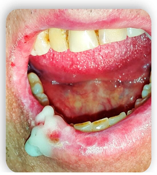
2. Systemic Treatment of Oral Lesions

The treatment of mucocutaneous lesions from acute and chronic GvHD is often a difficult problem for the clinician [18]. In addition to the standard systemic therapies for acute and chronic GvHD, it is possible to use an additional therapy in case of refractory and particularly compromising mucocutaneous manifestations [19]. Much interest has been aroused by the use of Rituximab (anti-CD20 monoclonal antibody) [20][21]. The role played by the cooperation between B and T cells in the activation of immune responses and in the pathogenesis of chronic GvHD could justify the effectiveness of this therapeutic option. Extracorporeal photochemotherapy (ECP), in combination with other immunosuppressive agents, is a very useful option [22]. With ECP, at an extracorporeal level, the blood is treated with a drug activated by UV light that causes direct apoptosis of leukocytes (especially lymphocytes) and is reinfused into the patient. This seems to generate immunological tolerance by interfering with the maturation of the dendritic cells, modulation of cytokine production, and Treg cell expansion. ECP is proposed not only as an alternative therapy but recently also as a first-line therapy for chronic GvHD [16]. The basis of ECP effectiveness is the immunomodulatory effect of the procedure which acts by inhibiting the population of activated donor lymphocytes and altering the interaction between these and the APCs. ECP is an invasive procedure, requiring a dedicated team and several weeks before signs of clinical improvement appear. Some scholars have recently obtained good results using etretinate (synthetic retinoid) [23], in combination with standard therapies, for the treatment of scleroderma resulting from refractory cGvHD. Other authors, especially in consideration of the failure of rescue therapies in multi-treated patients, have experimented with the use of Pentostatin (adenosine deaminase inhibitor), whose effects have already been tested for acute GvHD [21].

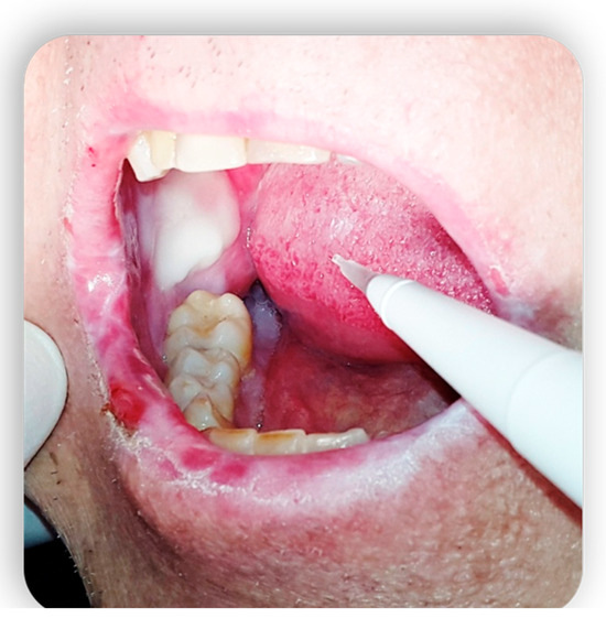
3. Topical Treatment of Oral Lesions

The systemic treatment of the disease consists of immunosuppressive drugs but only in a few cases leads to the healing of oral lesions [18]. These patients could benefit from topical agents, both to focus the action exclusively on the regions of interest and to reduce the state of systemic immunosuppression. In these cases, it is very common to use steroid preparations for topical use such as the combination of fluocinonide and clobetasol, or dexamethasone and betamethasone [24]. The topical application of CSA has also been used successfully in lichenoid manifestations of the mucous membranes and in cases of punctate keratitis. In addition, the application of topical preparations of azathioprine, in association with systemic immunosuppressive therapy, was evaluated in patients with skin ulcerative lesions that are resistant to topical treatment with CSA and cortisone [25]. The results, although obtained from a small number of patients, showed a good response to treatment considering both the extension of the ulcerated areas and pain. Several reports also indicate tacrolimus, already known in the dermatological field for its beneficial effects in atopic dermatitis, as a possible topical agent in the treatment of cutaneous cGVHD [26] (Figure 3 and Figure 4).
Figure 3. Topical treatment with non-transfusional hemocomponents.
Figure 4. Topical treatment with non-transfusional hemocomponents.

4. GvHD and Implantology: Case Reports in the Literature

In the current research, there are insufficient references in the literature regarding implant survival and success in patients with GvHD, in any of its forms [27]. Despite this, there are three case reports in the literature that demonstrate the possibility of carrying out an implant treatment plan on a GvHD patient [28].
The first case report is about a clinical case presented by Mahn D.H. of a 46-year-old non-smoker man with a medical history characterized by HSCT-treated non-Hodgkin’s lymphoma, which led to the development of cGvHD 1 year later. Clinical evaluation of the patient revealed remission of the disease, but poor oral hygiene and teeth that were severely compromised by carious disease. Severe gingivitis and gingival recessions were also present. The treatment plan, after evaluating the bone structure, provided for the simultaneous extraction of all the mandibular and maxillary dental elements and the contextual placement of five implants for the lower arch [22]. The treatment was temporarily completed with an immediate upper removable prosthesis and an inferior immediate hybrid prosthesis. After 6 months, the final prostheses were delivered. One year after the completion of the treatment plan, the implant sites were healthy and with stable bone levels. The patient had to maintain good oral hygiene, and no signs of cGvHD were present, apart from oral dryness [29].
In the second case report, Curtis J.W. described the case of a patient with GvHD developed after HSCT. The patient presented with severe caries pathology and sharp enamel projections causing chronic trauma to the tongue and adjacent mucosa. In this case, the teeth were extracted and replaced with removable total prostheses delivered 6 months after the extractions. Only after 2 years, five mandibular implants were inserted to create a complete overdenture delivered 14 months after the insertion of the implants [30].
In the third case report, A. Etebarian et al. [31] presented the case of a patient with cGvHD with a sclerotic phenotype developed after HSCT for the therapeutic treatment of acute myeloid leukemia. The patient is a 35-year-old man who, 1 year after the onset of cGvHD symptoms, has generalized carious pathology with non-recoverable teeth. Therefore, all the elements were extracted, and after healing, there were placed six mandibular endosseous implants. After 6 months, implant success was established according to the criteria of Albrektsson et al., and a fixed prosthesis was made on implants. After 2 years of follow-up, the maxilla was also rehabilitated through the insertion of four endosseous implants and after 6 months the delivery of an overdenture. After 6 years of HSCT transplant, cGvHD resulted in remission and there was a successful oral rehabilitation, significantly increasing the patient’s quality of life [32][33].
Mahn’s case report remains to date the only one that describes an implant success in a patient with GvHD in which dental extractions and simultaneous insertion of mandibular implants were performed [25][34].
In conclusion, what are the keys to successful case management? Attention to detail during the diagnostic, surgical, and restorative phases, management of patient expectations, and compliance with patient instructions.
Extremely important, then, is patient compliance; only then can predictable results be achieved and complications averted.

References

  1. Lee, S.J. Classification systems for chronic graft-versus-host disease. Blood 2017, 129, 30–37.
  2. Arora, M.; Nagaraj, S.; Witte, J.; DeFor, T.E.; MacMillan, M.; Burns, L.J.; Weisdorf, D.J. New classification of chronic GVHD: Added clarity from the consensus diagnoses. Bone Marrow Transpl. 2009, 43, 149–153.
  3. Flowers, M.E.D.; Inamoto, Y.; Carpenter, P.A.; Lee, S.J.; Kiem, H.-P.; Petersdorf, E.W.; Pereira, S.E.; Nash, R.A.; Mielcarek, M.; Fero, M.L.; et al. Comparative analysis of risk factors for acute graft-versus-host disease and for chronic graft-versus-host disease according to National Institutes of Health consensus criteria. Blood 2011, 117, 3214–3219.
  4. Pavletic, S.Z.; Martin, P.; Lee, S.J.; Mitchell, S.; Jacobsohn, D.; Cowen, E.W.; Turner, M.L.; Akpek, G.; Gilman, A.; McDonald, G.; et al. Measuring therapeutic response in chronic graft-versus-host disease: National Institutes of Health Consensus Development Project on Criteria for Clinical Trials in Chronic Graft-versus-Host Disease: IV. Response Criteria Working Group report. Biol. Blood Marrow Transplant. 2006, 12, 252–266.
  5. Jagasia, M.H.; Greinix, H.T.; Arora, M.; Williams, K.M.; Wolff, D.; Cowen, E.W.; Palmer, J.; Weisdorf, D.; Treister, N.S.; Cheng, G.S.; et al. National Institutes of Health Consensus Development Project on Criteria for Clinical Trials in Chronic Graft-versus-Host Disease: I. The 2014 Diagnosis and Staging Working Group report. Biol. Blood Marrow Transplant. 2015, 21, 389–401.e1.
  6. Ion, D.; Stevenson, K.; Woo, S.-B.; Ho, V.T.; Soiffer, R.; Antin, J.H.; Treister, N.S. Characterization of oral involvement in acute graft-versus-host disease. Biol. Blood Marrow Transplant. 2014, 20, 1717–1721.
  7. Zeiser, R.; Blazar, B.R. Pathophysiology of Chronic Graft-versus-Host Disease and Therapeutic Targets. N. Engl. J. Med. 2017, 377, 2565–2579.
  8. Elad, S.; Cheng, K.K.F.; Lalla, R.V.; Yarom, N.; Hong, C.; Logan, R.M.; Bowen, J.; Gibson, R.; Saunders, D.P.; Zadik, Y.; et al. MASCC/ISOO clinical practice guidelines for the management of mucositis secondary to cancer therapy. Cancer 2020, 126, 4423–4431.
  9. Yuan, A.; Chai, X.; Martins, F.; Arai, S.; Arora, M.; Correa, M.E.; Pidala, J.; Cutler, C.S.; Lee, S.J.; Treister, N.S. Oral chronic GVHD outcomes and resource utilization: A subanalysis from the chronic GVHD consortium. Oral. Dis. 2016, 22, 235–240.
  10. Brunstein, C.G.; Fuchs, E.J.; Carter, S.L.; Karanes, C.; Costa, L.J.; Wu, J.; Devine, S.M.; Wingard, J.R.; Aljitawi, O.S.; Cutler, C.S.; et al. Alternative donor transplantation after reduced intensity conditioning: Results of parallel phase 2 trials using partially HLA-mismatched related bone marrow or unrelated double umbilical cord blood grafts. Blood 2011, 118, 282–288.
  11. Al-Hashimi, I.; Schifter, M.; Lockhart, P.B.; Wray, D.; Brennan, M.; Migliorati, C.A.; Axéll, T.; Bruce, A.J.; Carpenter, W.; Eisenberg, E.; et al. Oral lichen planus and oral lichenoid lesions: Diagnostic and therapeutic considerations. Oral Surg. Oral Med. Oral Pathol. Oral Radiol. Endod. 2007, 103 (Suppl. S25), e1–e12.
  12. Elad, S.; Zadik, Y.; Yarom, N. Oral Complications of Nonsurgical Cancer Therapies. Atlas Oral Maxillofac. Surg. Clin. N. Am. 2017, 25, 133–147.
  13. Zadik, Y. Restricted mouth opening in chronic graft-versus-host disease. Oral Surg. Oral Med. Oral Pathol. Oral Radiol. 2016, 121, 201–202.
  14. Bertelli, L.; Di Nardo, G.; Zama, D.; Bardasi, G.; Morello, W.; Masetti, R.; Belotti, T.; Forchielli, M.L.; Prete, A.; Pession, A. A New Formulation of an Old Drug: A Potential New Therapy in the Management of Oral cGvHD. J. Pediatr. Hematol. Oncol. 2016, 38, e295–e297.
  15. Gomes, C.B.F.; Treister, N.S.; Miller, B.; Armand, P.; Friedland, B. Pulp Obliteration in a Patient with Sclerodermatous Chronic Graft-versus-Host Disease. J. Endod. 2016, 42, 678–680.
  16. Zadik, Y.; Elad, S.; Shapira, A.; Shapira, M.Y. Treatment of oral mucosal manifestations of chronic graft-versus-host disease: Dexamethasone vs. budesonide. Expert. Opin. Pharmacother. 2017, 18, 235–242.
  17. Schubert, M.M. Correa MEP Oral graft-versus-host disease. Dent. Clin. N. Am. 2008, 52, 79–109.
  18. Ramachandran, V.; Kolli, S.S.; Strowd, L.C. Review of Graft-Versus-Host Disease. Dermatol. Clin. 2019, 37, 569–582.
  19. Fall-Dickson, J.M.; Pavletic, S.Z.; Mays, J.W.; Schubert, M.M. Oral Complications of Chronic Graft-Versus-Host Disease. J. Natl. Cancer Inst. Monogr. 2019, 2019, lgz007.
  20. Jiang, H.; Fu, D.; Bidgoli, A.; Paczesny, S. T Cell Subsets in Graft Versus Host Disease and Graft Versus Tumor. Front. Immunol. 2021, 12, 761448.
  21. Janowiak-Majeranowska, A.; Osowski, J.; Mikaszewski, B.; Majeranowski, A. Secondary Oral Cancer after Systemic Treatment of Hematological Malignancies and Oral GVHD: A Systematic Review. Cancers 2022, 14, 2175.
  22. Bleakley, M.; Sehgal, A.; Seropian, S.; Biernacki, M.A.; Krakow, E.F.; Dahlberg, A.; Persinger, H.; Hilzinger, B.; Martin, P.J.; Carpenter, P.A.; et al. Naive T-Cell Depletion to Prevent Chronic Graft-Versus-Host Disease. J. Clin. Oncol. 2022, 40, 1174–1185.
  23. Bazinet, A.; Popradi, G. A General Practitioner’s Guide to Hematopoietic Stem-cell Transplantation. Curr. Oncol. 2019, 26, 187–191.
  24. Treister, N.; Li, S.; Lerman, M.A.; Lee, S.; Soiffer, R. Narrow-band UVB phototherapy for management of oral chronic graft-versus-host disease. Photodermatol. Photoimmunol. Photomed. 2015, 31, 75–82.
  25. Miranda, M.; Gianfreda, F.; Rosa, A.; Fiorillo, L.; Cervino, G.; Cicciù, M.; Bollero, P. Treatment of Oral Mucositis Using Platelet-Rich-Fibrin: A Retrospective Study on Oncological Patients. J. Craniofac. Surg. 2023, 34, 1527–1529.
  26. Sánchez, A.R.; Sheridan, P.J.; Rogers, R.S. Successful treatment of oral lichen planus-like chronic graft-versus-host disease with topical tacrolimus: A case report. J. Periodontol. 2004, 75, 613–619.
  27. Dean, D.; Sroussi, H. Oral Chronic Graft-Versus-Host Disease. Front. Oral Health 2022, 3, 903154.
  28. Brown, R.S.; Edwards, D.; Walsh-Chocolaad, T.; Childs, R.W. Topical tacrolimus with custom trays in the treatment of severe oral chronic graft-versus-host disease refractory to a potent topical steroid therapy: A case report. Oral Surg. Oral Med. Oral Pathol. Oral Radiol. 2013, 115, e26–e30.
  29. Vogelsang, G.B.; Wolff, D.; Altomonte, V.; Farmer, E.; Morison, W.L.; Corio, R.; Horn, T. Treatment of chronic graft-versus-host disease with ultraviolet irradiation and psoralen (PUVA). Bone Marrow Transplant. 1996, 17, 1061–1067.
  30. Curtis, J.R.; Yamaoka, K.; Chen, Y.-H.; Bhatt, D.L.; Gunay, L.M.; Sugiyama, N.; Connell, C.A.; Wang, C.; Wu, J.; Menon, S.; et al. Malignancy risk with tofacitinib versus TNF inhibitors in rheumatoid arthritis: Results from the open-label, randomised controlled ORAL Surveillance trial. Ann. Rheum. Dis. 2023, 82, 331–343.
  31. Etebarian, A.; Mirshamsi, H.; Sadeghi, H.S.; Hemmati, F. Oral rehabilitation in a patient with sclerotic-phenotype chronic graft versus host disease: A case report. Quintessence Int. 2019, 50, 208–213.
  32. Castellarin, P.; Stevenson, K.; Biasotto, M.; Yuan, A.; Woo, S.-B.; Treister, N.S. Extensive dental caries in patients with oral chronic graft-versus-host disease. Biol. Blood Marrow Transplant. 2012, 18, 1573–1579.
  33. Bollero, P.; Franco, R.; Gianfreda, F.; Gualtieri, P.; Miranda, M.; Barlattani, A. Epidemiology, Etiopathogenesis, Treatment and Prognosis of Oral Thermal Burns from Food and Drinks. Dent. Hypotheses 2019, 10, 80.
  34. Thi, H.N.; Manh, C.P.; Tuan, L.T.; Le Thi, L.A.; Thanh, N.N.; Vilaiyuk, S. Tumor-Induced Osteomalacia Associated with a Maxillary Tumor in Children: A Case Report and Review of the Literature. J. Clin. Res. Pediatr. Endocrinol. 2022.
More
Information
Contributors MDPI registered users' name will be linked to their SciProfiles pages. To register with us, please refer to https://encyclopedia.pub/register : , , , , ,
View Times: 88
Revisions: 2 times (View History)
Update Date: 10 Nov 2023
1000/1000