Submitted Successfully!
To reward your contribution, here is a gift for you: A free trial for our video production service.
Thank you for your contribution! You can also upload a video entry or images related to this topic.
Version Summary Created by Modification Content Size Created at Operation
1 -- 1939 2023-11-01 14:35:56 |
2 Format correct Meta information modification 1939 2023-11-02 02:28:41 |

Video Upload Options

Do you have a full video?

Confirm

Are you sure to Delete?
Cite
If you have any further questions, please contact Encyclopedia Editorial Office.
Dong, Z.; Wu, L.; Hong, H. Role of Mitochondrial Dysfunction in Oral Inflammatory Diseases. Encyclopedia. Available online: https://encyclopedia.pub/entry/51047 (accessed on 04 May 2024).
Dong Z, Wu L, Hong H. Role of Mitochondrial Dysfunction in Oral Inflammatory Diseases. Encyclopedia. Available at: https://encyclopedia.pub/entry/51047. Accessed May 04, 2024.
Dong, Zhili, Liping Wu, Hong Hong. "Role of Mitochondrial Dysfunction in Oral Inflammatory Diseases" Encyclopedia, https://encyclopedia.pub/entry/51047 (accessed May 04, 2024).
Dong, Z., Wu, L., & Hong, H. (2023, November 01). Role of Mitochondrial Dysfunction in Oral Inflammatory Diseases. In Encyclopedia. https://encyclopedia.pub/entry/51047
Dong, Zhili, et al. "Role of Mitochondrial Dysfunction in Oral Inflammatory Diseases." Encyclopedia. Web. 01 November, 2023.
Role of Mitochondrial Dysfunction in Oral Inflammatory Diseases
Edit

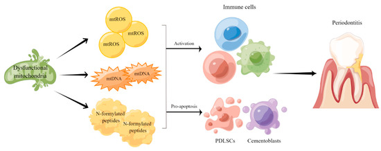
Oral inflammatory diseases (OIDs) include many common diseases such as periodontitis and pulpitis. The causes of OIDs consist microorganism, trauma, occlusal factors, autoimmune dis-eases and radiation therapy. When treated unproperly, such diseases not only affect oral health but also pose threat to people’s overall health condition. Therefore, identifying OIDs at an early stage and exploring new therapeutic strategies are important tasks for oral-related research. Mitochondria are crucial organelles for many cellular activities and disruptions of mitochondrial function not only affect cellular metabolism but also indirectly influence people’s health and life span. Increasing evidence suggests that mitochondrial dysfunction plays a critical role in the development and progression of OIDs and its associated systemic diseases. 

mitochondrial dysfunction oxidative stress redox balance oral inflammatory diseases (OIDs) periodontitis pathogenesis treatment

1. Introduction

A mitochondrion is a double membrane-enclosed organelle found in most eukaryotic cells, with a size ranging from 0.5 to 10 μm in diameter and the number of mitochondria per cell varying according to energy demand. Mitochondria are often referred to as the cellular power plants or energy center because they generate most of the adenosine-5′-triphosphate (ATP) via oxidative phosphorylation (OXPHOS) as the major source of chemical energy for physiological processes. Since mitochondria generate most reactive oxygen species (ROS) via OXPHOS and possess effective antioxidant systems, they play a central role in regulating oxidative stress and cellular redox homeostasis [1][2]. In addition to generating cellular energy, mitochondria are involved in many other processes, such as lipid metabolism, cellular differentiation, immune regulation, apoptosis, autophagy, cell growth and protein synthesis [3][4][5]. Given that mitochondria play pleiotropic roles in cellular physiology, mitochondrial dysfunction can directly regulate cell and tissue homeostasis and participate in the pathological process of many systemic diseases such as diabetes mellitus, cancer and autoimmune diseases [6][7][8].
Oral inflammatory diseases (OIDs) such as chronic periodontitis and pulpitis are becoming increasingly common, which not only damage orofacial tissue but also contribute to the increased risks of many systemic diseases [9][10]. Since OIDs have both high prevalence and recurrence, and present treatment methods for OIDs are mostly symptomatic treatment, many patients are constantly suffering from the negative impact of OIDs on life quality [11][12][13]. Therefore, it is imperative to explore the underlying causes of OIDs as well as develop new therapeutic strategies. Recently, mitochondrial dysfunction gained increasing attention in the pathogenesis and progression of OIDs by regulating oxidative stress and activating immune responses [14][15][16].

2. The Role of Mitochondrial Dysfunction in the Etiopathogenesis of the Chronic Periodontitis

As one of the most common human diseases, periodontitis is a chronic inflammatory disease that affects about 45% of adults, rising to over 60% in people aged over 65, which creates a significant healthcare, social and economic burden when left untreated or not treated appropriately [17][18]. Common features of periodontitis include gingival inflammation, clinical attachment loss and alveolar bone loss [19]. Although the main causative factor is microorganisms which colonize the subgingival dental plaque—inducing an exaggerated inflammatory response—genetic predisposition, smoking, poor oral hygiene and malnutrition are also important factors in the pathogenesis and progression of periodontitis [20][21][22][23]. Despite recent advances in the understanding of the pathological process, common treatments for periodontitis, including basic treatment, periodontal surgery and adjuvant drug administration, still provide insufficient periodontal tissue repair [24][25][26]. Recently, many studies have suggested that mitochondrial dysfunction could also contribute to the initiation of periodontitis and increase the risk of its related systemic diseases [21][27][28][29]. Govindaraj et al. conducted a study focusing on mitochondrial dysfunction in the periodontal tissue of chronic periodontitis (CP) patients and found that compared to healthy subjects, mitochondrial membrane potential and oxygen consumption rate of gingival cells from CP patients were reduced by four- and five-fold, respectively, whereas, ROS production was increased by 18%. Moreover, mitochondrial DNA sequencing revealed 14 mutations existed only in periodontal tissues but not in circulation, suggesting that mitochondrial dysfunction and genetic heterogeneity could contribute to the pathogenesis of CP [30]. These studies highlighted the value of mitochondrial function analysis in the early diagnosis of periodontitis.
Periodontal ligament stem cells (PDLSCs) are a kind of somatic mesenchymal stromal cell (MSC) which show typical mesenchymal stromal cell properties, such as self-renewal, multilineage differentiation and immunoregulation, which are essential for homeostasis in periodontal tissue [31]. Notably, PDLSCs are impaired under periodontitis and are involved in the progression of inflammation by aggravating immune response and stimulating osteoclast differentiation [32][33]. Li et al. adopted a quantitative proteomic technique to investigate the protein expression pattern during human PDLSCs osteogenesis. They found that protein related to OXPHOS may be essential in the osteogenesis process, which highlighted the role of mitochondria in regulating PDLSCs’ differentiation ability [34]. Chen et al. found that mitochondria abnormalities are present in the oxidative stress-induced periodontal ligament fibroblast apoptosis, judging by the increased mtROS amounts, upregulated mitochondrial membrane potential and ATP production [35]. Liu et al. showed that LPS-induced inflammatory responses in human gingival fibroblasts (HGFs) were partially dependent on the interaction between P53 and ROS. Upon activation, P53 and ROS formed a feedback loop and led to disrupted redox imbalance and mitochondrial dysfunction in periodontitis, triggering increased secretion of IL-1β, IL-6 and tumor necrosis factor (TNF)-α [36]. Liu et al. collected a gingiva tissue sample from CP patients and observed greater mitochondrial structure destruction, reduced mtDNA copy and lower mitochondrial protein PDK2 levels, compared with those samples from healthy individuals [37]. Similarly, HGFs from periodontitis patients exhibited increased levels of mitochondrial p53, enhanced mtROS production and secretion of pro-inflammatory cytokines, as compared to HGFs from healthy donors [38].
In an experimental periodontitis rat model, Franca et al. observed morphometric changes in renal tissues and disruption of the brush border in renal tubules, accompanied by an increase in oxidative stress and lipid peroxidation in kidneys [39]. In another study, melatonin treatment in periodontitis rats was found to be effective in restoring redox balance in gingival tissue and reducing oxidative stress levels in circulation, which could alleviate kidney injury [40]. Sun et al. observed that diabetic rats with periodontitis presented more severe mitochondrial dysfunction than non-diabetic rats with periodontitis, reflected by the decreased ATP production, reduced gene expression of electron transport chain complex I subunits and weaker mitochondrial biogenesis. They demonstrated a close correlation between these mitochondrial events and periodontal tissue damage, proving that impaired mitochondrial function contributed to the pathogenesis of periodontitis in diabetic rats [41]. Another study found that diabetic rats displayed enhanced macrophages infiltration and M1 polarization in periodontal lesions, compared with vehicle-treated rats. Under LPS or IL-4 stimulation, RAW264.7 macrophage cells showed elevated ROS levels and increased expression of M1 macrophage markers, which could be reversed by N-acetylcysteine treatment [42].
Atherosclerosis (AS) is a chronic artery disease characterized by plaque formation and chronic vascular inflammation. Many epidemiological studies have described the correlation between periodontitis and carotid AS [43][44]. Porphyromonas gingivalis (P. gingivalis), a well-known pathogen in periodontitis progression, has been shown to accelerate lipid droplet accumulation in macrophages, partially through the induction of ROS production, leading to disturbed lipid homeostasis and foam cell formation during AS [45]. P. gingivalis infection can also induce mitochondrial fragmentation, disrupt redox balance and decrease ATP concentration in vascular endothelial cells. Researchers suggested that the phosphorylation and recruitment of Drp1, a key protein involved in mitochondrial fission, might be the key events leading to mitochondrial dysfunction in P. gingivalis-infected endothelial cells, providing new insights into how periodontal pathogen-induced mitochondrial dysfunction exacerbates atherosclerotic lesions [46].
Taken together, the findings mentioned above indicate that mitochondrial dysfunction participates in the pathogenesis and progression of periodontitis by affecting oxidative stress and regulating inflammatory responses (Figure 1). Therefore, developing novel strategies to evaluate mitochondrial function in periodontitis patients may assist the diagnosis and treatment of such disease as well as its complications.
Figure 1. Mitochondrial dysfunction in the pathogenesis and progression of periodontitis. Mitochondrial dysfunction leads to metabolic disruptions and promotes the release of mtROS, mtDNA and N-formylated peptides to regulate immune responses and inflammation in periodontal tissue.

3. Mitochondrial Dysfunction-Targeted Therapies

Liu et al. identified TRPA1, an important transient receptor potential (TRP) cation channel, as an important factor in periodontium destruction in periodontitis. Inhibiting TRPA1 markedly reduced oxidative stress and apoptotic levels in LPS-treated PDLSCs, via the inhibition of endoplasmic reticulum (ER), and mitochondria stress, via downregulating PERK/eIF2α/ATF-4/CHOP pathways [47]. Periodontitis is closely related to hypoxic microenvironment. Previous studies have demonstrated that oxygen saturation in periodontal microenvironment reduced by 6% [48]. Cementoblasts possess similar characteristics with osteoblasts and generate cementum in the reconstruction process of periodontal tissue [49][50]. Wang et al. showed that overexpression of peroxisome proliferator-activated receptor gamma coactivator-1 alpha (PGC-1α), a critical regulator of mitochondrial biogenesis, can partially reverse the inhibition of cementoblasts mineralization and mitochondrial biogenesis caused by CoCl2-induced hypoxia [51].
Since oxidative stress is recognized as one of the key regulators in periodontitis, the therapeutic efficacy of several antioxidants on periodontitis are examined. Zhao et al. showed that rutin treatment inhibited the release of ROS, increased the secretion of antioxidative factors and promoted PDLSCs proliferation via the PIK3/AKT signaling pathway under an inflammatory environment [52]. Similar effects were also observed on hyperglycemic periodontitis rats [53]. Hydroxytyrosol (HT), a natural phenolic compound possessing antioxidative abilities, could inhibit mitochondrial dysfunction by decreasing optic atrophy 1 (OPA1) cleavage and by elevating AKT and GSK3β phosphorylation, which helped prevent oxidative stress-induced osteoblast apoptosis [54]. HT was also reported to exert a therapeutic effect on the periodontitis mice model via repressing RANKL-induced osteoclast maturation and promoting osteogenic differentiation. Such effect was partly dependent on attenuating mitochondrial dysfunction and inhibiting ERK and JNK pathways [55]. In addition to antioxidants, photodynamic therapy (PDT) has shown a protective effect on periodontitis by targeting mitochondria as well. Jiang et al. found methylene blue-mediated PDT could induce macrophage apoptosis in vitro and in rats with periodontitis via regulating ROS levels and reducing mitochondrial-dependent apoptosis, suggesting that the potential of PDT in treating periodontitis does not only rely on its antimicrobial capacity [56][57].
Recently, nanomaterials showed great potential in the field of periodontitis therapy [58][59]. Ren et al. synthesized a nanocomposite with ROS-scavenging activity by combining CeO2 nanoparticles (CeO2 NPs) onto the surface of mesoporous silica. Periodontal administration of such nanoparticles efficiently reduced ROS levels and improved the osteogenic differentiative capacity of hPDLSCs with H2O2-induced oxidative stress injury [60]. The same group synthesized controlled drug release nanoparticles by encasing mitoquinone (MitoQ, an autophagy enhancer) into tailor-made ROS-cleavable amphiphilic polymer nanoparticles. Once exposed to ROS under oxidative stress conditions, the ROS-cleavable structure disintegrated, promoting the release of the encapsulated MitoQ. The released MitoQ efficiently induced mitophagy through the PINK1-Parkin pathway and reduced oxidative stress, which contributed to a redox homeostasis and facilitated periodontal tissue regeneration [61]. Qiu et al. fabricated a ROS-cleavable nanoplatform by encapsulating N-acetylcysteine into tailor-made amphiphilic polymer nanoparticles which could decrease osteoclast activity and inflammation in the periodontitis rat model and improve the restoration of destroyed periodontal tissue [62]. Zhai et al. showed that obstructed mitophagy and Ca2+ overload led to dysfunctional mitochondria accumulation in MSCs isolated from periodontitis and osteoarthritis patients. They engineered mitochondria-targeting and intracellular microenvironment-responsive nanoparticles to attract Ca2+ around mitochondria in MSCs to regulate calcium flux into mitochondria, which successfully restored the mitochondrial function of diseased MSCs and rescued periodontal tissue damage [63].
In addition to alleviating periodontitis-induced periodontal tissue damage, targeting mitochondrial dysfunction in periodontitis also contributes to ameliorating the complications of periodontitis. Li et al. demonstrated that resveratrol could prevent periodontal tissue destruction, as indicated by the improvement in pocket depth, gingival bleeding and tooth mobility. Meanwhile, resveratrol administration also alleviated periodontitis-induced kidney injury by means of decreasing oxidative stress, regulating mitochondrial membrane potential and increasing mtDNA such as Sirtuin 1 and PGC-1α [1]. Febuxostat, a potent xanthine oxidase inhibitor, has been shown to attenuate the progression of periodontitis in rats by reducing inflammatory cytokine levels and oxidative stress. It also attenuated periodontitis-induced glucose intolerance and blood pressure elevations, suggesting its therapeutic potential in treating patients with both diabetes and periodontitis [64].

References

  1. Li, X.; Liu, X.C.; Ding, X.; Liu, X.M.; Cao, N.B.; Deng, Y.; Hou, Y.B.; Yu, W.X. Resveratrol protects renal damages induced by periodontitis via preventing mitochondrial dysfunction in rats. Oral. Dis. 2022, 29, 1812–1825.
  2. Willems, P.H.; Rossignol, R.; Dieteren, C.E.; Murphy, M.P.; Koopman, W.J. Redox Homeostasis and Mitochondrial Dynamics. Cell Metab. 2015, 22, 207–218.
  3. Abate, M.; Festa, A.; Falco, M.; Lombardi, A.; Luce, A.; Grimaldi, A.; Zappavigna, S.; Sperlongano, P.; Irace, C.; Caraglia, M.; et al. Mitochondria as playmakers of apoptosis, autophagy and senescence. Semin. Cell Dev. Biol. 2020, 98, 139–153.
  4. Spinelli, J.B.; Haigis, M.C. The multifaceted contributions of mitochondria to cellular metabolism. Nat. Cell Biol. 2018, 20, 745–754.
  5. Chan, D.C. Mitochondrial Dynamics and Its Involvement in Disease. Annu. Rev. Pathol. 2020, 15, 235–259.
  6. Sangwung, P.; Petersen, K.F.; Shulman, G.I.; Knowles, J.W. Mitochondrial Dysfunction, Insulin Resistance, and Potential Genetic Implications. Endocrinology 2020, 161, bqaa017.
  7. Moro, L. Mitochondrial Dysfunction in Aging and Cancer. J. Clin. Med. 2019, 8, 1983.
  8. Zhao, M.; Wang, Y.; Li, L.; Liu, S.; Wang, C.; Yuan, Y.; Yang, G.; Chen, Y.; Cheng, J.; Lu, Y.; et al. Mitochondrial ROS promote mitochondrial dysfunction and inflammation in ischemic acute kidney injury by disrupting TFAM-mediated mtDNA maintenance. Theranostics 2021, 11, 1845–1863.
  9. Gong, W.; Wang, F.; He, Y.; Zeng, X.; Zhang, D.; Chen, Q. Mesenchymal Stem Cell Therapy for Oral Inflammatory Diseases: Research Progress and Future Perspectives. Curr. Stem Cell Res. Ther. 2021, 16, 165–174.
  10. Bitencourt, F.V.; Nascimento, G.G.; Costa, S.A.; Andersen, A.; Sandbæk, A.; Leite, F.R.M. Co-occurrence of Periodontitis and Diabetes-Related Complications. J. Dent. Res. 2023, 102, 1088–1097.
  11. Vujovic, S.; Desnica, J.; Stevanovic, M.; Mijailovic, S.; Vojinovic, R.; Selakovic, D.; Jovicic, N.; Rosic, G.; Milovanovic, D. Oral Health and Oral Health-Related Quality of Life in Patients with Primary Sjögren’s Syndrome. Medicina 2023, 59, 473.
  12. Sødal, A.T.T.; Skudutyte-Rysstad, R.; Diep, M.T.; Koldsland, O.C.; Hove, L.H. Periodontitis in a 65-year-old population: Risk indicators and impact on oral health-related quality of life. BMC Oral Health 2022, 22, 640.
  13. Taha, N.A.; Abuzaid, A.M.; Khader, Y.S. A Randomized Controlled Clinical Trial of Pulpotomy versus Root Canal Therapy in Mature Teeth with Irreversible Pulpitis: Outcome, Quality of Life, and Patients’ Satisfaction. J. Endod. 2023, 49, 624–631.e622.
  14. Jiang, W.; Wang, Y.; Cao, Z.; Chen, Y.; Si, C.; Sun, X.; Huang, S. The role of mitochondrial dysfunction in periodontitis: From mechanisms to therapeutic strategy. J. Periodontal Res. 2023, 58, 853–863.
  15. Verma, A.; Azhar, G.; Zhang, X.; Patyal, P.; Kc, G.; Sharma, S.; Che, Y.; Wei, J.Y.P. gingivalis-LPS Induces Mitochondrial Dysfunction Mediated by Neuroinflammation through Oxidative Stress. Int. J. Mol. Sci. 2023, 24, 950.
  16. Zhou, L.; Zhang, Y.F.; Yang, F.H.; Mao, H.Q.; Chen, Z.; Zhang, L. Mitochondrial DNA leakage induces odontoblast inflammation via the cGAS-STING pathway. Cell Commun. Signal 2021, 19, 58.
  17. Demmer, R.T.; Papapanou, P.N. Epidemiologic patterns of chronic and aggressive periodontitis. Periodontol. 2000 2010, 53, 28–44.
  18. Genco, R.J.; Sanz, M. Clinical and public health implications of periodontal and systemic diseases: An overview. Periodontol. 2000 2020, 83, 7–13.
  19. Papapanou, P.N.; Sanz, M.; Buduneli, N.; Dietrich, T.; Feres, M.; Fine, D.H.; Flemmig, T.F.; Garcia, R.; Giannobile, W.V.; Graziani, F.; et al. Periodontitis: Consensus report of workgroup 2 of the 2017 World Workshop on the Classification of Periodontal and Peri-Implant Diseases and Conditions. J. Periodontol. 2018, 89 (Suppl. S1), S173–S182.
  20. Laine, M.L.; Crielaard, W.; Loos, B.G. Genetic susceptibility to periodontitis. Periodontol. 2000 2012, 58, 37–68.
  21. Tóthová, L.; Celec, P. Oxidative Stress and Antioxidants in the Diagnosis and Therapy of Periodontitis. Front. Physiol. 2017, 8, 1055.
  22. Kinane, D.F.; Stathopoulou, P.G.; Papapanou, P.N. Periodontal diseases. Nat. Rev. Dis. Primers 2017, 3, 17038.
  23. Genco, R.J.; Borgnakke, W.S. Risk factors for periodontal disease. Periodontol. 2000 2013, 62, 59–94.
  24. Graziani, F.; Karapetsa, D.; Alonso, B.; Herrera, D. Nonsurgical and surgical treatment of periodontitis: How many options for one disease? Periodontol. 2000 2017, 75, 152–188.
  25. Kwon, T.; Lamster, I.B.; Levin, L. Current Concepts in the Management of Periodontitis. Int. Dent. J. 2021, 71, 462–476.
  26. Prietto, N.R.; Martins, T.M.; Santinoni, C.D.S.; Pola, N.M.; Ervolino, E.; Bielemann, A.M.; Leite, F.R.M. Treatment of experimental periodontitis with chlorhexidine as adjuvant to scaling and root planing. Arch. Oral. Biol. 2020, 110, 104600.
  27. Xie, M.; Tang, Q.; Nie, J.; Zhang, C.; Zhou, X.; Yu, S.; Sun, J.; Cheng, X.; Dong, N.; Hu, Y.; et al. BMAL1-Downregulation Aggravates Porphyromonas Gingivalis-Induced Atherosclerosis by Encouraging Oxidative Stress. Circ. Res. 2020, 126, e15–e29.
  28. Bullon, P.; Newman, H.N.; Battino, M. Obesity, diabetes mellitus, atherosclerosis and chronic periodontitis: A shared pathology via oxidative stress and mitochondrial dysfunction? Periodontol. 2000 2014, 64, 139–153.
  29. Li, L.; Zhang, Y.L.; Liu, X.Y.; Meng, X.; Zhao, R.Q.; Ou, L.L.; Li, B.Z.; Xing, T. Periodontitis Exacerbates and Promotes the Progression of Chronic Kidney Disease through Oral Flora, Cytokines, and Oxidative Stress. Front. Microbiol. 2021, 12, 656372.
  30. Govindaraj, P.; Khan, N.A.; Gopalakrishna, P.; Chandra, R.V.; Vanniarajan, A.; Reddy, A.A.; Singh, S.; Kumaresan, R.; Srinivas, G.; Singh, L.; et al. Mitochondrial dysfunction and genetic heterogeneity in chronic periodontitis. Mitochondrion 2011, 11, 504–512.
  31. Tomokiyo, A.; Wada, N.; Maeda, H. Periodontal Ligament Stem Cells: Regenerative Potency in Periodontium. Stem Cells Dev. 2019, 28, 974–985.
  32. Zhang, Z.; Deng, M.; Hao, M.; Tang, J. Periodontal ligament stem cells in the periodontitis niche: Inseparable interactions and mechanisms. J. Leukoc. Biol. 2021, 110, 565–576.
  33. Zheng, Y.; Dong, C.; Yang, J.; Jin, Y.; Zheng, W.; Zhou, Q.; Liang, Y.; Bao, L.; Feng, G.; Ji, J.; et al. Exosomal microRNA-155-5p from PDLSCs regulated Th17/Treg balance by targeting sirtuin-1 in chronic periodontitis. J. Cell Physiol. 2019, 234, 20662–20674.
  34. Li, J.; Wang, Z.; Huang, X.; Wang, Z.; Chen, Z.; Wang, R.; Chen, Z.; Liu, W.; Wu, B.; Fang, F.; et al. Dynamic proteomic profiling of human periodontal ligament stem cells during osteogenic differentiation. Stem Cell Res. Ther. 2021, 12, 98.
  35. Chen, Y.; Ji, Y.; Jin, X.; Sun, X.; Zhang, X.; Chen, Y.; Shi, L.; Cheng, H.; Mao, Y.; Li, X.; et al. Mitochondrial abnormalities are involved in periodontal ligament fibroblast apoptosis induced by oxidative stress. Biochem. Biophys. Res. Commun. 2019, 509, 483–490.
  36. Liu, J.; Zeng, J.; Wang, X.; Zheng, M.; Luan, Q. P53 mediates lipopolysaccharide-induced inflammation in human gingival fibroblasts. J. Periodontol. 2018, 89, 1142–1151.
  37. Liu, J.; Wang, X.; Xue, F.; Zheng, M.; Luan, Q. Abnormal mitochondrial structure and function are retained in gingival tissues and human gingival fibroblasts from patients with chronic periodontitis. J. Periodontal Res. 2022, 57, 94–103.
  38. Liu, J.; Wang, X.; Zheng, M.; Luan, Q. Oxidative stress in human gingival fibroblasts from periodontitis versus healthy counterparts. Oral. Dis. 2021, 29, 1214–1225.
  39. França, L.F.C.; Vasconcelos, A.; da Silva, F.R.P.; Alves, E.H.P.; Carvalho, J.S.; Lenardo, D.D.; de Souza, L.K.M.; Barbosa, A.L.R.; Medeiros, J.R.; de Oliveira, J.S.; et al. Periodontitis changes renal structures by oxidative stress and lipid peroxidation. J. Clin. Periodontol. 2017, 44, 568–576.
  40. Kose, O.; Kurt Bayrakdar, S.; Unver, B.; Altin, A.; Akyildiz, K.; Mercantepe, T.; Bostan, S.A.; Arabaci, T.; Turker Sener, L.; Emre Kose, T.; et al. Melatonin improves periodontitis-induced kidney damage by decreasing inflammatory stress and apoptosis in rats. J. Periodontol. 2021, 92, 22–34.
  41. Sun, X.; Mao, Y.; Dai, P.; Li, X.; Gu, W.; Wang, H.; Wu, G.; Ma, J.; Huang, S. Mitochondrial dysfunction is involved in the aggravation of periodontitis by diabetes. J. Clin. Periodontol. 2017, 44, 463–471.
  42. Zhang, B.; Yang, Y.; Yi, J.; Zhao, Z.; Ye, R. Hyperglycemia modulates M1/M2 macrophage polarization via reactive oxygen species overproduction in ligature-induced periodontitis. J. Periodontal Res. 2021, 56, 991–1005.
  43. Zeng, X.T.; Leng, W.D.; Lam, Y.Y.; Yan, B.P.; Wei, X.M.; Weng, H.; Kwong, J.S. Periodontal disease and carotid atherosclerosis: A meta-analysis of 17,330 participants. Int. J. Cardiol. 2016, 203, 1044–1051.
  44. Priyamvara, A.; Dey, A.K.; Bandyopadhyay, D.; Katikineni, V.; Zaghlol, R.; Basyal, B.; Barssoum, K.; Amarin, R.; Bhatt, D.L.; Lavie, C.J. Periodontal Inflammation and the Risk of Cardiovascular Disease. Curr. Atheroscler. Rep. 2020, 22, 28.
  45. Rho, J.H.; Kim, H.J.; Joo, J.Y.; Lee, J.Y.; Lee, J.H.; Park, H.R. Periodontal Pathogens Promote Foam Cell Formation by Blocking Lipid Efflux. J. Dent. Res. 2021, 100, 1367–1377.
  46. Xu, T.; Dong, Q.; Luo, Y.; Liu, Y.; Gao, L.; Pan, Y.; Zhang, D. Porphyromonas gingivalis infection promotes mitochondrial dysfunction through Drp1-dependent mitochondrial fission in endothelial cells. Int. J. Oral. Sci. 2021, 13, 28.
  47. Liu, Q.; Guo, S.; Huang, Y.; Wei, X.; Liu, L.; Huo, F.; Huang, P.; Wu, Y.; Tian, W. Inhibition of TRPA1 Ameliorates Periodontitis by Reducing Periodontal Ligament Cell Oxidative Stress and Apoptosis via PERK/eIF2α/ATF-4/CHOP Signal Pathway. Oxid. Med. Cell Longev. 2022, 2022, 4107915.
  48. Gölz, L.; Memmert, S.; Rath-Deschner, B.; Jäger, A.; Appel, T.; Baumgarten, G.; Götz, W.; Frede, S. Hypoxia and P. gingivalis synergistically induce HIF-1 and NF-κB activation in PDL cells and periodontal diseases. Mediat. Inflamm. 2015, 2015, 438085.
  49. Zhao, J.; Faure, L.; Adameyko, I.; Sharpe, P.T. Stem cell contributions to cementoblast differentiation in healthy periodontal ligament and periodontitis. Stem Cells 2021, 39, 92–102.
  50. Arzate, H.; Zeichner-David, M.; Mercado-Celis, G. Cementum proteins: Role in cementogenesis, biomineralization, periodontium formation and regeneration. Periodontol. 2000 2015, 67, 211–233.
  51. Wang, H.; Wang, X.; Ma, L.; Huang, X.; Peng, Y.; Huang, H.; Gao, X.; Chen, Y.; Cao, Z. PGC-1 alpha regulates mitochondrial biogenesis to ameliorate hypoxia-inhibited cementoblast mineralization. Ann. N.Y. Acad. Sci. 2022, 1516, 300–311.
  52. Zhao, B.; Zhang, W.; Xiong, Y.; Zhang, Y.; Zhang, D.; Xu, X. Effects of rutin on the oxidative stress, proliferation and osteogenic differentiation of periodontal ligament stem cells in LPS-induced inflammatory environment and the underlying mechanism. J. Mol. Histol. 2020, 51, 161–171.
  53. Iova, G.M.; Calniceanu, H.; Popa, A.; Szuhanek, C.A.; Marcu, O.; Ciavoi, G.; Scrobota, I. The Antioxidant Effect of Curcumin and Rutin on Oxidative Stress Biomarkers in Experimentally Induced Periodontitis in Hyperglycemic Wistar Rats. Molecules 2021, 26, 1332.
  54. Cai, W.J.; Chen, Y.; Shi, L.X.; Cheng, H.R.; Banda, I.; Ji, Y.H.; Wang, Y.T.; Li, X.M.; Mao, Y.X.; Zhang, D.F.; et al. AKT-GSK3β Signaling Pathway Regulates Mitochondrial Dysfunction-Associated OPA1 Cleavage Contributing to Osteoblast Apoptosis: Preventative Effects of Hydroxytyrosol. Oxid. Med. Cell Longev. 2019, 2019, 4101738.
  55. Zhang, X.; Jiang, Y.; Mao, J.; Ren, X.; Ji, Y.; Mao, Y.; Chen, Y.; Sun, X.; Pan, Y.; Ma, J.; et al. Hydroxytyrosol prevents periodontitis-induced bone loss by regulating mitochondrial function and mitogen-activated protein kinase signaling of bone cells. Free Radic. Biol. Med. 2021, 176, 298–311.
  56. Jiang, C.; Yang, W.; Wang, C.; Qin, W.; Ming, J.; Zhang, M.; Qian, H.; Jiao, T. Methylene Blue-Mediated Photodynamic Therapy Induces Macrophage Apoptosis via ROS and Reduces Bone Resorption in Periodontitis. Oxid. Med. Cell Longev. 2019, 2019, 1529520.
  57. Chambrone, L.; Wang, H.L.; Romanos, G.E. Antimicrobial photodynamic therapy for the treatment of periodontitis and peri-implantitis: An American Academy of Periodontology best evidence review. J. Periodontol. 2018, 89, 783–803.
  58. Sui, L.; Wang, J.; Xiao, Z.; Yang, Y.; Yang, Z.; Ai, K. ROS-Scavenging Nanomaterials to Treat Periodontitis. Front. Chem. 2020, 8, 595530.
  59. Mok, Z.H.; Proctor, G.; Thanou, M. Emerging nanomaterials for dental treatments. Emerg. Top. Life Sci. 2020, 4, 613–625.
  60. Ren, S.; Zhou, Y.; Fan, R.; Peng, W.; Xu, X.; Li, L.; Xu, Y. Constructing biocompatible MSN@Ce@PEG nanoplatform for enhancing regenerative capability of stem cell via ROS-scavenging in periodontitis. Chem. Eng. J. 2021, 423, 130207.
  61. Li, X.; Zhao, Y.; Peng, H.; Gu, D.; Liu, C.; Ren, S.; Miao, L. Robust intervention for oxidative stress-induced injury in periodontitis via controllably released nanoparticles that regulate the ROS-PINK1-Parkin pathway. Front. Bioeng. Biotechnol. 2022, 10, 1081977.
  62. Qiu, X.; Yu, Y.; Liu, H.; Li, X.; Sun, W.; Wu, W.; Liu, C.; Miao, L. Remodeling the periodontitis microenvironment for osteogenesis by using a reactive oxygen species-cleavable nanoplatform. Acta Biomater. 2021, 135, 593–605.
  63. Zhai, Q.; Chen, X.; Fei, D.; Guo, X.; He, X.; Zhao, W.; Shi, S.; Gooding, J.J.; Jin, F.; Jin, Y.; et al. Nanorepairers Rescue Inflammation-Induced Mitochondrial Dysfunction in Mesenchymal Stem Cells. Adv. Sci. 2022, 9, e2103839.
  64. Nessa, N.; Kobara, M.; Toba, H.; Adachi, T.; Yamamoto, T.; Kanamura, N.; Pezzotti, G.; Nakata, T. Febuxostat Attenuates the Progression of Periodontitis in Rats. Pharmacology 2021, 106, 294–304.
More
Information
Contributors MDPI registered users' name will be linked to their SciProfiles pages. To register with us, please refer to https://encyclopedia.pub/register : , ,
View Times: 201
Revisions: 2 times (View History)
Update Date: 02 Nov 2023
1000/1000