Submitted Successfully!
To reward your contribution, here is a gift for you: A free trial for our video production service.
Thank you for your contribution! You can also upload a video entry or images related to this topic.
Version Summary Created by Modification Content Size Created at Operation
1 -- 2761 2023-10-07 10:55:48

Video Upload Options

Do you have a full video?

Confirm

Are you sure to Delete?
Cite
If you have any further questions, please contact Encyclopedia Editorial Office.
Justiz-Vaillant, A.A.; Williams-Persad, A.F.; Arozarena-Fundora, R.; Gopaul, D.; Soodeen, S.; Asin-Milan, O.; Thompson, R.; Unakal, C.; Akpaka, P.E. Pathogens, Diagnosis and Treatment of Chronic Granulomatous Disease. Encyclopedia. Available online: https://encyclopedia.pub/entry/49898 (accessed on 06 September 2024).
Justiz-Vaillant AA, Williams-Persad AF, Arozarena-Fundora R, Gopaul D, Soodeen S, Asin-Milan O, et al. Pathogens, Diagnosis and Treatment of Chronic Granulomatous Disease. Encyclopedia. Available at: https://encyclopedia.pub/entry/49898. Accessed September 06, 2024.
Justiz-Vaillant, Angel A., Arlene Faye-Ann Williams-Persad, Rodolfo Arozarena-Fundora, Darren Gopaul, Sachin Soodeen, Odalis Asin-Milan, Reinand Thompson, Chandrashekhar Unakal, Patrick Eberechi Akpaka. "Pathogens, Diagnosis and Treatment of Chronic Granulomatous Disease" Encyclopedia, https://encyclopedia.pub/entry/49898 (accessed September 06, 2024).
Justiz-Vaillant, A.A., Williams-Persad, A.F., Arozarena-Fundora, R., Gopaul, D., Soodeen, S., Asin-Milan, O., Thompson, R., Unakal, C., & Akpaka, P.E. (2023, October 07). Pathogens, Diagnosis and Treatment of Chronic Granulomatous Disease. In Encyclopedia. https://encyclopedia.pub/entry/49898
Justiz-Vaillant, Angel A., et al. "Pathogens, Diagnosis and Treatment of Chronic Granulomatous Disease." Encyclopedia. Web. 07 October, 2023.
Pathogens, Diagnosis and Treatment of Chronic Granulomatous Disease
Edit

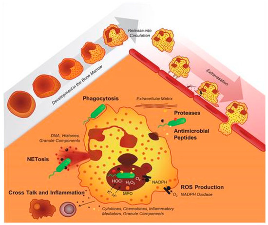
Chronic granulomatous disease (CGD) is a primary immunodeficiency caused by a defect in the phagocytic function of the innate immune system owing to mutations in genes encoding the five subunits of the nicotinamide adenine dinucleotide phosphatase (NADPH) oxidase enzyme complex. The most common microorganisms observed in the patients with CGD are Staphylococcus aureus, Aspergillus spp., Candida spp., Nocardia spp., Burkholderia spp., Serratia spp., and Salmonella spp. Antibacterial prophylaxis with trimethoprim-sulfamethoxazole, antifungal prophylaxis usually with itraconazole, and interferon gamma immunotherapy have been successfully used in reducing infection in CGD. Haematopoietic stem cell transplantation (HCT) have been successfully proven to be the treatment of choice in patients with CGD.

chronic granulomatous disease microorganisms neutrophils antimicrobials

1. Introduction

Chronic granulomatous disease (CGD) is a rare inherited primary immunodeficiency, that was first reported as a “Fatal granulomatous disease of childhood” owing to the early death of children with this condition, and has been described by some other authors [1][2][3][4][5][6][7]. A schematic representation of the neutrophil effector functions required to achieve an adequate primary immune defense has recently been described in this condition. It is displayed in reports by Kruger P et al. (Figure 1) [8]. Neutrophils develop in the bone marrow; band neutrophils are released into circulation and subsequently travel to tissues and organs to fight infections using various mechanisms, including phagocytosis, production of reactive oxygen species, and release of antimicrobial peptides, to destroy pathogens [8].
Figure 1. Schematic representation of normal phagocytic function reproduced from Kruger et al. (2015). Copyright: © 2015 Kruger et al. This is an open-access article distributed under the terms of the Creative Commons [8].
CGD is caused by the impaired phagocytic function of the innate immune system cells owing to mutations in genes encoding the five subunits of the nicotinamide adenine dinucleotide phosphate (NADPH) oxidase enzyme complex (OEC) [4]. The normal respiratory burst process is critical in killing pathogens, manifesting as CGD when it is dysfunctional.

2. Pathogenesis

The CGD’s inability to produce reactive oxygen species leads to pathognomonic systemic granuloma formation and increased susceptibility to recurrent and severe opportunistic bacterial and fungal infections. It causes unrestrained inflammation and autoimmunity. Since the condition was first described [1], there have been several improvements in treatment, such as antibacterial, antifungal, immunomodulatory, and hematopoietic stem cell transplantation, which extended the life expectancy of patients [9].
The severity of the phenotypes can vary depending on the mode of genotype inheritance, which is most severe in the X-linked type, followed by the autosomal recessive type [10]. The functional NADPH OEC comprises five subunits: two are localized in the cell membrane during the resting phase, and three are localized in the cytoplasm. The two membrane-bound subunits are gp91phox and p22phox. These proteins form a heterodimeric complex (cytochrome b558). The cell membrane’s contact with a pathogen activates the protein complex, and three cytoplasmic subunits (p47phox, p67phox, and p40phox) form a hetero-trimer translocating to cytochrome b558 [4].

3. Clinical Manifestations

Patients usually present with fever, malaise, or weight loss. Perirectal abscesses are also typical in patients with CGD and can persist for years despite aggressive antimicrobial treatments and intense local care. Several pathogens are associated with infections in CGD and these granulomas produce active lesions that in many cases are sterile with no pathogens involved. For example, in inflammatory bowel diseases (IBD), see below. In these circumstances, chronic inflammatory cell responses consisting of activated lymphocytes and histiocytes evolve and arrange to form granulomas, one of the hallmarks of CGD, provoking diverse clinical manifestations of obstruction such as delayed gastric emptying, antral narrowing of the stomach, dysphagia, emesis, weight loss, biliary tract or gastrointestinal obstruction [11].

3.1. CGD-Related Inflammatory Responses

There is an increased risk of autoimmune disorders such as inflammatory bowel colitis and inflammatory bowel disease among patients owing to increased activation of NF-kB, increasing the production of proinflammatory cytokines. The inflammatory manifestations of CGD are mainly observed in the GI and urogenital tracts, lungs, and eyes. Inflammation can be suppressed by blocking TNF-alpha and oral corticosteroids [12].

3.2. Hemophagocytic Lymphohistiocytosis (HLH)

In addition, patients with CGD experience infection-triggered hemophagocytic lymphohistiocytosis (HLH), which presents as pathological hyperactive inflammation [13]. Possible pathologies, including CGD, should be considered in children with HLH because it can indicate CGD. An optimal management strategy is yet to be developed for children with CGD who manifest with HLH. Early recognition and proper management of infectious triggers and HLH are crucial to reducing mortality [3].

4. CGD Incidence

Pediatric CGD is relatively rare; this genetic condition, which has variable ethnic associations, occurs in 1 out of every 200,000–250,000 births in the United States and is often diagnosed in the first three years of life [9][14]. According to data from European nations, approximately 65% of patients with CGD have a molecular defect in CYBB (most are hemizygous males). Autosomal recessive CGD accounts for approximately 30% of all CGD cases. Molecular defects in any of these five genes (CYBB for gp91phox (located on the X chromosome), CYBA for p22phox, NCF1 for p47phox, NCF2 for p67phox, and NCF4 for p40phox) can occur in 90% of patients with CGD. They harbour mutations in the CYBB (gp91phox) or NCF1 (p47phox) genes [15]. Symptoms may be delayed in some patients with residual activity [13]. In most countries, the offspring of CGD consanguineous marriages or unions is at increased risk of CGD due to expressing autosomal recessive gene mutations inherited from common CGD ancestors. Therefore, consanguineous unions increase the incidence of this condition, which sometimes is not detected quickly enough, and death from infectious diseases may occur [16][17][18][19].

5. Granulomas

In patients with CGD, microgranulomas, tissue eosinophilia, and brown-pigmented epithelioid histiocytes found in the lamina propria and inflammatory changes revealed these distinctive features from biopsy materials [20][21]. Alimchandani et al. (2013) conducted a study on 87 patients with CGD and observed using GI biopsy that 74% (64/87) of patients had prominent brown granular cytoplasmic pigmented inclusions in macrophages [22]. Multiple aseptic granulomas most frequently form in the skin. Granulomas are active lesions and in many cases are sterile [20].

6. Pathogens and CGD-Related Infectious Diseases

6.1. Pathogens

Owing to changes and the introduction of new prophylactic treatments after the initial emergence of CGD, the median age of death has increased over the last few decades, with fungal infections being the highest risk of mortality [14][23]. The five most common pathogens that infect North American patients are Staphylococcus aureus, Aspergillus spp., Nocardia spp., Burkholderia spp., and Serratia spp. [21][23] in contrast to patients from Europe, where the five most common pathogens are Staphylococcus aureus, Aspergillus, Salmonella, Candida, and Serratia species. The infections can be severe or opportunistic and unusual because of fungi and bacteria that cause suppurative lymphadenitis, pneumonia, and abscesses at various locations [18][23]. These are also discussed below regarding signs, symptoms, and complications.
Chronic Granulomatous Disease patients are susceptible to a subset of catalase-positive organisms (CPO) because CPO degrades host-produced hydrogen peroxide before its conversion to hypochlorous acid by myeloperoxidase [9]. These CPOs include bacteria such as Pseudomonas spp., and Enterobacteriaceae such as Klebsiella spp. [14][20][23].
Fungi including Neosartorya udagawae, and Sporothrix schenckii have been reported in patients with CGD treated with antifungal prophylaxis [21]. Other rare pathogens that have been cited, including sporadic cases of Cephalosporium, Streptococcus pneumoniae, Scedosporium, Paecilomyces aecilomyce, Staphylococcus epidermidis, Rhodococcus equi and Phialophora richardsiae [20]. Non-Aspergillus fungal infections are prevalent and can be associated with Rhizopus spp. and Trichosporon spp. They were reported in nine cases and the lung was the most commonly affected organ [24].

6.2. CGD-Related Infectious Diseases

Notably, severe recurrent bacterial and fungal infections usually present early in childhood (<5 years of age) in most patients with CGD. This is attributed to severe respiratory burst defects and the lack of EROS production. However, symptoms are delayed until adolescence and adulthood owing to the degree of residual NADPH oxidase activity [25].
The most frequent fungal infection is invasive aspergillosis caused by Aspergillus fumigatus, followed by Aspergillus nidulans and less common Aspergillus terreus, which has been isolated from bronchoalveolar lavage of patients with CGD [14][26].

7. Laboratory Diagnosis

7.1. Neutrophil-Function Testing

Patients suspected of suffering from CGD are diagnosed by the inability of their blood phagocytes to generate reactive oxygen species [5]. Initial diagnostic tests for CGD often rely on the various measurements of neutrophil superoxide production [27]. These include (a) direct measurement of superoxide production [28], (b) Cytochrome C Reduction Assay—This is based on a colorimetric assay that measures the reduction of cytochrome C by NADPH-Cytochrome C reductase in the presence of NADPH. The reduction of cytochrome C results in the formation of distinct bands in the absorption spectrum and the increase in absorbance at 550 nm is measured with time [29][30], (c) Nitroblue tetrazolium (NBT) reduction test—The nitroblue tetrazolium reduction test (NBT) is an assay based on the activation percentage of neutrophils in peripheral blood. It has been used to study the follow-up of microbial agents owing to the narrow relationship between the molecules involved in the oxidative burst and the organisms e.g., Leishmania activity in phagocytes [30][31][32][33][34][35][36], (d) Dihydrorhodamine (DHR) 123 Oxidation test—White blood cells are incubated with dihydrorhodamine 123 (DHR) and catalase, then stimulated with Phorbol 12-Myristate 13-Acetate (PMA). Dihydrorhodamine oxidation to rhodamine by the respiratory burst of the cell is measured by flow cytometry [32][37] and (e) Chemiluminescence [37][38].

7.2. Nitroblue Tetrazolium (NBT) Reduction Test

The oldest laboratory test for CGD is the NBT test, often the primary screening test for CGD [39]. This test uses light microscopy to provide a rapid but relatively qualitative analysis of phagocyte NADPH oxidase activity. Superoxide produced by normal peripheral blood neutrophils reduces yellow NBT to dark blue/black formazan, which forms a precipitate in the cells [40]. The test is performed on a microscope slide, to manually distinguish reducing (blue-black) from non-reducing (unstained) cells manually.

7.3. Flow Cytometric Dihydrorhodamine Assay

Dihydrorhodamine (DHR) assay testing is often the test of choice in diagnosing CGD [41]. DHR123 is a lipophilic nonfluorescent molecule that readily diffuses across cell membranes and localises in the mitochondria [29]. The molecule is oxidised to rhodamine 123 in stimulated phagocytes of the nicotinamide adenine dinucleotide phosphate (NADPH) oxidase and is trapped within cells in this form [30]. The quantitative nature of this assay allows for differentiation between oxidase-positive and oxidase-negative phagocyte CGD carriers [27] and diagnosis of gp91phox and p47phox deficiencies [31]. DHR also allows for measuring residual superoxide production and thus provides prognostic information for CGD patients [32]. DHR assays are relatively easier to perform, more reliable, more quantitative, and more sensitive when compared to NBT [37]. DHR assays can distinguish between X-linked and autosomal variants of CGD and detect gp91phox carriers [38][42]. The DHR test can also be used to determine chimerism status following hematopoietic cell transplantation [43] which is important to evaluate for early engraftment and graft failure and thus, guides for early intervention [44].

7.4. Luminol-Enhanced Chemiluminescence Assay

Chemiluminescence is the emission of light (luminescence) as the result of a chemical reaction (e.g., oxidation). It is essentially an oxyluminescence since molecular oxygen is necessary for the reaction. Luminol is widely used to detect reactive oxygen species produced in biological systems and is used in CGD patients to measure ROS production [45].

7.5. Genetic Testing

The diagnosis of CGD based on positive abnormal neutrophil function testing should be followed by genetic testing for confirmation. Sequencing of the patient’s phagocyte oxidase (phox) genes is done to determine the exact molecular defect [27]. The most common p47phox pathogenic defect in CGD is due to a pseudogene conversion and may be missed by standard sequencing [46]. Immunoblot (standard immunoblotting of neutrophils stained with phox-specific antibodies) or gene dose determination may be needed to confirm p47phox deficiency [28]. Pathogenic variants in the CYBB gene encoding gp91phox are mostly due to missense or nonsense mutations but can also be due to promoter, insertion, deletion or splice site mutations [47][48].

8. Management of CGD

8.1. Haematopoietic Stem Cell Transplantation (HSCT/HCT)

HCT is the principal treatment for managing CGD with favourable results regardless of symptoms, age, sex, or mutations [49][50][51]. Transplantation therapy has an overall survival rate of over 90% in children under 14 years and has improved in the last decade, particularly with early diagnosis [50].
However, the group of HCT-treated patients demonstrated excellent survival rates, although the risks and benefits still need to be assessed in individual patients. Based on the significant progress of patients with CGD treated with HCT, it is regarded as the only known curative treatment with an improved life expectancy owing to its improved implementation over time [52][53][54][55].
When Human Leucocyte Antigen (HLA)-matched donor is identified, the source of HCT could be cord blood, bone marrow, or peripheral blood [51]. Hematopoietic stem cells are drawn and infused into the patient. These are immature cells, and after they develop into platelets, red- and white-blood cells.

8.2. Drug-Based Treatment

Antimicrobial and antifungal prophylaxis are the most common management routes used to minimize the incidence of infections. However, treatment with antibiotics is contraindicated in healthy patients because of antibiotic resistance. Most studies suggest a link between aggressive antibiotic use and preventing the spread of infection in patients with CGD [56].
Drugs such as trimethoprim-sulfamethoxazole reduce the occurrence of bacterial infections in patients with CGD but do not interfere considerably with the gut microbiome [57]. Patients with sulfamethoxazole allergy have other options, such as cloxacillin and ciprofloxacin [50]. A concern arises in pregnancy since trimethoprim is a folic acid antagonist, which increases the high risk for congenital disabilities and is discontinued during pregnancy [49].
CGD treatment should start at the earliest, and before the microbiological cultures are available. Antimicrobials should be given parenterally. Bacterial infections such as S. aureus and gram-negative bacteria, including B. cepacia complex, can be treated with a combination of ceftazidime and nafcillin and or a carbapenem. However, Burkholderia is typically resistant to most aminoglycosides. If the infection persists for 24–48 h, then more diagnostic tests should be done to identify the responsible microorganism. 
If fungus is identified, antifungal treatment should be institutionalised even before the diagnosis is confirmed. Lung and bone aspergillosis are very prevalent and require prolonged therapy. The echinocandin antifungals including micafungin, caspofungin, and anidulafungin can effectively treat refractory Aspergillosis in patients unresponsive to lipid-formulated amphotericin B and azoles. Intravenous antifungals must be early considered in CGD patients [58].
Treatment using TNF-alpha inhibitors in patients with CGD could help improve the outcome of severe inflammatory complications despite the associated risk factors. This treatment could provide short-term benefits in selected patients with CGD with severe inflammatory complications awaiting HCT [59]. There is conflicting evidence regarding infliximab, a TNF-alpha inhibitor, causing rapid improvement; however, it is associated with an increased risk of severe infections and death in patients with CGD and should be strictly avoided. It is owing to a study involving five patients [50][60].

8.3. Gene Therapy

Gene therapy remains in the experimental stage. A recent human trial involved nine patients with X-linked CGD undergoing ex-vivo autologous CD34+ haematopoietic stem-and progenitor cell-based lentiviral gene therapy following myeloablative conditioning. Two of the nine patients died during the trial; however, prophylactic antibiotic treatment was no longer required in the surviving patients.
Current gene therapy trials, which remains experimental, have demonstrated that lentiviruses or gene editing can be used as curative therapy where HCT is inappropriate for a patient and removes the risk of graft-versus-host disease. Notably, in the future, gene therapy could be applied when human leukocyte antigen (HLA)-matched donors are difficult to identify, and HCT is not feasible. It is a promising method that involves the insertion of a functional copy of a gene into the correct cells, where success depends on viral vectors. Lentiviral systems are currently the main techniques used to deliver therapeutic genes in experimental gene therapy for treating CGD [61]. These advances in gene therapy have facilitated more accurate treatment procedures [62]

9. Conclusions

In conclusion, CGD is relatively rare, the most common microorganisms observed in patients with CGD are Staphylococcus aureus, Aspergillus spp., Candida spp., Nocardia spp., Burkholderia spp., Serratia spp., and Salmonella spp. Granulomas are active lesions, and in many cases are sterile. They provoke diverse clinical manifestations of obstruction such as delayed gastric emptying, antral narrowing of the stomach, dysphagia, emesis, weight loss, biliary tract, or gastrointestinal obstruction. The laboratory diagnosis of CGD includes state of the art techniques to measure ROS production in neutrophils and the detection of anomalies in the genome of CGD patients by testing, including the expression pattern of different NADPH components by flow cytometry as a screening tool to identify the underlying affected gene, next-generation sequencing (NGS), Sanger sequencing and Genescan analysis. The current management of patients with CGD involves a comprehensive multidisciplinary approach and its potential complications. Antibiotics and antifungals were once considered the most important treatment options for managing CGD. Despite starting as an experimental option, they helped achieve a high curative rate and longer life expectancy. Gene therapy may be considered an option to improve treatment outcomes but remains experimental. Although it initially led to clinical improvement, methylation of the viral promoter causes transgene silencing over time and the loss of therapeutic benefit.

References

  1. Bridges, R.A.; Berendes, H.; Good, R.A. A Fatal Granulomatous Disease of Childhood; the Clinical, Pathological, and Laboratory Features of a New Syndrome. AMA J. Dis. Child. 1959, 97, 387–408.
  2. O’Shea, P.A. Chronic Granulomatous Disease of Childhood. Perspect. Pediatr. Pathol. 1982, 7, 237–258.
  3. Good, R.A.; Quie, P.G.; Windhorst, D.B.; Page, A.R.; Rodey, G.E.; White, J.; Wolfson, J.J.; Holmes, B.H. Fatal (chronic) Granulomatous Disease of Childhood: A Hereditary Defect of Leukocyte Function. Semin. Hematol. 1968, 5, 215–254.
  4. Anjani, G.; Vignesh, P.; Joshi, V.; Shandilya, J.K.; Bhattarai, D.; Sharma, J.; Rawat, A. Recent Advances in Chronic Granulomatous Disease. Genes Dis. 2020, 7, 84–92.
  5. Roos, D. Chronic Granulomatous Disease. Br. Med. Bull. 2016, 118, 50–63.
  6. Mollin, M.; Beaumel, S.; Vigne, B.; Brault, J.; Roux-Buisson, N.; Rendu, J.; Barlogis, V.; Catho, G.; Dumeril, C.; Fouyssac, F.; et al. Clinical, Functional and Genetic Characterization of 16 Patients Suffering from Chronic Granulomatous Disease Variants—Identification of 11 Novel Mutations in CYBB. Clin. Exp. Immunol. 2021, 203, 247–266.
  7. Dinauer, M.C. Inflammatory Consequences of Inherited Disorders Affecting Neutrophil Function. Blood 2019, 133, 2130–2139.
  8. Kruger, P.; Saffarzadeh, M.; Weber, A.N.R.; Rieber, N.; Radsak, M.; von Bernuth, H.; Benarafa, C.; Roos, D.; Skokowa, J.; Hartl, D. Neutrophils: Between Host Defence, Immune Modulation, and Tissue Injury. PLoS Pathog. 2015, 11, e1004651.
  9. Rider, N.L.; Jameson, M.B.; Creech, C.B. Chronic Granulomatous Disease: Epidemiology, Pathophysiology, and Genetic Basis of Disease. J. Pediatr. Infect. Dis. Soc. 2018, 7, S2–S5.
  10. Mauch, L.; Lun, A.; O’Gorman, M.R.G.; Harris, J.S.; Schulze, I.; Zychlinsky, A.; Fuchs, T.; Oelschlägel, U.; Brenner, S.; Kutter, D.; et al. Chronic Granulomatous Disease (CGD) and Complete Myeloperoxidase Deficiency Both Yield Strongly Reduced Dihydrorhodamine 123 Test Signals but Can Be Easily Discerned in Routine Testing for CGD. Clin. Chem. 2007, 53, 890–896.
  11. Roos, D. Chronic Granulomatous Disease. In NADPH Oxidases: Methods and Protocols; Knaus, U.G., Leto, T.L., Eds.; Springer: New York, NY, USA, 2019; pp. 531–542. ISBN 9781493994243.
  12. Yu, H.-H.; Yang, Y.-H.; Chiang, B.-L. Chronic Granulomatous Disease: A Comprehensive Review. Clin. Rev. Allergy Immunol. 2021, 61, 101–113.
  13. Valentine, G.; Thomas, T.A.; Nguyen, T.; Lai, Y.-C. Chronic Granulomatous Disease Presenting as Hemophagocytic Lymphohistiocytosis: A Case Report. Pediatrics 2014, 134, e1727–e1730.
  14. Marciano, B.E.; Spalding, C.; Fitzgerald, A.; Mann, D.; Brown, T.; Osgood, S.; Yockey, L.; Darnell, D.N.; Barnhart, L.; Daub, J.; et al. Common Severe Infections in Chronic Granulomatous Disease. Clin. Infect. Dis. 2015, 60, 1176–1183.
  15. Curnutte, J.T.; Scott, P.J.; Mayo, L.A. Cytosolic Components of the Respiratory Burst Oxidase: Resolution of Four Components, Two of Which Are Missing in Complementing Types of Chronic Granulomatous Disease. Proc. Natl. Acad. Sci. USA 1989, 86, 825–829.
  16. Wolach, B.; Gavrieli, R.; de Boer, M.; Gottesman, G.; Ben-Ari, J.; Rottem, M.; Schlesinger, Y.; Grisaru-Soen, G.; Etzioni, A.; Roos, D. Chronic Granulomatous Disease in Israel: Clinical, Functional and Molecular Studies of 38 Patients. Clin. Immunol. 2008, 129, 103–114.
  17. Fattahi, F.; Badalzadeh, M.; Sedighipour, L.; Movahedi, M.; Fazlollahi, M.R.; Mansouri, S.D.; Khotaei, G.T.; Bemanian, M.H.; Behmanesh, F.; Hamidieh, A.A.; et al. Inheritance Pattern and Clinical Aspects of 93 Iranian Patients with Chronic Granulomatous Disease. J. Clin. Immunol. 2011, 31, 792–801.
  18. Kutukculer, N.; Aykut, A.; Karaca, N.E.; Durmaz, A.; Aksu, G.; Genel, F.; Pariltay, E.; Cogulu, Ö.; Azarsız, E. Chronic Granulamatous Disease: Two Decades of Experience from a Paediatric Immunology Unit in a Country with High Rate of Consangineous Marriages. Scand. J. Immunol. 2019, 89, e12737.
  19. Wolach, B.; Gavrieli, R.; de Boer, M.; van Leeuwen, K.; Berger-Achituv, S.; Stauber, T.; Ben Ari, J.; Rottem, M.; Schlesinger, Y.; Grisaru-Soen, G.; et al. Chronic Granulomatous Disease: Clinical, Functional, Molecular, and Genetic Studies. The Israeli Experience with 84 Patients. Am. J. Hematol. 2017, 92, 28–36.
  20. van den Berg, J.M.; van Koppen, E.; Ahlin, A.; Belohradsky, B.H.; Bernatowska, E.; Corbeel, L.; Español, T.; Fischer, A.; Kurenko-Deptuch, M.; Mouy, R.; et al. Chronic Granulomatous Disease: The European Experience. PLoS ONE 2009, 4, e5234.
  21. Slack, M.A.; Thomsen, I.P. Prevention of Infectious Complications in Patients With Chronic Granulomatous Disease. J. Pediatr. Infect. Dis. Soc. 2018, 7, S25–S30.
  22. Alimchandani, M.; Lai, J.-P.; Aung, P.P.; Khangura, S.; Kamal, N.; Gallin, J.I.; Holland, S.M.; Malech, H.L.; Heller, T.; Miettinen, M.; et al. Gastrointestinal Histopathology in Chronic Granulomatous Disease: A Study of 87 Patients. Am. J. Surg. Pathol. 2013, 37, 1365–1372.
  23. Winkelstein, J.A.; Marino, M.C.; Johnston, R.B., Jr.; Boyle, J.; Curnutte, J.; Gallin, J.I.; Malech, H.L.; Holland, S.M.; Ochs, H.; Quie, P.; et al. Chronic Granulomatous Disease. Report on a National Registry of 368 Patients. Medicine 2000, 79, 155–169.
  24. Dotis, J.; Pana, Z.D.; Roilides, E. Non-Aspergillus Fungal Infections in Chronic Granulomatous Disease. Mycoses 2013, 56, 449–462.
  25. Kobayashi, S.; Murayama, S.; Takanashi, S.; Takahashi, K.; Miyatsuka, S.; Fujita, T.; Ichinohe, S.; Koike, Y.; Kohagizawa, T.; Mori, H.; et al. Clinical Features and Prognoses of 23 Patients with Chronic Granulomatous Disease Followed for 21 Years by a Single Hospital in Japan. Eur. J. Pediatr. 2008, 167, 1389–1394.
  26. Mortaz, E.; Sarhifynia, S.; Marjani, M.; Moniri, A.; Mansouri, D.; Mehrian, P.; van Leeuwen, K.; Roos, D.; Garssen, J.; Adcock, I.M.; et al. An Adult Autosomal Recessive Chronic Granulomatous Disease Patient with Pulmonary Aspergillus terreus Infection. BMC Infect. Dis. 2018, 18, 552.
  27. Roos, D.; de Boer, M. Molecular Diagnosis of Chronic Granulomatous Disease. Clin. Exp. Immunol. 2014, 175, 139–149.
  28. Chen, Y.; Junger, W.G. Measurement of Oxidative Burst in Neutrophils. Methods Mol. Biol. 2012, 844, 115–124.
  29. Marrocco, I.; Altieri, F.; Peluso, I. Measurement and Clinical Significance of Biomarkers of Oxidative Stress in Humans. Oxid. Med. Cell. Longev. 2017, 2017, 6501046.
  30. Verbsky, J.W.; Routes, J.M. Recurrent Fever, Immune Deficiency, and Autoinflammatory Disorders. In Nelson Pediatric Symptom-Based Diagnosis: Common Diseases and Their Mimics; Elsevier: Amsterdam, The Netherlands, 2023; pp. 1015–1046.e1. ISBN 9780323761741.
  31. Jirapongsananuruk, O.; Malech, H.L.; Kuhns, D.B.; Niemela, J.E.; Brown, M.R.; Anderson-Cohen, M.; Fleisher, T.A. Diagnostic Paradigm for Evaluation of Male Patients with Chronic Granulomatous Disease, Based on the Dihydrorhodamine 123 Assay. J. Allergy Clin. Immunol. 2003, 111, 374–379.
  32. Kuhns, D.B.; Alvord, W.G.; Heller, T.; Feld, J.J.; Pike, K.M.; Marciano, B.E.; Uzel, G.; DeRavin, S.S.; Priel, D.A.L.; Soule, B.P.; et al. Residual NADPH Oxidase and Survival in Chronic Granulomatous Disease. N. Engl. J. Med. 2010, 363, 2600–2610.
  33. Blancas-Galicia, L.; Santos-Chávez, E.; Deswarte, C.; Mignac, Q.; Medina-Vera, I.; León-Lara, X.; Roynard, M.; Scheffler-Mendoza, S.C.; Rioja-Valencia, R.; Alvirde-Ayala, A.; et al. Genetic, Immunological, and Clinical Features of the First Mexican Cohort of Patients with Chronic Granulomatous Disease. J. Clin. Immunol. 2020, 40, 475–493.
  34. Sanabria, D.; Giménez, V.; de Cuéllar, C.M.; Carpinelli, M.; Benegas, S.; Insaurralde, S. Enfermedad Granulomatosa Crónica. Diagnóstico Mediante El Ensayo de Dihidrorodamina. Rev. Chil. Pediatría 2020, 91, 19–26.
  35. Ang, E.Y.; Soh, J.Y.; Liew, W.K.; Chan, K.W.; Thoon, K.C.; Chong, C.Y.; Lau, Y.L.; Lee, B.W. Reliability of Acute Illness Dihydrorhodamine-123 Testing for Chronic Granulomatous Disease. Clin. Lab. 2013, 59, 203–206.
  36. Lai, B.; Bernhardt, P.V.; Krömer, J.O. Cytochrome c Reductase Is a Key Enzyme Involved in the Extracellular Electron Transfer Pathway towards Transition Metal Complexes in Pseudomonas putida. ChemSusChem 2020, 13, 5308–5317.
  37. Yu, J.E.; Azar, A.E.; Chong, H.J.; Jongco, A.M., 3rd; Prince, B.T. Considerations in the Diagnosis of Chronic Granulomatous Disease. J. Pediatr. Infect. Dis. Soc. 2018, 7, S6–S11.
  38. Vowells, S.J.; Sekhsaria, S.; Malech, H.L.; Shalit, M.; Fleisher, T.A. Flow Cytometric Analysis of the Granulocyte Respiratory Burst: A Comparison Study of Fluorescent Probes. J. Immunol. Methods 1995, 178, 89–97.
  39. Leiding, J.W.; Holland, S.M. Chronic Granulomatous Disease; University of Washington: Seattle, WA, USA, 2022.
  40. Parvaneh, N.; Teimourian, S. Effectiveness of Nitroblue Tetrazolium (NBT) Test. Arch. Iran. Med. 2008, 11, 129–130; author reply 130.
  41. Güngör, T.; Teira, P.; Slatter, M.; Stussi, G.; Stepensky, P.; Moshous, D.; Vermont, C.; Ahmad, I.; Shaw, P.J.; Telles da Cunha, J.M.; et al. Reduced-Intensity Conditioning and HLA-Matched Haemopoietic Stem-Cell Transplantation in Patients with Chronic Granulomatous Disease: A Prospective Multicentre Study. Lancet 2014, 383, 436–448.
  42. Vowells, S.J.; Fleisher, T.A.; Sekhsaria, S.; Alling, D.W.; Maguire, T.E.; Malech, H.L. Genotype-Dependent Variability in Flow Cytometric Evaluation of Reduced Nicotinamide Adenine Dinucleotide Phosphate Oxidase Function in Patients with Chronic Granulomatous Disease. J. Pediatr. 1996, 128, 104–107.
  43. Kim, H.-Y.; Kim, H.-J.; Ki, C.-S.; Kim, D.W.; Yoo, K.H.; Kang, E.-S. Rapid Determination of Chimerism Status Using Dihydrorhodamine Assay in a Patient with X-Linked Chronic Granulomatous Disease Following Hematopoietic Stem Cell Transplantation. Ann. Lab. Med. 2013, 33, 288–292.
  44. Thiede, C. Diagnostic Chimerism Analysis after Allogeneic Stem Cell Transplantation: New Methods and Markers. Am. J. Pharmacogenom. 2004, 4, 177–187.
  45. Ríos, N.; Prolo, C.; Álvarez, M.N.; Piacenza, L.; Radi, R. Chapter 21—Peroxynitrite Formation and Detection in Living Cells. In Nitric Oxide, 3rd ed.; Ignarro, L.J., Freeman, B.A., Eds.; Academic Press: Cambridge, MA, USA, 2017; pp. 271–288. ISBN 9780128042731.
  46. Görlach, A.; Lee, P.L.; Roesler, J.; Hopkins, P.J.; Christensen, B.; Green, E.D.; Chanock, S.J.; Curnutte, J.T. A p47-Phox Pseudogene Carries the Most Common Mutation Causing p47-Phox- Deficient Chronic Granulomatous Disease. J. Clin. Investig. 1997, 100, 1907–1918.
  47. Kulkarni, M.; Hule, G.; de Boer, M.; van Leeuwen, K.; Kambli, P.; Aluri, J.; Gupta, M.; Dalvi, A.; Mhatre, S.; Taur, P.; et al. Approach to Molecular Diagnosis of Chronic Granulomatous Disease (CGD): An Experience from a Large Cohort of 90 Indian Patients. J. Clin. Immunol. 2018, 38, 898–916.
  48. Roos, D.; Kuhns, D.B.; Maddalena, A.; Roesler, J.; Lopez, J.A.; Ariga, T.; Avcin, T.; de Boer, M.; Bustamante, J.; Condino-Neto, A.; et al. Hematologically Important Mutations: X-Linked Chronic Granulomatous Disease (third Update). Blood Cells Mol. Dis. 2010, 45, 246–265.
  49. Yonkof, J.R.; Gupta, A.; Fu, P.; Garabedian, E.; Dalal, J. The United States Immunodeficiency Network Consortium Role of Allogeneic Hematopoietic Stem Cell Transplant for Chronic Granulomatous Disease (CGD): A Report of the United States Immunodeficiency Network. J. Clin. Immunol. 2019, 39, 448–458.
  50. Arnold, D.E.; Heimall, J.R. A Review of Chronic Granulomatous Disease. Adv. Ther. 2017, 34, 2543–2557.
  51. Chiesa, R.; Wang, J.; Blok, H.-J.; Hazelaar, S.; Neven, B.; Moshous, D.; Schulz, A.; Hoenig, M.; Hauck, F.; Al Seraihy, A.; et al. Hematopoietic Cell Transplantation in Chronic Granulomatous Disease: A Study of 712 Children and Adults. Blood 2020, 136, 1201–1211.
  52. Magnani, A.; Mahlaoui, N. Managing Inflammatory Manifestations in Patients with Chronic Granulomatous Disease. Paediatr. Drugs 2016, 18, 335–345.
  53. Yanagimachi, M.; Kato, K.; Iguchi, A.; Sasaki, K.; Kiyotani, C.; Koh, K.; Koike, T.; Sano, H.; Shigemura, T.; Muramatsu, H.; et al. Hematopoietic Cell Transplantation for Chronic Granulomatous Disease in Japan. Front. Immunol. 2020, 11, 1617.
  54. Marsh, R.A.; Leiding, J.W.; Logan, B.R.; Griffith, L.M.; Arnold, D.E.; Haddad, E.; Falcone, E.L.; Yin, Z.; Patel, K.; Arbuckle, E.; et al. Chronic Granulomatous Disease-Associated IBD Resolves and Does Not Adversely Impact Survival Following Allogeneic HCT. J. Clin. Immunol. 2019, 39, 653–667.
  55. Yi, E.S.; Choi, Y.B.; Lee, N.H.; Lee, J.W.; Sung, K.W.; Koo, H.H.; Kang, E.-S.; Kim, Y.-J.; Yoo, K.H. Allogeneic Hematopoietic Cell Transplantation in Patients with Primary Immunodeficiencies in Korea: Eleven-Year Experience in a Single Center. J. Clin. Immunol. 2018, 38, 757–766.
  56. Garcia-Eulate, R.; Hussain, N.; Heller, T.; Kleiner, D.; Malech, H.; Holland, S.; Choyke, P.L. CT and MRI of Hepatic Abscess in Patients with Chronic Granulomatous Disease. AJR Am. J. Roentgenol. 2006, 187, 482–490.
  57. Köker, M.Y.; Camcıoğlu, Y.; van Leeuwen, K.; Kılıç, S.Ş.; Barlan, I.; Yılmaz, M.; Metin, A.; de Boer, M.; Avcılar, H.; Patıroğlu, T.; et al. Clinical, Functional, and Genetic Characterization of Chronic Granulomatous Disease in 89 Turkish Patients. J. Allergy Clin. Immunol. 2013, 132, 1156–1163.e5.
  58. Ochs, H.D.; Edvard Smith, C.I.; Puck, J. Primary Immunodeficiency Diseases: A Molecular and Genetic Approach; Oxford University Press: Oxford, UK, 2007; ISBN 9780195147742.
  59. Conrad, A.; Neven, B.; Mahlaoui, N.; Suarez, F.; Sokol, H.; Ruemmele, F.M.; Rouzaud, C.; Moshous, D.; Lortholary, O.; Blanche, S.; et al. Infections in Patients with Chronic Granulomatous Disease Treated with Tumor Necrosis Factor Alpha Blockers for Inflammatory Complications. J. Clin. Immunol. 2021, 41, 185–193.
  60. Peixoto, A.; Coelho, R.; Maia, T.; Sarmento, A.; Magro, F.; Macedo, G. Chronic Granulomatous Disease Mimicking Colonic Crohn’s Disease Successfully Treated with Infliximab. ACG Case Rep. J. 2017, 4, e46.
  61. Jafarian, A.; Shokri, G.; Shokrollahi Barough, M.; Moin, M.; Pourpak, Z.; Soleimani, M. Recent Advances in Gene Therapy and Modeling of Chronic Granulomatous Disease. Iran. J. Allergy Asthma Immunol. 2019, 18, 131–142.
  62. Renga, G.; Oikonomou, V.; Moretti, S.; Stincardini, C.; Bellet, M.M.; Pariano, M.; Bartoli, A.; Brancorsini, S.; Mosci, P.; Finocchi, A.; et al. Thymosin β4 Promotes Autophagy and Repair via HIF-1α Stabilization in Chronic Granulomatous Disease. Life Sci. Alliance 2019, 2, e201900432.
More
Information
Subjects: Immunology
Contributors MDPI registered users' name will be linked to their SciProfiles pages. To register with us, please refer to https://encyclopedia.pub/register : , , , , , , , ,
View Times: 300
Revision: 1 time (View History)
Update Date: 07 Oct 2023
1000/1000
ScholarVision Creations