Submitted Successfully!
To reward your contribution, here is a gift for you: A free trial for our video production service.
Thank you for your contribution! You can also upload a video entry or images related to this topic.
Version Summary Created by Modification Content Size Created at Operation
1 -- 3545 2023-09-24 08:06:42 |
2 format correct Meta information modification 3545 2023-09-25 04:23:50 |

Video Upload Options

Do you have a full video?

Confirm

Are you sure to Delete?
Cite
If you have any further questions, please contact Encyclopedia Editorial Office.
Steadman, W.; Chapman, P.R.; Schuetz, M.; Schmutz, B.; Trampuz, A.; Tetsworth, K. Local Antibiotic Carriers in Prosthetic Joint Infection. Encyclopedia. Available online: https://encyclopedia.pub/entry/49565 (accessed on 18 May 2024).
Steadman W, Chapman PR, Schuetz M, Schmutz B, Trampuz A, Tetsworth K. Local Antibiotic Carriers in Prosthetic Joint Infection. Encyclopedia. Available at: https://encyclopedia.pub/entry/49565. Accessed May 18, 2024.
Steadman, William, Paul R. Chapman, Michael Schuetz, Beat Schmutz, Andrej Trampuz, Kevin Tetsworth. "Local Antibiotic Carriers in Prosthetic Joint Infection" Encyclopedia, https://encyclopedia.pub/entry/49565 (accessed May 18, 2024).
Steadman, W., Chapman, P.R., Schuetz, M., Schmutz, B., Trampuz, A., & Tetsworth, K. (2023, September 24). Local Antibiotic Carriers in Prosthetic Joint Infection. In Encyclopedia. https://encyclopedia.pub/entry/49565
Steadman, William, et al. "Local Antibiotic Carriers in Prosthetic Joint Infection." Encyclopedia. Web. 24 September, 2023.
Local Antibiotic Carriers in Prosthetic Joint Infection
Edit

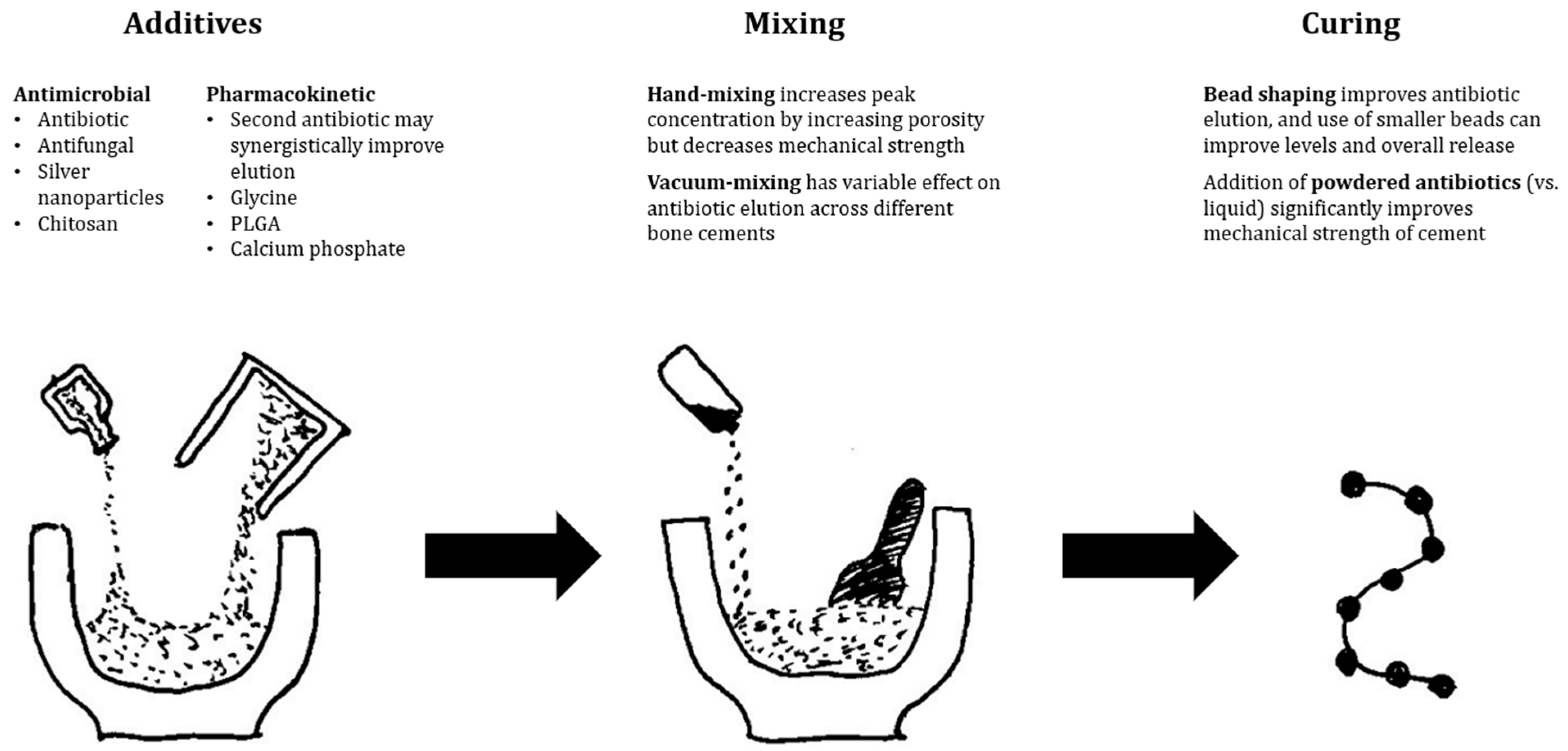
Prosthetic Joint Infection (PJI) causes significant morbidity and mortality for patients globally. Delivery of antibiotics to the site of infection has potential to improve the treatment outcomes and enhance biofilm eradication. These antibiotics can be delivered using an intra-articular catheter or combined with a carrier substance to enhance pharmacokinetic properties. Use of an antibiotic carrier is an alternative method of maintaining high local concentrations of antibiotic without systemic exposure. These substances incorporate an antimicrobial to prolong its half-life and provide predictable elution characteristics. They may also serve additional roles, such as filling dead space and providing mechanical support for limb alignment. The ideal antibiotic carrier would provide prolonged antibiotic concentrations at an effective level and achieve complete antibiotic release to minimise subtherapeutic elution time. It would also be versatile and have compatibility with the desired antibiotics, and fully resorbable with minimal risk of allergies, and systemic or local adverse effects.

prosthetic joint infection orthopaedic device related infection local antibiotics

1. PMMA

Polymethylmethacrylate (PMMA) bone cement has a long history of use as an antibiotic carrier, with Buchholz and Engelbrecht first publishing on antibiotics mixed with Palacos cement in 1970 [1]. Structural cement spacers that bear load are an important part of staged revision for PJI. The spacer is inserted following debridement at the first stage of TSE to allow weight-bearing, maintain bony alignment, and aid in mobility and pain management. Local antibiotic elution comes from mixing antibiotics with PMMA before curing, and is a balance between increasing antibiotic concentration and decreasing structural integrity. Antibiotics are available pre-mixed from multiple manufacturers, or may be mixed at the time of surgery. It is effective in reducing infection recurrence, and the use of an antibiotic loaded spacer in TSE is associated with a 7% absolute risk reduction [2]. Structural spacers have lower antibiotic elution when compared to beads of the same composition [3]. This is due to beads having greater surface area to volume ratio, with surface elution from PMMA responsible for initial burst levels [4]. Following this, slower elution is through the network of interconnecting pores and cracks in the material, and passive diffusion through the cement matrix [5]. Antibiotic beads have been shown to be non-inferior to conventional intravenous antibiotic therapy in osteomyelitis treatment [6][7]. In a randomised controlled trial of 28 hip and knee PJI patients, antibiotic beads had a lower infection recurrence rate (15%) compared to conventional parenteral therapy (30%) following prosthesis resection, although this was not statistically significant [8].
Effective local therapy has questioned the need for prolonged systemic antibiotic treatment in some studies. Stockley et al. provided targeted local antibiotic therapy with PMMA beads at the first stage of 114 TSE for chronic hip PJI [9]. Patients only received three doses of systemic peri-operative antibiotic prophylaxis, with a success rate of 87.7% at a minimum two-year follow-up. From the same centre, a case series of 53 chronic knee PJIs undergoing TSE or staged arthrodesis reported an infection eradication rate of 89% with use of targeted antibiotics in PMMA beads and spacer [10]. These patients received a mean of only 4.6 days of intravenous therapy, and only three patients received subsequent oral antibiotics. Hart et al. reported on 48 TSEs for knee PJI using PMMA spacers containing gentamicin and vancomycin, with 88% infection eradication following only two weeks of post-operative intravenous therapy after the first stage [11].
Variable antibiotic pharmacokinetics have been reported when using PMMA as an antibiotic carrier. Local antibiotic level peaks between day one and three, indicating an initial burst-release followed by a slow decline in concentration; however, the peak levels and length of time above the MIC is variable between studies and difficult to control [12][13]. Variability is due to multiple factors, including antibiotic type and concentration, PMMA type, mixing technique, physical wear, use of prefabricated or handmade products, and anatomic location with local fluid turnover [4][14]. Nonetheless, peak local antibiotic levels vastly exceed those achievable with systemic therapy [15]. PMMA antibiotic release is poor, with only 25–50% of contained antibiotic eluted in bead form [6]. Shaping smaller beads with greater surface area can improve elution, with 3 × 5 mm ‘mini-beads’ releasing 93% of contained antibiotics over 14 days and achieving local concentrations seven times higher than 7 × 7 mm beads [16].
In PJI, studies that measure antibiotic release from PMMA are predominantly in structural spacers. Some studies demonstrate several months of continuous antibiotic release, while others reveal that local concentrations drop below MIC after seven to fourteen days [7][17]. A systematic review of in-vivo cement spacer antibiotic levels by Anagnostakos and Meyer found no clearly superior cement or antibiotic mix with significant heterogeneity of studies in cement spacer composition and antibiotic level sampling technique [12]. Invitro studies to define the superior composition of cement in spacers unfortunately have conflicting results. Palacos with gentamicin has shown highest elution in initial studies; however, recent literature has shown other cements to have similar elution kinetics, with variable superiority when changing or adding impregnated antibiotics [12][13][17][18][19][20][21][22]. Antibiotic elution may be increased through altering composition by increasing antibiotic concentration or putting other additives in the cement such as glycine, Poly(lactic-co-glycolic) Acid (PLGA), or calcium phosphate [4][17][23]. Stevens et al. demonstrated that spacers with higher antibiotic doses achieved higher burst levels and eluted antibiotics above the MIC for over 80 days [17].
Mixing technique can alter cement porosity, which subsequently affects antibiotic release. Hand mixing, in comparison to vacuum mixing, can incorporate more air which can increase peak antibiotic concentration up to five-fold in vitro [19][24]. Vacuum mixing has a variable effect on antibiotic elution, and in vitro studies confirm improved antimicrobial activity with Cobalt G-HV, Palacos R+G, and Simplex P, while declining performance when using Cemex Genta, Smartset GMV, and Versabond AB [25]. Using multiple antibiotics can similarly increase porosity and synergistically improve release of both antibiotics [24]. However, contradicting studies have also found no difference between mixing techniques or with the use of multiple antibiotics [26][27].
PMMA may be incompatible with antibiotics due to its effects on the antibiotics themselves, or vice versa. The exothermic polymerisation reaction of PMMA can generate temperatures up to 90 degrees Celsius [4]. This may degrade contained antibiotics, and heat stability should be considered in antibiotic choice. In vitro, beta-lactams are highly fragile, while gentamicin has only a slight decrease in activity after heat treatment. Other antibiotics including aminoglycosides, glycopeptides, tetracyclines and quinolones are stable over six weeks following initial heat treatment [28]. Mechanical changes can also occur in PMMA induced by some antibiotics. Antibiotics in liquid form have a greater impact on the structural integrity of cement compared to powdered equivalents, and some antibiotics such as rifampicin cause significant changes in cement consistency during polymerisation resulting in decreased mechanical strength [4].
Local adverse effects of PMMA appear to be minimal once the cement is cured and the risk of thermal injury has passed. However systemic adverse effects due to supratherapeutic antibiotic levels are being increasingly recognised, especially aminoglycoside nephrotoxicity. Serum levels of gentamicin and vancomycin from spacers are detectable for up to eight weeks [29], and spacers with high dose vancomycin or aminoglycoside (over 3.6 g antibiotic per 40 g PMMA) have been associated with almost two-fold increase in acute kidney injury (AKI) risk compared to lower dose spacers [30]. In two systematic reviews, overall rate of AKI following TSE with an antibiotic spacer is 4.18%, compared to 0.55% in primary arthroplasty [2][31]. However, this may simply reflect intraoperative hypovolemia and the published studies are clearly susceptible to multiple biases, primarily selection bias. These revision cases are of course far more complicated and prolonged than standard primary arthroplasty, and these results must be interpreted carefully and viewed with caution.
Mechanical studies have demonstrated that PMMA does not appear to lose strength due to elution of antibiotics; however, persistence of the carrier has been associated with the emergence of antibiotic resistance [32]. Small colony variants emerge following prolonged exposure to subtherapeutic levels of antibiotic, and resistant organisms have been isolated in retrieved cement spacers, although the incidence is unclear [33]. Persistent cement may also be a focus for clinical infection recurrence [34]. Removal of PMMA generally requires a second surgery, which carries the risk of local or systemic complications.
Regardless, PMMA remains an essential antibiotic carrier in the treatment of PJI and is by far the most studied. It is well understood as a biomaterial in orthopaedic surgery due to its long history of use and has effective elution of local antibiotics with proven reduction in infection recurrence in clinical studies. Its ability to bear load is not shared by any other available antibiotic carrier. It is not without issues, with variability in antibiotic levels, incompatibility with several classes of antibiotics, and the requirement for removal following infection clearance. Finally, antibiotic elution from PMMA, despite much lower systemic levels than parenteral therapy, has still been associated with occasional systemic adverse effects. Figure 1 illustrates several evidence-based recommendations from the authors to improve elution characteristics.
Figure 1. Evidence-based recommendations to improve antibiotic elution from PMMA-based antibiotic delivery vehicles.

2. Resorbable Carriers

Resorbable carriers aim to completely degrade to overcome the persistence issues of PMMA and do not require a second surgery for removal. This avoids subtherapeutic levels associated with carrier persistence in PMMA, as all antibiotics are released when the carrier is fully resorbed. In PJI, their resorbable nature makes them more attractive in DAIR or SSE, when there is no planned return to theatre to facilitate removal, as in TSE. Cements or gels may also be mouldable to fit defects, manage surgical dead space and increase antibiotic penetrance.

3. Calcium Sulphate

Calcium sulphate (CS) may be used as an antibiotic carrier, and its effectiveness in chronic osteomyelitis and fracture-related infection has led to further investigation in PJI [6][35]. It is promising as a bioabsorbable antibiotic carrier with favourable elution qualities, although it carries a risk of persistent wound leakage, heterotrophic ossification, or life-threatening hypercalcaemia [36]. It is available as cement which can be shaped into beads to suit a variety of clinical applications (Figure 2). It releases 100% of loaded antibiotics over time, and elutes over a period of several weeks [37]. When combined with targeted antibiotics, it has been reported to successfully inhibit biofilm formation and eradicate established biofilms in vitro in multiple bacterial species [38]. It has also been shown in vitro to cause minor third-body wear when used in prosthetic joints; however, rates are much lower than with PMMA or ceramic particles [39]. A systematic review of wound leakage in prosthetic joint surgeries using CS beads revealed a rate of 3.8%, even with deep usage inside the joint, and the included larger studies suggested that higher volumes of CS or medical co-morbidities posed an increased risk of this complication [36][40][41]. A separate systematic review of hypercalcaemia demonstrated a 4.2% overall rate; however, only 0.28% required management, with one case out of 1049 being life-threatening and needing intensive care [42]. Unfortunately, the comparative studies assessing success rates are limited to case series or case-control studies in DAIR procedures. A matched case-control study of 40 DAIRs for acute hip or knee PJI demonstrated no significant improvement with the use of vancomycin and tobramycin CS beads at 90 days or two years [43]. This study reported a 45% failure rate, consistent with other smaller published case series of CS DAIRs [44]. However, a recently published cohort study by Reinisch et al. identified significant improvement in 41 DAIRs for hip PJI, with vancomycin, ceftriaxone, or tobramycin CS beads [45]. The CS group had a significantly lower revision rate of 15% compared to 64% in the standard group. Studies of CS in SSE or TSE for PJI either do not have re-infection rate as their primary outcome, have no comparator group, or have grouped aseptic and septic revision. Reported re-infection rates range from 0% to 6.7% [40][41][46]. While further higher-quality studies need to be performed, the significant cost and risk of adverse effects of CS carriers should be considered before recommending their routine clinical use in the absence of any demonstrated benefit.
Figure 2. Antibiotic-impregnated Calcium sulphate as cement and shaped into beads (from Ene et al. [47]).

4. Hydroxyapatite

Calcium hydroxyapatite (CHA) has seen recent renewed interest as an antibiotic carrier, despite first publication of its use over 20 years ago [47]. A number of calcium apatite compounds are available, including hydroxyapatite and tricalcium phosphate [47]. These can be prepared as blocks, with an encased antibiotic powder reservoir, or as a cement mixed with antibiotic powder. Isothermic hardening of cements has the advantage of avoiding thermal damage to antibiotics, and its porous nature promotes biological reactivity for bone formation and antibiotic release [47][48]. However, the slow and incomplete degradation of these cements causes incomplete and inconsistent antibiotic elution, and promotes bacterial colonisation [47]. A case series of fourteen knee PJI patients with antibiotic-impregnated CHA pellets (Bone Ceram P, Olympus Terumo Biomaterials Corp, Tokyo, Japan) at the first stage of TSE reported a recurrence rate of 21% at mean follow-up of 5.1 years [49]. They reported no complications related to use of CHA. However, inconsistent antibiotic elution needs to be addressed before it can be used as a reliable carrier.
Combinations of CHA and CS are more clinically studied in bone and joint infection. Cerament® (BoneSupport AB, Lund, Sweden) is a cement mixture of 40% CHA and 60% CS, and is commercially available with gentamicin or vancomycin (Figure 3) [50]. In vitro, the substance has been shown to increase prosthesis pull-out strength and to inhibit biofilm formation with antibiotics [51][52]. When used clinically in PJI patients, it can deliver high local antibiotic levels, with drain effluent levels well above MIC at mean 90 h post-op [53]. However, this study also noted that presence of a post-operative drain significantly reduces available local antibiotic by measuring renal antibiotic clearance. Logoluso et al. found a 95% infection eradication rate with targeted antibiotic-laden Cerament® applied to stemmed components at second-stage of TSE for PJI [54]. Across two large case series of chronic osteomyelitis, including 71% following infected fracture fixation, McNally et al. reported 94% infection eradication at four to eight years follow-up in higher risk Cierny–Mader type B hosts [50][55]. PerOssal® (Osartis GmBH, Dieburg, Germany), a mix of 51.5% nanocrystalline CHA and 48.5% CS, has been used in PJI at first-stage of TSE combined with antibiotics within the intramedullary canal [56]. The PerOssal group reported a trend towards improved infection recurrence rate (6.67% vs. 16.13%), and significantly lower serum inflammatory marker values from the second to sixth postoperative weeks. There was no difference in complication rate between groups, although they did note delayed wound healing when the substance was used outside of the intramedullary canal. Overall, this combination carrier has significant potential in PJI given its promising results in osteomyelitis; however, similarly to CS, it must be used with caution to avoid local wound healing complications.

5. Bioactive Glass

Bioactive glass (BAG) carries inherent antibacterial activity following implantation and can also act as an antibiotic carrier. It is an attractive option in osteomyelitis and fracture-related infection due to stimulation of bone formation to heal defects, and has been shown to improve local vascularity with proangiogenic properties [57]. Once implanted, calcium is released into the local environment from the glass, which reacts with interstitial phosphate to form a calcium phosphate layer on the surface of the BAG. Ongoing growth of this layer continues with further dissolution of the glass, which then undergoes crystallisation to hydroxyapatite allowing cellular adhesion [57][58][59]. Sodium and potassium are also released from the substance and exchanged for ionic hydrogen. This increases osmotic pressure and local pH up to 11.65, creating a hostile environment for microbes [57][59]. BAG-S53P4 (commercially available as Bonalive®, Turku, Finland) is a silicate glass that provides bactericidal activity against a broad-spectrum of planktonic and biofilm-phase bacteria, including multi-resistant organisms, and also prevents biofilm formation [60][61][62]. In vitro bacterial killing has been shown to be equivalent to antibiotic-loaded PMMA [63]. Resistance to BAG has not been reported during in vitro studies [59]. The porous nature of mesoporous and sol-gel glasses allows for release of metal ions and larger molecules, including antibiotics. Metal ions can be incorporated at the time of manufacture and silver, gallium, magnesium, copper, strontium, and zinc have shown positive in vitro results [58][59][64]. Antibiotic compounds have been incorporated into BAG during in vitro studies, with total antibiotic release up to 90%; however, similar to PMMA, heterogeneity exists in both BAG composition and antibiotic used, leading to variability in the level released and the duration [65][66][67][68][69]. Newer composite borate glasses exhibit promising elution kinetics compared to silicate glasses and possess tailorable elution qualities based on their composition [70]. In animal models, antibiotic-loaded borate glass revealed significant improvement in infection clearance and histological tissue quality over glass alone, suggesting that antibiotic therapy provides extra antibacterial activity [66][67][68][70][71]. Antibiotic release has been detected in rabbit models out to 21 days for teicoplanin, and fourteen days for vancomycin [66][67].
There are no published studies of clinical use of BAG in prosthetic joint infection currently, but non-antibiotic-laden BAG has been used in osteomyelitis treatment following surgical debridement. Lindfors et al. reported an overall cure rate of 89.7% at 31 months in a multinational register of 116 patients, predominantly involving post-traumatic long bone osteomyelitis [72]. In this study 84.5% of patients received BAG-S53P4 without antibiotics as a single-stage procedure, while 15.5% initially received antibiotic-loaded PMMA beads followed by BAG-S53P4 at the second stage. They also reported significantly more early poor outcomes in the two-stage treatment group. Another prospective study of BAG-S53P4 in 27 cases of long bone osteomyelitis found 88.9% of patients were infection-free at mean 17.8 months follow-up [61]. A comparative retrospective study of long bone osteomyelitis compared BAG-S53P4 to hydroxyapatite plus calcium sulphate plus antibiotics and tricalcium phosphate plus demineralised bone matrix plus antibiotics [73]. They found 92.6% of patients who received BAG-S53P4 were infection-free at mean 21.8 months with equivalent infection clearance across the three groups, but significantly lower wound complication rates in the BAG group. Two smaller retrospective case series of eleven and three patients reported infection clearance rates of 91% and 100% after treatment with BAG-S53P4 following debridement [57][74]. Importantly, all patients in all studies discussed received systemic antibiotic therapy. Despite these results in the treatment of osteomyelitis, further research is required to understand the utility of bioactive glass in prosthetic joint infection, including its effect on articulated surfaces. Newer composite and borate glasses to deliver antibiotics also have potential to augment the natural antibacterial activity of BAG.

6. Hydrogels

Hydrogels carry significant promise as a fully resorbable carrier that can be tailored to a specific clinical indication. They can be manufactured from multiple biodegradable polymers, with variable antibiotic release kinetics and degradation time of the gel [75][76]. Their consistency also makes them versatile for application to implant surfaces or fill dead space, as seen in Figure 3. They can also carry a wide variety of antimicrobial substances as, in contrast to PMMA, hydrogel can be mixed at room temperature, without thermal damage to the contained antimicrobial. They have been combined in experimental studies or case reports with a variety of antibiotics, antifungals, chitosan, and tyrosol [77][78][79]. The high water content inside the hydrogel matrix is also an appropriate environment for containing and releasing bacteriophages [80]. Animal models have found that loaded hydrogels are able to achieve the MBEC for multiple common ODRI pathogens [81]. The forefront of hydrogel technology has produced layered microparticles within a hydrogel to enable multiphase release, in a rabbit model releasing vancomycin and ceftazidime at stable therapeutic level out to 56 days, while simultaneously administering lidocaine for pain relief to 14 days [82].
Clinical studies are limited to the use of Defensive Antibacterial Coating (DAC®, Novagenit Srl, Mezzo Lombardo, Italy), which is the most studied and is already commercially available (Figure 3). DAC® is recommended as a prophylactic implant coating, as it changes the implant surface from hydrophobic to hydrophilic, to impair bacterial adhesion, but is completely resorbed in 72 h [83]. It can also carry antibiotics to improve its capacity for infection prevention or for PJI treatment. The largest clinical study in PJI treatment is a 1:1 case-control study of cementless TSE by Zagra of 54 patients. At 2.7 years mean follow-up, they reported four recurrences in the control group (14.8%), compared to none in the group which received DAC with targeted antibiotics at the second stage [84]. Another matched case-control study of 44 patients found equivalence in infection recurrence rate between TSE compared to SSE with DAC and antibiotics, with a significant reduction in hospital stay and antibiotic duration in the DAC group [85]. However, importantly, all the SSE patients in this study met their criteria for SSE, for which other authors have found equivalent results between SSE and TSE (no large soft tissue defect, previously identified and antibiotic sensitive pathogen) [86]. In the largest study of DAC use with antibiotics, an RCT of 380 primary and revision arthroplasty patients, the treatment group was not found to have any complications attributable to the hydrogel, with equivalent wound healing, lab biomarkers, and cementless implant osseointegration [87]. This is consistent with the other smaller clinical and safety studies that have not found any significant complications associated with DAC use. These promising early clinical results for DAC suggest that a dedicated hydrogel for PJI could safely deliver therapeutic levels over a prolonged period.

7. Nanocarriers

Nanocarriers are complex molecular moieties that respond to internal or external stimuli for activation. Self-targeting carriers can respond to local changes in infected tissue, such as pH, hypoxia, macrophage presence, increased reactive oxygen species, local ligands, bacterial enzymes, or increased local temperature [88]. Nanocarriers can also respond to external stimuli such as photothermal radiation or ultrasound waves [89][90]. These carriers, once activated, change their structure from a mobile hydrophilic compound, becoming hydrophobic and releasing their antimicrobial agent. This can be an antibiotic, antifungal, heavy metal nanoparticle, or other bacterio-toxic substance. They can also be designed to release their antimicrobial substance with enhanced pharmacokinetics compared to current options [88]. Self-propelled ‘Nanorobots’ can be guided using photothermal stimulation to release their antimicrobial substances into a target area [91].
Figure 3. Defensive Antibacterial Coating (DAC) applied to acetabular and femoral components (from Romano et al. [87]).

References

  1. Buchholz, H.W.; Engelbrecht, H. Depot effects of various antibiotics mixed with palacos resins. Chirurg 1970, 41, 511–515.
  2. Luu, A.; Syed, F.; Raman, G.; Bhalla, A.; Muldoon, E.; Hadley, S.; Smith, E.; Rao, M. Two-Stage Arthroplasty for Prosthetic Joint Infection. J. Arthroplast. 2013, 28, 1490–1498.
  3. Anagnostakos, K.; Wilmes, P.; Schmitt, E.; Kelm, J. Elution of gentamicin and vancomycin from polymethylmethacrylate beads and hip spacers in vivo. Acta Orthop. 2009, 80, 193–197.
  4. Wall, V.; Nguyen, T.-H.; Nguyen, N.; Tran, P. Controlling Antibiotic Release from Polymethylmethacrylate Bone Cement. Biomedicines 2021, 9, 26.
  5. Zalavras, C.G.; Patzakis, M.J.; Holtom, P. Local Antibiotic Therapy in the Treatment of Open Fractures and Osteomyelitis. Clin. Orthop. Relat. Res. 2004, 427, 86–93.
  6. E Kent, M.; Rapp, R.P.; Smith, K.M. Antibiotic Beads and Osteomyelitis: Here Today, What’s Coming Tomorrow? Orthopedics 2006, 29, 599–603.
  7. van Vugt, T.A.G.; Arts, J.J.; Geurts, J.A.P. Antibiotic-Loaded Polymethylmethacrylate Beads and Spacers in Treatment of Orthopedic Infections and the Role of Biofilm Formation. Front. Microbiol. 2019, 10, 1626.
  8. Nelson, C.L.; Evans, R.P.; Blaha, J.D.; Calhoun, J.; Henry, S.L.; Patzakis, M.J. A comparison of gentamicin-impregnated polymethylmethacrylate bead implantation to conventional parenteral antibiotic therapy in infected total hip and knee arthroplasty. Clin. Orthop. Relat. Res. 1993, 295, 96–101.
  9. Stockley, I.; Mockford, B.J.; Hoad-Reddick, A.; Norman, P. The use of two-stage exchange arthroplasty with depot antibiotics in the absence of long-term antibiotic therapy in infected total hip replacement. J. Bone Jt. Surg. 2008, 90, 145–148.
  10. Hoad-Reddick, D.A.; Evans, C.R.; Norman, P.; Stockley, I. Is there a role for extended antibiotic therapy in a two-stage revision of the infected knee arthroplasty? J. Bone Jt. Surg. 2005, 87, 171–174.
  11. Hart, W.J.; Jones, R.S. Two-stage revision of infected total knee replacements using articulating cement spacers and short-term antibiotic therapy. J. Bone Jt. Surg. 2006, 88, 1011–1015.
  12. Anagnostakos, K.; Meyer, C. Antibiotic Elution from Hip and Knee Acrylic Bone Cement Spacers: A Systematic Review. BioMed Res. Int. 2017, 2017, 4657874.
  13. Walenkamp, G.H.I.M. Antibiotic Loaded Cement: From Research to Clinical Evidence. In Infection and Local Treatment in Orthopedic Surgery; Springer: Berlin/Heidelberg, Germany, 2007; pp. 170–175.
  14. Dodds, S.; Smith, T.J.; Akid, R.; Stephenson, J.; Nichol, T.; Banerjee, R.D.; Stockley, I.; Townsend, R. Contrasting Effects of Physical Wear on Elution of Two Antibiotics from Orthopedic Cement. Antimicrob. Agents Chemother. 2012, 56, 1471–1475.
  15. Roy, M.E.; Peppers, M.P.; Whiteside, L.A.; LaZear, R.M. Vancomycin Concentration in Synovial Fluid: Direct Injection into the Knee vs. Intravenous Infusion. J. Arthroplast. 2014, 29, 564–568.
  16. Walenkamp, G. Small PMMA beads improve gentamicin release. Acta Orthop. 1989, 60, 668–669.
  17. Stevens, C.M.; Tetsworth, K.D.; Calhoun, J.H.; Mader, J.T. An articulated antibiotic spacer used for infected total knee arthroplasty: A comparative in vitro elution study of Simplex® and Palacos® bone cements. J. Orthop. Res. 2005, 23, 27–33.
  18. van De Belt, H.; Neut, D.; Schenk, W.; van Horn, J.R.; van Der Mei, H.C.; Busscher, H.J. Gentamicin release from polymethylmethacrylate bone cements and Staphylococcus aureus biofilm formation. Acta Orthop. 2000, 71, 625–629.
  19. Neut, D.; van de Belt, H.; van Horn, J.; van der Mei, H.; Busscher, H. The effect of mixing on gentamicin release from polymethylmethacrylate bone cements. Acta Orthop. 2003, 74, 670–676.
  20. Cerretani, D.; Giorgi, G.; Fornara, P.; Bocchi, L.; Neri, L.; Ceffa, R.; Ghisellini, F.; Ritter, M.A. The in vitro elution characteristics of vancomycin combined with imipenem-cilastatin in acrylic bone–cements: A pharmacokinetic study. J. Arthroplast. 2002, 17, 619–626.
  21. Bitsch, R.; Kretzer, J.; Vogt, S.; Büchner, H.; Thomsen, M.; Lehner, B. Increased antibiotic release and equivalent biomechanics of a spacer cement without hard radio contrast agents. Diagn. Microbiol. Infect. Dis. 2015, 83, 203–209.
  22. Penner, M.J.; Duncan, C.P.; Masri, B.A. The in vitro elution characteristics of antibiotic-loaded CMW and Palacos-R bone cements. J. Arthroplast. 1999, 14, 209–214.
  23. McLaren, A.C.; Nelson, C.L.; McLaren, S.G.; DeClerk, G.R. The Effect of Glycine Filler on the Elution Rate of Gentamicin from Acrylic Bone Cement. Clin. Orthop. Relat. Res. 2004, 427, 25–27.
  24. Frew, N.M.; Cannon, T.; Nichol, T.; Smith, T.; Stockley, I. Comparison of the elution properties of commercially available gentamicin and bone cement containing vancomycin with ‘home-made’ preparations. Bone Jt. J. 2017, 99-B, 73–77.
  25. Meyer, J.; Piller, G.; Spiegel, C.A.; Hetzel, S.; Squire, M. Vacuum-Mixing Significantly Changes Antibiotic Elution Characteristics of Commercially Available Antibiotic-Impregnated Bone Cements. J. Bone Jt. Surg. 2011, 93, 2049–2056.
  26. Minelli, E.B.; Della Bora, T.; Benini, A. Different microbial biofilm formation on polymethylmethacrylate (PMMA) bone cement loaded with gentamicin and vancomycin. Anaerobe 2011, 17, 380–383.
  27. McLaren, A.C.; Nugent, M.; Economopoulos, K.; Kaul, H.; Vernon, B.L.; McLemore, R. Hand-mixed and Premixed Antibiotic-loaded Bone Cement Have Similar Homogeneity. Clin. Orthop. Relat. Res. 2009, 467, 1693–1698.
  28. Samara, E.; Moriarty, T.F.; Decosterd, L.; Richards, R.G.; Gautier, E.; Wahl, P. Antibiotic stability over six weeks in aqueous solution at body temperature with and without heat treatment that mimics the curing of bone cement. Bone Jt. Res. 2017, 6, 296–306.
  29. Edelstein, A.I.; Okroj, K.T.; Rogers, T.; Della Valle, C.J.; Sporer, S.M. Systemic Absorption of Antibiotics From Antibiotic-Loaded Cement Spacers for the Treatment of Periprosthetic Joint Infection. J. Arthroplast. 2018, 33, 835–839.
  30. Dagneaux, L.; Limberg, A.K.; Osmon, D.R.; Leung, N.; Berry, D.J.; Abdel, M.P. Acute Kidney Injury When Treating Periprosthetic Joint Infections After Total Knee Arthroplasties with Antibiotic-Loaded Spacers. J. Bone Jt. Surg. 2021, 103, 754–760.
  31. Jafari, S.M.; Huang, R.; Joshi, A.; Parvizi, J.; Hozack, W.J. Renal Impairment Following Total Joint Arthroplasty: Who Is at Risk? J. Arthroplast. 2010, 25, 49–53.
  32. Duey, R.E.; Chong, A.C.M.; McQueen, D.A.; Womack, J.L.; Song, Z.; Steinberger, T.A.; Wooley, P.H. Mechanical properties and elution characteristics of polymethylmethacrylate bone cement impregnated with antibiotics for various surface area and volume constructs. Iowa Orthop. J. 2012, 32, 104–115.
  33. Van de Belt, H.; Neut, D.; Schenk, W.; van Horn, J.R.; van der Mei, H.C.; Busscher, H.J. Infection of orthopedic implants and the use of antibiotic-loaded bone cements: A review. Acta Orthop. Scand. 2001, 72, 557–571.
  34. Buttaro, M.; Valentini, R.; Piccaluga, F. Persistent infection associated with residual cement after resection arthroplasty of the hip. Acta Orthop. 2004, 75, 427–429.
  35. Shi, X.; Wu, Y.; Ni, H.; Li, M.; Zhang, C.; Qi, B.; Wei, M.; Wang, T.; Xu, Y. Antibiotic-loaded calcium sulfate in clinical treatment of chronic osteomyelitis: A systematic review and meta-analysis. J. Orthop. Surg. Res. 2022, 17, 104.
  36. Tarar, M.Y.; Khalid, A.; Usman, M.; Javed, K.; Shah, N.; Abbas, M.W. Wound Leakage With the Use of Calcium Sulphate Beads in Prosthetic Joint Surgeries: A Systematic Review. Cureus 2021, 13, e19650.
  37. Abosala, A.; Ali, M. The Use of Calcium Sulphate beads in Periprosthetic Joint Infection, a systematic review. J. Bone Jt. Infect. 2020, 5, 43–49.
  38. Howlin, R.P.; Brayford, M.J.; Webb, J.S.; Cooper, J.J.; Aiken, S.S.; Stoodley, P. Antibiotic-Loaded Synthetic Calcium Sulfate Beads for Prevention of Bacterial Colonization and Biofilm Formation in Periprosthetic Infections. Antimicrob. Agents Chemother. 2015, 59, 111–120.
  39. Heuberger, R.; Wahl, P.; Krieg, J.; Gautier, E. Low in vitro third-body wear on total hip prostheses induced by calcium sulphate used for local antibiotic therapy. Eur. Cells Mater. 2014, 28, 246–257.
  40. McPherson, F.E.; Dipane, B.M.; Sherif, S. Dissolvable Antibiotic Beads in Treatment of Periprosthetic Joint Infection and Revision Arthroplasty—The Use of Synthetic Pure Calcium Sulfate (Stimulan®) Impregnated with Vancomycin & Tobramycin. Reconstr. Rev. 2013, 3, 32–43.
  41. Kallala, R.; Harris, W.E.; Ibrahim, M.; Dipane, M.; McPherson, E. Use of Stimulan absorbable calcium sulphate beads in revision lower limb arthroplasty. Bone Jt. Res. 2018, 7, 570–579.
  42. Tarar, M.Y.; Toe, K.K.Z.; Javed, K.; Shah, N.; Khalid, A. The Risk of Iatrogenic Hypercalcemia in Patients Undergoing Calcium Sulphate Beads Implantation in Prosthetic Joint Surgery: A Systematic Review. Cureus 2021, 13, e18777.
  43. Tarity, T.D.; Xiang, W.; Jones, C.W.; Gkiatas, I.; Nocon, A.; Selemon, N.A.; Carli, A.; Sculco, P.K. Do Antibiotic-Loaded Calcium Sulfate Beads Improve Outcomes After Debridement, Antibiotics, and Implant Retention? A Matched Cohort Study. Arthroplast. Today 2022, 14, 90–95.
  44. Flierl, M.A.; Culp, B.M.; Okroj, K.T.; Springer, B.D.; Levine, B.R.; Della Valle, C.J. Poor Outcomes of Irrigation and Debridement in Acute Periprosthetic Joint Infection With Antibiotic-Impregnated Calcium Sulfate Beads. J. Arthroplast. 2017, 32, 2505–2507.
  45. Reinisch, K.; Schläppi, M.; Meier, C.; Wahl, P. Local antibiotic treatment with calcium sulfate as carrier material improves the outcome of debridement, antibiotics, and implant retention procedures for periprosthetic joint infections after hip arthroplasty—A retrospective study. J. Bone Jt. Infect. 2022, 7, 11–21.
  46. Lum, Z.; Pereira, G.C. Local bio-absorbable antibiotic delivery in calcium sulfate beads in hip and knee arthroplasty. J. Orthop. 2018, 15, 676–678.
  47. Ene, R.; Nica, M.; Ene, D.; Cursaru, A.; Cirstoiu, C. Review of calcium-sulphate-based ceramics and synthetic bone substitutes used for antibiotic delivery in PJI and osteomyelitis treatment. EFORT Open Rev. 2021, 6, 297–304.
  48. Wassif, R.K.; Elkayal, M.; Shamma, R.N.; Elkheshen, S.A. Recent advances in the local antibiotics delivery systems for management of osteomyelitis. Drug Deliv. 2021, 28, 2392–2414.
  49. Hasegawa, M.; Tone, S.; Naito, Y.; Wakabayashi, H.; Sudo, A. Use of antibiotic-impregnated hydroxyapatite for infection following total knee arthroplasty. Mod. Rheumatol. 2021, 31, 1073–1077.
  50. McNally, M.A.; Ferguson, J.Y.; Lau, A.C.K.; Diefenbeck, M.; Scarborough, M.; Ramsden, A.J.; Atkins, B.L. Single-stage treatment of chronic osteomyelitis with a new absorbable, gentamicin-loaded, calcium sulphate/hydroxyapatite biocomposite. Bone Jt. J. 2016, 98-B, 1289–1296.
  51. Bidossi, A.; Bottagisio, M.; Logoluso, N.; De Vecchi, E. In Vitro Evaluation of Gentamicin or Vancomycin Containing Bone Graft Substitute in the Prevention of Orthopedic Implant-Related Infections. Int. J. Mol. Sci. 2020, 21, 9250.
  52. Zampelis, V.; Tägil, M.; Lidgren, L.; Isaksson, H.; Atroshi, I.; Wang, J.-S. The effect of a biphasic injectable bone substitute on the interface strength in a rabbit knee prosthesis model. J. Orthop. Surg. Res. 2013, 8, 25.
  53. Colding-Rasmussen, T.; Horstmann, P.; Petersen, M.M.; Hettwer, W. Antibiotic Elution Characteristics and Pharmacokinetics of Gentamicin and Vancomycin from a Mineral Antibiotic Carrier: An in vivo Evaluation of 32 Clinical Cases. J. Bone Jt. Infect. 2018, 3, 234–240.
  54. Logoluso, N.; Drago, L.; Gallazzi, E.; George, D.A.; Morelli, I.; Romanò, C.L. Calcium-Based, Antibiotic-Loaded Bone Substitute as an Implant Coating: A Pilot Clinical Study. J. Bone Jt. Infect. 2016, 1, 59–64.
  55. McNally, M.A.; Ferguson, J.Y.; Scarborough, M.; Ramsden, A.; Stubbs, D.A.; Atkins, B.L. Mid- to long-term results of single-stage surgery for patients with chronic osteomyelitis using a bioabsorbable gentamicin-loaded ceramic carrier. Bone Jt. J. 2022, 104-B, 1095–1100.
  56. Sakellariou, V.I.; Savvidou, O.; Markopoulos, C.; Drakou, A.; Mavrogenis, A.F.; Papagelopoulos, P.J. Combination of Calcium Hydroxyapatite Antibiotic Carrier with Cement Spacers in Peri-Prosthetic Knee Infections. Surg. Infect. 2015, 16, 748–754.
  57. Lindfors, N.; Hyvönen, P.; Nyyssönen, M.; Kirjavainen, M.; Kankare, J.; Gullichsen, E.; Salo, J. Bioactive glass S53P4 as bone graft substitute in treatment of osteomyelitis. Bone 2010, 47, 212–218.
  58. Rahaman, M.N.; Bal, B.S.; Huang, W. Review: Emerging developments in the use of bioactive glasses for treating infected prosthetic joints. Mater. Sci. Eng. C 2014, 41, 224–231.
  59. Drago, L.; Toscano, M.; Bottagisio, M. Recent Evidence on Bioactive Glass Antimicrobial and Antibiofilm Activity: A Mini-Review. Materials 2018, 11, 326.
  60. Bortolin, M.; De Vecchi, E.; Romanò, C.L.; Toscano, M.; Mattina, R.; Drago, L. Antibiofilm agents against MDR bacterial strains: Is bioactive glass BAG-S53P4 also effective? J. Antimicrob. Chemother. 2015, 71, 123–127.
  61. Drago, L.; Romanò, D.; De Vecchi, E.; Vassena, C.; Logoluso, N.; Mattina, R.; Romano, C.L. Bioactive glass BAG-S53P4 for the adjunctive treatment of chronic osteomyelitis of the long bones: An in vitroand prospective clinical study. BMC Infect. Dis. 2013, 13, 584.
  62. Pérez-Tanoira, R.; García-Pedrazuela, M.; Hyyrynen, T.; Soininen, A.; Aarnisalo, A.; Nieminen, M.; Tiainen, V.-M.; Konttinen, Y.T.; Kinnari, T. Effect of S53P4 bone substitute on staphylococcal adhesion and biofilm formation on other implant materials in normal and hypoxic conditions. J. Mater. Sci. Mater. Med. 2015, 26, 239.
  63. Cunha, M.T.; Murça, M.A.; Nigro, S.; Klautau, G.B.; Salles, M.J.C. In vitro antibacterial activity of bioactive glass S53P4 on multiresistant pathogens causing osteomyelitis and prosthetic joint infection. BMC Infect. Dis. 2018, 18, 157.
  64. Gupta, S.; Majumdar, S.; Krishnamurthy, S. Bioactive glass: A multifunctional delivery system. J. Control Release 2021, 335, 481–497.
  65. Meseguer-Olmo, L.; Ros-Nicolás, M.J.; Clavel-Sainz, M.; Vicente-Ortega, V.; Alcaraz-Baños, M.; Lax-Pérez, A.; Arcos, D.; Ragel, C.V.; Vallet-Regí, M. Biocompatibility andin vivogentamicin release from bioactive sol-gel glass implants. J. BioMed Mater. Res. 2002, 61, 458–465.
  66. Xie, Z.; Liu, X.; Jia, W.; Zhang, C.; Huang, W.; Wang, J. Treatment of osteomyelitis and repair of bone defect by degradable bioactive borate glass releasing vancomycin. J. Control Release 2009, 139, 118–126.
  67. Jia, W.-T.; Zhang, X.; Luo, S.-H.; Liu, X.; Huang, W.-H.; Rahaman, M.N.; Day, D.E.; Zhang, C.-Q.; Xie, Z.-P.; Wang, J.-Q. Novel borate glass/chitosan composite as a delivery vehicle for teicoplanin in the treatment of chronic osteomyelitis. Acta Biomater. 2010, 6, 812–819.
  68. Zhang, X.; Jia, W.; Gu, Y.; Xiao, W.; Liu, X.; Wang, D.; Zhang, C.; Huang, W.; Rahaman, M.N.; Day, D.E.; et al. Teicoplanin-loaded borate bioactive glass implants for treating chronic bone infection in a rabbit tibia osteomyelitis model. Biomaterials 2010, 31, 5865–5874.
  69. Domingues, R.; Cortés, M.; Gomes, T.; Diniz, H.; Freitas, C.; Gomes, J.; Faria, A.M.; Sinisterra, R. Bioactive glass as a drug delivery system of tetracycline and tetracycline associated with β-cyclodextrin. Biomaterials 2004, 25, 327–333.
  70. Ding, H.; Zhao, C.-J.; Cui, X.; Gu, Y.-F.; Jia, W.-T.; Rahaman, M.N.; Wang, Y.; Huang, W.-H.; Zhang, C.-Q. A Novel Injectable Borate Bioactive Glass Cement as an Antibiotic Delivery Vehicle for Treating Osteomyelitis. PLoS ONE 2014, 9, e85472.
  71. Jia, W.-T.; Fu, Q.; Huang, W.-H.; Zhang, C.-Q.; Rahaman, M.N. Comparison of Borate Bioactive Glass and Calcium Sulfate as Implants for the Local Delivery of Teicoplanin in the Treatment of Methicillin-Resistant Staphylococcus aureus-Induced Osteomyelitis in a Rabbit Model. Antimicrob. Agents Chemother. 2015, 59, 7571–7580.
  72. Lindfors, N.; Geurts, J.; Drago, L.; Arts, J.J.; Juutilainen, V.; Hyvönen, P.; Suda, A.J.; Domenico, A.; Artiaco, S.; Alizadeh, C.; et al. Antibacterial Bioactive Glass, S53P4, for Chronic Bone Infections—A Multinational Study. In A Modern Approach to Biofilm-Related Orthopaedic Implant Infections: Advances in Microbiology, Infectious Diseases and Public Health Volume 5; Drago, L., Ed.; Springer International Publishing: Cham, Switzerland, 2017; pp. 81–92.
  73. Romanò, C.L.; Logoluso, N.; Meani, E.; Romanò, D.; De Vecchi, E.; Vassena, C.; Drago, L. A comparative study of the use of bioactive glass S53P4 and antibiotic-loaded calcium-based bone substitutes in the treatment of chronic osteomyelitis. Bone Jt. J. 2014, 96-B, 845–850.
  74. McAndrew, J.; Efrimescu, C.; Sheehan, E.; Niall, D. Through the looking glass; bioactive glass S53P4 (BonAlive®) in the treatment of chronic osteomyelitis. Ir. J. Med Sci. 2013, 182, 509–511.
  75. Overstreet, D.J.; Huynh, R.; Jarbo, K.; McLemore, R.Y.; Vernon, B.L. In situforming, resorbable graft copolymer hydrogels providing controlled drug release. J. BioMed Mater. Res. Part A 2012, 101A, 1437–1446.
  76. Censi, R.; Casadidio, C.; Dubbini, A.; Cortese, M.; Scuri, S.; Grappasonni, I.; Golob, S.; Vojnovic, D.; Sabbieti, M.G.; Agas, D.; et al. Thermosensitive hybrid hydrogels for the controlled release of bioactive vancomycin in the treatment of orthopaedic implant infections. Eur. J. Pharm. Biopharm. 2019, 142, 322–333.
  77. Lai, P.-L.; Hong, D.-W.; Ku, K.-L.; Lai, Z.-T.; Chu, I.-M. Novel thermosensitive hydrogels based on methoxy polyethylene glycol-co-poly(lactic acid-co-aromatic anhydride) for cefazolin delivery. Nanomed. Nanotechnol. Biol. Med. 2014, 10, 553–560.
  78. Romanò, C.L.; Scarponi, S.; Gallazzi, E.; Romanò, D.; Drago, L. Antibacterial coating of implants in orthopaedics and trauma: A classification proposal in an evolving panorama. J. Orthop. Surg. Res. 2015, 10, 157.
  79. Tsikopoulos, K.; Bidossi, A.; Drago, L.; Petrenyov, D.; Givissis, P.; Mavridis, D.; Papaioannidou, P. Is Implant Coating With Tyrosol- and Antibiotic-loaded Hydrogel Effective in Reducing Cutibacterium (Propionibacterium) acnes Biofilm Formation? A Preliminary In Vitro Study. Clin. Orthop. Relat. Res. 2019, 477, 1736–1746.
  80. Kim, H.; Chang, R.; Morales, S.; Chan, H.-K. Bacteriophage-Delivering Hydrogels: Current Progress in Combating Antibiotic Resistant Bacterial Infection. Antibiotics 2021, 10, 130.
  81. Overstreet, D.J.; Badha, V.S.; Heffernan, J.M.; Childers, E.P.; Moore, R.C.; Vernon, B.L.; McLaren, A.C. Temperature-responsive PNDJ hydrogels provide high and sustained antimicrobial concentrations in surgical sites. Drug Deliv. Transl. Res. 2019, 9, 802–815.
  82. Hsu, Y.-H.; Yu, Y.-H.; Lee, D.; Chou, Y.-C.; Wu, C.-K.; Lu, C.-J.; Liu, S.-J. Pharmaceutical-eluting hybrid degradable hydrogel/microparticle loaded sacs for finger joint interpositional arthroplasty. Biomater. Adv. 2022, 137, 212846.
  83. De Meo, D.; Ceccarelli, G.; Iaiani, G.; Torto, F.L.; Ribuffo, D.; Persiani, P.; Villani, C. Clinical Application of Antibacterial Hydrogel and Coating in Orthopaedic and Traumatology Surgery. Gels 2021, 7, 126.
  84. Zagra, L.; Gallazzi, E.; Romanò, D.; Scarponi, S.; Romanò, C. Two-stage cementless hip revision for peri-prosthetic infection with an antibacterial hydrogel coating: Results of a comparative series. Int. Orthop. 2018, 43, 111–115.
  85. Capuano, N.; Logoluso, N.; Gallazzi, E.; Drago, L.; Romanò, C.L. One-stage exchange with antibacterial hydrogel coated implants provides similar results to two-stage revision, without the coating, for the treatment of peri-prosthetic infection. Knee Surg. Sport. Traumatol. Arthrosc. 2018, 26, 3362–3367.
  86. Thakrar, R.R.; Horriat, S.; Kayani, B.; Haddad, F.S. Indications for a single-stage exchange arthroplasty for chronic prosthetic joint infection. Bone Jt. J. 2019, 101-B, 19–24.
  87. Romanò, C.L.; Malizos, K.; Capuano, N.; Mezzoprete, R.; D’Arienzo, M.; Van Der Straeten, C.; Scarponi, S.; Drago, L. Does an Antibiotic-Loaded Hydrogel Coating Reduce Early Post-Surgical Infection After Joint Arthroplasty? J. Bone Jt. Infect. 2016, 1, 34–41.
  88. Wang, D.-Y.; Su, L.; Yang, G.; Ren, Y.; Zhang, M.; Jing, H.; Zhang, X.; Bayston, R.; van der Mei, H.C.; Busscher, H.J.; et al. Self-targeting of zwitterion-based platforms for nano-antimicrobials and nanocarriers. J. Mater. Chem. B 2022, 10, 2316–2322.
  89. Huang, B.; Wang, L.; Tang, K.; Chen, S.; Xu, Y.; Liao, H.; Niu, C. IR780 Based Sonotherapeutic Nanoparticles to Combat Multidrug-Resistant Bacterial Infections. Front. Chem. 2022, 10, 840598.
  90. Yu, Q.; Deng, T.; Lin, F.-C.; Zhang, B.; Zink, J.I. Supramolecular Assemblies of Heterogeneous Mesoporous Silica Nanoparticles to Co-deliver Antimicrobial Peptides and Antibiotics for Synergistic Eradication of Pathogenic Biofilms. ACS Nano 2020, 14, 5926–5937.
  91. Cui, T.; Wu, S.; Sun, Y.; Ren, J.; Qu, X. Self-Propelled Active Photothermal Nanoswimmer for Deep-Layered Elimination of Biofilm In Vivo. Nano Lett. 2020, 20, 7350–7358.
More
Information
Contributors MDPI registered users' name will be linked to their SciProfiles pages. To register with us, please refer to https://encyclopedia.pub/register : , , , , ,
View Times: 357
Entry Collection: Biopharmaceuticals Technology
Revisions: 2 times (View History)
Update Date: 25 Sep 2023
1000/1000