Submitted Successfully!
To reward your contribution, here is a gift for you: A free trial for our video production service.
Thank you for your contribution! You can also upload a video entry or images related to this topic.
Version Summary Created by Modification Content Size Created at Operation
1 -- 4561 2023-09-11 13:03:32 |
2 format correct + 4 word(s) 4565 2023-09-12 03:00:21 | |
3 format correct Meta information modification 4565 2023-09-12 03:01:39 |

Video Upload Options

Do you have a full video?

Confirm

Are you sure to Delete?
Cite
If you have any further questions, please contact Encyclopedia Editorial Office.
Cavallaro, P.A.; De Santo, M.; Belsito, E.L.; Longobucco, C.; Curcio, M.; Morelli, C.; Pasqua, L.; Leggio, A. HER2-Positive Breast Cancer Targeted Therapies. Encyclopedia. Available online: https://encyclopedia.pub/entry/49026 (accessed on 19 May 2024).
Cavallaro PA, De Santo M, Belsito EL, Longobucco C, Curcio M, Morelli C, et al. HER2-Positive Breast Cancer Targeted Therapies. Encyclopedia. Available at: https://encyclopedia.pub/entry/49026. Accessed May 19, 2024.
Cavallaro, Palmira Alessia, Marzia De Santo, Emilia Lucia Belsito, Camilla Longobucco, Manuela Curcio, Catia Morelli, Luigi Pasqua, Antonella Leggio. "HER2-Positive Breast Cancer Targeted Therapies" Encyclopedia, https://encyclopedia.pub/entry/49026 (accessed May 19, 2024).
Cavallaro, P.A., De Santo, M., Belsito, E.L., Longobucco, C., Curcio, M., Morelli, C., Pasqua, L., & Leggio, A. (2023, September 11). HER2-Positive Breast Cancer Targeted Therapies. In Encyclopedia. https://encyclopedia.pub/entry/49026
Cavallaro, Palmira Alessia, et al. "HER2-Positive Breast Cancer Targeted Therapies." Encyclopedia. Web. 11 September, 2023.
HER2-Positive Breast Cancer Targeted Therapies
Edit

Breast cancer represents the most common cancer type and one of the major leading causes of death in the female worldwide population. Overexpression of human epidermal growth factor (HER2), a transmembrane glycoprotein related to the epidermal growth factor receptor, results in a biologically and clinically aggressive breast cancer subtype. It is also the primary driver for tumor detection and progression and, in addition to being an important prognostic factor in women diagnosed with breast cancer, HER2 is a widely known therapeutic target for drug development. In breast cancer, the overexpression of the HER2 receptor makes it a reliable biomarker and a successful therapeutic target. Several strategies have been developed to target HER2, using various targeting molecules including monoclonal antibodies and tyrosine kinase inhibitors, antibody–drug conjugates, small molecules, and peptides.

targeting peptides HER2-positive breast cancer monoclonal antibodies

1. HER2-Targeting Monoclonal Antibodies

HER2-targeted therapies have revolutionized the strategy for HER2-positive breast cancer treatment, both in metastatic and early stage disease. HER2 monoclonal antibodies (mAbs) represent highly specific therapies with moderate toxicity, which significantly improve the clinical outcome for breast cancer patients. However, some patients experienced relapse due to an acquired therapeutic resistance to mAbs. In response, the development of antibody–drug conjugates (ADCs) has emerged as a promising class of therapeutics that combines the specificity of monoclonal antibodies with the antitumor activity of cytotoxic agents, known as payloads linked to mAbs by means molecular linkers [1], thereby maximizing the antitumoral activity [2].
Trastuzumab, also known as Herceptin, is a recombinant humanized monoclonal antibody that binds domain IV of the extracellular segment of the HER2 tyrosine kinase receptor [3][4]. The antitumor efficacy of trastuzumab, in combination with established therapeutic agents, has been extensively investigated through in vitro and in vivo studies on SKBR3, MCF7, and BT-474 cell lines. In 1998, the U.S. Food and drug Administration (FDA) approved Trastuzumab as one of the first available targeted chemotherapies. It received approval as an adjuvant therapy when used in combination with anthracycline- or a taxane-based chemotherapy for HER2-positive breast cancer treatment, and in metastatic HER2-positive breast cancer as a monotherapy or in combination with paclitaxel [5]. It selectively exerts anticancer effects in HER2-positive breast cancer patients [6]. Trastuzumab, when used in combination with standard chemotherapy, has shown significantly improved response rates compared to chemotherapy alone [7][8]. As a result, treatment regimens that include trastuzumab have become the standard of care for patients with HER2-overexpressing breast cancer [9]. Despite the fact that treatment with trastuzumab is considered clinically very effective in HER2-overexpressing breast cancer, its mechanism of action is not yet well-understood. In vivo breast cancer models and clinical trials indicated that antibody-dependent cell-mediated cytotoxicity (ADCC) is the most supported mechanism of action [10][11].
However, several clinical studies showed a 48% clinical benefit rate in patients treated with trastuzumab monotherapy, indicating that a significant number of HER2-amplified metastatic breast cancers do not respond favorably to monotherapy [12]. Furthermore, another important concern is the development of acquired resistance, which frequently occurs and restricts the effectiveness of trastuzumab-based therapy to the duration of the treatment [13][14]. Overall, due to breast cancer heterogeneity and the accumulation of intracellular alterations, multiple mechanisms are responsible for resistance occurrence [15]. Both innate and acquired resistance, in trastuzumab treatments, are progressively becoming a major clinical issue [16][17][18]; therefore, further and new approaches are continuously explored in order to develop more specific and effective combination therapies.
In 2013, the conjugated emtansine-trastuzumab (T-DM1) was globally approved for treating trastuzumab-resistant patients [19][20][21]. T-DM1 is a therapeutic agent composed of trastuzumab conjugated to DM1 (Figure 1), a potent microtubule-disrupting drug. This conjugated form efficiently inhibits the growth of cells and tumors that are refractory to trastuzumab [22]. It acts by combining the targeted internalization of the cytotoxic molecule (DM1) with the inherent antitumor properties of trastuzumab. Data from pivotal trials such as EMILIA and TH3RESA indicated T-DM1 as a second-line treatment for metastatic conditions [23][24][25].
Figure 1. T-DM1 structure. T-DM1 is composed of the monoclonal antibody Trastuzumab (shown in green), which is conjugated to the potent tubulin polymerization inhibitor DM1 (N2’-deacetyl-N2’-(3-mercapto-1-oxopropyl)-maytansine (shown in blue) through a non-cleavable MCC (4-[N-maleimidomethyl]-cyclohexane-1-carbonyl) thioether linker (shown in black).
Trastuzumab-deruxtecan (T-DXd) was the second HER2 ADC to obtain FDA approval in 2019, for treating advanced HER2-positive disease in patients who have previously received at least two anti-HER2-based regiments in the metastatic setting [26].
T-DXd is a unique combination of trastuzumab and a topoisomerase-I-inhibitor, DX-8951 (exatecan), linked by a tetrapeptide-based linker that is highly stable in plasma thus reducing the potential for systemic toxicity. Once T-DXd has reached the tumor tissue and is internalized by HER2-positive cells [27], the linker is selectively cleaved by lysosomal cathepsins, releasing the cytotoxic payload and enabling targeted anticancer effects. Compared to T-DM1, T-DXd presents a higher drug to antibody ratio and incorporates a cleavable linker, which likely contributes to its enhanced and more efficient anticancer activity [28][29].
The Phase 2 TUXEDO-1 trial, conducted as an open-label single-arm study, was undertaken to explore the therapeutic efficacy of T-DXd treatment in patients afflicted with active brain metastases originating from HER-positive breast cancer. The study also served as a demonstration that antibody–drug conjugates (ADCs) can effectively exhibit activity within the intracranial compartment. The results of the TUXEDO-1 study showed the clinically pertinent efficacy of ADC trastuzumab deruxtecan with comparable response rates observed both within the intracranial and extracranial domains, in a population that had previously received pretreatment. Additionally, the data concerning progression-free survival (PFS) suggest an important extension in disease control despite the presence of brain metastases [30].
Pertuzumab (rhuMAb-2C4) is a humanized mAb, based on human immunoglobulin G1 (IgG1) [20], belonging to a new class of drugs known as dimerization inhibitors. It binds the human epidermal growth factor receptor 2 (HER2), inhibiting the heterodimerization of HER2 with other HER receptors. Pertuzumab, which has a mechanism of action that is complementary to that of trastuzumab, is able to circumvent different mechanisms of resistance to trastuzumab, showing promising efficacy when combined with trastuzumab in several treatment settings. This mAb targets a different extracellular subdomain of HER2 (subdomain II) than Trastuzumab (subdomain IV) and, when it is administered together with trastuzumab and taxanes, shows a strong survival benefit for patients with metastatic HER2+ breast cancer [31]. In 2012, recognizing its potential, the FDA granted approval for Pertuzumab use in combination with trastuzumab and docetaxel in patients with HER2-positive metastatic breast cancer (MBC) who had not previously received anti-HER2 therapy or chemotherapy for metastatic disease.
In April 2013, the FDA granted accelerated approval for Pertuzumab as a neoadjuvant treatment. This was the first application for the neoadjuvant treatment of breast cancer [32]. The efficacy of the combined treatment with trastuzumab and Pertuzumab was further confirmed through in vitro studies conducted on BT474 breast cancer cells, as well as in vivo studies using Calu-3 and KPL-4 xenograft tumor models in mice. These studies showed that the combined trastuzumab and Pertuzumab treatment is additive and gives results that are superior to the monotherapy resulting in enhanced antitumor activity [20].
Margetuximab is a chimeric anti-HER2 monoclonal antibody, based on the murine precursor of trastuzumab with a modified Fc domain that results in an increased binding capacity to CD16A (FcγRIIIA) and a reduced binding capacity to inhibitory FcγRIIB (CD32B) compared to trastuzumab. Margetuximab not only retains the antiproliferative effects observed in trastuzumab, but also possesses the ability to enhance the immune response [33][34]. Results from an initial Phase 1 human clinical trial of Margetuximab in patients with HER2-expressing tumors showed that Margetuximab monotherapy is well tolerated and has promising activity [35]. In the phase 3 SOPHIA clinical trial, the clinical efficacy of Margetuximab was investigated in comparison to trastuzumab, both in combination with chemotherapy, for patients with metastatic HER2-positive breast cancer. This study showed a highly favorable benefit–risk profile for Margetuximab when administered alongside chemotherapy [36][37]. The SOPHIA study’s findings led to the US FDA approval of Margetuximab in 2020. This approval was based on the evidence of improved progression-free survival (PFS) observed in patients treated with Margetuximab in comparison to those receiving trastuzumab plus chemotherapy.
Approximately fifty percent of breast tumors designated as HER2-negative show an expression of HER2-low-positivity. Dieci and coworkers [38] conducted an extensive investigation into the dynamics of HER2-low-positive breast cancer, exploring its progression from the primary tumor stage to the residual disease state in a large cohort of patients undergoing neoadjuvant treatment. By analysing the molecular alterations and histological transformations that occur in HER2-low-positive breast cancer, the study aims to provide a deeper understanding of its clinical behavior and therapeutic implications. Through the tracing of this evolution from primary tumor to residual disease, the research offers valuable insights to enhance the precision of treatment strategies and improve patient outcomes in this specific breast cancer subset.
In the context of clinical trials involving novel anti-HER2 antibody–drug conjugates (ADC), the presence of HER2-low-positive expression on residual disease (RD) holds the potential to steer tailored adjuvant treatments for patients at high risk.

2. HER2 Receptor-Targeting Peptides

Monoclonal antibodies are stable molecules known for their high target selectivity. However, their production costs can be significant, making them relatively expensive. Furthermore, they have properties that may limit their use in some cases, such as large size, reduced tissue penetration due to steric hindrance, and poor efficacy for targeted applications to the brain, since in general they cannot cross the blood–brain barrier [39].
Small peptides have emerged as a highly attractive alternative to mAbs as targeting agents for human cancers since they offer a unique combination of advantageous properties that make them promising candidates for therapeutic applications. Combining the advantages of small molecules, including oral bioavailability and high membrane permeability, with those of proteins, such as target specificity, high potency of action, and relatively few off-target side effects, small peptides present a compelling option [40][41].
Over the last few decades, tumor-targeting peptides (TTPs) have become appealing tools in targeted cancer therapy. Due to their lower molecular weights, peptides are much easier and cost-effective to produce compared to mAbs. The unique properties of TTPs, such as their low immunogenicity, easy modification, high tumor penetration, tumor-homing capacity, and low bone marrow accumulation, highlight their potential as promising building blocks for the development of targeted drug delivery systems as well as probes for molecular imaging for cancer treatment [39][42][43]. To this aim, the solid-phase peptide synthesis method [44] emerges as a highly efficient technique, demanding only moderate reaction conditions. This method allows for the straightforward production of peptides with well-defined functions, showing specific bioactivities such as receptor recognition, cellular pathway activation, or inhibition. For cancer treatment, peptides can be used directly as drugs or as delivery vehicles of imaging agents and cytotoxic compounds to tumor tissues. In this case, cytotoxic drugs are conjugated to specific tumor-homing peptides with the aim of decreasing the off-target toxicity of the compound and delivering higher concentrations to the target. An important feature of drug-containing conjugates is that the antineoplastic agent is guided and easily released from the conjugate at the target site in order to achieve effective inhibition of tumor growth. To this aim, the insertion of pH-sensitive or enzymatically cleavable linkers between the drug and the peptide plays a key role in the circulation time of the conjugate and drug release at the target site [45][46]. Over the past decade, there has been significant discovery and reporting of various HER2-targeting peptides.
Qiaojun Fang et al., conducted an investigation based on the interactions between HER2 and its affibody Z(HER2:342) [47] leading to the identification of 2 novel peptides containing 27 amino acid residues, pep27 (NKFNKGMRGYWGALGGGNGKRGIRGYD), and pep27-24M (NKFNKGMRGYWGALGGGNGKRGIMGYD). Both peptides showed high affinity and specificity against HER2. ZHER2, an affibody composed of 58 amino acids, adopts a stable three-helix structure consisting of helix1 (Asn6-Leu18), helix2 (Asn24-Asp36), and helix3 (Ala42-Gln55) (Figure 2). The crystal structure of the HER2/ZHER2 complex (PDB ID: 3MZW) revealed that ZHER2 binds to HER-2 at distinct sites, showing alternative properties compared to HER2 antibodies (Trastuzumab and Pertuzumab).
Figure 2. Structure of affibody Z(HER2:342). The affibody Z(HER2:342), comprising 58 amino acids, is structured into three distinct α-helices: helix1 (Asn6-Leu18), helix2 (Asn24-Asp36), and helix3 (Ala42-Gln55).
ZHER2 is useful for the development of imaging agents or as a vector to direct drugs towards the HER2 target. The interactions between HER2 and the two short peptides pep27 and pep27-24M were studied by combining molecular dynamics (MD) modeling with MM/GBSA binding free energy calculations and binding free energy decomposition analyses. This study showed that both peptides, pep27 and pep27-24M, are able to bind with high affinity to the extracellular domain of the HER2 protein with dissociation constants (KD) of 346 and 293 nmol/L, respectively. In vivo and ex vivo fluorescence imaging of tumors targeted by pep27 and pep27-24M showed that both peptides have strong affinity and specificity for HER2-positive tumors. In addition, both peptides showed no significant cytotoxicity, measured by MTT assay, even at high concentrations (50–100 μM) against SKBR3 (HER2 high expression) breast cancer cells [48][49].
In a subsequent work, the same research group identified four additional peptide sequences: P51, P25, P47, and P40. These peptides displayed high affinity against the HER2 protein, making them promising tools for applications in HER2-positive breast cancer imaging and targeted drug delivery [50]. The screening process of these peptides targeting the HER2 receptor involved the design of an OBOC (computational-aided one-bead-one-compound) peptide library combined with in situ single-bead sequencing microarray methods. As a result, 72 peptides were identified, and 4 of them in particular, 2 peptides with the lowest binding free energy (P51: CDTFPYLGWWNPNEYRY and P25: CKTIYYLGYYNPNEYRY) and 2 with the highest (P47: CDYIPYLAYYNPNTYFQ and P40: CKKIPPLGWWNPNTWRY), were synthesized and analysed by the SPRi (Surface Plasmon Resonance Imaging) method in order to determine their binding affinity toward HER2. The results of the SPRi analysis indicated that all four peptides have high binding affinity to the HER2 protein, with the best affinity of P51 with a KD value as low as 18.6 nmol/L. The high affinity and specificity of peptides P51 and P25 to HER2-positive cells were confirmed in vitro by flow cytometry analysis on the HER2-positive human breast cancer cell line SKBR3 and the HER2-negative human embryonic kidney cell line 293A treated with fluorescein isothiocyanate (FITC) labeled peptides P51 and P25. Furthermore, confocal fluorescence imaging analysis on HER2-positive SKBR3 cells and HER1-positive but HER2-negative 468 cells provided additional evidence of the peptides’ selective targeting. In addition, in vivo and ex vivo imaging were consistent with the in vitro findings, confirming the ability of P51 and P25 peptides to effectively target HER2-positive tumors.
In this study, the authors observed an enhanced cytotoxicity against HER2-positive cells when using doxorubicin (DOX)-loaded liposome nanoparticles that were modified by P51 and P25 peptides. This finding indicates that these peptides can be successfully used in targeted drug delivery for cancer treatment. Confocal microscopy images of SKBR3 cells treated with DOX-loaded liposome with peptides (P51-LS-DOX and P25-LS-DOX) or without peptides (LS-DOX) showed that the targeting effect of peptides occurs at the early stage of binding (within the first 5 min). In addition, an MTT Cell Viability Assay of SKBR3, treated with P51 and P25-modified DOX-loaded liposomes, demonstrated that the cytotoxicity of targeted liposomes, P25-LS-DOX and P51-LS-DOX, was significantly higher than that of non-targeted liposomes when the concentration of DOX exceeded 50 μg/mL. This suggests that the endocytosis of peptide-modified liposomes most likely occurs via receptor-mediated and non-specific uptake, while LS-DOX liposomes enter the cells only through a non-specific pathway.
In a recent work, Hai Qian et al. [51], investigating the binding mode of the Trastuzumab antibody with HER2 protein, designed and synthesized a cyclic peptide, Cyclo-GCGPep1, with good affinity towards HER2 which was able to specifically target camptothecin to HER2-positive cells via a peptide–drug conjugate (PDC). The authors first identified a lead sequence, Leadpep, containing 10 residues (RIYPTNGYTR) that is crucial for the interaction between trastuzumab and HER2 protein. Next, the researchers systematically replaced each residue of the Leadpep sequence with other natural amino acids and, through in silico analysis, evaluated the effect on binding affinity by calculating the mutation energy. As a result, they identified five peptides with triple mutations that exhibited the lowest mutation energy and selected them as potential candidates to target the HER2 protein (Table 1). Among these peptides, Pep1 showed the best binding affinity with HER2 protein, as confirmed by surface plasmon resonance (SPR) experiments, which determined a Kd (dissociation constant) value of 7.595 μM.
Table 1. Sequences of five potential HER2-targeting peptides.
In order to introduce conformational restrictions in the peptide and improve its stability, the Pep1 sequence was cyclized incorporating the Gly-Cys-Gly (GCG) sequence into the Pep1 chain, which was required for the subsequent conjugation of the drug. The obtained cyclic peptide, Cyclo-GCGPep1, showed a smaller Kd (2.555 μM) compared to the linear Pep1, indicating a higher binding affinity which could therefore result in a better HER2-targeting ability. Therefore, Cyclo-GCGPep1 was used to develop peptide–camptothecin conjugates by linking camptothecin to the cysteine residue of Cyclo-GCGPep1 via disulphide bonds. Among the synthesized conjugates, Conjugate 1 (Figure 3) showed the most potent antiproliferative activity against SK-BR-3 and NCI-N87 cells. Additionally, it demonstrated excellent specific delivery capacity to HER2-positive cells, as well as better penetration compared to camptothecin used alone. These promising results suggest that Conjugate 1 represents a promising therapeutic option for the treatment of HER2-positive cancer.
Figure 3. Structure of camptothecin (a) and structure of Conjugate I (Cyclo-GCGPep1-Camptothecin) (b).
Another group [39] used the monoclonal antibody Trastuzumab (Fab) to design peptide ligands specifically targeting the HER2 receptor by studying, through computational approaches, the interactions between these ligands and HER2-DIVMP (HER2-domain IV-mimicking peptide), a specific model system of HER2 domain IV [52]. Binding experiments between the selected ligands and the receptor fragment HER2-DIVMP were performed by using the receptor fragment approach by means of two techniques: the fluorescence spectroscopy and surface plasmon resonance (SPR) [53]. Among the peptides studied, the low molecular weight peptide A9 (Ac-Q27-D28-V29-N30-T31-A32-V33-A34-W35-NH2), selected from the Fab heavy chain of trastuzumab, showed a dissociation constant in the low nanomolar range; moreover, further structural investigations (computational method and NMR validation) on the molecular interaction between A9 and the receptor model confirmed the high binding affinity of A9 towards HER2-DIVMP [54]. Hence, the A9 peptide ligand was chosen as a suitable candidate for the development of HER2-specific radioactive probes [55]. For this purpose, the N-terminus of A9 was conjugated, via an amide bond, with the acyclic chelator DTPA (diethylenetriaminepentaacetic acid) and subsequently radiolabeled with 111In; thus, the affinity of the 111In-DTPA-A9 conjugate to HER2-positive human breast cancer BT474 cells has been investigated. The radioactive probe showed a high interaction target-specific (KD = 4.9 nM) to HER2-overexpressing cancer cells. Furthermore, biodistribution data of 111In-DTPA-A9 in normal mice showed that it does not bind healthy organs and tissues to any significant measure. The collected data confirmed that the A9 peptide is able to target the HER2 receptor with high affinity, paving the way for the use of this peptide as a promising probe for molecular imaging diagnostics and active targeting of anticancer drugs.
To address the problem of drug resistance, Koji Kawakami et al. developed a novel molecular-targeted drug, the so-called “hybrid peptide”, consisting of a target-binding peptide and a lytic peptide rich in cationic amino acid residues that disrupt the cell membrane inducing cancer cell death through membrane lysis [56]. In 2013 [57], the same group, relying on this hybrid system, developed a new HER2-targeting peptide, called HER2-lytic hybrid peptide, by combining a previously identified HER2-binding peptide sequence [58] with a lytic peptide, and investigated its cytotoxic activity in vitro and in vivo. The HER2-lytic hybrid peptide consists of two functional domains, the HER2-targeting peptide KCCYSL and the lytic peptide KLLLKLLKKLLKLLKKK (bold and underlined letters indicate D-amino acids) linked together to form the hybrid peptide KCCYSLGGGKLLLKLLKKLLKLLKKK capable of binding to HER2 and to provoke cell death by lysis. The HER2-lytic hybrid peptide was tested in 13 cell lines (1 ovarian cancer, 10 breast cancers, and 2 normal). The results of this study suggested that it specifically binds to HER2 and selectively kills HER2-overexpressing cancer cells, including trastuzumab- and/or lapatinib-resistant MDA-MB-453 and MDA-MB-361 cells, but not normal cells, and that it inhibits HER2 signaling. The antitumor activity of HER2-lytic peptide was assessed in mouse BT-474 and MDA-MB-453 xenograft tumor models. The results of the in vivo study showed that HER2-lytic hybrid peptide significantly inhibited tumor progression at the dose of 3 mg/kg.
Recently, Hosseinimehr S.J. and colleagues [59] developed a novel peptide-based 68Ga-PET radiotracer (68Ga-DOTA-(Ser)3-LTVSPWY) for HER-2 detection in cancer. Over the years, different targeted ligands such as antibodies, nanobodies, small scaffold proteins and peptides have been combined with various radioisotopes (18F, 124I, 44Sc, 89Zr, 64Cu, and 68Ga) in order to obtain PET radiotracers for imaging HER2-positive tumors [60][61]. Small peptides have many advantageous characteristics for the development of imaging agents over macromolecules such as protein and antibodies. These benefits include higher tissue penetration and faster circulation time in the blood resulting in quicker imaging time when radionuclide peptides are used for imaging of HER2-expressing tumors [62].
The LTVSPWY peptide sequence, identified using phage display technology, [63] or peptides with LTVSPWY incorporated into the sequence, have shown specific binding to HER2-overexpressing cancer cells. Hosseinimehr and colleagues labeled the core peptide LTVSPWY with the addition of the bifunctional chelator HYNIC (hydrazinonicotinamide) for imaging studies of HER2-positive tumors [64][65]. They also used 99mTc (Technetium-99m) to label two peptides with an LTVSPWY sequence conjugated to cysteine-based chelators (CGGG and CSSS) by obtaining the 99mTc-CGGG-LTVSPWY and 99mTc-CSSS-LTVSPWY peptides that showed significantly higher binding to SKOV-3 (HER2+) cells compared with A549 and MCF-7 (HER2-) cells [66]. In a recent study [59], the same team developed a HER2-binding peptide (DOTA-(Ser)3-LTVSPWY) (Figure 4) labeled with 68Ga (Gallium-68) and evaluated its properties in vitro and in vivo.
Figure 4. Chemical structure of the 68Ga-DOTA-(Ser)3-LTVSPWY peptide. The peptide (LTVSPWY) linked to a spacer consisting of three serine residues ((Ser)3) (blue), is conjugated to the radionuclide (68Ga) contained within the DOTA chelator (orange).
The imaging agent, 68Ga-DOTA-(Ser)3-LTVSPWY, consists of the HER2-targeted peptide, LTVSPWY, conjugated, through a spacer of three serine amino acid residues, to the bifunctional chelator DOTA labeled with 68Ga-radionuclide. 68Ga-DOTA-(Ser)3-LTVSPWY was evaluated in vitro in the HER2-positive human ovarian cancer cell line SKOV-3a. Subsequent in vivo studies encompassed biodistribution and imaging analyses in mice harboring xenografted SKOV-3 tumors. The developed PET (positron emission tomography) imaging probe exhibited good stability and specific binding to the HER2 receptor in the low-nanomolar range; furthermore, 68Ga-DOTA-(Ser)3-LTVSPWY revealed specific tumor accumulation and high-contrast imaging in HER2-expressing xenografts.
The KCCYSL peptide, first discovered by Quinn et al. [58], and peptides containing the KCCYSL sequence, also specifically target the HER2 receptor. These peptides, radiolabeled with 111In and 64Cu, have been assessed in numerous studies in both in vitro and in vivo [67][68][69].
Gábor Mező et al. [70] chose this peptide as the starting sequence to obtain new modified peptides able to bind HER2-overexpressing breast cancer cells with high affinity and specificity. They combined the modified KCCYSL sequence with the GYYNPN peptide, taken from the computational-aided one-bead-one-compound (OBOC) peptide library [50], to yield peptide analogues targeting the extracellular region of HER2 and compared their binding to HER2-expressing cells. A set of hexa- and 12-mer peptide analogues were synthesized by solid-phase peptide synthesis (Table 2). In the last step, the fluorescent dye 5(6)-carboxyfluorescein (CF) was conjugated to the N-terminal residue of the peptide chain (Table 2).
Table 2. Sequences of HER2-binding peptides.
Their extracellular localization and specificity were measured and confirmed by flow cytometry and confocal microscopy using HER2-overexpressing MDAMB-453 breast cancer cells. Changes in the amino acid sequence of the hexapeptide KCCYSL were investigated based on the fluorescence intensity measured after incubation of cells with CF-labeled peptides. The best analogues P(SC) and P(AA), showing the highest uptake in this set, were selected for the design of combined peptides. The most promising combined peptide cP(AA)_P(YY) showed ten times higher fluorescence intensity values than the hexapeptide P(AA) in the cellular uptake study. Furthermore, the peptides were detected at the membrane of MDA-MB-453 cells demonstrating their binding to the extracellular domain of HER2. The specificity of the peptides that showed the highest cellular uptake, cP(AA)_P(YY) and cP(SC)_P(YY), was monitored pre-incubating the cells with unlabeled peptides before adding CF-labeled derivatives. The flow cytometry results demonstrated that the fluorescent signal was decreased, suggesting that unlabeled peptides bind to the specific receptors on the cell surface inhibiting the binding of the CF-labeled peptides. A reversed version (cP(YY)_(P(AA)) of the best combined peptide cP(AA)_P(YY) was also analysed by flow cytometric and confocal microscopic analysis. The obtained results confirmed the importance of the order of the two peptides since the reversed peptide cP(YY)_(P(AA) binds to cells to a much lower extent. By modifying and combining two sets of known HER2-binding peptides, a 12-mer peptide was developed that binds to HER2-overexpressing cells with high affinity and specificity for use in cancer diagnostics and drug targeting.
Peptide-based therapies represent promising avenues for cancer diagnosis and therapy. Nevertheless, it is essential to acknowledge the existing limitations and challenges. Peptides, while offering immense potential, can encounter issues such as stability concerns, susceptibility to enzymatic degradation, and poor membrane permeability due to their size and hydrophobic nature. Their short circulation half-life within plasma and rapid in vivo clearance further impact their efficacy [41][71][72].
These intrinsic properties often limit their administration to the intravenous route, affecting patient compliance [73]. To overcome these limitations, strategies involving peptide modification and conjugation with stabilizing agents emerge as viable solutions to enhance peptide stability. Addressing the challenge of peptide biodistribution is equally critical, as their structural flexibility can lead to off-target interactions and trigger unwanted side effects [74].
Therefore, different chemical strategies have been explored to refine peptide design, focusing on increasing secondary structure stability and improving overall bioavailability. These techniques include amino acids substitution (D- or β-amino acids), cyclization, N-methylation [75][76][77][78][79], PEGylation, lipidation, and stapling to stabilize α-helices [80][81][82].
An additional aspect deserving attention is the potential for synthetic peptides to trigger immunogenic responses within the body [83][84]. Such immune reactions can compromise therapeutic efficacy and lead to potential adverse effects, necessitating careful modification to mitigate these challenges.
Furthermore, it is pertinent to highlight the economic aspect, as the complex synthesis and purification processes involved in creating these peptides can be laborious, time-consuming, and expensive [85][86]. Ensuring reproducibility, scalability, and product quality is crucial for successful clinical translation.
The risk of target cells developing resistance or adaptations, especially in the context of cancer treatment, underscores the importance of combining therapies or developing innovative targeting approaches. Additionally, challenges related to tissue penetration and intracellular delivery may be mitigated through the use of innovative delivery systems such as nanoparticles.

References

  1. Ferraro, E.; Drago, J.Z.; Modi, S. Implementing antibody-drug conjugates (ADCs) in HER2-positive breast cancer: State of the art and future directions. Breast Cancer Res. 2021, 23, 84.
  2. Fu, Z.; Li, S.; Han, S.; Shi, C.; Zhang, Y. Antibody drug conjugate: The “biological missile” for targeted cancer therapy. Signal Transduct. Target. Ther. 2022, 7, 93.
  3. Goldenberg, M.M. Trastuzumab, a recombinant DNA-derived humanized monoclonal antibody, a novel agent for the treatment of metastatic breast cancer. Clin. Ther. 1999, 21, 309–318.
  4. Cho, H.S.; Mason, K.; Ramyar, K.X.; Stanley, A.M.; Gabelli, S.B.; Denney, D.W., Jr.; Leahy, D.J. Structure of the extracellular region of HER2 alone and in complex with the Herceptin Fab. Nature 2003, 421, 756–760.
  5. Greenblatt, K.; Khaddour, K. Trastuzumab. StatPearls: Treasure Island (FL). 2022. Available online: https://www.ncbi.nlm.nih.gov/books/NBK532246/ (accessed on 1 May 2023).
  6. Spector, N.L.; Blackwell, K.L. Understanding the mechanisms behind trastuzumab therapy for human epidermal growth factor receptor 2-positive breast cancer. J. Clin. Oncol. 2009, 27, 5838–5847.
  7. Slamon, D.J.; Leyland-Jones, B.; Shak, S.; Fuchs, H.; Paton, V.; Bajamonde, A.; Fleming, T.; Eiermann, W.; Wolter, J.; Pegram, M.; et al. Use of chemotherapy plus a monoclonal antibody against HER2 for metastatic breast cancer that overexpresses HER2. N. Engl. J. Med. 2001, 344, 783–792.
  8. Vogel, C.L.; Cobleigh, M.A.; Tripathy, D.; Gutheil, J.C.; Harris, L.N.; Fehrenbacher, L.; Slamon, D.J.; Murphy, M.; Novotny, W.F.; Burchmore, M.; et al. Efficacy and safety of trastuzumab as a single agent in first-line treatment of HER2-overexpressing metastatic breast cancer. J. Clin. Oncol. 2002, 20, 719–726.
  9. Hudis, C.A. Trastuzumab--mechanism of action and use in clinical practice. N. Engl. J. Med. 2007, 357, 39–51.
  10. Kute, T.; Stehle, J.R., Jr.; Ornelles, D.; Walker, N.; Delbono, O.; Vaughn, J.P. Understanding key assay parameters that affect measurements of trastuzumab-mediated ADCC against HER2 positive breast cancer cells. Oncoimmunology 2012, 1, 810–821.
  11. Arnould, L.; Gelly, M.; Penault-Llorca, F.; Benoit, L.; Bonnetain, F.; Migeon, C.; Cabaret, V.; Fermeaux, V.; Bertheau, P.; Garnier, J.; et al. Trastuzumab-based treatment of HER2-positive breast cancer: An antibody-dependent cellular cytotoxicity mechanism? Br. J. Cancer 2006, 94, 259–267.
  12. Gajria, D.; Chandarlapaty, S. HER2-amplified breast cancer: Mechanisms of trastuzumab resistance and novel targeted therapies. Expert Rev. Anticancer Ther. 2011, 11, 263–275.
  13. Cobleigh, M.A.; Vogel, C.L.; Tripathy, D.; Robert, N.J.; Scholl, S.; Fehrenbacher, L.; Wolter, J.M.; Paton, V.; Shak, S.; Lieberman, G.; et al. Multinational study of the efficacy and safety of humanized anti-HER2 monoclonal antibody in women who have HER2-overexpressing metastatic breast cancer that has progressed after chemotherapy for metastatic disease. J. Clin. Oncol. 1999, 17, 2639–2648.
  14. Seidman, A.D.; Fornier, M.N.; Esteva, F.J.; Tan, L.; Kaptain, S.; Bach, A.; Panageas, K.S.; Arroyo, C.; Valero, V.; Currie, V.; et al. Weekly trastuzumab and paclitaxel therapy for metastatic breast cancer with analysis of efficacy by HER2 immunophenotype and gene amplification. J. Clin. Oncol. 2001, 19, 2587–2595.
  15. Vu, T.; Claret, F.X. Trastuzumab: Updated mechanisms of action and resistance in breast cancer. Front. Oncol. 2012, 2, 62.
  16. Rexer, B.N.; Arteaga, C.L. Intrinsic and acquired resistance to HER2-targeted therapies in HER2 gene-amplified breast cancer: Mechanisms and clinical implications. Crit. Rev. Oncog. 2012, 17, 1–16.
  17. Menyhart, O.; Santarpia, L.; Gyorffy, B. A comprehensive outline of trastuzumab resistance biomarkers in HER2 overexpressing breast cancer. Curr. Cancer Drug Targets 2015, 15, 665–683.
  18. Slamon, D.; Pegram, M. Rationale for trastuzumab (Herceptin) in adjuvant breast cancer trials. Semin. Oncol. 2001, 28, 13–19.
  19. Ballantyne, A.; Dhillon, S. Trastuzumab emtansine: First global approval. Drugs. 2013, 73, 755–765.
  20. Eiger, D.; Pondè, N.F.; De Azambujia, E. Pertuzumab in HER2-positive early breast cancer: Current use and perspectives. Future Oncol. 2019, 15, 1823–1843.
  21. Amiri-Kordestani, L.; Blumenthal, G.M.; Xu, Q.C.; Zhang, L.; Tang, S.W.; Ha, L.; Weinberg, W.C.; Chi, B.; Candau-Chacon, R.; Hughes, P.; et al. FDA Approval: Ado-Trastuzumab Emtansine for the Treatment of Patients with HER2-Positive Metastatic Breast Cancer. Clin. Cancer Res. 2014, 20, 4436–4441.
  22. Lewis Phillips, G.D.; Li, G.; Dugger, D.L.; Crocker, L.M.; Parsons, K.L.; Mai, E.; Blättler, W.A.; Lambert, J.M.; Chari, R.V.; Lutz, R.J.; et al. Targeting HER2-positive breast cancer with trastuzumab-DM1, an antibody-cytotoxic drug conjugate. Cancer Res. 2008, 68, 9280–9290.
  23. Diéras, V.; Miles, D.; Verma, S.; Pegram, M.; Welslau, M.; Baselga, J.; Krop, I.E.; Blackwell, K.; Hoersch, S.; Xu, J.; et al. Trastuzumab emtansine versus capecitabine plus lapatinib in patients with previously treated HER2-positive advanced breast cancer (EMILIA): A descriptive analysis of final overall survival results from a randomized, open-label, phase 3 trial. Lancet Oncol. 2017, 18, 732–742.
  24. Krop, I.E.; Kim, S.B.; González-Martín, A.; Lo Russo, P.M.; Ferrero, J.M.; Smitt, M.; Yu, R.; Leung, A.C.; Wildiers, H. TH3RESA study collaborators. Trastuzumab emtansine versus treatment of physician’s choice for pretreated HER2-positive advanced breast cancer (TH3RESA): A randomised, open-label, phase 3 trial. Lancet Oncol. 2014, 15, 689–699.
  25. Krop, I.E.; Kim, S.B.; Martin, A.G.; LoRusso, P.M.; Ferrero, J.M.; Badovinac-Crnjevic, T.; Hoersch, S.; Smitt, M.; Wildiers, H. Trastuzumab emtansine versus treatment of physician’s choice in patients with previously treated HER2-positive metastatic breast cancer (TH3RESA): Final overall survival results from a randomized open-label phase 3 trial. Lancet Oncol. 2017, 18, 743–754.
  26. Narayan, P.; Osgood, C.L.; Singh, H.; Chiu, H.J.; Ricks, T.K.; Chiu Yuen Chow, E.; Qiu, J.; Song, P.; Yu, J.; Namuswe, F.; et al. FDA Approval Summary: Fam-Trastuzumab Deruxtecan-Nxki for the Treatment of Unresectable or Metastatic HER2-Positive Breast Cancer. Clin Cancer Res. 2021, 27, 4478–4485.
  27. Mezni, E.; Vicier, C.; Guerin, M.; Sabatier, R.; Bertucci, F.; Gonçalves, A. New Therapeutics in HER2-Positive Advanced Breast Cancer: Towards a Change in Clinical Practices? Cancers 2020, 12, 1573.
  28. Modi, S.; Saura, C.; Yamashita, T.; Park, Y.H.; Kim, S.B.; Tamura, K.; Andre, F.; Iwata, H.; Ito, Y.; Tsurutani, J.; et al. DESTINY-Breast01 Investigators. Trastuzumab Deruxtecan in Previously Treated HER2-Positive Breast Cancer. N. Engl. J. Med. 2020, 382, 610–621.
  29. Modi, S.; Saura, C.; Yamashita, T.; Park, Y.H.; Kim, S.B.; Tamura, K.; Andre, F.; Iwata, H.; Ito, Y.; Tsurutani, J.; et al. Abstract PD3–06: Updated results from DESTINY-breast01, a phase 2 trial of trastuzumab deruxtecan (T-DXd) in HER2 positive metastatic breast cancer. Cancer Res. 2021, 8, PD3-06.
  30. Bartsch, R.; Berghoff, A.S.; Furtner, J.; Marhold, M.; Bergen, E.S.; Roider-Schur, S.; Starzer, A.M.; Forstner, H.; Rottenmanner, B.; Dieckmann, K.; et al. Trastuzumab deruxtecan in HER2-positive breast cancer with brain metastases: A single-arm, phase 2 trial. Nat. Med. 2022, 28, 1840–1847.
  31. Swain, S.M.; Baselga, J.; Kim, S.B.; Ro, J.; Semiglazov, V.; Campone, M.; Ciruelos, E.; Ferrero, J.M.; Schneeweiss, A.; Heeson, S.; et al. CLEOPATRA Study Group. Pertuzumab, trastuzumab, and docetaxel in HER2-positive metastatic breast cancer. N. Engl. J. Med. 2015, 372, 724–734.
  32. Amiri-Kordestani, L.; Wedam, S.; Zhang, L.; Tang, S.; Tilley, A.; Ibrahim, A.; Justice, R.; Pazdur, R.; Cortazar, P. First FDA approval of neoadjuvant therapy for breast cancer: Pertuzumab for the treatment of patients with HER2-positive breast cancer. Clin. Cancer Res. 2014, 20, 5359–5364.
  33. Nordstrom, J.L.; Gorlatov, S.; Zhang, W.; Yang, Y.; Huang, L.; Burke, S.; Li, H.; Ciccarone, V.; Zhang, T.; Stavenhagen, J.; et al. Anti-tumor activity and toxicokinetics analysis of MGAH22, an anti-HER2 monoclonal antibody with enhanced Fcγ receptor binding properties. Breast Cancer Res. 2011, 13, R123.
  34. Schlam, I.; Nunes, R.; Lynce, F. Profile of Margetuximab: Evidence to Date in the Targeted Treatment of Metastatic HER2-positive Breast Cancer. Onco Targets Ther. 2022, 15, 471–478.
  35. Bang, Y.J.; Giaccone, G.; Im, S.A.; Oh, D.Y.; Bauer, T.M.; Nordstrom, J.L.; Li, H.; Chichili, G.R.; Moore, P.A.; Hong, S.; et al. First-in-human phase 1 study of margetuximab (MGAH22), an Fc-modified chimeric monoclonal antibody, in patients with HER2-positive advanced solid tumors. Ann. Oncol. 2017, 28, 855–861.
  36. Rugo, H.S.; Im, S.A.; Cardoso, F.; Cortés, J.; Curigliano, G.; Musolino, A.; Pegram, M.D.; Wright, G.S.; Saura, C.; Escrivá-de-Romaní, S.; et al. SOPHIA Study Group. Efficacy of Margetuximab vs Trastuzumab in Patients With Pretreated ERBB2-Positive Advanced Breast Cancer: A Phase 3 Randomized Clinical Trial. JAMA Oncol. 2021, 7, 573–584.
  37. Verdaguer, H.; Saurí, T.; Acosta, D.A.; Guardiola, M.; Sierra, A.; Hernando, J.; Nuciforo, P.; Miquel, J.M.; Molero, C.; Peiró, S.; et al. ESMO Scale for Clinical Actionability of Molecular Targets Driving Targeted Treatment in Patients with Cholangiocarcinoma. Clin. Cancer Res. 2022, 28, 1662–1671.
  38. Miglietta, F.; Griguolo, G.; Bottosso, M.; Giarratano, T.; Lo Mele, M.; Fassan, M.; Cacciatore, M.; Genovesi, E.; De Bartolo, D.; Vernaci, G.; et al. HER2-low-positive breast cancer: Evolution from primary tumor to residual disease after neoadjuvant treatment. NPJ Breast Cancer 2022, 8, 66.
  39. De Luca, S.; Verdoliva, V.; Saviano, M. Peptide Ligands Specifically Targeting HER2 Receptor and the Role Played by a Synthetic Model System of the Receptor Extracellular Domain: Hypothesized Future Perspectives. J. Med. Chem. 2020, 63, 15333–15343.
  40. Craik, D.J.; Fairlie, D.P.; Liras, S.; Price, D. The future of peptide-based drugs. Chem. Biol. Drug Des. 2013, 81, 136–147.
  41. Fosgerau, K.; Hoffmann, T. Peptide therapeutics: Current status and future directions. Drug Discov. Today 2015, 20, 122–128.
  42. Eberle, A.N.; Mild, G. Receptor-mediated tumor targeting with radiopeptides. Part 1. General principles and methods. J. Recept. Signal Transduct. Res. 2009, 29, 1–37.
  43. Spector, N.; Xia, W.; El-Hariry, I.; Yarden, Y.; Bacus, S. HER2 therapy. Small molecule HER-2 tyrosine kinase inhibitors. Breast Cancer Res. 2007, 9, 205.
  44. Di Gioia, M.; Leggio, A.; Liguori, A.; Perri, F. Solid-Phase Synthesis of N-Nosyl- and N-Fmoc-N-Methyl-α-amino Acids. J. Org. Chem. 2007, 72, 3723–3728.
  45. Ulbrich, K.; Etrych, T.; Chytil, P.; Jelínková, M.; Ríhová, B. HPMA copolymers with pH-controlled release of doxorubicin: In vitro cytotoxicity and in vivo antitumor activity. J. Control. Release 2003, 87, 33–47.
  46. Alas, M.; Saghaeidehkordi, A.; Kaur, K. Peptide-Drug Conjugates with Different Linkers for Cancer Therapy. J. Med. Chem. 2021, 64, 216–232.
  47. Geng, L.; Wang, Z.; Yang, X.; Li, D.; Lian, W.; Xiang, Z.; Wang, W.; Bu, X.; Lai, W.; Hu, Z.; et al. Structure-based Design of Peptides with High Affinity and Specificity to HER2 Positive Tumors. Theranostics 2015, 5, 1154–1165.
  48. Eigenbrot, C.; Ultsch, M.; Dubnovitsky, A.; Abrahmsén, L.; Härd, T. Structural basis for high-affinity HER2 receptor binding by an engineered protein. Proc. Natl. Acad. Sci. USA 2010, 107, 15039–15044.
  49. Ekerljung, L.; Lindborg, M.; Gedda, L.; Frejd, F.Y.; Carlsson, J.; Lennartsson, J. Dimeric HER2-specific affibody molecules inhibit proliferation of the SKBR-3 breast cancer cell line. Biochem. Biophys. Res. Commun. 2008, 377, 489–494.
  50. Geng, L.; Wang, Z.; Jia, X.; Han, Q.; Xiang, Z.; Li, D.; Yang, X.; Zhang, D.; Bu, X.; Wang, W.; et al. HER2 Targeting Peptides Screening and Applications in Tumor Imaging and Drug Delivery. Theranostics 2016, 6, 1261–1273.
  51. Zhou, J.; Zou, Y.; Cai, Y.; Chi, F.; Huang, W.; Shi, W.; Qian, H. A designed cyclic peptide based on Trastuzumab used to construct peptide-drug conjugates for its HER2-targeting ability. Bioorganic Chem. 2021, 117, 105453.
  52. Langella, E.; Calce, E.; Saviano, M.; De Luca, S. Structural identification of an HER2 receptor model binding pocket to optimize lead compounds: A combined experimental and computational approach. Mol. BioSyst. 2016, 12, 2159–2167.
  53. Calce, E.; Monfregola, L.; Sandomenico, A.; Saviano, M.; De Luca, S. Fluorescence study for selecting specific ligands toward HER2 receptor: An example of receptor fragment approach. Eur. J. Med. Chem. 2013, 61, 116–121.
  54. De Luca, S.; Verdoliva, V.; Saviano, M.; Fattorusso, R.; Diana, D. SPR and NMR characterization of the molecular interaction between A9 peptide and a model system of HER2 receptor: A fragment approach for selecting peptide structures specific for their target. J. Pept. Sci. 2020, 26, e3231.
  55. Honarvar, H.; Calce, E.; Doti, N.; Langella, E.; Orlova, A.; Buijs, J.; D’Amato, V.; Bianco, R.; Saviano, M.; Tolmachev, V.; et al. Evaluation of HER2-specific peptide ligand for its employment as radiolabeled imaging probe. Sci. Rep. 2018, 8, 2998.
  56. Kohno, M.; Horibe, T.; Haramoto, M.; Yano, Y.; Ohara, K.; Nakajima, O.; Matsuzaki, K.; Kawakami, K. A novel hybrid peptide targeting EGFR-expressing cancers. Eur. J. Cancer 2011, 47, 773–783.
  57. Kawamoto, M.; Horibe, T.; Kohno, M.; Kawakami, K. HER2-targeted hybrid peptide that blocks HER2 tyrosine kinase disintegrates cancer cell membrane and inhibits tumor growth in vivo. Mol. Cancer Ther. 2013, 12, 384–393.
  58. Karasseva, N.G.; Glinsky, V.V.; Chen, N.X.; Komatireddy, R.; Quinn, T.P. Identification and characterization of peptides that bind human ErbB-2 selected from a bacteriophage display library. J. Protein Chem. 2002, 21, 287–296.
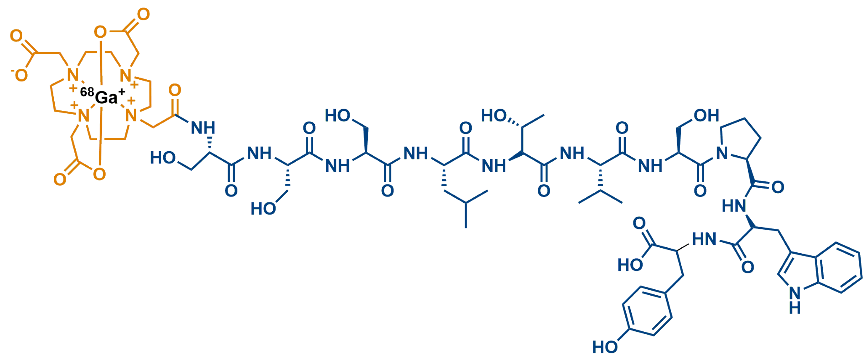
  59. Biabani Ardakani, J.; Akhlaghi, M.; Nikkholgh, B.; Hosseinimehr, S.J. Targeting and imaging of HER2 overexpression tumor with a new peptide-based 68Ga-PET radiotracer. Bioorganic Chem. 2021, 106, 104474.
  60. Ge, S.; Li, J.; Yu, Y.; Chen, Z.; Yang, Y.; Zhu, L.; Sang, S.; Deng, S. Review: Radionuclide Molecular Imaging Targeting HER2 in Breast Cancer with a Focus on Molecular Probes into Clinical Trials and Small Peptides. Molecules 2021, 26, 6482.
  61. Chen, K.; Conti, P.S. Target-specific delivery of peptide-based probes for PET imaging. Adv. Drug Deliv. Rev. 2010, 62, 1005–1022.
  62. Fani, M.; Maecke, H.R.; Okarvi, S.M. Radiolabeled peptides: Valuable tools for the detection and treatment of cancer. Theranostics 2012, 2, 481–501.
  63. Shadidi, M.; Sioud, M. Identification of novel carrier peptides for the specific delivery of therapeutics into cancer cells. FASEB J. 2003, 17, 256–258.
  64. Mikulová, M.B.; Mikuš, P. Advances in Development of Radiometal Labeled Amino Acid-Based Compounds for Cancer Imaging and Diagnostics. Pharmaceuticals 2021, 14, 167.
  65. Khodadust, F.; Ahmadpour, S.; Aligholikhamseh, N.; Abedi, S.M.; Hosseinimehr, S.J. An improved 99mTc-HYNIC-(Ser)3-LTVSPWY peptide with EDDA/tricine as co-ligands for targeting and imaging of HER2 overexpression tumor. Eur. J. Med. Chem. 2018, 144, 767–773.
  66. Sabahnoo, H.; Noaparast, Z.; Abedi, S.M.; Hosseinimehr, S.J. New small 99mTc-labeled peptides for HER2 receptor imaging. Eur. J. Med. Chem. 2017, 127, 1012–1024.
  67. Kumar, S.R.; Gallazzi, F.A.; Ferdani, R.; Anderson, C.J.; Quinn, T.P.; Deutscher, S.L. In vitro and in vivo evaluation of ⁶⁴Cu-radiolabeled KCCYSL peptides for targeting epidermal growth factor receptor-2 in breast carcinomas. Cancer Biother. Radiopharm. 2010, 25, 693–703.
  68. Larimer, B.M.; Thomas, W.D.; Smith, G.P.; Deutscher, S.L. Affinity maturation of an ERBB2-targeted SPECT imaging peptide by in vivo phage display. Mol. Imaging Biol. 2014, 16, 449–458.
  69. Kumar, S.R.; Quinn, T.P.; Deutscher, S.L. Evaluation of an 111In-radiolabeled peptide as a targeting and imaging agent for ErbB-2 receptor expressing breast carcinomas. Clin. Cancer Res. 2007, 13, 6070–6079.
  70. Biri-Kovács, B.; Adorján, A.; Szabó, I.; Szeder, B.; Bősze, S.; Mező, G. Structure–Activity Relationship of HER2 Receptor Targeting Peptide and Its Derivatives in Targeted Tumor Therapy. Biomolecules 2020, 10, 183.
  71. Luo, X.; Chen, H.; Song, Y.; Qin, Z.; Xu, L.; He, N.; Tan, Y.; Dessie, W. Advancements, challenges and future perspectives on peptide-based drugs: Focus on antimicrobial peptides. Eur. J. Pharm. Sci. 2023, 181, 106363.
  72. Qian, Z.; Rhodes, C.A.; McCroskey, L.C.; Wen, J.; Appiah-Kubi, G.; Wang, D.J.; Guttridge, D.C.; Pei, D. Enhancing the cell permeability and metabolic stability of peptidyl drugs by reversible bicyclization. Angew. Chem. Int. Ed. Engl. 2017, 56, 1525–1529.
  73. Aguirre, T.A.; Teijeiro-Osorio, D.; Rosa, M.; Coulter, I.; Alonso, M.; Brayden, D. Current status of selected oral peptide technologies in advanced preclinical development and in clinical trials. Adv. Drug Deliv. Rev. 2016, 106, 223–241.
  74. Pichereau, C.; Allary, C. Therapeutic peptides under the spotlight. Eur. Biopharm. 2005, 5, 88–91.
  75. Belsito, E.; Di Gioia, M.L.; Greco, A.; Leggio, A.; Liguori, A.; Perri, F.; Siciliano, C.; Viscomi, M.C. N-Methyl-N-nosyl-β3-amino Acids. J. Org. Chem. 2007, 72, 4798–4802.
  76. Di Gioia, M.L.; Leggio, A.; Malagrinò, F.; Romio, E.; Siciliano, C.; Liguori, A. N-Methylated α-Amino Acids And Peptides: Synthesis And Biological Activity. Mini-Rev. Med. Chem. 2016, 16, 683–690.
  77. Leggio, A.; Belsito, E.L.; De Marco, R.; Liguori, A.; Perri, F.; Viscomi, M.C. An Efficient Preparation of N-Methyl-α-amino Acids from N-Nosyl-α-amino Acid Phenacyl Esters. J. Org. Chem. 2010, 75, 1386–1392.
  78. Zhu, Q.; Chen, Z.; Paul, P.K.; Lu, Y.; Wu, W.; Qi, J. Oral delivery of proteins and peptides: Challenges, status quo and future perspectives. Acta Pharm. Sin. B 2021, 11, 2416–2448.
  79. Barman, P.; Joshi, S.; Sharma, S.; Preet, S.; Sharma, S.; Saini, A. Strategic Approaches to Improvise Peptide Drugs as Next Generation Therapeutics. Int. J. Pept. Res. Ther. 2023, 29, 61.
  80. Veronese, F.M.; Mero, A. The impact of PEGylation on biological therapies. Bio. Drugs 2008, 22, 315–329.
  81. Freire Haddad, H.; Burke, J.A.; Scott, E.A.; Ameer, G.A. Clinical relevance of pre-existing and treatment-induced anti-poly(ethylene glycol) antibodies. Regen. Eng. Transl. Med. 2021, 8, 32–42.
  82. Costa, A.R.; Rodrigues, M.E.; Henriques, M.; Oliveira, R.; Azeredo, J. Glycosylation: Impact, control and improvement during therapeutic protein production. Crit. Rev. Biotechnol. 2014, 34, 281–299.
  83. Van Regenmorte, M.H. Antigenicity and immunogenicity of synthetic peptides. Biologicals 2001, 29, 209–213.
  84. Sauna, Z.E. Immunogenicity of Protein-Based Therapeutics. U.S. Food and Drug Administration. 2020. Available online: https://www.fda.gov/vaccines-blood-biologics/biologics-research-projects/immunogenicity-protein-based-therapeutics (accessed on 26 August 2023).
  85. Bray, B.L. Large-scale manufacture of peptide therapeutics by chemical synthesis. Nat. Rev. Drug Discov. 2003, 2, 587–593.
  86. Vlieghe, P.; Lisowski, V.; Martinez, J.; Khrestchatisky, M. Synthetic therapeutic peptides: Science and market. Drug Discov. Today 2010, 15, 40–56.
More
Information
Contributors MDPI registered users' name will be linked to their SciProfiles pages. To register with us, please refer to https://encyclopedia.pub/register : , , , , , , ,
View Times: 221
Entry Collection: Peptides for Health Benefits
Revisions: 3 times (View History)
Update Date: 12 Sep 2023
1000/1000