Submitted Successfully!
To reward your contribution, here is a gift for you: A free trial for our video production service.
Thank you for your contribution! You can also upload a video entry or images related to this topic.
Version Summary Created by Modification Content Size Created at Operation
1 -- 2045 2023-09-07 15:56:03 |
2 format change Meta information modification 2045 2023-09-08 04:47:52 |

Video Upload Options

Do you have a full video?

Confirm

Are you sure to Delete?
Cite
If you have any further questions, please contact Encyclopedia Editorial Office.
Suga, N.; Ikeda, Y.; Yoshikawa, S.; Taniguchi, K.; Sawamura, H.; Matsuda, S. Non-Coding RNAs and Gut Microbiota in Cardiac Arrhythmias. Encyclopedia. Available online: https://encyclopedia.pub/entry/48936 (accessed on 08 September 2024).
Suga N, Ikeda Y, Yoshikawa S, Taniguchi K, Sawamura H, Matsuda S. Non-Coding RNAs and Gut Microbiota in Cardiac Arrhythmias. Encyclopedia. Available at: https://encyclopedia.pub/entry/48936. Accessed September 08, 2024.
Suga, Naoko, Yuka Ikeda, Sayuri Yoshikawa, Kurumi Taniguchi, Haruka Sawamura, Satoru Matsuda. "Non-Coding RNAs and Gut Microbiota in Cardiac Arrhythmias" Encyclopedia, https://encyclopedia.pub/entry/48936 (accessed September 08, 2024).
Suga, N., Ikeda, Y., Yoshikawa, S., Taniguchi, K., Sawamura, H., & Matsuda, S. (2023, September 07). Non-Coding RNAs and Gut Microbiota in Cardiac Arrhythmias. In Encyclopedia. https://encyclopedia.pub/entry/48936
Suga, Naoko, et al. "Non-Coding RNAs and Gut Microbiota in Cardiac Arrhythmias." Encyclopedia. Web. 07 September, 2023.
Non-Coding RNAs and Gut Microbiota in Cardiac Arrhythmias
Edit

Non-coding RNAs (ncRNAs) are indispensable for adjusting gene expression and genetic programming throughout development and for health as well as cardiovascular diseases. Cardiac arrhythmia is a frequent cardiovascular disease that has a complex pathology. Studies have shown that ncRNAs are also associated with cardiac arrhythmias. Many non-coding RNAs and/or genomes have been reported as genetic background for cardiac arrhythmias. In general, arrhythmias may be affected by several functional and structural changes in the myocardium of the heart. Therefore, ncRNAs might be indispensable regulators of gene expression in cardiomyocytes, which could play a dynamic role in regulating the stability of cardiac conduction and/or in the remodeling process.

ncRNA lncRNA miRNA cardiac arrhythmia atrial fibrillation gut microbiota APRO family protein

1. Introduction

Cardiac arrhythmias are a group of heterogeneous disorders in the heartbeat, which may be often defined as any variations in the rate of the normal heart. Abnormal impulse formations and disturbances in conduction may be two major reasons for arrhythmias. However, the physiological dysfunctions linked to the specific arrhythmias are very intricate. The primary pace-making activity of the heart is determined by a spontaneous action potential within atrial node cells [1], which generates cardiac excitation. At present, it has been suggested that the distinctive pace generation might characteristically rely on a probable membrane clock mechanism. The membrane clock consists of plasma membrane ion channels carrying an inward Na+/Ca2+ exchange current, which might stimulate the surface sarcolemmal electrogenic membrane clock [2]. Therefore, the rhythm-associated ion channels related to the instigation or progression of the action potential have been well recognized. Almost certainly, arrhythmias are mostly caused by imbalances of these calcium ion channels and dysregulations of conduction in heart muscles. The identification of genetic components underlying cardiac arrhythmias has also highlighted the specific role of ion channels. Most ion channels are typically protein complexes that are positioned in the cardiomyocyte sarcolemma, and they might play a role in calcium ion flow conduction [3]. Shifts in the balance of these currents could either increase or decrease the duration of the action potential. Inhomogeneous polarization, influencing the heart, may potentially lead to arrhythmias [3].
Non-coding RNAs (ncRNAs) have been revealed to regulate a diversity of ion channels and/or intercellular linking proteins/molecules, suggesting that ncRNAs may be an important regulator of cardiovascular diseases, including arrhythmias [4]. In fact, it has been revealed that ncRNA could regulate the development of various cardiac diseases [5]. Therefore, recent findings on the implication of ncRNAs in the development of the most common forms of cardiac arrhythmias with a focus on therapeutic and/or clinical application have been discussed here. The ncRNAs are around 200-nt base sequences that might regulate genetic and epigenetic gene expression as well as intracellular signaling mechanisms [6]. In general, ncRNAs have been used as biomarkers for diagnosis and/or treatment due to their involvement in the instigation of diseases. Hence, ncRNA has also been investigated in the field of cardiovascular diseases [7] (Figure 1).
Figure 1. Schematic representation of the players of ncRNAs involved in cardiac arrhythmias. Example ncRNA molecules are shown for each cardiac arrhythmia.

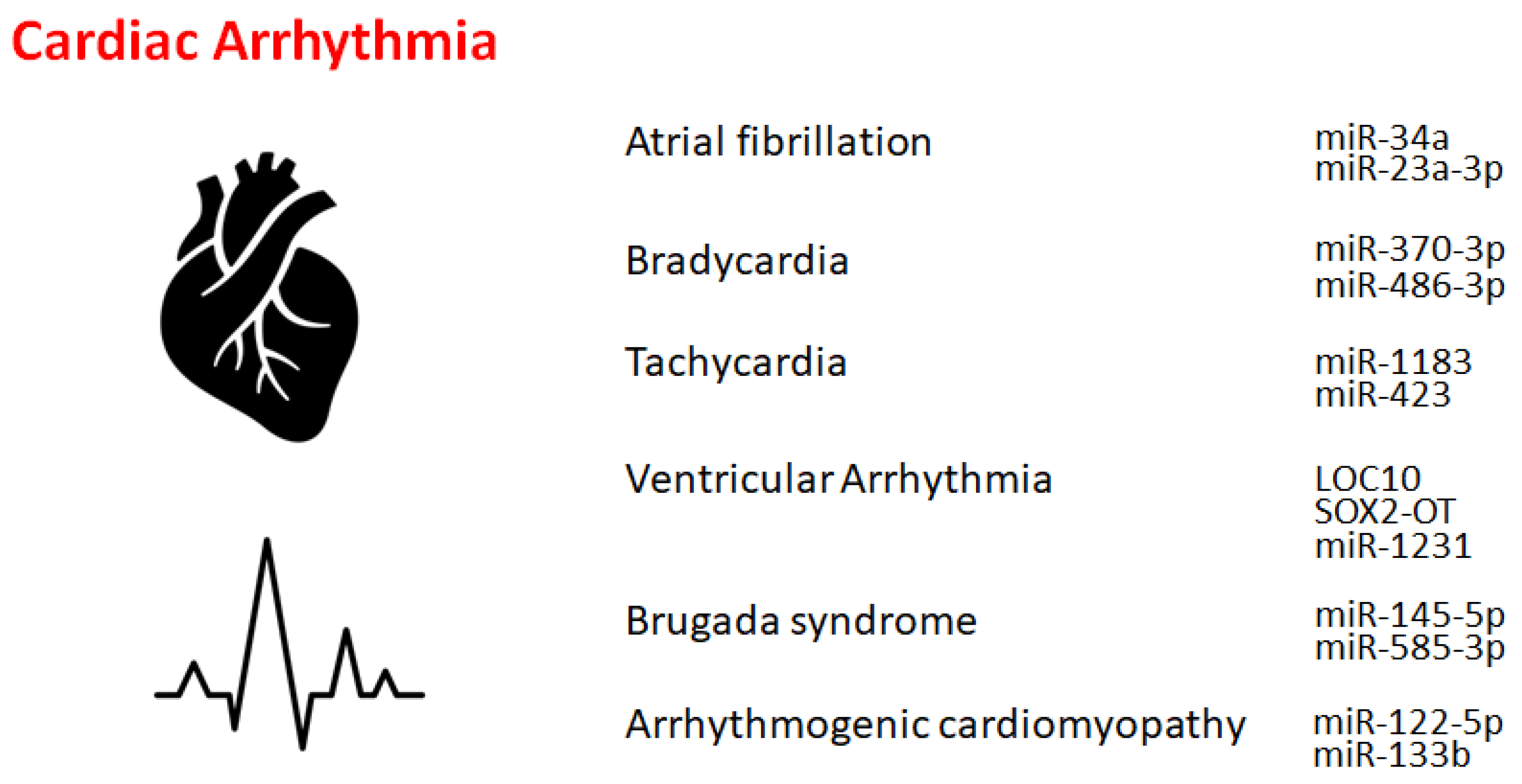
2. Atrial Fibrillation

Atrial fibrillation is an asymmetrical heart rhythm that might commonly lead to heart palpitations. Interestingly, individuals carrying the non-coding 4q25 locus near the PITX2 gene were 60% more susceptible to this abnormal heart rhythm [8]. Similarly, lncRNAs may be potentially associated with the development of atrial fibrillation. Several lncRNAs, including LINC00844, RP11-532N4.2, UNC5B-AS1, RP3-332B22.1, RP11-432J24.5, and RP11-557H15.4, have been shown to be differentially expressed in patients with atrial fibrillation compared to normal individuals, whose target might be related to the calcium signaling pathway and/or toll-like receptor signaling pathway [9][10]. In addition, several miRNAs, including miR-135a, might be involved in the process of atrial fibrillation. The physiological roles of these differentially expressed ncRNAs might be associated with the pathogenesis of atrial fibrillation, which might provide a new therapeutic target and/or a prognosis marker for patients with atrial fibrillation [9].

3. Bradycardia and Tachycardia

Bradycardia might be a heart dysfunction described by an extremely decreased heart rate, typically lower than 50 beats/min. Key etiological factors of bradycardia may include the dysregulation of sinus, atrial, or junctional bradycardia and/or an intricate transmission system with atrioventricular block. The most frequent form of bradycardia occurs asymptomatically during sleep and/or in athletes [11]. The only treatment for insistent bradycardia may be the placement of an everlasting pacemaker. Bradycardia is usually regulated by a range of molecules, including ncRNAs, at molecular levels. Transcription factors might also control the expression of genes involved in several bradycardias by modulating a variety of effectors, including miRNAs. For example, it has been demonstrated that the function of miR-370-3p could contribute to the development of bradycardia [12], which might be triggered via the reduced function of sinus node pacemaker channels with a related reduction of the ionic current. The miR-370-3p could directly bind to the mRNA of HCN4 to suppress its activity [12]. Similarly, miR-486-3p could suppress HCN4, inducing sinus node dysregulation such as bradycardia [13][14]. Supraventricular arrhythmia may be an example of tachycardia that begins in the upper compartments of the heart. Tachycardia is known to be considered a fast heartbeat, which might also be regulated by a range of molecules, including ncRNAs, at molecular levels.

4. Other Cardiac Rhythm Disorders

Ventricular arrhythmias have been described as irregular heartbeats that may derive from the ventricles. The origin of ventricular arrhythmias might be sympathetic remodeling that originates in myocardial infarction. Various ncRNAs are important regulators of inflammation and/or sympathetic remodeling following myocardial infarction. Several lncRNAs are potentially implicated in ventricular arrhythmias following myocardial infarction. For example, the lncRNA LOC100911717 (LOC10) might be increased in cardiac cells and macrophages in the infarcted heart area [15]. Another important factor in ventricular arrhythmias may be NLRP3 inflammasomes. It has been demonstrated that the role of SOX2-OT lncRNA in regulating NLRP3 inflammasome-mediated ventricular arrhythmias. The levels of SOX2-OT and NLRP3 inflammasomes could be intensified after ventricular arrhythmias [16]. Similarly, miR-2355-3p and miR-1231 may play key roles in the regulation of ventricular arrhythmias [17][18], which might provide a potential diagnostic marker as well as a potential target for various treatments [17].
Long QT and short QT syndromes are known as the entities of Brugada syndrome [19]. Risk stratification for sudden cardiac death in patients with Brugada syndrome remains a major challenge. Brugada patients display a distinct miRNA expression profile compared with unaffected control individuals.

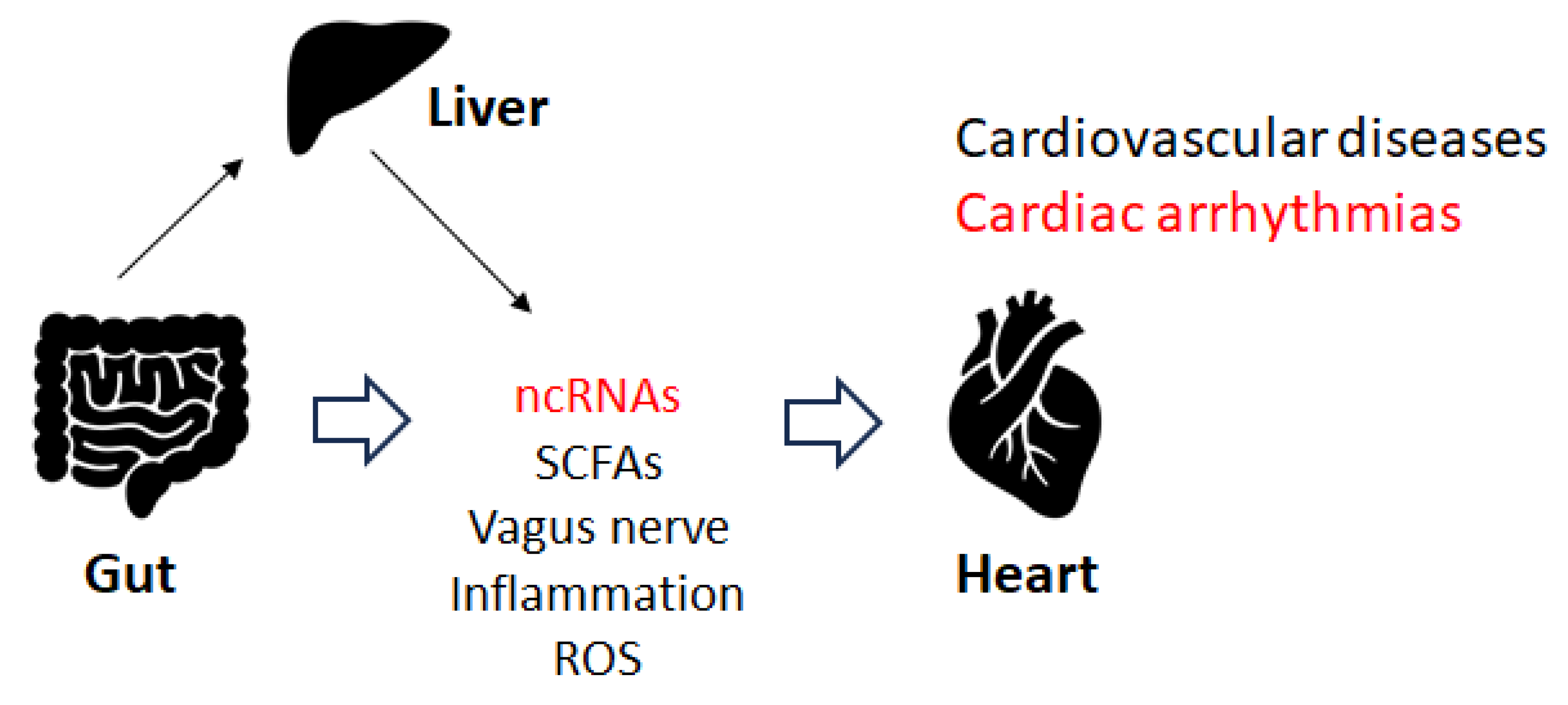
5. Gut Microbiota and Cardiac Arrhythmia

Disruption of the gut microbiota could induce cardiorespiratory morbidity. Consistently, modulation of autonomic homeostasis via the gut microbiota-brain axis could control the heart rate, independent of carotid body plasticity [20]. Sympathetic neuronal communication between the hypothalamic paraventricular nucleus and the gut might also be involved in the regulation of blood pressure [20]. The adult gut system might be an extremely diverse and dynamic ecosystem [21][22], which might employ specific physiological functions including immune regulation, gut mucosal protection, and conservation of nutritional metabolisms, in addition to the development of several diseases [23]. Some elements of the gut microbiota could stimulate the afferent fiber of the vagus nerve through the gut endocrine cells, which could also inspire the central nervous system (CNS). Depending on the material, the different SCFAs produced by the gut microbiota could activate the vagal afferent fibers in several ways. For example, butyric acid, a short fatty acid, directly affects afferent terminals [24][25]. An increase in the concentration of butyric acid in the colon may produce a significant hypotensive effect, which depends on afferent colonic vagus nerve signaling and GPR41/43 receptors [25]. Several investigations have considered the effect of food components on gut microbiota, which could be an imperative target for the forthcoming treatment of cardiac arrhythmias through the alteration of gut microbiota. For example, patients with arrhythmias may prefer to obtain more energy from animal fat [26]. Therefore, patients with cardiac arrhythmias may be frequently diagnosed with atherosclerosis. In addition, diabetes may also be a frequent co-morbidity in individuals with arrhythmias. Furthermore, the interaction between some drugs and their impact on the gut microbiota may result in uncontrolled arrhythmias [27][28][29]. Recent increasing data suggest that regulatory non-coding RNAs such as miRNAs, circular RNAs, and lncRNAs may affect host-microbe interactions. These ncRNAs have also been suggested as potential biomarkers in microbiome-associated disorders with a direct cross-talk between microbiome composition and ncRNAs [30]. Therefore, gut microbiota could affect responses to stimuli by host cells with modifications to their epigenome and/or gene expression. Recent data suggest that regulatory ncRNAs such as miRNAs, circular RNAs, and lncRNAs might affect host-microbe interactions. For example, miR-155 has been associated with inflammatory bowel diseases and might also be involved in cardiac remodeling following acute myocardial infarction [30]. Epigenetically, miR-23a-3p could lead to a Th17/Treg imbalance and participate in the progression of Graves’ disease [31], which might also be involved in the mechanism underlying atrial fibrillation. Consequently, gut microbiota could play a vital role in the pathogenesis of both atrial fibrillation and Graves’ disease [31]. The imbalance of Th17/Treg cells induced by the alteration of gut microbiota could also play a dynamic role in the pathogenesis of arrhythmias [32][33], suggesting that the immune response and arrhythmias are closely related. In particular, Th17 cells might be involved in the pathogenesis of atrial fibrillation, since increased levels of Th17-associated cytokines may be independently associated with an increased risk of atrial fibrillation [32]. The balance between Th17 and Treg cells has been deliberated to represent a paradigm for several inflammatory and auto-immune diseases. The inflammatory response might play a key role in the pathogenesis of atrial fibrillation, and the restoration of the Th17/Treg balance might represent a promising therapeutic target for treatment [33]. Interestingly, altered composition of the gut microbiota could change the expression of hepatic miR-34a [34], which might also be involved in the development of atrial fibrillation. In addition, there is a negative correlation between miR-122-5p and intestinal bacteria, including Bacteriodes. uniformis and Phascolarctobacterium. Faecium has been shown [35], suggesting that the crosstalk between miRNA and certain gut microbiota could regulate intracellular signal transduction by controlling the expression of key genes related to the development of arrhythmias [35]. As mentioned before, miR-122-5p could possess great diagnostic potential in arrhythmogenic cardiomyopathy patients [36][37] (Figure 2).
Figure 2. Several factors and/or inflammation with ROS may affect the development of cardiovascular diseases, including cardiac arrhythmias. Note that some critical pathways have been omitted for clarity.

6. Immune Pathway and Cardiac Arrhythmias

There is accumulative attention on mechanisms molecularly directing the onset and/or progression of cardiac arrhythmias due to the intricate interplay that could elicit several immune cells, enhancing atrial fibrosis [38]. The dysregulation of the immune pathway might meaningfully contribute to ion channel dysfunction and the initiation of cardiac arrhythmias [39]. In other words, the potential molecular mechanism of cardiac arrhythmias may involve inflammatory and immune pathways that cause ion channel dysfunction, in which a variety of signaling systems might be involved [39]. Consecutively, the interaction between immune cells and atrial myocytes might augment atrial electrical and/or practical remodeling, which might be responsible for the progression of cardiac arrhythmias [40]. Interestingly, some ncRNAs are involved in the ribonucleolytic cleavage of the target mRNA [41], and several members of the APRO family have been revealed to be associated with the cytoplasmic mRNA deadenylation [42][43]. Similarly, several APRO family proteins could cooperate with the poly (A) ribonuclease complex [44][45]. These APRO family proteins (Btg1, Btg2, Tob1, and Tob2) might be key modulators of microRNAs [46], which may be extremely involved in the regulation of immune cells [47]. Therefore, APRO family proteins have been shown to be involved in immune-related disorders, including ulcerative colitis and cancer [48][49][50]. Interestingly, it has been shown that one of the APRO family members, BTG1, may be involved in the regulation of ion channel-related gene expression [51][52]. Remarkably, it has been shown that BTG1 could promote the deadenylation and/or degradation of mRNA to secure T cell quiescence [53], suggesting that BTG1 might be involved in a key mechanism underlying T cell quiescence. In addition, another APRO family member, Tob1, might be a key regulatory molecule in the process of endothelialization in the repair of atrial septal defects [54]. Tob1 might also play a fundamental role in keeping cells in a quiescent state, thereby obstructing the cellular proliferation of normal and/or cancer cells [55]. Tob1 may be inactivated in the cells of many human cancers. Therefore, Tob1 has been considered a tumor-suppressor protein [56].

References

  1. Fan, W.; Sun, X.; Yang, C.; Wan, J.; Luo, H.; Liao, B. Pacemaker activity and ion channels in the sinoatrial node cells: MicroRNAs and arrhythmia. Prog. Biophys. Mol. Biol. 2023, 177, 151.
  2. Yaniv, Y.; Lakatta, E.G.; Maltsev, V.A. From two competing oscillators to one coupled-clock pacemaker cell system. Front. Physiol. 2015, 6, 28.
  3. Wilde, A.A.; Bezzina, C.R. Genetics of cardiac arrhythmias. Heart 2005, 91, 1352.
  4. Kawaguchi, S.; Moukette, B.; Hayasaka, T.; Haskell, A.K.; Mah, J.; Sepúlveda, M.N.; Tang, Y.; Kim, I.M. Noncoding RNAs as Key Regulators for Cardiac Development and Cardiovascular Diseases. J. Cardiovasc. Dev. Dis. 2023, 10, 166.
  5. Sallam, T.; Sandhu, J.; Tontonoz, P. Long Noncoding RNA Discovery in Cardiovascular Disease: Decoding Form to Function. Circ. Res. 2018, 122, 155.
  6. Zhang, C.; Han, B.; Xu, T.; Li, D. The biological function and potential mechanism of long non-coding RNAs in cardiovascular disease. J. Cell. Mol. Med. 2020, 24, 12900.
  7. Correia, C.C.M.; Rodrigues, L.F.; de Avila Pelozin, B.R.; Oliveira, E.M.; Fernandes, T. Long Non-Coding RNAs in Cardiovascular Diseases: Potential Function as Biomarkers and Therapeutic Targets of Exercise Training. Non-Coding RNA 2021, 7, 65.
  8. Gudbjartsson, D.F.; Arnar, D.O.; Helgadottir, A.; Gretarsdottir, S.; Holm, H.; Sigurdsson, A.; Jonasdottir, A.; Baker, A.; Thorleifsson, G.; Kristjansson, K. Variants conferring risk of atrial fibrillation on chromosome 4q25. Nature 2007, 448, 353.
  9. Ruan, Z.B.; Wang, F.; Gongben, B.D.; Chen, G.C.; Zhu, L. Identification of Circulating lncRNA Expression Profiles in Patients with Atrial Fibrillation. Dis. Mark. 2020, 2020, 8872142.
  10. Xie, L.; Huang, G.L.; Gao, M.L.; Huang, J.L.; Li, H.L.; Xia, H.L.; Xiang, X.L.; Wu, S.L.; Ruan, Y. Identification of Atrial Fibrillation-Related lncRNA Based on Bioinformatic Analysis. Dis. Mark. 2022, 2022, 8307975.
  11. Barstow, C.; McDivitt, J.D. Cardiovascular Disease Update: Bradyarrhythmias. FP Essent. 2017, 454, 18.
  12. Yanni, J.; D’Souza, A.; Wang, Y.; Li, N.; Hansen, B.J.; Zakharkin, S.O.; Smith, M.; Hayward, C.; Whitson, B.A.; Mohler, P.J.; et al. Silencing miR-370-3p rescues funny current and sinus node function in heart failure. Sci. Rep. 2020, 10, 11279.
  13. Petkova, M.; Atkinson, A.J.; Yanni, J.; Stuart, L.; Aminu, A.J.; Ivanova, A.D.; Pustovit, K.B.; Geragthy, C.; Feather, A.; Li, N.; et al. Identification of Key Small Non-Coding MicroRNAs Controlling Pacemaker Mechanisms in the Human Sinus Node. J. Am. Heart Assoc. 2020, 9, e016590.
  14. Aminu, A.J.; Petkova, M.; Atkinson, A.J.; Yanni, J.; Morris, A.D.; Simms, R.T.; Chen, W.; Yin, Z.; Kuniewicz, M.; Holda, M.K.; et al. Further insights into the molecular complexity of the human sinus node—The role of ‘novel’ transcription factors and microRNAs. Prog. Biophys. Mol. Biol. 2021, 166, 86.
  15. Zhao, H.; Tan, Z.; Zhou, J.; Wu, Y.; Hu, Q.; Ling, Q.; Ling, J.; Liu, M.; Ma, J.; Zhang, D.; et al. The regulation of circRNA and lncRNAprotein binding in cardiovascular diseases: Emerging therapeutic targets. Biomed. Pharmacother. 2023, 165, 115067.
  16. Liang, Y.; Wang, B.; Huang, H.; Wang, M.; Wu, Q.; Zhao, Y.; He, Y. Silenced SOX2-OT alleviates ventricular arrhythmia associated with heart failure by inhibiting NLRP3 expression via regulating miR-2355-3p. Immun. Inflamm. Dis. 2021, 9, 255.
  17. Xiong, F.; Mao, R.; Zhang, L.; Zhao, R.; Tan, K.; Liu, C.; Xu, J.; Du, G.; Zhang, T. CircNPHP4 in monocyte-derived small extracellular vesicles controls heterogeneous adhesion in coronary heart atherosclerotic disease. Cell Death Dis. 2021, 12, 948.
  18. Shi, Y.; Qiao, L.; Han, F.; Xie, X.; Wang, W. MiR-1231 regulates L-calcium in ventricular arrhythmia in chronic heart failure. Minerva Med. 2021, 112, 305.
  19. Sarquella-Brugada, G.; Cesar, S.; Zambrano, M.D.; Fernandez-Falgueras, A.; Fiol, V.; Iglesias, A.; Torres, F.; Garcia-Algar, O.; Arbelo, E.; Brugada, J.; et al. Electrocardiographic Assessment and Genetic Analysis in Neonates: A Current Topic of Discussion. Curr. Cardiol. Rev. 2019, 15, 30.
  20. Lucking, E.F.; O’Connor, K.M.; Strain, C.R.; Fouhy, F.; Bastiaanssen, T.F.S.; Burns, D.P.; Golubeva, A.V.; Stanton, C.; Clarke, G.; Cryan, J.F.; et al. Chronic intermittent hypoxia disrupts cardiorespiratory homeostasis and gut microbiota composition in adult male guinea-pigs. eBioMedicine 2018, 38, 191.
  21. Anthony, W.E.; Wang, B.; Sukhum, K.V.; D’Souza, A.W.; Hink, T.; Cass, C.; Seiler, S.; Reske, K.A.; Coon, C.; Dubberke, E.R.; et al. Acute and persistent effects of commonly used antibiotics on the gut microbiome and resistome in healthy adults. Cell Rep. 2022, 39, 110649.
  22. Tierney, B.T.; Yang, Z.; Luber, J.M.; Beaudin, M.; Wibowo, M.C.; Baek, C.; Mehlenbacher, E.; Patel, C.J.; Kostic, A.D. The Landscape of Genetic Content in the Gut and Oral Human Microbiome. Cell Host Microbe 2019, 26, 283.
  23. Sonnenburg, E.D.; Smits, S.A.; Tikhonov, M.; Higginbottom, S.K.; Wingreen, N.S.; Sonnenburg, J.L. Diet-induced extinctions in the gut microbiota compound over generations. Nature 2016, 529, 212.
  24. Lal, S.; Kirkup, A.J.; Brunsden, A.M.; Thompson, D.G.; Grundy, D. Vagal afferent responses to fatty acids of different chain length in the rat. Am. J. Physiol. Gastrointest. Liver Physiol. 2001, 281, G907.
  25. Onyszkiewicz, M.; Gawrys-Kopczynska, M.; Konopelski, P.; Aleksandrowicz, M.; Sawicka, A.; Koźniewska, E.; Samborowska, E.; Ufnal, M. Butyric acid, a gut bacteria metabolite, lowers arterial blood pressure via colon-vagus nerve signaling and GPR41/43 receptors. Pflug. Arch. 2019, 471, 1441.
  26. Tabata, T.; Yamashita, T.; Hosomi, K.; Park, J.; Hayashi, T.; Yoshida, N.; Saito, Y.; Fukuzawa, K.; Konishi, K.; Murakami, H.; et al. Gut microbial composition in patients with atrial fibrillation: Effects of diet and drugs. Heart Vessels 2021, 36, 105.
  27. Celikyurt, I.; Meier, C.R.; Kühne, M.; Schaer, B. Safety and Interactions of Direct Oral Anticoagulants with Antiarrhythmic Drugs. Drug Saf. 2017, 40, 1091.
  28. Aliabadi, T.; Saberi, E.A.; Motameni Tabatabaei, A.; Tahmasebi, E. Antibiotic use in endodontic treatment during pregnancy: A narrative review. Eur. J. Transl. Myol. 2022, 32, 10813.
  29. Valdivielso, J.M.; Balafa, O.; Ekart, R.; Ferro, C.J.; Mallamaci, F.; Mark, P.B.; Rossignol, P.; Sarafidis, P.; Del Vecchio, L.; Ortiz, A. Hyperkalemia in Chronic Kidney Disease in the New Era of Kidney Protection Therapies. Drugs 2021, 81, 1467.
  30. Fardi, F.; Khasraghi, L.B.; Shahbakhti, N.; Salami Naseriyan, A.; Najafi, S.; Sanaaee, S.; Alipourfard, I.; Zamany, M.; Karamipour, S.; Jahani, M.; et al. An interplay between non-coding RNAs and gut microbiota in human health. Diabetes Res. Clin. Pract. 2023, 201, 110739.
  31. Zhou, F.; Wang, X.; Wang, L.; Sun, X.; Tan, G.; Wei, W.; Zheng, G.; Ma, X.; Tian, D.; Yu, H. Genetics, Epigenetics, Cellular Immunology, and Gut Microbiota: Emerging Links with Graves’ Disease. Front. Cell Dev. Biol. 2022, 9, 794912.
  32. Wang, X.; Fan, H.; Wang, Y.; Yin, X.; Liu, G.; Gao, C.; Li, X.; Liang, B. Elevated Peripheral T Helper Cells Are Associated with Atrial Fibrillation in Patients with Rheumatoid Arthritis. Front. Immunol. 2021, 12, 744254.
  33. He, Y.; Chen, X.; Guo, X.; Yin, H.; Ma, N.; Tang, M.; Liu, H.; Mei, J. Th17/Treg Ratio in Serum Predicts Onset of Postoperative Atrial Fibrillation After Off-Pump Coronary Artery Bypass Graft Surgery. Heart Lung Circ. 2018, 27, 1467.
  34. Jia, N.; Lin, X.; Ma, S.; Ge, S.; Mu, S.; Yang, C.; Shi, S.; Gao, L.; Xu, J.; Bo, T.; et al. Amelioration of hepatic steatosis is associated with modulation of gut microbiota and suppression of hepatic miR-34a in Gynostemma pentaphylla (Thunb.) Makino treated mice. Nutr. Metab. 2018, 15, 86.
  35. Li, L.; Li, C.; Lv, M.; Hu, Q.; Guo, L.; Xiong, D. Correlation between alterations of gut microbiota and miR-122-5p expression in patients with type 2 diabetes mellitus. Ann. Transl. Med. 2020, 8, 1481.
  36. Bueno Marinas, M.; Celeghin, R.; Cason, M.; Bariani, R.; Frigo, A.C.; Jager, J.; Syrris, P.; Elliott, P.M.; Bauce, B.; Thiene, G.; et al. A microRNA Expression Profile as Non-Invasive Biomarker in a Large Arrhythmogenic Cardiomyopathy Cohort. Int. J. Mol. Sci. 2020, 21, 1536.
  37. Khudiakov, A.A.; Panshin, D.D.; Fomicheva, Y.V.; Knyazeva, A.A.; Simonova, K.A.; Lebedev, D.S.; Mikhaylov, E.N.; Kostareva, A.A. Different Expressions of Pericardial Fluid MicroRNAs in Patients with Arrhythmogenic Right Ventricular Cardiomyopathy and Ischemic Heart Disease Undergoing Ventricular Tachycardia Ablation. Front. Cardiovasc. Med. 2021, 8, 647812.
  38. Desantis, V.; Potenza, M.A.; Sgarra, L.; Nacci, C.; Scaringella, A.; Cicco, S.; Solimando, A.G.; Vacca, A.; Montagnani, M. microRNAs as Biomarkers of Endothelial Dysfunction and Therapeutic Target in the Pathogenesis of Atrial Fibrillation. Int. J. Mol. Sci. 2023, 24, 5307.
  39. Hao, H.; Dai, C.; Han, X.; Li, Y. A novel therapeutic strategy for alleviating atrial remodeling by targeting exosomal miRNAs in atrial fibrillation. Biochim. Biophys. Acta Mol. Cell Res. 2022, 1869, 119365.
  40. Tan, A.Y.; Zimetbaum, P. Atrial fibrillation and atrial fibrosis. J. Cardiovasc. Pharmacol. 2011, 57, 625–629.
  41. Lee, Y.; Choe, J.; Park, O.H.; Kim, Y.K. Molecular Mechanisms Driving mRNA Degradation by m6A Modification. Trends Genet. 2020, 36, 177–188.
  42. Winkler, G.S. The mammalian anti-proliferative BTG/Tob protein family. J. Cell. Physiol. 2010, 222, 66–72.
  43. Matsuda, S.; Rouault, J.; Magaud, J.; Berthet, C. In search of a function for the TIS21/PC3/BTG1/TOB family. FEBS Lett. 2001, 497, 67–72.
  44. Tirone, F. The gene PC3TIS21/BTG2, prototype member of the PC3/BTG/TOB family: Regulator in control of cell growth, differentiation, and DNA repair? J. Cell. Physiol. 2001, 187, 155–165.
  45. Ezzeddine, N.; Chang, T.-C.; Zhu, W.; Yamashita, A.; Chen, C.-Y.A.; Zhong, Z.; Yamashita, Y.; Zheng, D.; Shyu, A.-B. Human TOB, an Antiproliferative Transcription Factor, Is a Poly(A)-Binding Protein-Dependent Positive Regulator of Cytoplasmic mRNA Deadenylation. Mol. Cell. Biol. 2007, 27, 7791–7801.
  46. Ikeda, Y.; Taniguchi, K.; Nagase, N.; Tsuji, A.; Kitagishi, Y.; Matsuda, S. Reactive oxygen species may influence on the crossroads of stemness, senescence, and carcinogenesis in a cell via the roles of APRO family proteins. Explor. Med. 2021, 2, 443–454.
  47. Ikeda, Y.; Taniguchi, K.; Sawamura, H.; Yoshikawa, S.; Tsuji, A.; Matsuda, S. Presumed Roles of APRO Family Proteins in Cancer Invasiveness. Cancers 2022, 14, 4931.
  48. Zhang, J.; Dong, W. Expression of B Cell Translocation Gene 1 Protein in Colon Carcinoma and its Clinical Significance. Recent Pat. Anti-Cancer Drug Discov. 2020, 15, 78–85.
  49. Zhao, S.; Xue, H.; Hao, C.L.; Jiang, H.M.; Zheng, H.C. BTG1 Overexpression Might Promote Invasion and Metastasis of Colorectal Cancer via Decreasing Adhesion and Inducing Epithelial-Mesenchymal Transition. Front. Oncol. 2020, 10, 598192.
  50. Jung, Y.Y.; Sung, J.Y.; Kim, J.Y.; Kim, H.S. Down-regulation of B-Cell Translocation Gene 1 by Promoter Methylation in Colorectal Carcinoma. Anticancer Res. 2018, 38, 691–697.
  51. Ishida, Y.; Kawakami, H.; Kitajima, H.; Nishiyama, A.; Sasai, Y.; Inoue, H.; Muguruma, K. Vulnerability of Purkinje Cells Generated from Spinocerebellar Ataxia Type 6 Patient-Derived iPSCs. Cell Rep. 2016, 17, 1482–1490.
  52. Luan, S.H.; Yang, Y.Q.; Ye, M.P.; Liu, H.; Rao, Q.F.; Kong, J.L.; Wu, F.R. ASIC1a promotes hepatic stellate cell activation through the exosomal miR-301a-3p/BTG1 pathway. Int. J. Biol. Macromol. 2022, 211, 128–139.
  53. Hwang, S.S.; Lim, J.; Yu, Z.; Kong, P.; Sefik, E.; Xu, H.; Harman, C.C.D.; Kim, L.K.; Lee, G.R.; Li, H.B.; et al. mRNA destabilization by BTG1 and BTG2 maintains T cell quiescence. Science 2020, 367, 1255–1260.
  54. Li, B.N.; Tang, Q.D.; Tan, Y.L.; Yan, L.; Sun, L.; Guo, W.B.; Qian, M.Y.; Chen, A.; Luo, Y.J.; Zheng, Z.X.; et al. Key Regulatory Differentially Expressed Genes in the Blood of Atrial Septal Defect Children Treated with Occlusion Devices. Front. Genet. 2021, 12, 790426.
  55. Tzachanis, D.; Boussiotis, V.A. TOB, a member of the APRO family, regulates immunological quiescence and tumor suppression. Cell Cycle 2009, 8, 1019–1025.
  56. Lee, H.S.; Kundu, J.; Kim, R.N.; Shin, Y.K. Transducer of ERBB2.1 (TOB1) as a Tumor Suppressor: A Mechanistic Perspective. Int. J. Mol. Sci. 2015, 16, 29815–29828.
More
Information
Contributors MDPI registered users' name will be linked to their SciProfiles pages. To register with us, please refer to https://encyclopedia.pub/register : , , , , ,
View Times: 249
Revisions: 2 times (View History)
Update Date: 08 Sep 2023
1000/1000
ScholarVision Creations