Submitted Successfully!
To reward your contribution, here is a gift for you: A free trial for our video production service.
Thank you for your contribution! You can also upload a video entry or images related to this topic.
Version Summary Created by Modification Content Size Created at Operation
1 -- 6478 2023-09-07 10:57:06 |
2 format change Meta information modification 6478 2023-09-07 11:13:33 |

Video Upload Options

Do you have a full video?

Confirm

Are you sure to Delete?
Cite
If you have any further questions, please contact Encyclopedia Editorial Office.
Prete, S.; Dattilo, M.; Patitucci, F.; Pezzi, G.; Parisi, O.I.; Puoci, F. Natural and Synthetic Polymeric Biomaterials in Wound Management. Encyclopedia. Available online: https://encyclopedia.pub/entry/48911 (accessed on 23 July 2024).
Prete S, Dattilo M, Patitucci F, Pezzi G, Parisi OI, Puoci F. Natural and Synthetic Polymeric Biomaterials in Wound Management. Encyclopedia. Available at: https://encyclopedia.pub/entry/48911. Accessed July 23, 2024.
Prete, Sabrina, Marco Dattilo, Francesco Patitucci, Giuseppe Pezzi, Ortensia Ilaria Parisi, Francesco Puoci. "Natural and Synthetic Polymeric Biomaterials in Wound Management" Encyclopedia, https://encyclopedia.pub/entry/48911 (accessed July 23, 2024).
Prete, S., Dattilo, M., Patitucci, F., Pezzi, G., Parisi, O.I., & Puoci, F. (2023, September 07). Natural and Synthetic Polymeric Biomaterials in Wound Management. In Encyclopedia. https://encyclopedia.pub/entry/48911
Prete, Sabrina, et al. "Natural and Synthetic Polymeric Biomaterials in Wound Management." Encyclopedia. Web. 07 September, 2023.
Natural and Synthetic Polymeric Biomaterials in Wound Management
Edit

Biomaterials are at the forefront of the future, finding a variety of applications in the biomedical field, especially in wound healing, thanks to their biocompatible and biodegradable properties. Wounds spontaneously try to heal through a series of interconnected processes involving several initiators and mediators such as cytokines, macrophages, and fibroblasts. The combination of biopolymers with wound healing properties may provide opportunities to synthesize matrices that stimulate and trigger target cell responses crucial to the healing process. 

wound healing wound management dressings biopolymers natural polymers synthetic polymers hydrogels electrospinning 3D printing

1. Introduction

Biomaterials exert a significant influence on various stages of wound healing, including cell proliferation, migration, and differentiation, thereby offering innovative avenues for tissue regeneration and repair. Many of these agents exhibit multifunctionality, contributing to different phases of the wound healing process [1]. Among these, synthetic polymer delivery systems stand out as particularly promising materials for tissue regeneration, enabling controlled and sustained drug release [2].

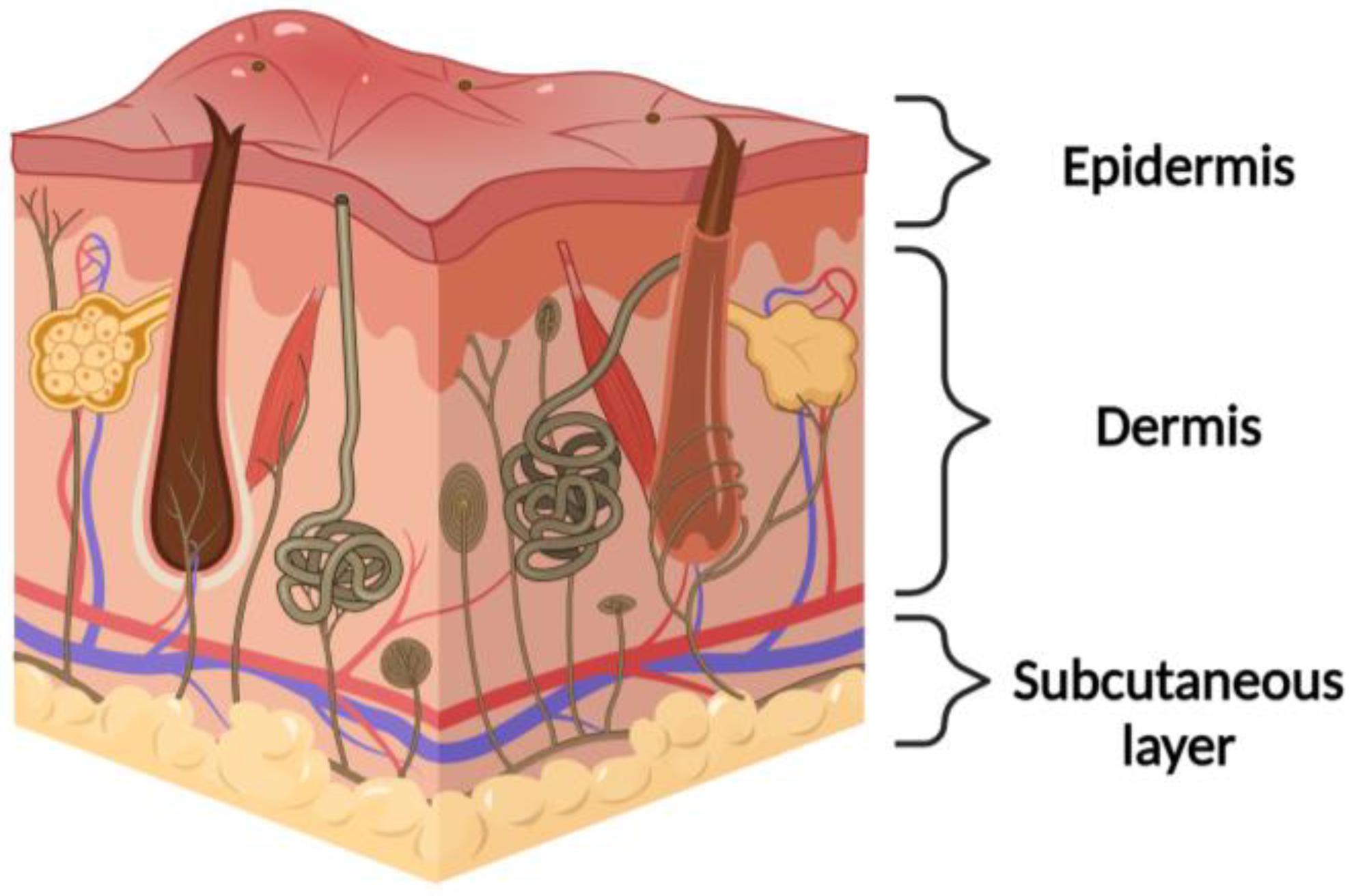
1.1. Physiological Native Skin

The skin is the largest organ of the body and covers the entire external body surface. It is a continuous tissue, belonging to the integumentary apparatus. It is composed of three main layers that, from the outside towards the inside, are called the epidermis, dermis, and hypodermis (or subcutaneous layer) (Figure 1). The epidermis has a thin and cellular structure that forms the superficial layer of the skin. The basement membrane under the epidermis is the dermis composed of a collagen-rich extracellular matrix (ECM), elastin, fibroblasts, and glycosaminoglycans [3]. Throughout the dermis, there is a network of nerve fibers that has a sensory role in the skin and influences immune and inflammatory responses. The hypodermis is the layer beneath the dermis and contains a large amount of adipose tissue that is well vascularized [4]. The skin has many properties, such as the capacity to self-repair (it regenerates after an injury) and extensibility (it adapts perfectly to the variations in body size that occur throughout life). It provides a defensive barrier against external physical influences such as cold, heat, electricity, and radiation, protecting us from trauma, ultraviolet (UV) radiation, microorganisms, and chemical agents. It prevents the loss of liquids and participates in the mechanism of thermoregulation, exploiting the intervention of the sweat glands and the ability to regulate blood flow, increasing (vasodilation) or slowing down (vasoconstriction) the dispersion of heat [5]. The hypodermis, the deepest layer of the skin, holds significant lipid reserves, contributing to enhanced heat retention in proportion to its lipid thickness [6]. Moreover, the skin has metabolic properties, and the synthesis of vitamin D takes place there. It also has a very important sensory function: with its most superficial layer, it registers and transmits pressure (tactile), pain, and thermal stimuli, while with its deepest layer it also perceives thermal and vibratory signals [7].
Figure 1. Cross section of the skin: this diagram illustrates the intricate composition of human skin, showcasing the epidermis, dermis, and subcutaneous tissue layers. Key components such as hair follicles, sweat glands, blood vessels, nerve endings, and sensory receptors are also shown, emphasizing their roles in protection, sensation, thermoregulation, and more.

1.2. Wound Healing Process

The physiological response of an organism to an injury is characterized by a complex mechanism of articulated events, regardless of the type of wound, be it acute or chronic, and the extent of the status of the patient. The healing process of each wound proceeds through many phases that overlap in time and cannot be separated from each other [8]. The process involves the interaction of immune cells (neutrophils, monocytes, macrophages, lymphocytes), non-immune cells (endothelial, fibroblasts, keratinocytes), soluble mediators (cytokines and growth factors), and ECM components [9]. The rate of healing of an acute wound differs from the chronic wound and is dependent on the immunological response [10].
Under normal circumstances, wounds heal by themselves following four major phases: hemostasis, inflammation, proliferation, and remodeling (Figure 2) [11].
Figure 2. Wound healing phases: (1) hemostasis (the body initiates blood clotting to control bleeding at the wound site), (2) inflammation (white blood cells migrate to the wound to eliminate pathogens and debris, creating an optimal environment for healing), (3) proliferation (new tissue formation occurs as fibroblasts produce collagen, essential for wound strength), (4) remodeling (collagen reorganizes and matures, enhancing tissue strength, and granulation tissue is formed, helping in wound contraction and epithelial cell migration).
The initial phase is referred to as hemostasis, during which a newly formed fibrin clot acts as a protective barrier against external elements, ensuring optimal moisture retention [12]. Following hemostasis is the inflammatory stage, where pro-inflammatory cytokines are released from the damaged tissues, attracting circulating white blood cells, and de-granulated platelets and activated macrophages then release various growth factors [13].
The subsequent proliferation phase encompasses crucial processes such as angiogenesis, granulation tissue formation, synthesis of extracellular matrix (ECM) components, epithelialization, and wound contraction [14]. As this phase advances, collagen molecules self-assemble into a distinct triple helical structure and are then released into the extracellular space. Here, they form stable cross-links, imparting strength and stability to the tissue. Dermal collagen typically possesses strong and well-organized properties, whereas scar tissue exhibits smaller and weaker collagen structures. It is important to note that injured tissue never fully regains the characteristics of uninjured skin [15].
The concluding phase, known as remodeling, involves the maturation of granulation tissue into scar tissue over a period of 6–12 months (Figure 3) [16]. Excessive granulation tissue formation and an abundance of collagen can result in the development of scars, including hypertrophic scars like keloids [17].
Figure 3. The four phases of acute wound healing immediately proceed for steps from a few mins after an injury to days or months [16].

1.3. Wound Management

The primary goal of wound management is to facilitate the swiftest possible healing of the wound while minimizing patient discomfort and scarring [18]. Effective wound treatment should aim to minimize scar tissue formation, reduce necrotic tissue production, and thwart the infiltration of microbes. Historically, wound management involved the application of basic gauze materials that did not actively promote the wound healing process. At that time, there was limited consideration for creating an optimal environment conducive to wound healing, or for addressing the functional needs of the wound itself [19].
In recent years, different types of medication have been developed, and they are divided into two major categories: traditional dressings and advanced dressings. Traditional dressing refers to a material placed in direct contact with the injured tissue and presents the functions of hemostasis, coverage, and protection, while advanced dressing also aims to maintain a moist microenvironment and constant temperature, remove exudates and necrotic material, protect against exogenous infection, be permeable to oxygen, and reduce trauma on change [20].
The skin’s natural capacity to mend minor injuries is truly remarkable. Yet, in cases of significant damage or when the affected skin area is extensive, the utilization of an appropriate device or dressing becomes crucial. These aids play a pivotal role in safeguarding the wound and expediting the healing trajectory. A wide array of wound management solutions are accessible, spanning diverse physiological forms [21][22].
Wound dressing can be classified according to their nature of action as passive products, interactive products or bioactive products [23].
Gauze and conventional dressings are characterized as passive elements, providing basic coverage. In contrast, polymeric films and foams, distinguished by their transparency, permeability to water vapor and oxygen, and sometimes biodegradability, fall into the category of interactive solutions [24]. Dressings possessing the capability to convey active substances to the wound site are classified as bioactive products. Additionally, specialized devices with distinct attributes, harmoniously blending various constituents in optimized proportions, often involving polymers and hydrocolloids, also play a significant role in wound management [25].
According to the types and stages of wounds, medical dressings are essential in healthcare. A therapeutic effect is documented for traditional dry dressings (gauze, lint, bandage) which are economical and offer physical protection, but their benefits are limited to the prevention of infection. The current generation of modern dressings (foam, hydrogel, film, scaffold, etc.) offers comfort and many benefits (Figure 4) [26].
Figure 4. Wound dressing forms.
Ensuring effective wound healing relies on accurately selecting the most suitable materials tailored to the unique requirements of each specific wound [27]. An acute wound is a sudden injury that progresses through the normal stages of healing, including hemostasis, inflammation, proliferation, and remodeling. The healing process typically occurs as expected and follows a predictable timeline. Dressings for acute wounds (hydrogels, foams, or films) often focus on protecting the wound, promoting a moist environment, and preventing infection [28].
Chronic wounds, like venous ulcers or arterial ulcers, are wounds that do not progress or progress slowly through the stages of healing. These wounds often fail to respond to normal healing mechanisms due to underlying issues such as poor circulation, diabetes, or infection [29]. Dressings for chronic wounds aim to address the underlying causes, manage exudate, promote tissue growth, and combat infection to stimulate healing over an extended period [30].
A surgical wound is a wound that occurs as a result of a surgical procedure, such as an incision made by a scalpel or the placement of a surgical drain. These wounds are typically clean and planned. Dressings for surgical wounds focus on preventing infection, securing the wound edges, and maintaining a sterile environment to support healing (post-surgery, sterile adhesive strips or surgical dressings) [31].
A non-surgical wound is any wound that is not a result of a surgical procedure. This category encompasses various types of wounds, including those resulting from trauma, accidents, or underlying medical conditions. Dressings for non-surgical wounds depend on the wound type, depth, and underlying health factors. They can range from simple protective coverings, like sterile gauze or adhesive strips, to more advanced dressings that address infection, exudate, and tissue regeneration [32].
The safety and toxicity of developed wound dressings are critical considerations in their practical application. It is crucial to assess the potential adverse effects or allergic reactions that the dressings might induce in patients. Comprehensive biocompatibility studies should be conducted to ensure that the dressings do not cause irritation, inflammation, or any other undesirable reactions when in contact with the skin. Additionally, the release of any incorporated antimicrobial agents or other active substances should be evaluated to ensure that they remain within safe limits and do not lead to toxicity.

2. Biomaterial-Based Dressings

Employing biomaterials in wound care offers a multitude of advantages stemming from their exceptional characteristics such as biocompatibility, capacity to foster cell growth, regenerative potential, biodegradability, and durability. These biomaterials encompass both natural and synthetic polymers, harnessing the benefits of both domains [33][34].

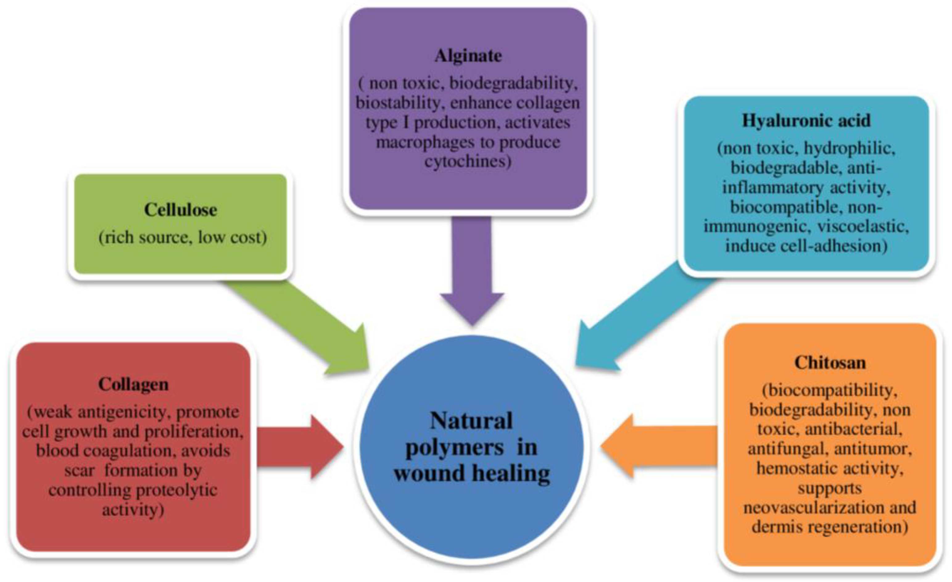
2.1. Natural Polymers

Natural polymers, also known as biopolymers, are organic compounds synthesized by living organisms. These molecules have a structural arrangement characterized by sequences of repeating units or monomers, typically amino acids or monosaccharides, which combine to form peptides and polysaccharides. Biopolymers originate from various sources, including plants, animals, fungi, bacteria, and algae. Their diverse origins make them applicable across numerous fields due to their distinct properties (Figure 5) [35]. The natural source of these polymers positions them as optimal substitutes for the extracellular matrix (ECM) and the original cellular environment of native skin. Natural polymers offer several advantages over synthetic materials, including high biocompatibility, biodegradability, reduced antigenicity, and renewability [36].
Figure 5. Some functional biomaterials identified as promising wound healing applications and their main characteristics.

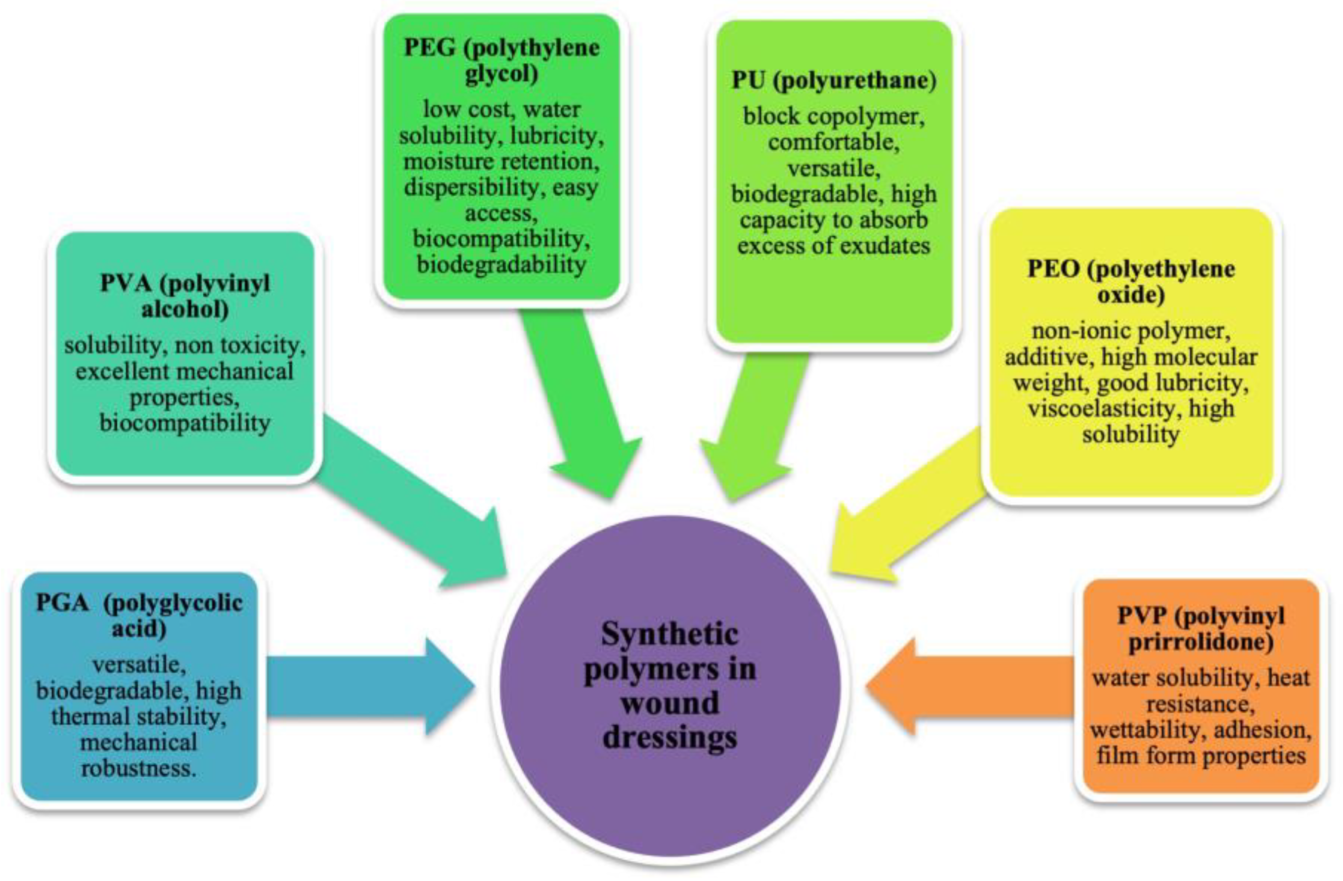
2.2. Synthetic Polymers

The class of synthetic polymers includes those polymers that are biocompatible, bioresorbable, and, being synthetic, have reproducible properties that can be adapted to a given application.
The difference between synthetic and natural polymers is that the former can be synthesized and modified in a controlled manner to achieve specific properties or stability. This type of polymers includes PVA, polyethylene oxide (PEO), PEG, poly(ε-caprolactone) (PCL), polyurethane (PU), poly lactic acid (PLA), poly vinyl pyrrolidone (PVP), and polyglycolic acid (PGA) (Figure 6) [37]. The physical, chemical, mechanical, and kinetic characteristics of these materials, unlike natural materials, can be adapted to the type of desired application. Some of the main disadvantages of synthetic polymers include the high cost to use and sometimes have a very different structure from the extracellular matrix [38]. It is possible to functionalize the material to give it the property of bioactivity by immobilizing biomolecules within the structure or on its surface, such as peptide sequences, adhesion proteins, or polysaccharides [39]. Alternatively, blends can be made by mixing synthetic materials with a natural polymer or with biomolecules to create a complex (Interpolymer complex) [40].
Figure 6. Schematic properties of the main synthetic polymers used in wound healing.

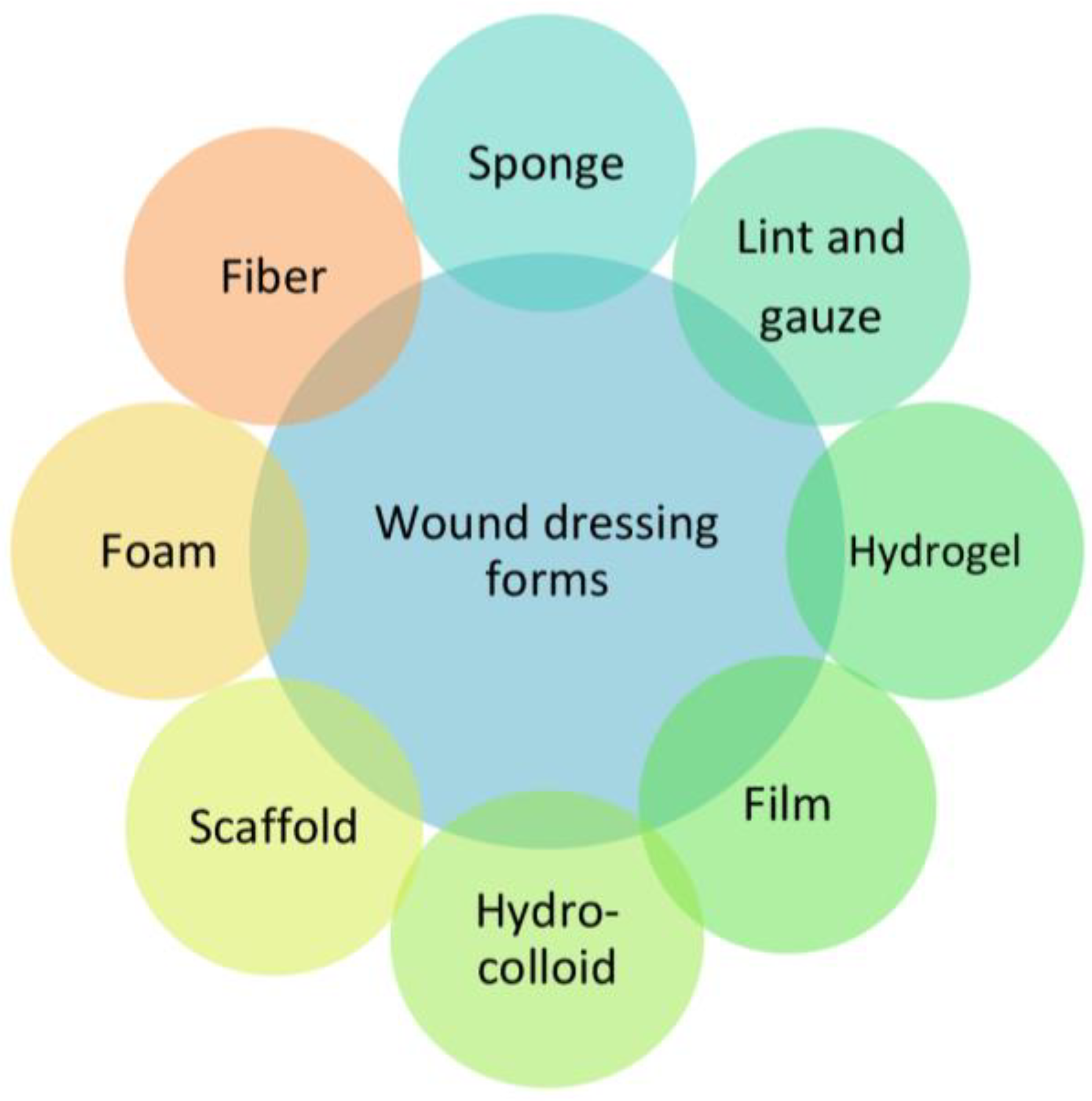
3. Classification of the Dressings by Physical Form

3.1. Bandages

Conventional approaches to wound treatment involve the utilization of bandages and gauze, serving as materials to absorb exudates and offer physical shielding. These dressings are inherently dry and lack the capacity to maintain a moist environment. As the fluid content decreases, they tend to adhere to the wound site, resulting in discomfort and pain when removal is attempted [41].

3.2. Lyophilized Wafers

Freeze-dried wafers are created by transforming polymeric solutions or gels into solid, porous structures. These wafers possess the unique ability to incorporate drugs within their matrix. They exhibit a remarkable capacity to absorb fluids, including excessive exudates, while also facilitating the gradual diffusion of the drug within the wound bed [42].

3.3. Hydrogels

Hydrogels are insoluble polymers in water, which creates a hydrophobic crosslinked network. They are highly absorbent and do not lose their network structure; they can donate water molecules and maintain a moist environment at the wound bed. Hydrogels transmit moisture vapor and oxygen. Moreover, they are non-toxic, biocompatible, and reversible for medical application. They promote wound debridement by rehydration of non-viable tissue, thus facilitating the process of natural autolysis. Hydrogels have been used as standard form in the management of necrotic wounds. They are not indicated for wounds with high levels of exudate or in the presence of gangrenous tissue, which should be kept dry to reduce the risk of infection [43]. Hydrogels also possess a degree of flexibility similar to tissue and offer the advantage of incorporating many bioactive agents in a specific space, thanks to their tridimensional structure, and releasing them in the environment, usually by a gel-sol transition to the liquid state [44][45].

3.4. Films

Film polymers may include co-polymers, homopolymers, and plasticized polymers consisting of a series of sheets, like sheets of PU coated with hypoallergenic acrylic adhesive. They should be impermeable to fluids and bacteria and permeable to air and water vapor. Through this mechanism, dressings are able to create a moist wound environment. In wound healing, they can be used as primary or secondary dressing or can be incorporated into hydrogels or foams to create composite dressing [46]. In the past, dressing films were usually made of nylon and supported in an adhesive polyethylene frame, but they were much occlusive. Modern dressing films are semi-permeable adhesive sheets, very flexible, and they are good for wounds but also over joints [47]. However, they are unable to cope with large amounts of exudate and may cause maceration of the skin surrounding the wound bed if they are used injudiciously. Films are used for superficial wounds and wounds with light exudates [45].

3.5. Patches

Wound patches have shown immense potential in offering properties such as ultra-adhesion, self-healing capabilities, biosensing functionality, antibacterial effects, and anti-inflammatory attributes. Recent times have witnessed a significant surge of interest in wearable patches, driven by their distinct advantages including flexibility, non-invasiveness, real-time monitoring, seamless integration, sensitivity, and robust stability. As the demand for personalized medical care continues to rise, wearable patches have emerged as a focal point due to their substantial potential in shaping the next phase of wound management [48]. These bio-sensorial patches rely on biochemical and physiological sensing mechanisms, which encompass parameters like pH variation, glucose levels, and temperature shifts. For instance, unhealed wounds typically exhibit an alkaline pH compared to the skin’s acidic pH. Additionally, the concentration of blood glucose in a wound serves as not only an indicator of a diabetic patient’s physical state but also influences the extent of bacterial infection within the wound.
The fabrication of these patches often involves 3D printing technology. The selection of an appropriate bioink is of paramount importance in the production of 3D-printed patches. This bioink must exhibit high biocompatibility, mechanical stability, and exceptional post-printing shape fidelity. As wearable patches continue to evolve, they hold significant promise for revolutionizing wound management and ushering in a new era of personalized healthcare [49].

3.6. Scaffolds

In the last few years, researchers have included biomaterials in the creation of 3D scaffolds. Deep wounds are unable to regenerate themselves, so the development of specific scaffolds is essential to promote the natural sequence of healing events by providing mechanical support for the growth of new tissue. A scaffold is a structural framework that provides support and cohesion to living tissue. It interacts with cells and initiates the natural physiological processes involved in the healing and regeneration of tissue. It supports the delivery and the retention of biochemical factors facilitating proper cell attachment and migration [50]. Biomaterial types and processing techniques are important for the properties of the resultant scaffolds. Moreover, a scaffold necessitates being either bioresorbable or pro-regenerative, thus displaying attributes such as notable porosity, a substantial surface area-to-volume ratio, a distinct geometry, and a pliable nature to adapt to the wound’s contour. These scaffolds can be fashioned from natural, synthetic, or hybrid polymers, and are amenable to functionalization with various agents aimed at augmenting cellular reactions and accelerating the wound healing trajectory. The manufacturing of these scaffolds can be achieved through techniques like electrospinning or 3D printing [51].

3.7. Hydrocolloids

Among advanced dressings, those based on hydrocolloids create a moist environment and absorb medium amounts of exudate. They are available in plaques and pastes and promote the growth of granulation tissue, favoring the healing process. In the presence of exudate, they absorb the malodorous liquid and produce a gel, which is why they are so used in the treatment of pressure sores. A hydrocolloid dressing consists of a thin dressing that contains gelling agents in an adhesive compound, laminated to a flexible, water-resistant outer layer. The sheets are self-adhering and available with or without an adhesive border, in different thicknesses and shapes pre-cut for various areas of the body such as the sacrum, elbows, and heels. Hydrocolloid dressings are occlusive, thus providing a moist healing environment, autolytic debridement, and isolation, without permeability to bacteria and other contaminants. They are easy to apply, self-adherent moldable dressings (do not adhere to the wound, only to the intact skin around the wound) that can be used under venous compression products. Essential to keep the decubitus wound clean, they can remain in place for 3 to 7 days (depending on the amount of wound exudate), greatly limiting the trauma produced by the operation of changing the dressing while avoiding disturbing the healing [52]. The ideal wounds for the use of hydrocolloid dressings are low-exudating and non-infected skin lesions. They can also be used as an alternative tool in wound prevention, to protect fragile skin or recently healed wounds with re-epithelialized skin. Among the best hydrocolloid dressings, there is DUODERM CGF; it retains exudate very well and it is used in the treatment of wounds with low exudate production [53].

3.8. Foam Dressings

Foam dressings are crafted from polymer solutions, creating structures with open cells capable of retaining fluids. These foams are commonly composed of materials like PU or silicone. PU foam dressings typically consist of two or three layers, featuring a hydrophilic surface in direct contact with the wound and a highly absorbent hydrophobic layer. These dressings offer multiple benefits such as thermal insulation for the wound bed, facilitating the exchange of water vapor and oxygen, effective dispersion of exudate within the absorbent layer, and preventing its escape to the external environment. In some instances, PU foam dressings are available in the form of cavity dressing-chips enclosed within a perforated polymeric film membrane.
On the other hand, silicone foams are produced using a silicone elastomer derived from the combination of two liquid components. When mixed, these components create a foam that conforms to the contours of the wound, resulting in a soft, open-cell foam dressing. These silicone foam dressings also contribute to efficient fluid management and wound protection [54]. In conclusion, the major advantages of foams are that they promote permeability to vapor and gases and create an impervious barrier to fluids and bacteria, protecting the wound. The main forms of dressing materials are shown in Table 1.
Table 1. Classification of wound dressings according to their physical form.

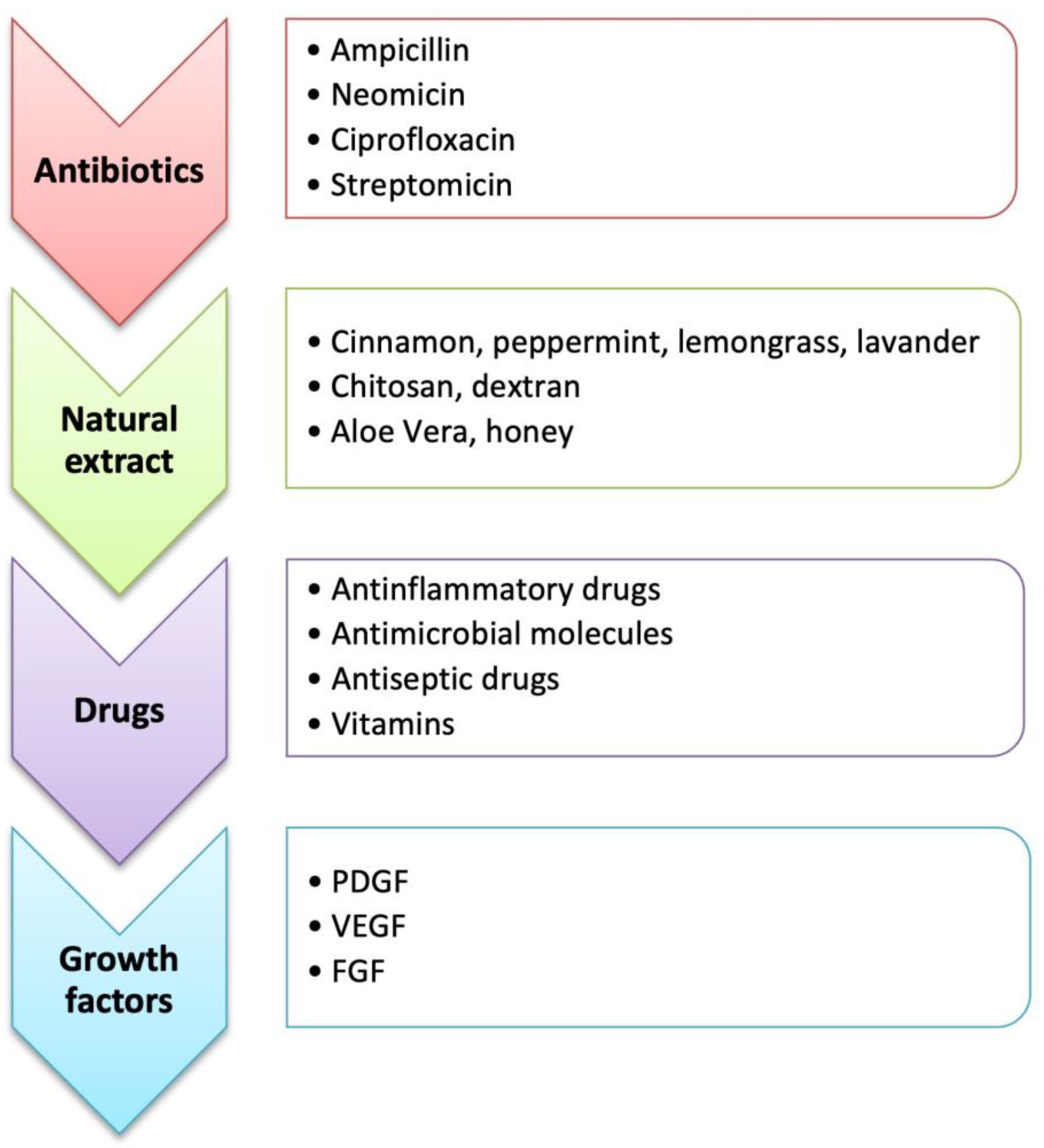
4. Bioactive Molecules

The incorporation of biomolecules or active pharmaceutical ingredients (APIs) in different types of wound dressings and structures prevents skin infections and promotes the healing process. The 3D wound dressing architecture, which mimics the ECM and has a porous structure, can be functionalized with bioactive molecules. Different bioactive molecules can be included in a polymeric matrix, highlighting the antibacterial biomolecules (e.g., antibiotics, silver NPs, and natural extracts-derivate products) and molecules capable of enhancing the healing process (e.g., growth factors, vitamins, and anti-inflammatory molecules) (Figure 7) [2][83].
Figure 7. Main bioactive molecules used in wound healing.

5. Methods of Preparation for Wound Dressings

Wound dressings are produced by different methods which depend on the desired structure and the materials used. The three methods commonly employed are solvent casting, electrospinning, and extrusion-type three-dimensional (3D) printing.

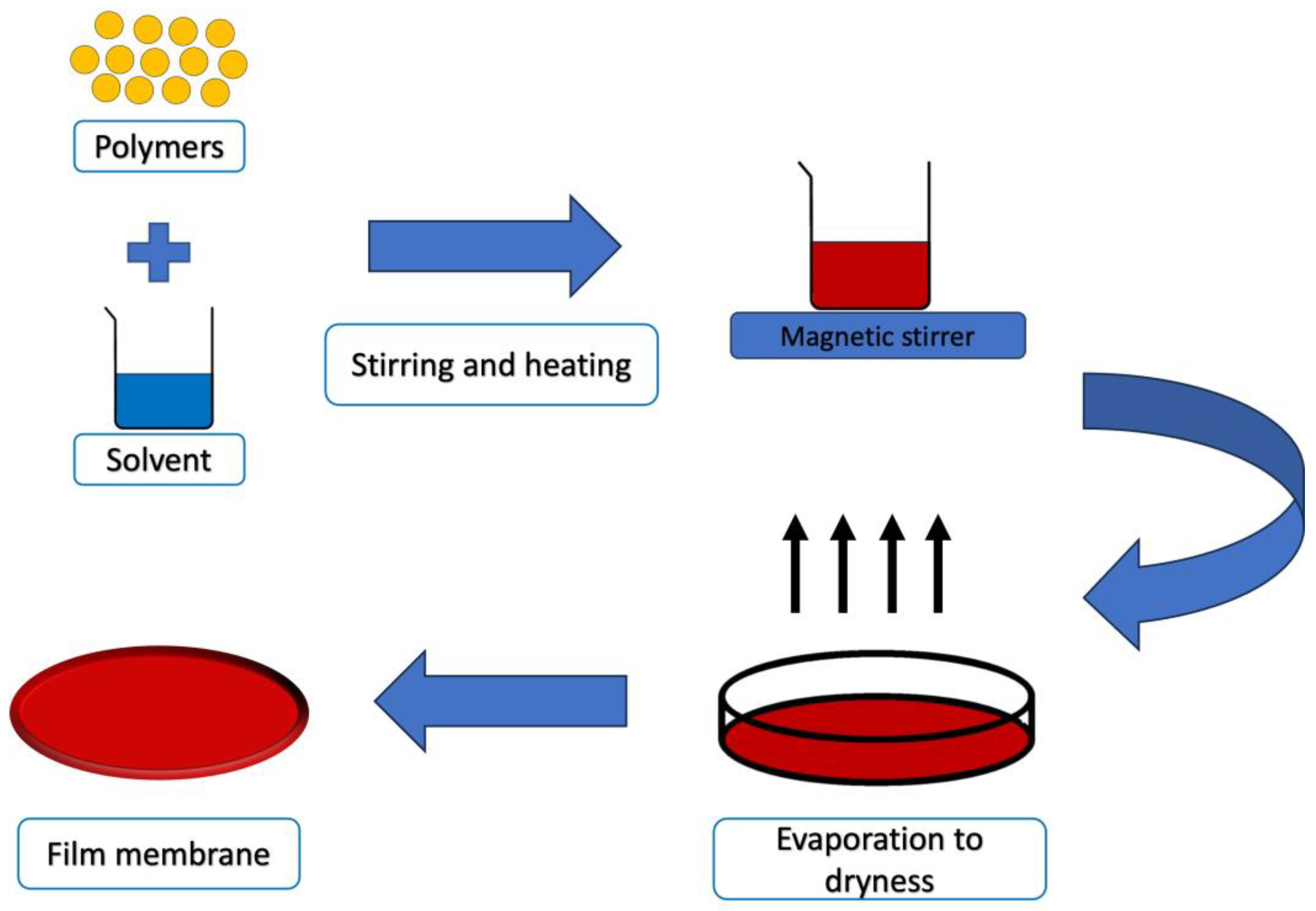
5.1. Solvent Casting Technique

The solvent casting technique involves creating polymeric films by drying viscous solutions containing polymers, additives, and active substances in a uniformly spread layer (Figure 8). While this method is straightforward, it can be time-intensive, and the properties and stability of the resulting films are primarily influenced by the chosen materials [68]. Typically, polymers are dissolved or dispersed in an organic solvent, which is then poured onto a supporting surface. As the solvent evaporates during drying, a solid layer forms on the substrate. Occasionally, particles of specific sizes, often salts, are incorporated into the solution. The dried mixture is then shaped into its final form. Alternatively, the composite material can be immersed in a bath to dissolve the particles, leaving behind a porous structure. These solvent-cast polymer films exhibit enhanced structural integrity and are well-suited as the foundational layer in multi-layered formulations [84].
Figure 8. Solvent casting technique.
In a recent study, a PVA/dextran hydrogel was developed using the solvent casting method in order to provide an efficient wound dressing. Zataria essential oil was loaded in the hydrogel as an antioxidant and antibacterial agent. PVA/dextran gels were crosslinked with GA and the mixture was poured in a petri dish and stored at 60 °C for 5 h. In vitro studies revealed the dressing’s ability to prevent wound inflammation and infection at the same time [85].

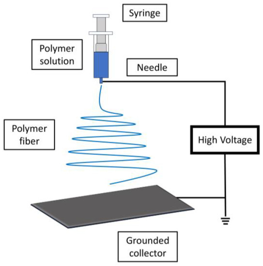
5.2. Electrospinning

Electrospinning is a versatile technology utilized to create polymeric fibers with excellent diffusion properties and a high surface area to volume ratio. These attributes make it valuable for wound care applications, aiding in controlling bleeding, absorbing excess wound fluid, and fostering tissue regeneration [86].
The electrospinning procedure employs a high Direct Current (DC) voltage, typically within the range of several kilovolts (10–20 kV). This voltage is applied to generate electrical charges within a stream of polymer solution, which then dries, resulting in the formation of polymer nano fibers (Figure 9).
Figure 9. Electrospinning system.
The stability, consistency, and production efficiency of the electrospinning process hinge upon the compatibility of various components: the chosen polymer(s), solvent(s), and any additional additives or active substances.
Indeed, the process of electrospinning involves several key steps to create polymer nanofibers for wound care applications: the chosen polymer is first dissolved in a suitable solvent to form a homogeneous solution. The properties of the polymer, solvent, and their compatibility play a significant role in achieving successful electrospinning. The polymer solution is then carefully introduced into the syringe tube that is part of the electrospinning setup. The solution will be dispensed through a small-diameter needle or spinneret. The positive terminal of a DC power supply is connected to the hollow needle or spinneret, while the negative terminal is connected to a metal collector, often in the form of a rotating drum or plate. The solution within the syringe is subjected to a high electric field generated by the voltage difference between the needle and the collector. The electric field causes charges to accumulate at the surface of the droplet of polymer solution at the tip of the needle. The electric field forces the charged droplet to overcome the surface tension and elongate into a conical shape known as a Taylor cone. A fine jet of polymer solution is emitted from the tip of the cone. As the jet travels toward the collector, the solvent evaporates, leaving behind a solid fiber. During its flight from the needle to the collector, the solvent in the jet evaporates due to the temperature and pressure conditions in the surrounding environment. This process leads to the solidification of the polymer into a continuous nanofiber. The nanofibers are collected on the metal collector, which may be a rotating drum or stationary plate. The fibers form a nonwoven mat on the collector’s surface. Stable environmental conditions, including temperature and humidity, are crucial to ensure the quality and uniformity of the nanofibers. Factors such as the distance between the needle and collector, solution vapor pressure, and chamber temperature are optimized to achieve the desired fiber properties.
Overall, electrospinning is a complex yet highly controlled process that enables the production of fine polymer nanofibers with tailored properties for various applications, including wound care. The resulting nanofibrous mats can provide enhanced capabilities for wound healing, such as improved absorption, moisture management, and cell interaction [87].
Habibi et al. reported the synthesis of a bi-layer chitosan/PVA nanofiber loaded with bupivacaine and mupirocin. Smooth and uniform nanofibrous mats were obtained using an electrospinning apparatus. Bupivacaine was used as a topical anesthetic to reduce the pain, while mupirocin exerted antibacterial activity. Cell viability studies highlighted the ability of the engineered wound dressing to mimic the ECM and histopathology studies confirmed its potential to accelerate the wound healing process [88].

5.3. Melt-Blowing

Melt blowing is a versatile technique used in the preparation of wound dressings. In this process, thermoplastic polymers are melted and then forced through fine nozzles to form microfibers. These microfibers are rapidly cooled by high-velocity air streams, causing them to solidify and create a nonwoven web of interconnected fibers. The resulting nonwoven material has a unique combination of porosity, surface area, and mechanical strength, making it suitable for wound dressing applications [89]. Melt blowing offers several advantages for wound dressing production. The technique allows for the incorporation of various materials, such as antimicrobial agents or growth factors, into the polymer melt, enhancing the functional properties of the dressings. The resulting nonwoven structure promotes moisture management and breathability, creating a favorable environment for wound healing [90]. Additionally, the scalability and efficiency of the melt blowing process make it suitable for large-scale production of wound dressings with consistent quality [91].
The versatility of melt-blown wound dressings makes them suitable for various wound types, from minor abrasions to more severe injuries. The ability to tailor the material properties and composition to specific wound requirements highlights melt blowing as a valuable method for preparing wound dressings that support optimal healing conditions [92].
Wang et al. developed microfiber-based nonwovens using a melt blowing technique, combining PLA and PEG with sodium dodecyl sulfate (SDS). The influence of melt blowing technology parameters, including die temperature and hot air pressure, was examined in relation to the structure and characteristics of the microfibers nonwovens. Elevating either the die temperature or the hot air pressure led to improved melt flow, facilitating thorough fiber stretching and gradually enhancing the fabric’s mechanical properties. Decreased fiber diameters yielded smoother surfaces and smaller pores, resulting in increased liquid pressure generation, thereby enhancing fabric wettability. The findings demonstrated the potential of utilizing PLA/PEG/SDS microfiber-based nonwovens as raw materials for wound dressing production, owing to their remarkable liquid absorption capacity. Furthermore, their inherent ability to undergo natural degradation post-use offers an environmentally friendly solution, reducing their environmental impact [93].

5.4. Thermal Annealing

Thermal annealing is a process used in wound dressing preparation to enhance the mechanical properties, stability, and functionality of the materials used. It involves subjecting a polymer film or material to controlled heating and cooling cycles to induce structural changes at the molecular level instead of using chemical and harmful cross-linkers. This process can lead to improvements in the wound dressing’s properties, making it more suitable for its intended application [94].
In a recent study, electrospun PVP-based fibers loaded with hydroxycinnamic acid derivatives (p-coumaric and ferulic acids) were synthesized. The fibrous mats were transformed into hydrogels via thermal annealing. The phenolic compounds in the fibers maintained their antioxidant activity even after annealing and the fibers demonstrated antioxidant effects in vitro and cell experiments, protecting against oxidative stress. The fiber mats showed suitable swelling properties, forming hydrogels when immersed in water. Moreover, in mice with UV-B-induced burns, the fibers reduced pro-inflammatory marker levels. These thermally treated PVP fibers loaded with phenolic acids showed potential as active skin dressing materials for healing skin injuries with oxidative stress [95].

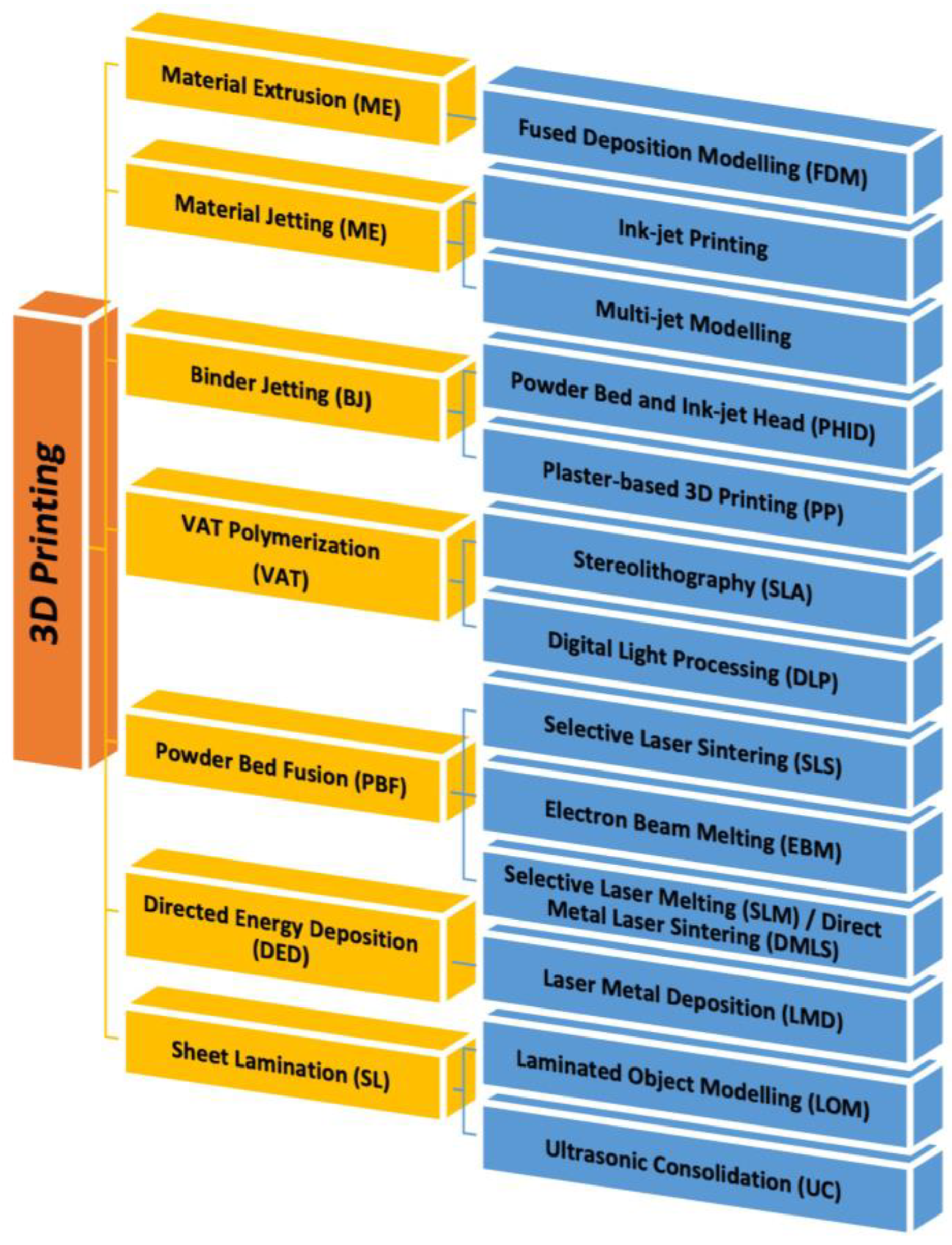
5.5. 3D Printing Technology

The technology of 3D printing creates layers of materials to form a three-dimensional structure. This technique allows you to print different materials, from plastic to metal, from resins to different polymers. The use of 3D printing for wound dressing made possible the generation of skin tissue and constructs similar to physiological skin. It has been used for on-demand therapies for the production of complex pharmaceutical forms. Today, 3D printing technology is the most interesting manufacturing technology thanks to the possibility of obtaining highly customizable products. For this characteristic, a 3D printer is able to produce a product with a bottom-up fabrication by a layer-by-layer method. The object to be printed is created using a computer-aided design (CAD) software package which is then exported as a file to be printed. The exported file splits the 3D object into a series of layers and then starts printing. The growth in the number of publications over the last years confirms the interest in 3D printing. It can be used in many areas, from engineering to the biomedical and pharmacological fields. In particular, in the development of pharmaceutical and biomedical products, 3D printing can be used for dressing up three-dimensional supports covered in or containing active ingredients to be delivered to the skin [96]. The emergence of 3D printing technology and its diverse applications in various fields offers a multitude of benefits, particularly in enhancing the quality of life for patients, promoting treatment adherence, and boosting the effectiveness of therapies, notably in cases of chronic wounds [97]. The intrinsic qualities can closely mimic the natural environment of the skin, thereby fostering an optimal milieu for the treatment of diseases and injuries [98].
Recent advancements have seen 3D printing employed for the production of scaffolds and patches tailored for transdermal applications [96]. Furthermore, as bioprinting techniques have continue to advance, enabling the printing of living cells, these systems have been harnessed for the realm of skin tissue engineering [99].
The basic components of a 3D system can be divided in three groups: hardware (which is the 3D printer itself), software (used to communicate with hardware and also allow conversation of CAD images into stereolithography images which are recognized by the printers), and materials used to print objects [100].
Based on the specific application and the desired material to be used, three main groups of 3D printing techniques can be distinguished: extrusion-based methods, powder-based methods, and photopolymerization methods. Each of these groups include different approaches with slight mechanical or chemical variations, such as Material Extrusion (ME), Binder Jetting (BJ), Powder Bed Fusion (PBF), Sheet Lamination (SL), Material Jetting [101], VAT polymerization, or Directed Energy Deposition (DED) (Figure 10) [102][103].
Figure 10. Classification of different 3D Printing processes.
ME is an additive manufacturing methodology where a spool of material (usually thermoplastic polymer) is pushed through a heated nozzle in a continuous stream and selectively deposited layer by layer to build a 3D object. In fact, it is also known as fused filament fabrication or fused deposition modeling [104]. BJ is the second most used 3D printing technology and is a process in which a binder is printed onto a powdered material, binding it together to develop a 3D printed structure [105]. PBF uses an electron beam, laser, or other heat source to selectively consolidate the powder in each layer into 3D objects. PBF technologies comprise Selective Laser Sintering (SLS), Selective Laser Melting (SLM)/Direct Metal Laser Sintering (DMLS), Electron Beam Melting (EBM), and Multi-Jet Fusion (MJF) [106]. SL is another process in additive manufacturing in which a laser is used to cut the material, joined to a substrate with a heated roller, to the desired shape [107]. MJ is a process in which tiny droplets are propelled from specialized drop-on-demand print heads. These print heads employ thermal, piezoelectric, or electromagnetic mechanisms to create pressure pulses. The resulting droplets are meticulously positioned on a substrate at designated spots. Following each layer’s deposition, UV light triggers polymerization, solidifying the material. This layer-by-layer approach gradually constructs a three-dimensional object, with successive layers fusing together to form the final structure [108]. Vat polymerization is a method used to print 3D objects by using photopolymerization. The process exposes liquid polymers to UV light to turn liquid into solids. Stereolithography [109] is the primary technique of vat polymerization, and it enables fast production with a precise architecture [110]. Finally, DED uses a focused energy source, such as a laser or an electron beam, to fuse the feedstock material and continuously deposit it, layer by layer [111].

3D Bioprinting

3D bioprinting technology is widely employed in regenerative medicine. It is used to generate different types of tissue, creating grafts directly on the patient’s skin in the shortest possible time and at reduced costs. Bioprinting creates customized treatments and minimizes the risk of organ rejection after transplantation. It is a type of additive manufacturing that integrates living cells into the printing procedure, often in conjunction with biomaterials. This field can be categorized into two key technologies: extrusion and inkjet bioprinting [112]. In extrusion bioprinting, a continuous flow of bioink—a viscous solution containing living cells and biomaterials—is precisely dispensed in strands. These strands are layered on top of one another to create intricate three-dimensional structures [113]. In inkjet bioprinting, droplets containing cells are deposited with or without the addition of biomaterials onto a receiving substrate [114]. Natural and synthetic polymers, metal, and ceramic are the most common materials used for this application [115].
In the last few years, 3D bioprinting has played a key role in tissue regeneration. It allows the development of biocompatible structures able to mimic the natural systems with good reproducibility in terms of size, shape, geometry, and orientation with high precision, while additionally providing the option to use a variety of bioink material and cell types (cells, growth factors, biomolecules, and biopolymers) in a controlled manner. A bioink should be biocompatible to enhance cell growth and proliferation and possess high chemical and physical fidelity to preserve its shape after deposition and post crosslinking [116]. Many natural polymers, such as collagen, chitosan, alginate, and cellulose, as well as several synthetic biopolymers have been used as bioinks to develop constructs with a specific design [117]. Finding application in wound treatment, 3D printing enables the synthesis of 3D devices able to enhance skin regeneration and the healing process. Recently, Hao et al. developed a gelatin–alginate-based hydrogel using 3D extrusion bioprinting, and hUC-MSCs (human umbilical cord mesenchymal stem cells) were laden in the sterilized bioink. The hydrogel showed good biocompatibility properties, ensuring cell functions in therapeutic applications. Moreover, a full-thickness skin defect repair experiment in mice was performed and the 3D printed cell-laden hydrogel significantly promoted wound healing by modulating inflammatory response and accelerating angiogenesis [118]. A double-crosslinked alginate/chondroitin sulfate patch was realized by the 3D printing approach, adding acrylated VEGF to the bioink. VEGF represents an alternative for skin wound treatment because of its ability to regulate angiogenesis during organism trauma repair. The hydrogel patches were first physical crosslinked using CaCl2 and then exposed to UV light for photocrosslinking. The VEGF-loaded patch prolonged the growth factor release, exhibiting excellent angiogenic ability and promoting wound healing [119].

References

  1. Shi, J.; Votruba, A.R.; Farokhzad, O.C.; Langer, R. Nanotechnology in drug delivery and tissue engineering: From discovery to applications. Nano Lett. 2010, 10, 3223–3230.
  2. Rambhia, K.J.; Ma, P.X. Controlled drug release for tissue engineering. J. Control. Release 2015, 219, 119–128.
  3. Zhong, S.; Zhang, Y.; Lim, C. Tissue scaffolds for skin wound healing and dermal reconstruction. Wiley Interdiscip. Rev. Nanomed. Nanobiotechnol. 2010, 2, 510–525.
  4. Kim, B.S.; Gao, G.; Kim, J.Y.; Cho, D.W. 3D cell printing of perfusable vascularized human skin equivalent composed of epidermis, dermis, and hypodermis for better structural recapitulation of native skin. Adv. Healthc. Mater. 2019, 8, 1801019.
  5. Joodaki, H.; Panzer, M.B. Skin mechanical properties and modeling: A review. Proc. Inst. Mech. Eng. Part H J. Eng. Med. 2018, 232, 323–343.
  6. Grover, S.; Ranyal, R.K.; Bedi, M.K. A cross section of skin diseases in rural Allahabad. Indian J. Dermatol. 2008, 53, 179.
  7. Mostafa, W.Z.; Hegazy, R.A. Vitamin D and the skin: Focus on a complex relationship: A review. J. Adv. Res. 2015, 6, 793–804.
  8. Tottoli, E.M.; Dorati, R.; Genta, I.; Chiesa, E.; Pisani, S.; Conti, B. Skin wound healing process and new emerging technologies for skin wound care and regeneration. Pharmaceutics 2020, 12, 735.
  9. Diegelmann, R.F.; Evans, M.C. Wound healing: An overview of acute, fibrotic and delayed healing. Front. Biosci. 2004, 9, 283–289.
  10. Raziyeva, K.; Kim, Y.; Zharkinbekov, Z.; Kassymbek, K.; Jimi, S.; Saparov, A. Immunology of acute and chronic wound healing. Biomolecules 2021, 11, 700.
  11. Gurtner, G.C.; Werner, S.; Barrandon, Y.; Longaker, M.T. Wound repair and regeneration. Nature 2008, 453, 314–321.
  12. Golebiewska, E.M.; Poole, A.W. Platelet secretion: From haemostasis to wound healing and beyond. Blood Rev. 2015, 29, 153–162.
  13. Eming, S.A.; Krieg, T.; Davidson, J.M. Inflammation in wound repair: Molecular and cellular mechanisms. J. Investig. Dermatol. 2007, 127, 514–525.
  14. Solarte David, V.A.; Güiza-Argüello, V.R.; Arango-Rodríguez, M.L.; Sossa, C.L.; Becerra-Bayona, S.M. Decellularized tissues for wound healing: Towards closing the gap between scaffold design and effective extracellular matrix remodeling. Front. Bioeng. Biotechnol. 2022, 10, 821852.
  15. Mehrabi, T.; Mesgar, A.S.; Mohammadi, Z. Bioactive glasses: A promising therapeutic ion release strategy for enhancing wound healing. ACS Biomater. Sci. Eng. 2020, 6, 5399–5430.
  16. Tabriz, A.G.; Douroumis, D. Recent advances in 3D printing for wound healing: A systematic review. J. Drug Deliv. Sci. Technol. 2022, 74, 103564.
  17. Stramer, B.M.; Mori, R.; Martin, P. The inflammation–fibrosis link? A Jekyll and Hyde role for blood cells during wound repair. J. Investig. Dermatol. 2007, 127, 1009–1017.
  18. Aisa, J.; Parlier, M. Local wound management: A review of modern techniques and products. Vet. Dermatol. 2022, 33, 463–478.
  19. Schultz, G.S.; Sibbald, R.G.; Falanga, V.; Ayello, E.A.; Dowsett, C.; Harding, K.; Romanelli, M.; Stacey, M.C.; Teot, L.; Vanscheidt, W. Wound bed preparation: A systematic approach to wound management. Wound Repair Regen. 2003, 11, S1–S28.
  20. Tavakoli, S.; Klar, A.S. Advanced hydrogels as wound dressings. Biomolecules 2020, 10, 1169.
  21. Boateng, J.S.; Matthews, K.H.; Stevens, H.N.; Eccleston, G.M. Wound healing dressings and drug delivery systems: A review. J. Pharm. Sci. 2008, 97, 2892–2923.
  22. Brumberg, V.; Astrelina, T.; Malivanova, T.; Samoilov, A. Modern wound dressings: Hydrogel dressings. Biomedicines 2021, 9, 1235.
  23. Weller, C.D.; Team, V.; Sussman, G. First-line interactive wound dressing update: A comprehensive review of the evidence. Front. Pharmacol. 2020, 11, 155.
  24. Andreu, V.; Mendoza, G.; Arruebo, M.; Irusta, S. Smart dressings based on nanostructured fibers containing natural origin antimicrobial, anti-inflammatory, and regenerative compounds. Materials 2015, 8, 5154–5193.
  25. Saghazadeh, S.; Rinoldi, C.; Schot, M.; Kashaf, S.S.; Sharifi, F.; Jalilian, E.; Nuutila, K.; Giatsidis, G.; Mostafalu, P.; Derakhshandeh, H. Drug delivery systems and materials for wound healing applications. Adv. Drug Deliv. Rev. 2018, 127, 138–166.
  26. Dong, R.; Guo, B. Smart wound dressings for wound healing. Nano Today 2021, 41, 101290.
  27. Wang, M.; Huang, X.; Zheng, H.; Tang, Y.; Zeng, K.; Shao, L.; Li, L. Nanomaterials applied in wound healing: Mechanisms, limitations and perspectives. J. Control. Release 2021, 337, 236–247.
  28. Shi, C.; Wang, C.; Liu, H.; Li, Q.; Li, R.; Zhang, Y.; Liu, Y.; Shao, Y.; Wang, J. Selection of appropriate wound dressing for various wounds. Front. Bioeng. Biotechnol. 2020, 8, 182.
  29. Heyer, K.; Augustin, M.; Protz, K.; Herberger, K.; Spehr, C.; Rustenbach, S. Effectiveness of advanced versus conventional wound dressings on healing of chronic wounds: Systematic review and meta-analysis. Dermatology 2013, 226, 172–184.
  30. Ongarora, B.G. Recent technological advances in the management of chronic wounds: A literature review. Health Sci. Rep. 2022, 5, e641.
  31. Dumville, J.C.; Gray, T.A.; Walter, C.J.; Sharp, C.A.; Page, T.; Macefield, R.; Blencowe, N.; Milne, T.K.; Reeves, B.C.; Blazeby, J. Dressings for the prevention of surgical site infection. Cochrane Database Syst. Rev. 2016, 12, CD003091.
  32. Enoch, S.; Grey, J.E.; Harding, K.G. Non-surgical and drug treatments. BMJ 2006, 332, 900–903.
  33. Agrawal, P.; Soni, S.; Mittal, G.; Bhatnagar, A. Role of polymeric biomaterials as wound healing agents. Int. J. Low. Extrem. Wounds 2014, 13, 180–190.
  34. Gardikiotis, I.; Cojocaru, F.-D.; Mihai, C.-T.; Balan, V.; Dodi, G. Borrowing the Features of Biopolymers for Emerging Wound Healing Dressings: A Review. Int. J. Mol. Sci. 2022, 23, 8778.
  35. Dattilo, M.; Patitucci, F.; Prete, S.; Parisi, O.I.; Puoci, F. Polysaccharide-Based Hydrogels and Their Application as Drug Delivery Systems in Cancer Treatment: A Review. J. Funct. Biomater. 2023, 14, 55.
  36. Moholkar, D.N.; Sadalage, P.S.; Peixoto, D.; Paiva-Santos, A.C.; Pawar, K.D. Recent advances in biopolymer-based formulations for wound healing applications. Eur. Polym. J. 2021, 160, 110784.
  37. Qiu, L.Y.; Bae, Y.H. Polymer architecture and drug delivery. Pharm. Res. 2006, 23, 1–30.
  38. Reddy, M.S.B.; Ponnamma, D.; Choudhary, R.; Sadasivuni, K.K. A comparative review of natural and synthetic biopolymer composite scaffolds. Polymers 2021, 13, 1105.
  39. Williams, D.F. Biocompatibility pathways and mechanisms for bioactive materials: The bioactivity zone. Bioact. Mater. 2022, 10, 306–322.
  40. Mndlovu, H.; du Toit, L.C.; Kumar, P.; Choonara, Y.E.; Marimuthu, T.; Kondiah, P.P.; Pillay, V. Bioplatform fabrication approaches affecting chitosan-based interpolymer complex properties and performance as wound dressings. Molecules 2020, 25, 222.
  41. Falabella, A.F. Debridement and wound bed preparation. Dermatol. Ther. 2006, 19, 317–325.
  42. Tabassum, A.; Furtado, S.C.; Bharath, S. Development of antimicrobial colloidal silver incorporated lyophilized biopolymer wafers for wound care. Wound Med. 2018, 21, 1–7.
  43. Kim, M.H.; Park, H.; Nam, H.C.; Park, S.R.; Jung, J.-Y.; Park, W.H. Injectable methylcellulose hydrogel containing silver oxide nanoparticles for burn wound healing. Carbohydr. Polym. 2018, 181, 579–586.
  44. Xie, Z.; Aphale, N.V.; Kadapure, T.D.; Wadajkar, A.S.; Orr, S.; Gyawali, D.; Qian, G.; Nguyen, K.T.; Yang, J. Design of antimicrobial peptides conjugated biodegradable citric acid derived hydrogels for wound healing. J. Biomed. Mater. Res. Part A 2015, 103, 3907–3918.
  45. Jones, V.; Grey, J.E.; Harding, K.G. Wound dressings. BMJ 2006, 332, 777–780.
  46. Arroyo, A.A.; Casanova, P.L.; Soriano, J.V.; Torra i Bou, J.E. Open-label clinical trial comparing the clinical and economic effectiveness of using a polyurethane film surgical dressing with gauze surgical dressings in the care of post-operative surgical wounds. Int. Wound J. 2015, 12, 285–292.
  47. Gopinath, D.; Ahmed, M.R.; Gomathi, K.; Chitra, K.; Sehgal, P.; Jayakumar, R. Dermal wound healing processes with curcumin incorporated collagen films. Biomaterials 2004, 25, 1911–1917.
  48. Pillai, M.M.; Dandia, H.; Checker, R.; Rokade, S.; Sharma, D.; Tayalia, P. Novel combination of bioactive agents in bilayered dermal patches provides superior wound healing. Nanomed. Nanotechnol. Biol. Med. 2022, 40, 102495.
  49. Smandri, A.; Nordin, A.; Hwei, N.M.; Chin, K.-Y.; Abd Aziz, I.; Fauzi, M.B. Natural 3D-printed bioinks for skin regeneration and wound healing: A systematic review. Polymers 2020, 12, 1782.
  50. Hutmacher, D.W. Scaffolds in tissue engineering bone and cartilage. Biomaterials 2000, 21, 2529–2543.
  51. Lam, C.X.F.; Mo, X.; Teoh, S.-H.; Hutmacher, D. Scaffold development using 3D printing with a starch-based polymer. Mater. Sci. Eng. C 2002, 20, 49–56.
  52. Takeuchi, T.; Ito, M.; Yamaguchi, S.; Watanabe, S.; Honda, M.; Imahashi, T.; Yamada, T.; Kokubo, T. Hydrocolloid dressing improves wound healing by increasing M2 macrophage polarization in mice with diabetes. Nagoya J. Med. Sci. 2020, 82, 487.
  53. Pott, F.S.; Meier, M.J.; Stocco, J.G.D.; Crozeta, K.; Ribas, J.D. The effectiveness of hydrocolloid dressings versus other dressings in the healing of pressure ulcers in adults and older adults: A systematic review and meta-analysis. Rev. Lat.-Am. Enferm. 2014, 22, 511–520.
  54. Priddy-Arrington, T.R.; Ward, M.S.; Edwards, R.E.; Caldorera-Moore, M.E. Proactive biomaterials for chronic wound management and treatment. Curr. Opin. Biomed. Eng. 2021, 20, 100327.
  55. Parkale, R.; Pulugu, P.; Kumar, P. Nanomaterials decoration on commercial cotton bandages for pain and infection management. arXiv 2021, arXiv:2105.10273.
  56. Mohamad, N.; Loh, E.Y.X.; Fauzi, M.B.; Ng, M.H.; Mohd Amin, M.C.I. In vivo evaluation of bacterial cellulose/acrylic acid wound dressing hydrogel containing keratinocytes and fibroblasts for burn wounds. Drug Deliv. Transl. Res. 2019, 9, 444–452.
  57. Jeong, J.-O.; Park, J.-S.; Kim, E.J.; Jeong, S.-I.; Lee, J.Y.; Lim, Y.-M. Preparation of radiation cross-linked poly (Acrylic acid) hydrogel containing metronidazole with enhanced antibacterial activity. Int. J. Mol. Sci. 2019, 21, 187.
  58. Shin, J.Y.; Lee, D.Y.; Kim, B.Y.; Yoon, J.I. Effect of polyethylene glycol molecular weight on cell growth behavior of polyvinyl alcohol/carboxymethyl cellulose/polyethylene glycol hydrogel. J. Appl. Polym. Sci. 2020, 137, 49568.
  59. Murakami, K.; Aoki, H.; Nakamura, S.; Nakamura, S.-I.; Takikawa, M.; Hanzawa, M.; Kishimoto, S.; Hattori, H.; Tanaka, Y.; Kiyosawa, T. Hydrogel blends of chitin/chitosan, fucoidan and alginate as healing-impaired wound dressings. Biomaterials 2010, 31, 83–90.
  60. Hemmingsen, L.M.; Julin, K.; Ahsan, L.; Basnet, P.; Johannessen, M.; Škalko-Basnet, N. Chitosomes-in-chitosan hydrogel for acute skin injuries: Prevention and infection control. Mar. Drugs 2021, 19, 269.
  61. Xie, Y.; Gao, P.; He, F.; Zhang, C. Application of alginate-based hydrogels in hemostasis. Gels 2022, 8, 109.
  62. Da Silva, L.; Cerqueira, M.; Correlo, V.; Reis, R.; Marques, A. Engineered hydrogel-based matrices for skin wound healing. In Wound Healing Biomaterials; Elsevier: Amsterdam, The Netherlands, 2016; pp. 227–250.
  63. Makvandi, P.; Ali, G.W.; Della Sala, F.; Abdel-Fattah, W.I.; Borzacchiello, A. Biosynthesis and characterization of antibacterial thermosensitive hydrogels based on corn silk extract, hyaluronic acid and nanosilver for potential wound healing. Carbohydr. Polym. 2019, 223, 115023.
  64. Kenawy, E.-R.S.; Kamoun, E.A.; Ghaly, Z.S.; Shokr, A.-b.M.; El-Meligy, M.A.; Mahmoud, Y.A.-G. Novel physically cross-linked curcumin-loaded PVA/Aloe vera hydrogel membranes for acceleration of topical wound healing: In vitro and in vivo experiments. Arab. J. Sci. Eng. 2023, 48, 497–514.
  65. Khan, M.I.; Paul, P.; Behera, S.K.; Jena, B.; Tripathy, S.K.; Lundborg, C.S.; Mishra, A. To decipher the antibacterial mechanism and promotion of wound healing activity by hydrogels embedded with biogenic Ag@ ZnO core-shell nanocomposites. Chem. Eng. J. 2021, 417, 128025.
  66. Capanema, N.S.; Mansur, A.A.; de Jesus, A.C.; Carvalho, S.M.; de Oliveira, L.C.; Mansur, H.S. Superabsorbent crosslinked carboxymethyl cellulose-PEG hydrogels for potential wound dressing applications. Int. J. Biol. Macromol. 2018, 106, 1218–1234.
  67. Hosseini, M.; Razavi, S.; Mousavi, M. Antimicrobial, physical and mechanical properties of chitosan-based films incorporated with thyme, clove and cinnamon essential oils. J. Food Process. Preserv. 2009, 33, 727–743.
  68. Buanz, A.B.; Belaunde, C.C.; Soutari, N.; Tuleu, C.; Gul, M.O.; Gaisford, S. Ink-jet printing versus solvent casting to prepare oral films: Effect on mechanical properties and physical stability. Int. J. Pharm. 2015, 494, 611–618.
  69. Panayi, A.C.; Haug, V.; Liu, Q.; Wu, M.; Karvar, M.; Aoki, S.; Ma, C.; Hamaguchi, R.; Endo, Y.; Orgill, D.P. Novel application of autologous micrografts in a collagen-glycosaminoglycan scaffold for diabetic wound healing. Biomed. Mater. 2021, 16, 035032.
  70. Diaz-Gomez, L.; Gonzalez-Prada, I.; Millan, R.; Da Silva-Candal, A.; Bugallo-Casal, A.; Campos, F.; Concheiro, A.; Alvarez-Lorenzo, C. 3D printed carboxymethyl cellulose scaffolds for autologous growth factors delivery in wound healing. Carbohydr. Polym. 2022, 278, 118924.
  71. Ahmed, S.; Ikram, S. Chitosan based scaffolds and their applications in wound healing. Achiev. Life Sci. 2016, 10, 27–37.
  72. Mobaraki, M.; Bizari, D.; Soltani, M.; Khshmohabat, H.; Raahemifar, K.; Akbarzade Amirdehi, M. The effects of curcumin nanoparticles incorporated into collagen-alginate scaffold on wound healing of skin tissue in trauma patients. Polymers 2021, 13, 4291.
  73. Ji, W.; Sun, Y.; Yang, F.; van den Beucken, J.J.; Fan, M.; Chen, Z.; Jansen, J.A. Bioactive electrospun scaffolds delivering growth factors and genes for tissue engineering applications. Pharm. Res. 2011, 28, 1259–1272.
  74. Song, E.-H.; Jeong, S.-H.; Park, J.-U.; Kim, S.; Kim, H.-E.; Song, J. Polyurethane-silica hybrid foams from a one-step foaming reaction, coupled with a sol-gel process, for enhanced wound healing. Mater. Sci. Eng. C 2017, 79, 866–874.
  75. Li, S.; Zhang, Y.; Ma, X.; Qiu, S.; Chen, J.; Lu, G.; Jia, Z.; Zhu, J.; Yang, Q.; Chen, J. Antimicrobial lignin-based Polyurethane/Ag composite foams for improving wound healing. Biomacromolecules 2022, 23, 1622–1632.
  76. Jinno, C.; Morimoto, N.; Ito, R.; Sakamoto, M.; Ogino, S.; Taira, T.; Suzuki, S. A comparison of conventional collagen sponge and collagen-gelatin sponge in wound healing. BioMed Res. Int. 2016, 2016, 4567146.
  77. Lin, W.-C.; Lien, C.-C.; Yeh, H.-J.; Yu, C.-M.; Hsu, S.-H. Bacterial cellulose and bacterial cellulose–chitosan membranes for wound dressing applications. Carbohydr. Polym. 2013, 94, 603–611.
  78. Silva, S.S.; Caridade, S.; Mano, J.; Reis, R. Effect of crosslinking in chitosan/aloe vera-based membranes for biomedical applications. Carbohydr. Polym. 2013, 98, 581–588.
  79. Pang, M.; Huang, Y.; Meng, F.; Zhuang, Y.; Liu, H.; Du, M.; Ma, Q.; Wang, Q.; Chen, Z.; Chen, L. Application of bacterial cellulose in skin and bone tissue engineering. Eur. Polym. J. 2020, 122, 109365.
  80. Miao, J.; Pangule, R.C.; Paskaleva, E.E.; Hwang, E.E.; Kane, R.S.; Linhardt, R.J.; Dordick, J.S. Lysostaphin-functionalized cellulose fibers with antistaphylococcal activity for wound healing applications. Biomaterials 2011, 32, 9557–9567.
  81. Moydeen, A.M.; Padusha, M.S.A.; Thamer, B.M.; Ahamed, N.A.; Al-Enizi, A.M.; El-Hamshary, H.; El-Newehy, M.H. Single-nozzle core-shell electrospun nanofibers of PVP/dextran as drug delivery system. Fibers Polym. 2019, 20, 2078–2089.
  82. Merrell, J.G.; McLaughlin, S.W.; Tie, L.; Laurencin, C.T.; Chen, A.F.; Nair, L.S. Curcumin loaded poly (ε-caprolactone) nanofibers: Diabetic wound dressing with antioxidant and anti-inflammatory properties. Clin. Exp. Pharmacol. Physiol. 2009, 36, 1149.
  83. Dwivedi, C.; Pandey, H.; Pandey, A.C.; Patil, S.; Ramteke, P.W.; Laux, P.; Luch, A.; Singh, A.V. In vivo biocompatibility of electrospun biodegradable dual carrier (antibiotic+ growth factor) in a mouse model—Implications for rapid wound healing. Pharmaceutics 2019, 11, 180.
  84. Palo, M.; Rönkönharju, S.; Tiirik, K.; Viidik, L.; Sandler, N.; Kogermann, K. Bi-layered polymer carriers with surface modification by electrospinning for potential wound care applications. Pharmaceutics 2019, 11, 678.
  85. Mostafavi Esfahani, M.; Koupaei, N.; Hassanzadeh-Tabrizi, S.A. Synthesis and characterization of polyvinyl alcohol/dextran/Zataria wound dressing with superior antibacterial and antioxidant properties. J. Vinyl Addit. Technol. 2023, 29, 380–394.
  86. Rodríguez-Tobías, H.; Morales, G.; Grande, D. Comprehensive review on electrospinning techniques as versatile approaches toward antimicrobial biopolymeric composite fibers. Mater. Sci. Eng. C 2019, 101, 306–322.
  87. Ghosal, K.; Chandra, A.; Roy, S.; Agatemor, C.; Thomas, S.; Provaznik, I. Electrospinning over solvent casting: Tuning of mechanical properties of membranes. Sci. Rep. 2018, 8, 5058.
  88. Habibi, S.; Mohammadi, T.; HMTShirazi, R.; Atyabi, F.; Kiani, M.; Asadi, A.A. A bilayer mupirocin/bupivacaine-loaded wound dressing based on chitosan/poly (vinyl alcohol) nanofibrous mat: Preparation, characterization, and controlled drug release. Int. J. Biol. Macromol. 2023, 240, 124399.
  89. Ellison, C.J.; Phatak, A.; Giles, D.W.; Macosko, C.W.; Bates, F.S. Melt blown nanofibers: Fiber diameter distributions and onset of fiber breakup. Polymer 2007, 48, 3306–3316.
  90. Hiremath, N.; Bhat, G. Melt blown polymeric nanofibers for medical applications-an overview. Nanosci. Technol. 2015, 2, 1–9.
  91. Gao, Y.; Zhang, J.; Su, Y.; Wang, H.; Wang, X.-X.; Huang, L.-P.; Yu, M.; Ramakrishna, S.; Long, Y.-Z. Recent progress and challenges in solution blow spinning. Mater. Horiz. 2021, 8, 426–446.
  92. Yu, B.; Han, J.; Sun, H.; Zhu, F.; Zhang, Q.; Kong, J. The Preparation and property of poly (lactic acid)/tourmaline blends and melt-blown nonwoven. Polym. Compos. 2015, 36, 264–271.
  93. Wang, R.; Zhang, H.; Cao, Y.; Zhai, Q.; Hu, J.; Zhen, Q.; Qian, X. Preparation of PLA/PEG@ SDS microfibers-based nonwovens via melt-blown process parameters: Wound dressings with enhanced water wetting performance. J. Appl. Polym. Sci. 2023, 140, e54234.
  94. Hajinasrollah, K.; Habibi, S.; Nazockdast, H. Fabrication of gelatin–chitosan–gum tragacanth with thermal annealing cross-linking strategy. J. Eng. Fibers Fabr. 2019, 14, 18033.
  95. Contardi, M.; Kossyvaki, D.; Picone, P.; Summa, M.; Guo, X.; Heredia-Guerrero, J.A.; Giacomazza, D.; Carzino, R.; Goldoni, L.; Scoponi, G. Electrospun polyvinylpyrrolidone (PVP) hydrogels containing hydroxycinnamic acid derivatives as potential wound dressings. Chem. Eng. J. 2021, 409, 128144.
  96. de Oliveira, R.S.; Fantaus, S.S.; Guillot, A.J.; Melero, A.; Beck, R.C.R. 3D-printed products for topical skin applications: From personalized dressings to drug delivery. Pharmaceutics 2021, 13, 1946.
  97. Maver, T.; Smrke, D.; Kurečič, M.; Gradišnik, L.; Maver, U.; Kleinschek, K.S. Combining 3D printing and electrospinning for preparation of pain-relieving wound-dressing materials. J. Sol-Gel Sci. Technol. 2018, 88, 33–48.
  98. Chen, G.; Xu, Y.; Kwok, P.C.L.; Kang, L. Pharmaceutical applications of 3D printing. Addit. Manuf. 2020, 34, 101209.
  99. Rajesh, N.U.; Coates, I.; Driskill, M.M.; Dulay, M.T.; Hsiao, K.; Ilyin, D.; Jacobson, G.B.; Kwak, J.W.; Lawrence, M.; Perry, J. 3D-Printed Microarray Patches for Transdermal Applications. JACS Au 2022, 2, 2426–2445.
  100. Yeong, W.Y.; Chua, C.K.; Leong, K.F.; Chandrasekaran, M.; Lee, M.W. Indirect fabrication of collagen scaffold based on inkjet printing technique. Rapid Prototyp. J. 2006, 12, 229–237.
  101. Awasthi, A.; Gulati, M.; Kumar, B.; Kaur, J.; Vishwas, S.; Khursheed, R.; Porwal, O.; Alam, A.; Kr, A.; Corrie, L. Recent progress in development of dressings used for diabetic wounds with special emphasis on scaffolds. BioMed Res. Int. 2022, 2022, 1659338.
  102. Jandyal, A.; Chaturvedi, I.; Wazir, I.; Raina, A.; Haq, M.I.U. 3D printing–A review of processes, materials and applications in industry 4.0. Sustain. Oper. Comput. 2022, 3, 33–42.
  103. Palmara, G.; Frascella, F.; Roppolo, I.; Chiappone, A.; Chiadò, A. Functional 3D printing: Approaches and bioapplications. Biosens. Bioelectron. 2021, 175, 112849.
  104. Penumakala, P.K.; Santo, J.; Thomas, A. A critical review on the fused deposition modeling of thermoplastic polymer composites. Compos. Part B Eng. 2020, 201, 108336.
  105. Sen, K.; Mehta, T.; Sansare, S.; Sharifi, L.; Ma, A.W.; Chaudhuri, B. Pharmaceutical applications of powder-based binder jet 3D printing process—A review. Adv. Drug Deliv. Rev. 2021, 177, 113943.
  106. Awad, A.; Fina, F.; Goyanes, A.; Gaisford, S.; Basit, A.W. Advances in powder bed fusion 3D printing in drug delivery and healthcare. Adv. Drug Deliv. Rev. 2021, 174, 406–424.
  107. Jadhav, A.; Jadhav, V.S. A review on 3D printing: An additive manufacturing technology. Mater. Today Proc. 2022, 62, 2094–2099.
  108. Tyagi, S.; Yadav, A.; Deshmukh, S. Review on mechanical characterization of 3D printed parts created using material jetting process. Mater. Today Proc. 2022, 51, 1012–1016.
  109. Rahman, M.A.; Islam, M.S.; Haque, P.; Khan, M.N.; Takafuji, M.; Begum, M.; Chowdhury, G.W.; Khan, M.; Rahman, M.M. Calcium ion mediated rapid wound healing by nano-ZnO doped calcium phosphate-chitosan-alginate biocomposites. Materialia 2020, 13, 100839.
  110. Pagac, M.; Hajnys, J.; Ma, Q.-P.; Jancar, L.; Jansa, J.; Stefek, P.; Mesicek, J. A review of vat photopolymerization technology: Materials, applications, challenges, and future trends of 3D printing. Polymers 2021, 13, 598.
  111. Svetlizky, D.; Das, M.; Zheng, B.; Vyatskikh, A.L.; Bose, S.; Bandyopadhyay, A.; Schoenung, J.M.; Lavernia, E.J.; Eliaz, N. Directed energy deposition (DED) additive manufacturing: Physical characteristics, defects, challenges and applications. Mater. Today 2021, 49, 271–295.
  112. Cubo-Mateo, N.; Gelinsky, M. Wound and skin healing in space: The 3D bioprinting perspective. Front. Bioeng. Biotechnol. 2021, 9, 720217.
  113. Ramesh, S.; Harrysson, O.L.; Rao, P.K.; Tamayol, A.; Cormier, D.R.; Zhang, Y.; Rivero, I.V. Extrusion bioprinting: Recent progress, challenges, and future opportunities. Bioprinting 2021, 21, e00116.
  114. Gudapati, H.; Dey, M.; Ozbolat, I. A comprehensive review on droplet-based bioprinting: Past, present and future. Biomaterials 2016, 102, 20–42.
  115. Karvinen, J.; Kellomäki, M. Design aspects and characterization of hydrogel-based bioinks for extrusion-based bioprinting. Bioprinting 2023, 32, e00274.
  116. Temirel, M.; Hawxhurst, C.; Tasoglu, S. Shape fidelity of 3D-bioprinted biodegradable patches. Micromachines 2021, 12, 195.
  117. Masri, S.; Fauzi, M.B. Current insight of printability quality improvement strategies in natural-based bioinks for skin regeneration and wound healing. Polymers 2021, 13, 1011.
  118. Hao, L.; Zhao, S.; Hao, S.; He, Y.; Feng, M.; Zhou, K.; He, Y.; Yang, J.; Mao, H.; Gu, Z. Functionalized gelatin-alginate based bioink with enhanced manufacturability and biomimicry for accelerating wound healing. Int. J. Biol. Macromol. 2023, 240, 124364.
  119. Liao, W.; Duan, X.; Xie, F.; Zheng, D.; Yang, P.; Wang, X.; Hu, Z. 3D-bioprinted double-crosslinked angiogenic alginate/chondroitin sulfate patch for diabetic wound healing. Int. J. Biol. Macromol. 2023, 236, 123952.
More
Information
Contributors MDPI registered users' name will be linked to their SciProfiles pages. To register with us, please refer to https://encyclopedia.pub/register : , , , , ,
View Times: 259
Revisions: 2 times (View History)
Update Date: 07 Sep 2023
1000/1000
Video Production Service