Submitted Successfully!
To reward your contribution, here is a gift for you: A free trial for our video production service.
Thank you for your contribution! You can also upload a video entry or images related to this topic.
Version Summary Created by Modification Content Size Created at Operation
1 -- 1732 2023-09-06 08:06:48 |
2 format correct Meta information modification 1732 2023-09-06 08:27:08 |

Video Upload Options

Do you have a full video?

Confirm

Are you sure to Delete?
Cite
If you have any further questions, please contact Encyclopedia Editorial Office.
Maier, J.A.; Andrés, V.; Castiglioni, S.; Giudici, A.; Lau, E.S.; Nemcsik, J.; Seta, F.; Zaninotto, P.; Catalano, M.; Hamburg, N.M. Vascular Aging. Encyclopedia. Available online: https://encyclopedia.pub/entry/48864 (accessed on 10 May 2024).
Maier JA, Andrés V, Castiglioni S, Giudici A, Lau ES, Nemcsik J, et al. Vascular Aging. Encyclopedia. Available at: https://encyclopedia.pub/entry/48864. Accessed May 10, 2024.
Maier, Jeanette A., Vicente Andrés, Sara Castiglioni, Alessandro Giudici, Emily S. Lau, János Nemcsik, Francesca Seta, Paola Zaninotto, Mariella Catalano, Naomi M. Hamburg. "Vascular Aging" Encyclopedia, https://encyclopedia.pub/entry/48864 (accessed May 10, 2024).
Maier, J.A., Andrés, V., Castiglioni, S., Giudici, A., Lau, E.S., Nemcsik, J., Seta, F., Zaninotto, P., Catalano, M., & Hamburg, N.M. (2023, September 06). Vascular Aging. In Encyclopedia. https://encyclopedia.pub/entry/48864
Maier, Jeanette A., et al. "Vascular Aging." Encyclopedia. Web. 06 September, 2023.
Vascular Aging
Edit

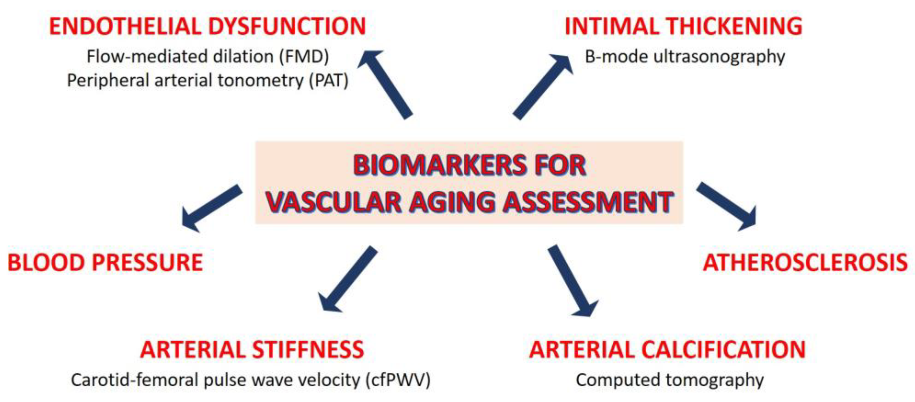
Vascular aging, i.e., the deterioration of the structure and function of the arteries over the life course, predicts cardiovascular events and mortality. Vascular degeneration can be recognized before becoming clinically symptomatic; therefore, its assessment allows the early identification of individuals at risk. This opens the possibility of minimizing disease progression. Vascular remodeling with advancing age is characterized by arterial stiffening and calcification. Arterial stiffening affects the macro- and micro-vasculature in unique ways. The main function of large arteries is to dampen the pulsatility of cardiac contraction by elastic recoiling after systolic expansion, so that constant, rather than pulsatile, blood flow is propelled to organs and tissues, including the coronary arteries.

aging arteries cardiovascular diseases

1. Biological vs. Chronological Aging: Lessons from Progeria

The extent of cardiovascular functional decline is highly variable among individuals of the same chronological age. Therefore, the identification of individuals for early prevention of CVD and other age-related disorders must rely on biomarkers of biological aging, also called physiological or functional aging, rather than chronologic age [1]. Because of the significant contribution of atherosclerosis and heart failure to CVD-related morbimortality in the elderly, substantial research efforts have been placed in understanding the genetic, molecular, and cellular mechanisms underlying CVD initiation, progression, and complications. Some hints derive from studies on Hutchinson–Gilford progeria syndrome (HGPS, OMIM 176670), an ultrarare (prevalence 1 in 18–20 million) genetic and fatal pediatric disorder characterized by segmental severe premature aging and early death and for which no cure exists [2]. The disease is caused by a de novo heterozygous dominant mutation in the LMNA gene (encoding nuclear A-type lamin) which causes the production of a truncated version of prelamin A called progerin. The accumulation of progerin causes multiple cellular alterations, including abnormal nuclear morphology, heterochromatin loss, mislocalization and loss of DNA damage repair proteins and chromatin-associated proteins, and mitochondrial and telomere dysfunction [3]. This results in accelerated VSMC senescence and loss, paralleled by reduced contractility, excessive deposition of extracellular matrix, and medial calcification [2][4]. In a murine model of progeria, vascular calcification is triggered by reduced extracellular deposition of pyrophosphate, a well-known inhibitor of vascular calcification [5]. Although patients with progeria lack or are only mildly affected by traditional cardiovascular risk factors, they develop CVD and die mainly from complications of atherosclerosis (myocardial infarction, heart failure, or stroke) at a mean age of 14.5 years [2]. HGPS research may therefore help identify mechanisms underlying CVD independently of risk factors or aging-associated chronic diseases that can influence cardiovascular health. Notably, progerin is expressed at low level in aged tissues of non-HGPS individuals, suggesting a role in normal aging [2]. Understanding how progerin causes CVD and premature aging may therefore shed some light on normal aging.

2. Sex Differences in Cardiovascular Aging

Sex differences in CVD are well established, and the mechanistic insights involved have been unraveled only recently [6]. While men and women have similar lifetime risks of developing CVD, women develop CVD later in life than men. Moreover, manifestations of CVD differ between men and women. For example, women with heart failure (HF) are more likely to have preserved left ventricular (LV) ejection fraction (EF) and non-ischemic etiology, while men often present with HF with reduced EF. Differences in cardiovascular aging between men and women may contribute to sex-based differences in CVD [7]. Early in life, men experience greater age-related vascular structural changes (intimal thickening, wall stiffening, calcium deposition, and atherosclerosis) and functional changes (endothelial dysfunction). However, after the sixth decade of life, age-related vascular dysfunction typically progresses at a faster rate in women than men. This observation has been consistently demonstrated in epidemiologic studies examining subclinical changes in vascular structure. The Baltimore Longitudinal Study of Aging measured carotid intima–media thickness (IMT)—a marker of early arterial wall alteration—over a 20-year period in 1067 men and women. The results showed that while men had higher baseline IMT than women, differences between men and women narrowed over time due to more pronounced acceleration of IMT in women later in life [8]. A similar pattern has been observed for other subclinical vascular measures, including coronary artery calcium scores [8].
Age-related decline in vascular function follows a similar trajectory to changes in vascular structure. Endothelial dysfunction decreases with age in men but is preserved in women until the onset of menopause, after which endothelial-dependent vasodilation markedly declines [9]. Arterial stiffness displays similar age-related trajectories in men and women, with lower autonomic tone, reduced baroreceptor response, and greater vascular function in pre-menopausal women vs. age-matched men. However, following menopause, women develop stiffer arteries than males. These differential patterns of vascular aging are reflected clinically in blood pressure (BP) trajectories over the life course. Data derived from the Framingham Heart Study enrolling 4993 individuals over a 28-year period show that early in life, male sex was positively associated with an increase in all BP measures, including systolic, diastolic, and mean BP, and pulse pressure, while the association with BP measures was attenuated in women. However, this association was attenuated in older individuals, as BP trajectories accelerated in women later in life [10].
Until recently, most investigations of sex differences in cardiovascular aging have operated under the prevailing hypothesis that cardiovascular aging is fundamentally the same in men and women, only delayed by 10–20 years in women. However, a recent investigation that examined BP trajectories over the life course in relation to sex-specific baseline values in 32,833 healthy individuals found that BP increases at a faster rate in women compared with men beginning early in life [11]. These results stand in contrast with the notion that cardiovascular changes in women lag behind men until the menopause transition, at which point cardiovascular aging accelerates preferentially in women. These data highlight the need to reimagine the design of studies examining sex differences in cardiovascular aging.

3. Assessment of Vascular Aging

Biomarkers of biological aging include a variety of molecular and cellular factors, such as telomere length, epigenetic alterations, somatic mutations, gut dysbiosis, inflammatory and omic-based biomarkers [12]. These factors can be integrated with functional and structural ones, such as arterial stiffness, blood pressure, endothelial dysfunction, intimal thickening, atherosclerosis, and arterial calcification. In the last decades, various invasive and non-invasive methods have been proposed to measure vascular aging (recently reviewed in [1][13]), some of which are summarized in Figure 1.
Figure 1. Biomarkers to assess vascular aging. Some invasive and non-invasive methods to measure vascular aging are reported.
Endothelial dysfunction can be assessed by various non-invasive methods and is considered a good predictor of age-related vascular disease [14]. Flow-mediated dilation (FMD), which measures endothelium-dependent response to shear stress, is a well validated measure of endothelial dysfunction, which consistently declines over the lifespan until the age of 70 in men and age 80 in women. Sex differences in FMD correspond to age-related differences in coronary heart disease incidence and are present a decade before clinical CVD [9]. Remarkably, sex also affects the impact of some risk factors in (un)medicated individuals [15]. The endothelial function in microvascular blood vessels can also be noninvasively assessed using peripheral arterial tonometry (PAT) [16]. PAT quantifies the pulsatile volume change in the arteries at the fingertip, a phenomenon influenced in part by nitric oxide (NO) availability [17]. Despite the fact that PAT is reported to predict cardiovascular events and stroke [18], it is unsuitable for monitoring endothelial function in aging males [19]. The possibility of using serum biomarkers to evaluate endothelial function—soluble cell adhesion molecules, asymmetric dimethylarginine, glycocalix degradation products—is challenging [20]. There are few studies on these markers in the elderly population. While it is known that vascular endothelial glycocalyx is more vulnerable in older than in younger individuals [21], the circulating levels of glycocalyx breakdown products in the elderly remain poorly investigated [22].
The IMT, noninvasively and reproducibly measured through B-mode carotid ultrasound [23], is widely used to detect subclinical alterations in wall structure and to predict future overt cardiovascular events [24]. Cross-sectional studies show that IMT increases linearly with age [25]. Accordingly, individuals older than 65 years show higher IMT than younger people [26]. In both sexes, IMT increases with age and frailty [27].
Carotid–femoral pulse wave velocity (cfPWV) is the most accepted clinical marker of arterial stiffness, and is used for the detection of early vascular ageing [28]. While the stiffening of elastic arteries (e.g., the large arteries located in the proximity of the heart) is a normal process that characterizes chronological ageing, individuals with faster biological aging have accelerated progression of arterial stiffening, leading to increased cardiovascular risk compared with age-matched individuals. The SPARTE study has shown that the measurement of cfPWV allows for the enacting of strategies to slow down accelerated vascular ageing, potentially reverting the physiological trends [29]. A study calculating vascular age based on cfPWV was published on the Malmö Diet and Cancer Study Cohort [30]. However, an exact vascular age metric based on cfPWV is not freely available yet.
Another method to ascertain vascular age is via assessment of coronary artery calcium content (CAC) via computed tomography. CAC quantifies an individual’s risk of coronary heart disease. By comparing the individual’s CAC with age trends in the normal population, vascular age can be determined with an equivalent risk score. A simple conversion of CAC to vascular age can be achieved via the formula: vascular age = 39.1 + 7.25log(CAC + 1) [31].
As a single biomarker is often suboptimal to estimate biological age, a compounded scores which combines vascular imaging, functional tests, and physical, genetic, and biochemical parameters have been developed to improve CVD risk prediction (13). For example, the Framingham Risk Score (FRS) and the Systematic COronary Risk Evaluation (SCORE) calculate the CVD risk score, taking into account age (FRS and SCORE), total cholesterol (FRS and SCORE), HDL cholesterol (FRS), brachial systolic blood pressure (FRS and SCORE), sex (FRS and SCORE), smoking status (FRS and SCORE), and ongoing treatment of hypertension and diabetes (FRS) [32][33]. These scores provide a simple way to assess the difference between chronological age and vascular age. The vascular age can be interpreted as the chronologic age that carries the same estimated risk when all other risk factors are set to physiological values [33][34]. However, as composite predictors of biological age become more complex, their application in the general population becomes impractical and costly. In addition, a current limitation of different measurement-based or risk-score-based methods is that they do not provide equal estimations of the vascular age. As demonstrated in two recent papers [35][36], different methods may lead to different clinical decisions in preventive strategies. These observations suggest that studies aiming to define the “gold standard” method for vascular age calculation are compelling. Further studies are warranted to move vascular age calculation from bench to bedside [13].

References

  1. Hamczyk, M.R.; Nevado, R.M.; Barettino, A.; Fuster, V.; Andrés, V. Biological Versus Chronological Aging: JACC Focus Seminar. J. Am. Coll. Cardiol. 2020, 75, 919–930.
  2. Hamczyk, M.R.; del Campo, L.; Andrés, V. Aging in the Cardiovascular System: Lessons from Hutchinson-Gilford Progeria Syndrome. Annu. Rev. Physiol. 2018, 80, 27–48.
  3. Benedicto, I.; Dorado, B.; Andrés, V. Molecular and Cellular Mechanisms Driving Cardiovascular Disease in Hutchinson-Gilford Progeria Syndrome: Lessons Learned from Animal Models. Cells 2021, 10, 1157.
  4. St Hilaire, C. Medial Arterial Calcification: A Significant and Independent Contributor of Peripheral Artery Disease. Arterioscler. Thromb. Vasc. Biol. 2022, 42, 253–260.
  5. Villa-Bellosta, R.; Rivera-Torres, J.; Osorio, F.G.; Acín-Pérez, R.; Enriquez, J.A.; López-Otín, C.; Andrés, V. Defective extracellular pyrophosphate metabolism promotes vascular calcification in a mouse model of Hutchinson-Gilford progeria syndrome that is ameliorated on pyrophosphate treatment. Circulation 2013, 127, 2442–2451.
  6. Dela Justina, V.; Miguez, J.S.G.; Priviero, F.; Sullivan, J.C.; Giachini, F.R.; Webb, R.C. Sex Differences in Molecular Mechanisms of Cardiovascular Aging. Front. Aging 2021, 2, 725884.
  7. Ji, H.; Kwan, A.C.; Chen, M.T.; Ouyang, D.; Ebinger, J.E.; Bell, S.P.; Niiranen, T.J.; Bello, N.A.; Cheng, S. Sex Differences in Myocardial and Vascular Aging. Circ. Res. 2022, 130, 566–577.
  8. Karikkineth, A.C.; AlGhatrif, M.; Oberdier, M.T.; Morrell, C.; Palchamy, E.; Strait, J.B.; Ferrucci, L.; Lakatta, E.G. Sex Differences in Longitudinal Determinants of Carotid Intima Medial Thickening with Aging in a Community-Dwelling Population: The Baltimore Longitudinal Study on Aging. J. Am. Heart Assoc. 2020, 9, e015396.
  9. Skaug, E.-A.; Aspenes, S.T.; Oldervoll, L.; Mørkedal, B.; Vatten, L.; Wisløff, U.; Ellingsen, O. Age and gender differences of endothelial function in 4739 healthy adults: The HUNT3 Fitness Study. Eur. J. Prev. Cardiol. 2013, 20, 531–540.
  10. Cheng, S.; Xanthakis, V.; Sullivan, L.M.; Vasan, R.S. Blood pressure tracking over the adult life course: Patterns and correlates in the Framingham heart study. Hypertension 2012, 60, 1393–1399.
  11. Ji, H.; Kim, A.; Ebinger, J.E.; Niiranen, T.J.; Claggett, B.L.; Bairey Merz, C.N.; Cheng, S. Sex Differences in Blood Pressure Trajectories Over the Life Course. JAMA Cardiol. 2020, 5, 19–26.
  12. López-Otín, C.; Blasco, M.A.; Partridge, L.; Serrano, M.; Kroemer, G. Hallmarks of aging: An expanding universe. Cell 2023, 186, 243–278.
  13. Climie, R.E.; Alastruey, J.; Mayer, C.C.; Schwarz, A.; Laucyte-Cibulskiene, A.; Voicehovska, J.; Bianchini, E.; Bruno, R.M.; Charlton, P.; Grillo, A.; et al. Vascular Ageing—Moving from Bench towards Bedside. Eur. J. Prev. Cardiol. 2023, 30, zwad028.
  14. Poredoš, P.; Cífková, R.; Marie Maier, J.A.; Nemcsik, J.; Šabovič, M.; Jug, B.; Ježovnik, M.K.; Schernthaner, G.H.; Antignani, P.L.; Catalano, M.; et al. Preclinical atherosclerosis and cardiovascular events: Do we have a consensus about the role of preclinical atherosclerosis in the prediction of cardiovascular events? Atherosclerosis 2022, 348, 25–35.
  15. Holder, S.M.; Bruno, R.M.; Shkredova, D.A.; Dawson, E.A.; Jones, H.; Hopkins, N.D.; Hopman, M.T.E.; Bailey, T.G.; Coombes, J.S.; Askew, C.D.; et al. Reference Intervals for Brachial Artery Flow-Mediated Dilation and the Relation with Cardiovascular Risk Factors. Hypertension 2021, 77, 1469–1480.
  16. Schnall, R.P.; Sheffy, J.K.; Penzel, T. Peripheral arterial tonometry-PAT technology. Sleep Med. Rev. 2022, 61, 101566.
  17. Nohria, A.; Gerhard-Herman, M.; Creager, M.A.; Hurley, S.; Mitra, D.; Ganz, P. Role of nitric oxide in the regulation of digital pulse volume amplitude in humans. J. Appl. Physiol. 2006, 101, 545–548.
  18. Sena, C.M.; Gonçalves, L.; Seiça, R. Methods to evaluate vascular function: A crucial approach towards predictive, preventive, and personalised medicine. EPMA J. 2022, 13, 209–235.
  19. Babcock, M.C.; DuBose, L.E.; Witten, T.L.; Brubaker, A.; Stauffer, B.L.; Hildreth, K.L.; Moreau, K.L. Assessment of macrovascular and microvascular function in aging males. J. Appl. Physiol. 2021, 130, 96–103.
  20. Alberro, A.; Iribarren-Lopez, A.; Sáenz-Cuesta, M.; Matheu, A.; Vergara, I.; Otaegui, D. Inflammaging markers characteristic of advanced age show similar levels with frailty and dependency. Sci. Rep. 2021, 11, 4358.
  21. Machin, D.R.; Bloom, S.I.; Campbell, R.A.; Phuong, T.T.T.; Gates, P.E.; Lesniewski, L.A.; Rondina, M.T.; Donato, A.J. Advanced age results in a diminished endothelial glycocalyx. Am. J. Physiol. Heart Circ. Physiol. 2018, 315, H531–H539.
  22. Patterson, E.K.; Cepinskas, G.; Fraser, D.D. Endothelial Glycocalyx Degradation in Critical Illness and Injury. Front. Med. 2022, 9, 898592.
  23. Gaarder, M.; Seierstad, T. Measurements of carotid intima media thickness in non-invasive high-frequency ultrasound images: The effect of dynamic range setting. Cardiovasc. Ultrasound 2015, 13, 5.
  24. Willeit, P.; Tschiderer, L.; Allara, E.; Reuber, K.; Seekircher, L.; Gao, L.; Liao, X.; Lonn, E.; Gerstein, H.C.; Yusuf, S.; et al. Carotid Intima-Media Thickness Progression as Surrogate Marker for Cardiovascular Risk: Meta-Analysis of 119 Clinical Trials Involving 100 667 Patients. Circulation 2020, 142, 621–642.
  25. Sarkola, T.; Manlhiot, C.; Slorach, C.; Bradley, T.J.; Hui, W.; Mertens, L.; Redington, A.; Jaeggi, E. Evolution of the arterial structure and function from infancy to adolescence is related to anthropometric and blood pressure changes. Arterioscler. Thromb. Vasc. Biol. 2012, 32, 2516–2524.
  26. van den Munckhof, I.C.L.; Jones, H.; Hopman, M.T.E.; de Graaf, J.; Nyakayiru, J.; van Dijk, B.; Eijsvogels, T.M.H.; Thijssen, D.H.J. Relation between age and carotid artery intima-medial thickness: A systematic review. Clin. Cardiol. 2018, 41, 698–704.
  27. O’Brien, M.W.; Kimmerly, D.S.; Theou, O. Sex-specific frailty and chronological age normative carotid artery intima-media thickness values using the Canadian longitudinal study of aging. Vascular 2023, 31, 17085381231157124.
  28. Nilsson, P.M.; Boutouyrie, P.; Laurent, S. Vascular aging: A tale of EVA and ADAM in cardiovascular risk assessment and prevention. Hypertension 2009, 54, 3–10.
  29. Laurent, S.; Chatellier, G.; Azizi, M.; Calvet, D.; Choukroun, G.; Danchin, N.; Delsart, P.; Girerd, X.; Gosse, P.; Khettab, H.; et al. SPARTE Study: Normalization of Arterial Stiffness and Cardiovascular Events in Patients with Hypertension at Medium to Very High Risk. Hypertension 2021, 78, 983–995.
  30. Bruno, R.M.; Nilsson, P.M.; Engström, G.; Wadström, B.N.; Empana, J.-P.; Boutouyrie, P.; Laurent, S. Early and Supernormal Vascular Aging: Clinical Characteristics and Association with Incident Cardiovascular Events. Hypertension 2020, 76, 1616–1624.
  31. McClelland, R.L.; Nasir, K.; Budoff, M.; Blumenthal, R.S.; Kronmal, R.A. Arterial age as a function of coronary artery calcium (from the Multi-Ethnic Study of Atherosclerosis ). Am. J. Cardiol. 2009, 103, 59–63.
  32. D’Agostino, R.B.S.; Vasan, R.S.; Pencina, M.J.; Wolf, P.A.; Cobain, M.; Massaro, J.M.; Kannel, W.B. General cardiovascular risk profile for use in primary care: The Framingham Heart Study. Circulation 2008, 117, 743–753.
  33. Conroy, R.M.; Pyörälä, K.; Fitzgerald, A.P.; Sans, S.; Menotti, A.; De Backer, G.; De Bacquer, D.; Ducimetière, P.; Jousilahti, P.; Keil, U.; et al. Estimation of ten-year risk of fatal cardiovascular disease in Europe: The SCORE project. Eur. Heart J. 2003, 24, 987–1003.
  34. Cuende, J.I.; Cuende, N.; Calaveras-Lagartos, J. How to calculate vascular age with the SCORE project scales: A new method of cardiovascular risk evaluation. Eur. Heart J. 2010, 31, 2351–2358.
  35. Gyöngyösi, H.; Kőrösi, B.; Batta, D.; Nemcsik-Bencze, Z.; László, A.; Tislér, A.; Cseprekál, O.; Torzsa, P.; Eörsi, D.; Nemcsik, J. Comparison of Different Cardiovascular Risk Score and Pulse Wave Velocity-Based Methods for Vascular Age Calculation. Heart Lung Circ. 2021, 30, 1744–1751.
  36. Vecsey-Nagy, M.; Szilveszter, B.; Kolossváry, M.; Boussoussou, M.; Vattay, B.; Merkely, B.; Maurovich-Horvat, P.; Radovits, T.; Nemcsik, J. Correlation between Coronary Artery Calcium- and Different Cardiovascular Risk Score-Based Methods for the Estimation of Vascular Age in Caucasian Patients. J. Clin. Med. 2022, 11, 1111.
More
Information
Contributors MDPI registered users' name will be linked to their SciProfiles pages. To register with us, please refer to https://encyclopedia.pub/register : , , , , , , , , ,
View Times: 372
Revisions: 2 times (View History)
Update Date: 06 Sep 2023
1000/1000