Your browser does not fully support modern features. Please upgrade for a smoother experience.
Submitted Successfully!
 +1 credit
Thank you for your contribution! You can also upload a video entry or images related to this topic.
For video creation, please contact our Academic Video Service.
| Version | Summary | Created by | Modification | Content Size | Created at | Operation |
|---|---|---|---|---|---|---|
| 1 | Alexandre Almeida | -- | 4918 | 2023-09-03 12:11:05 | | | |
| 2 | Camila Xu | + 9 word(s) | 4927 | 2023-09-04 03:17:38 | | |
Video Upload Options
We provide professional Academic Video Service to translate complex research into visually appealing presentations. Would you like to try it?
Cite
If you have any further questions, please contact Encyclopedia Editorial Office.
Almeida, A.; Ofir, H.; Zeltzer, A.A. The Lymphatic System. Encyclopedia. Available online: https://encyclopedia.pub/entry/48771 (accessed on 15 July 2026).
Almeida A, Ofir H, Zeltzer AA. The Lymphatic System. Encyclopedia. Available at: https://encyclopedia.pub/entry/48771. Accessed July 15, 2026.
Almeida, Alexandre, Hagit Ofir, Assaf A. Zeltzer. "The Lymphatic System" Encyclopedia, https://encyclopedia.pub/entry/48771 (accessed July 15, 2026).
Almeida, A., Ofir, H., & Zeltzer, A.A. (2023, September 03). The Lymphatic System. In Encyclopedia. https://encyclopedia.pub/entry/48771
Almeida, Alexandre, et al. "The Lymphatic System." Encyclopedia. Web. 03 September, 2023.
Copy Citation
Lymphedema is characterized by an abnormal accumulation of protein-rich fluid within the interstitium, resulting in swelling of the affected area. It can manifest as primary lymphedema when it results from a structural or developmental defect in the lymphatic system, or as secondary lymphedema, which is due to iatrogenic causes.
lymphedema
preoperative assessment
surgical treatment
liposuction
direct excision
1. Introduction
Lymphedema is characterized by an abnormal accumulation of protein-rich fluid within the interstitium, resulting in swelling of the affected area. It can manifest as primary lymphedema when it results from a structural or developmental defect in the lymphatic system, or as secondary lymphedema, which is due to iatrogenic causes. Most cases of lymphedema in developed countries are secondary, resulting from damage to the lymphatic system induced by cancer or cancer treatment [1][2]. The diagnosis of lymphedema is primarily clinical and may be confirmed by imaging studies. Nonsurgical treatment remains the cornerstone of early-stage management, with the aim of increasing interstitial pressure and decreasing capillary filtration, preventing the progression to clinical lymphedema. There is also some evidence that conservative treatment, which is widely accepted as the universal first-line therapy for extremity lymphedema, provides benefits in volume reduction for mild lymphedema. However, this approach does not address the underlying lymphatic dysfunction or pathophysiology of disease progression [3]. In cases where nonsurgical management is no longer effective, surgical options are considered, including debulking and physiological procedures. As our ability to understand the pathogenesis of the disease has increased, along with advances in microsurgical techniques, new physiological procedures have been developed, with the goal of restoring lymphatic function and flow within the affected area. These procedures include lymphaticovenous anastomosis and vascularized lymph node transfers. When the disease continues to progress, irreversible damage to the tissue occurs, including extensive fibrosis and the accumulation of adipose tissue, which is a condition that can only be managed via debulking procedures. The goal of those procedures is to reduce limb volume and improve the patient’s symptoms and discomfort, but without the ability to restore lymphatic flow. Although these procedures have shown significant benefits for lymphedema patients, there is a lack of clear guidelines and reproducible studies [2][3][4][5][6].
2. Imaging for Diagnosis
A clinical evaluation is the primary method of diagnosing lymphedema, and during the physical exam, it is important to exclude other conditions that can cause limb swelling like congestive heart failure, renal failure, malignancy, thyroid disease, and more. Lymphatic imaging studies, such as lymphoscintigraphy and indocyanine green lymphography, can assist in the accurate differential diagnosis of lymphedema, disease staging, and choosing the appropriate treatment modality.
2.1. Lymphoscintigraphy
Lymphoscintigraphy (LSG) gives a general overview of the lymphatic function and has been considered the gold standard for confirming lymphedema diagnosis [7]. This technique involves subdermally injecting a technetium-labeled colloid in the distal limb, followed by nuclear scanning to assess the lymphatic system. In patients with lymphedema, colloid transport through the lymphatics to the nodal basin is often compromised, resulting in delayed uptake and fluid leakage into the subcutaneous tissue [8]. Although LSG is helpful in evaluating central lymphatic system abnormalities and the extent of the disease, it has several limitations, including a poor anatomic/spatial resolution, the inability to assess interstitial tissues and accompany the vasculature of the lymphatic system, radiation exposure, and lengthy examination times [1][2][7] (Figure 1).
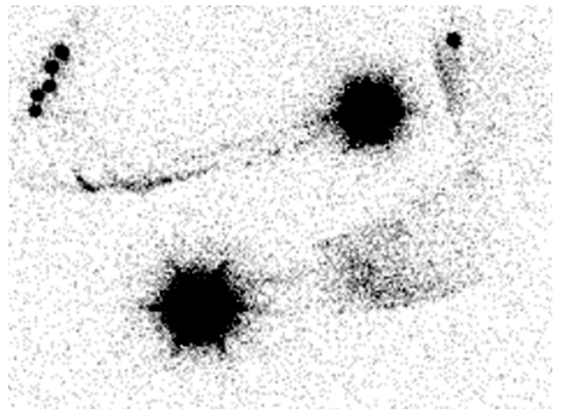
Figure 1. Lymphoscintigraphy of the upper limb. It depicts severe left lymphedema.
2.2. Indocyanine Green Lymphography
Indocyanine green lymphography (ICG-L) is currently the preferred diagnostic imaging technique for the lymphatic system among surgeons [8]. It has higher sensitivity and specificity for diagnosing lymphedema compared to lymphoscintigraphy [7]. It is also used for the selection of the surgical treatment modality and to give guidance to conservative treatment via manual lymphatic drainage. An intradermal injection of an ICG tracer in the distal limb is followed by the visualization of the lymphatic vessels under a near-infrared camera. It is a minimally invasive, simple, and highly accurate method for assessing lymphatic system status [9]. It enables the evaluation of the functional status of the superficial lymphatic vessels and the determination of their location, collateral lymphatic circulation, and dermal backflow [2][9][10]. A staging system correlating the disease severity with the pattern of dermal backflow on ICG-L was developed by Yamamoto et al. [11]. A linear pattern is considered to be normal, while splash, stardust, and diffuse patterns represent abnormalities with increasing levels of deterioration in lymphatic function (Figure 2). In severe cases of dermal backflow, the lymphatic flow beneath it is masked and cannot be detected by ICG-L. Other limitations include an inability to visualize lymphatic vessels deeper than 1.5–2.0 cm from the skin’s surface and an inability to assess interstitial tissues, the venous system, or the lymph nodes (except during surgery) [1][7][8][10]. Furthermore, the evaluation method is operator-dependent.
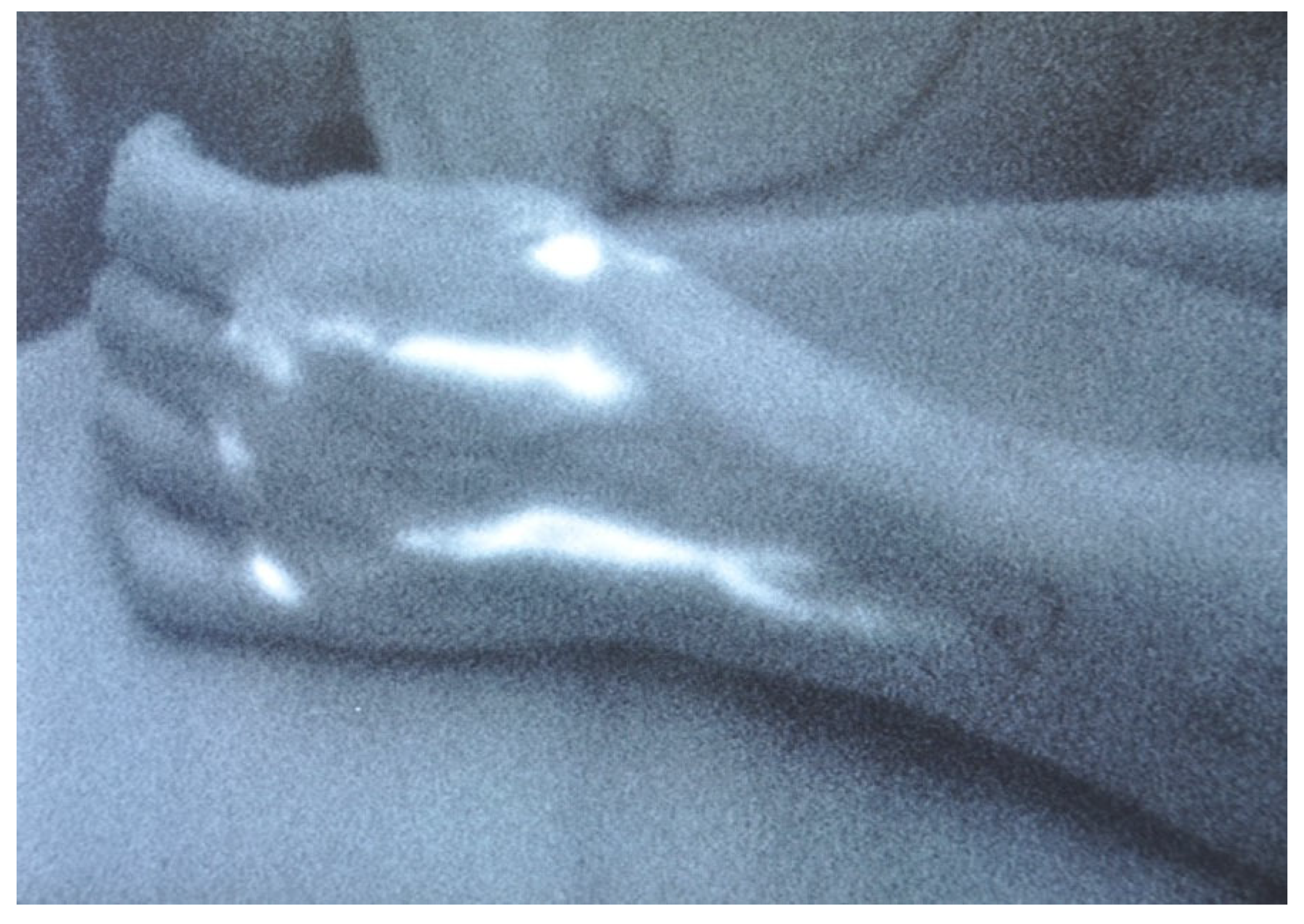
Figure 2. Linear pattern demonstrated by ICG lymphography.
3. Imaging for Treatment
After establishing the diagnosis of lymphedema, the next step is choosing the appropriate treatment option, and the possibility of performing a lymphaticovenous anastomosis (LVA) becomes an important aspect of surgical decision making. Functional lymphatic vessels and nearby receiving veins are important requirements for LVA, and therefore, preoperative imaging plays a significant role in substantiating the treatment choice. An ideal imaging study should evaluate the anatomy and function of the lymph nodes and lymphatic vessels, show their course in three dimensions, and display the venous network that will function as an anastomotic acceptor site [12]. In addition to being a diagnostic tool, indocyanine green lymphography (ICG-L) is also part of the preoperative evaluation for lymphatic surgery. Other helpful imaging modalities include magnetic resonance lymphangiography (MRL), single-photon emission computed tomography/computed tomography (SPECT/CT), ultra-high-frequency ultrasound (UHF-US), and photoacoustic (PA) imaging (Table 1).
Table 1. Overview of the imaging modalities and their advantages and limitations in evaluating the lymphatic system.
| Imaging Modalities | Advantages | Limitations |
|---|---|---|
| LSG |
|
|
| ICG-L |
|
|
| MRL |
|
|
| SPECT/CT |
|
|
| UHF-US |
|
|
| PA imaging |
|
|
3.1. Indocyanine Green Lymphography
As discussed before, indocyanine green lymphography can serve as a valuable tool in the outpatient setting for diagnosis and guiding treatment decisions. During LVA surgery, ICG-L facilitates the localization of lymphatic vessels, distinguishes between normal and abnormal drainage pathways, and optimizes the LVA surgical efficacy by assessing the anastomosis patency. These benefits make ICG-L a valuable intraoperative tool for surgeons performing LVA procedures [11].
3.2. Magnetic Resonance Lymphangiography (MRL)
Magnetic resonance lymphangiography (MRL) is typically performed by intradermally injecting a gadolinium-based contrast agent in the interdigital web spaces. There is also a possibility of performing MRL without the injection of a contrast agent, but no information is obtained about the functional status of the lymphatics and veins, and it has a lesser spatial resolution when compared to contrast-enhanced MRL [12]. Contrast-enhanced MRL produces high-resolution images of the lymphatic channels, including the number, size, depth, trajectory, and regions of dermal backflow [9]. In fact, MRL has a higher sensitivity to detect lymphatic vessel abnormalities than other imaging modalities such as lymphoscintigraphy (LSG), indocyanine green lymphangiography (ICG-L), and Ultrasound Doppler [9]. As opposed to ICG-L, MRL provides a three-dimensional image of the entire extremity and provides information about the quantity and quality of both the superficial and deep lymphatic systems. It also allows for the visualization of the lymph node basin, the venous system, and the quality of the interstitial fluid [1][2]. This capacity to identify and map functional lymphatic channels preoperatively makes MRL useful in determining suitable targets for performing lymphovenous anastomosis (LVA) (Figure 3). Studies [9][13] found that the concordant use of MRL and ICG-L in identifying functional lymphatic vessels correlates with a higher probability of successful LVA. Additionally, MRL can evaluate the composition of a lymphedematous limb, aiding in the determination of appropriate surgical treatment. Patients with normal subcutaneous tissues or fluid-dominant edema may benefit from microsurgical reconstruction, while adipose-dominant edema is usually treated with liposuction, and fibrosclerotic-dominant edema is treated with direct excision [1]. A study performed by Dayan et al. [14] found that even patients within the same International Society of Lymphology class may have different percentages of fat and fluid, which can impact the treatment choice, and demands an appropriate preoperative assessment. However, MRL is an expensive tool compared to ICG-L, it may produce images with venous enhancement due to venous uptake of the contrast agent, it is time-consuming, difficult for patients with claustrophobia, and might be impossible for patients with non-compatible implants [15].
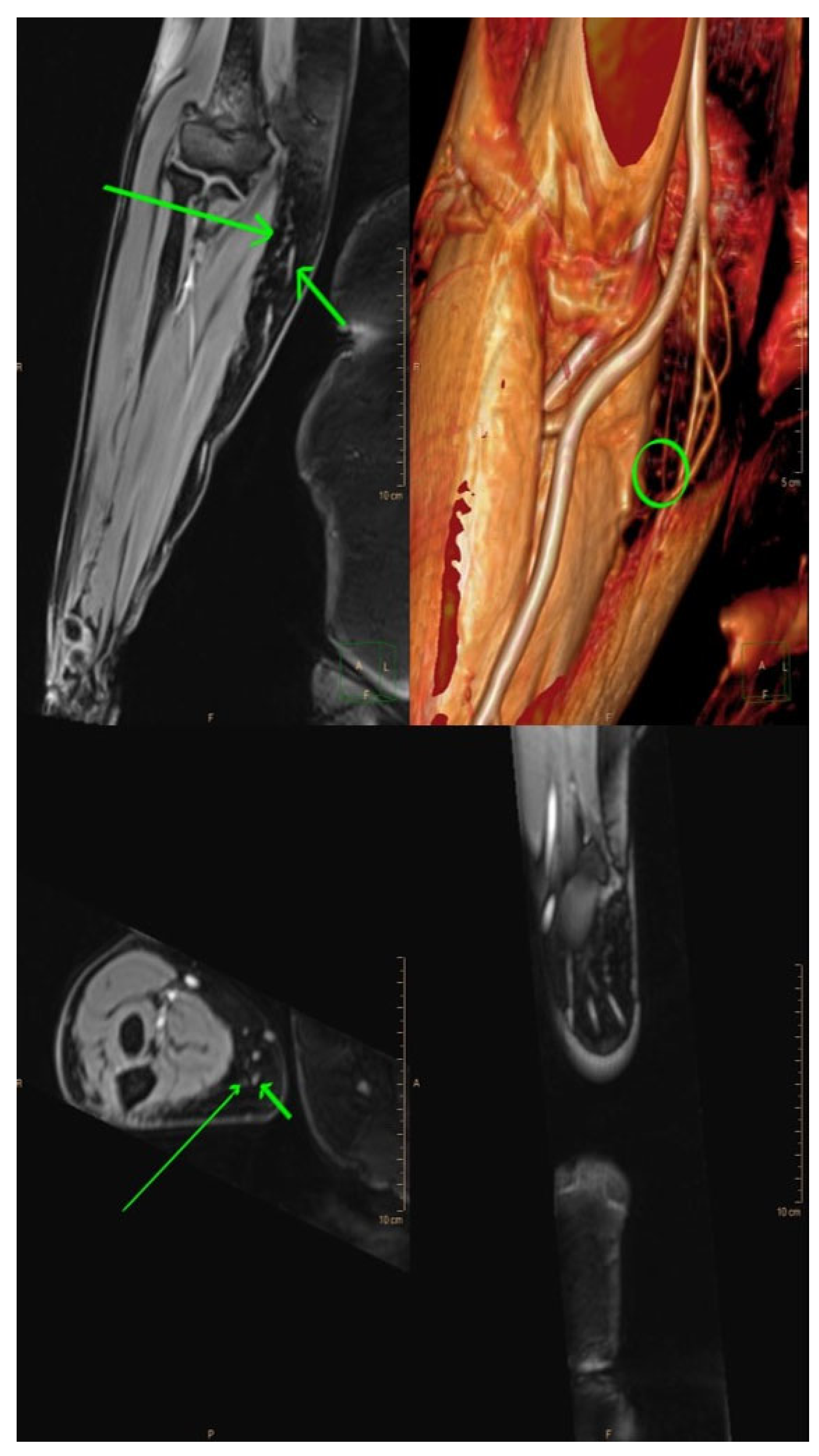
Figure 3. Magnetic resonance lymphangiography provides anatomic delineation and 3D reconstruction images of lymphatics and adjacent veins. Long arrows = lymphatic channels; short arrows = vein; and circle = the crossing of both (the possible place of incision).
3.3. Single-Photon Emission Computed Tomography/Computed Tomography (SPECT/CT)
SPECT/CT is a hybrid imaging modality that combines the planar imaging of SPECT with CT. This technique provides an anatomical localization of radio-activated lymph nodes and provides functional and three-dimensional information about the lymphatic system [16][17]. SPECT/CT can differentiate between lymphatic vessels and veins, and between tracer uptake in lymph nodes and lymphoceles. It is also capable of identifying dermal backflow from lymphatic vessel leakage and locating appropriate lymphatic vessels for LVA [18][19]. However, SPECT/CT has some drawbacks including a high cost and exposure to radiation, as well as low resolution for localizing lymphatic channels [17].
3.4. Ultra-High-Frequency Ultrasound (UHF-US)
Ultra-high frequency ultrasound (UHF-US) with a frequency range of 48–70 MHz provides an improved resolution compared to conventional ultrasound (15–24 MHz). It enables the accurate, real-time visualization of the lymphatic vessels, even those with diameters smaller than 0.3 mm, and allows operators to distinguish them from the subcutaneous veins or the nerves [20]. Lymphatic vessels are differentiated based on their shape, echogenic texture, color, Doppler collapsibility, convergence, and location [21]. UHF-US can also classify lymphatic vessels into two types: type I (less obstructed lymphatic channels, including normal and ectasis types) and type II (more obstructed lymphatic channels, including contraction and sclerosis types). Differentiating between these types is important for selecting suitable lymphatic vessels for LVA intraoperatively. Over time, the damaged lymphatic vessels become sclerotic and lose their ability to drain lymph fluid effectively; therefore, type I anastomosing lymphatic vessels have a significant advantage in LVA surgery [22]. UHF-US limitations include operator dependency and limited distance reach, as it can obtain images only up to 10 mm deep from the skin’s surface. To detect lymphatic vessels deeper than 10 mm, a transducer with a frequency of 48 MHz (max image depth: 23.5 mm) is recommended [20].
3.5. Photoacoustic (PA) or Optoacoustic Imaging
Photoacoustic imaging is a novel technique that utilizes the photoacoustic effect to visualize the lymphatic and vascular systems in three dimensions with a high resolution. This imaging modality, known as PA lymphangiography, involves the absorption of a specific wavelength of light by chromophores such as melanin, hemoglobin, or ICG, leading to thermoelastic expansion and the production of acoustic waves that are detected using an ultrasound transducer [23][24]. Unlike ICG-L, which also relies on ICG to identify lymphatic vessels, PA imaging can accurately differentiate between veins and lymphatic vessels, asses the three-dimensional relationship between them, and is less influenced by dermal backflow [23]. Consequently, using PA imaging intraoperatively enables the rapid identification of optimal sites for LVA and the assessment of anastomosis patency. In fact, PA imaging fulfills many of the criteria for an ideal imaging modality for surgical planning. However, it is still a developing technology, and handheld PA imaging devices are limited to a depth of 1 cm. Additionally, accurately adjusting the positions of the PA lymphangiography figures and the patient’s limb for clinical application remains challenging [23][24]. Despite these challenges, PA lymphangiography shows great promise as a valuable tool in lymphatic surgery.
4. Surgical Treatment
Complex decongestive therapy has been widely accepted as a conservative treatment strategy for lymphedema. In cases where it is no longer effective, surgical intervention may be considered. The surgical options can be classified as debulking or physiologic procedures. The former is typically performed in the later stages of the disease when there has been a transition from fluid-dominated edema to adipose or fibrosclerotic-dominated edema. Commonly used debulking procedures include suction-assisted lipectomy and direct excision, both focused on reducing the volume of the limb rather than restoring lymphatic flow. Physiologic treatment involves microsurgery techniques and focuses on restoring lymphatic flow and function. A treatment algorithm for patients with symptoms of lymphedema is provided in Figure 4.
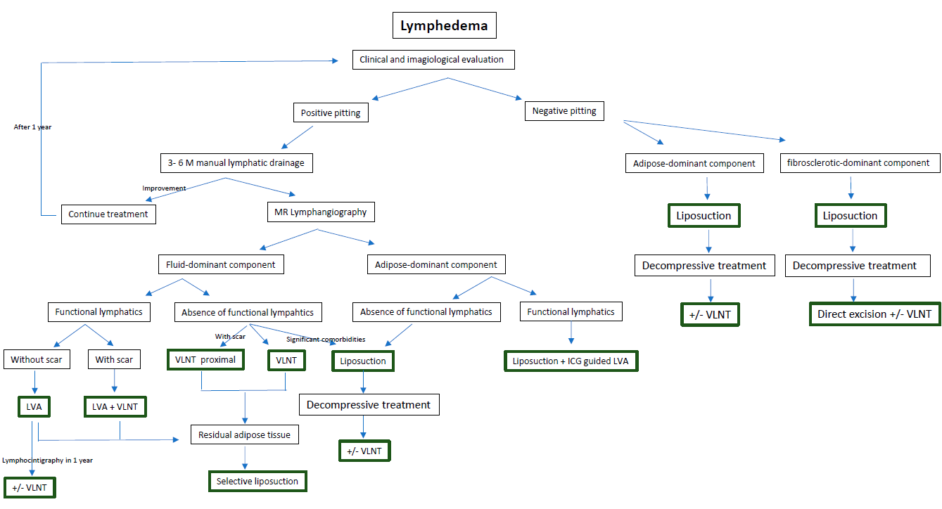
Figure 4. A treatment algorithm for patients with symptoms of lymphedema.
4.1. Debulking Procedures
4.1.1. Suction-Assisted Lipectomy (SAL)
Suction-assisted lipectomy (SAL) is a minimally invasive surgical procedure that removes fibrotic subcutaneous adipose tissue using suction cannulas. This technique involves making small skin incisions, and power-assisted devices may be used to aid in the removal of fibrous soft tissue. The procedure is performed circumferentially from the distal end to the proximal end and in a longitudinal direction to minimize damage to the remaining lymphatics, although some damage is inevitable. The goal of SAL is to remove the maximal amount of adipose tissue, and the incisions are left open to drain externally. Intraoperatively, custom compression garments are applied, and the lifelong use of compression garments is required to prevent recurrence [25][26]. Several studies have confirmed the long-term reduction in volume, an improvement in quality of life, and a decrease in infection rates with SAL [26][27][28]. In selected patients, SAL may be performed in conjunction with physiologic surgery to reduce the dependence on compression garments and to improve outcomes [26]. However, SAL is not effective for patients with end-stage fibrosclerotic lymphedema, and direct excision is required in those cases.
4.1.2. Direct Excision
In cases of advanced fibrosclerotic lymphedema, the direct excision of the diseased interstitial tissues may be necessary, with or without skin resection. The Charles procedure involves removing subcutaneous tissues and skin circumferentially, followed by applying skin grafts over the muscle fascia. Modified versions of this procedure incorporate negative pressure wound therapy and delayed skin grafting to improve graft take and wound recovery [13]. Alternatively, the modified Homan’s procedure [26] enables primary skin closure after performing the excision in a staged manner. These procedures are indicated for patients with irreversible fibrosclerotic lymphedema and significant symptoms, such as recurrent infections, impaired mobility, ulcerations, and malignancies. Although direct excision surgery provides consistent results with an improvement in well-being and function [29], it is associated with significant morbidity, scarring, a risk of graft loss, skin flap necrosis, lymphedema distal to the excised area, and sensory loss [1]. Additionally, patients may still need to use compression garments after surgery, and in cases where lymphedema recurs in the extremity, amputations may be necessary.
4.2. Physiologic Procedures
Advancements in the treatment of lymphedema have been achieved through the implementation of microsurgery, which allows for a targeted treatment of the underlying cause of the disease. Physiological procedures have been developed to restore the lymphatic flow, with the two main techniques being lymphaticovenular anastomosis (LVA) and vascularized lymph node transfer (VLNT).
4.2.1. LVA
The goal of lymphaticovenular anastomosis (LVA) is to restore lymphatic circulation by connecting functional lymphatic channels to subdermal venules of similar size, creating peripheral shunts within the lymphedematous limb. This allows for unidirectional flow from the congested high-pressure lymphatic system to the lower-pressure venous system [30]. To achieve successful long-term LVA, certain principles must be followed. Candidates for LVA must have functional or at least draining lymphatic vessels and a venule in proximity without reflux. As described before, there are many imaging modalities available to evaluate the functional status of lymphatics and, consequently, to predict the outcome of LVA. The most used modality is ICG-L. Since all of the lymphatic pathways do not deteriorate concurrently or to the same extent, early-stage patients are more likely to have functional lymphatics in superficial distribution that are easily visualized via ICG-L. On the other hand, patients in advanced stages may benefit from methods such as MRL or SPECT/CT to reveal deeper functional lymphatics. Therefore, the integration of different imaging modalities (ex., ICG-L and MRL) increases the reliability of the preoperative localization of functional lymphatics and may predict the outcome of LVA. Regarding the existence of a venule in proximity without reflux, as dynamic venous stenosis may contribute to lymphedema, some authors recommend screening for venous compression or reflux as part of the perioperative evaluation. This allows for the evaluation of the utility of scar fibrosis release, angioplasty, or venous stenting in the treatment of venous insufficiency [31][32]. A meticulous surgical technique is critical for a successful long-term LVA [26]. At selected cutaneous sites, two-centimeter skin incisions are made, and lymphatic channels and venules are carefully dissected under a microscope using super-microsurgery instruments. Nylon sutures of 11–0 or 12–0 are used for the anastomosis, and the anastomotic technique is selected depending on the relative calibers of the vessels found. Veins and lymphatics can mainly be anastomosed in the end-to-side, end-to-end, side-to-side, and side-to-end arrangements. The most efficient bypass is considered by some authors to be the side-to-end arrangement, as it allows for bidirectional lymph flow into a recipient vein while preserving the native lymph flow of the vessels and preventing damage to existing vessels [6]. However, there is no established consensus regarding the best technique to use or the number of anastomoses to perform [33][34]. After performing the anastomosis, its patency can be tested with ICG-L or patent blue dye (Figure 5). LVA is more effective in patients with early stages of lymphedema, although there can be a synergistic benefit when performed synchronously with vascularized lymph node transplantation in advanced-stage lymphedema. In fact, lymphedema treatment is a highly individualized process, and its effectiveness is consistently enhanced in combined approaches [30]. The eligibility of the LVA procedure is mainly determined by the presence of healthy, functional lymphatic vessels rather than the stage of lymphedema [32][35]. LVA can also be helpful in the treatment of lymphorrhea, as demonstrated by some studies [36][37]. The complications of LVA are minimal, with rates reported at 5.9% [30]. LVA offers significant advantages for the patient, being a minimally invasive procedure that can be performed as an outpatient procedure under local or general anesthesia, with a short recovery time and minimal postoperative restrictions [32] (Figure 6).
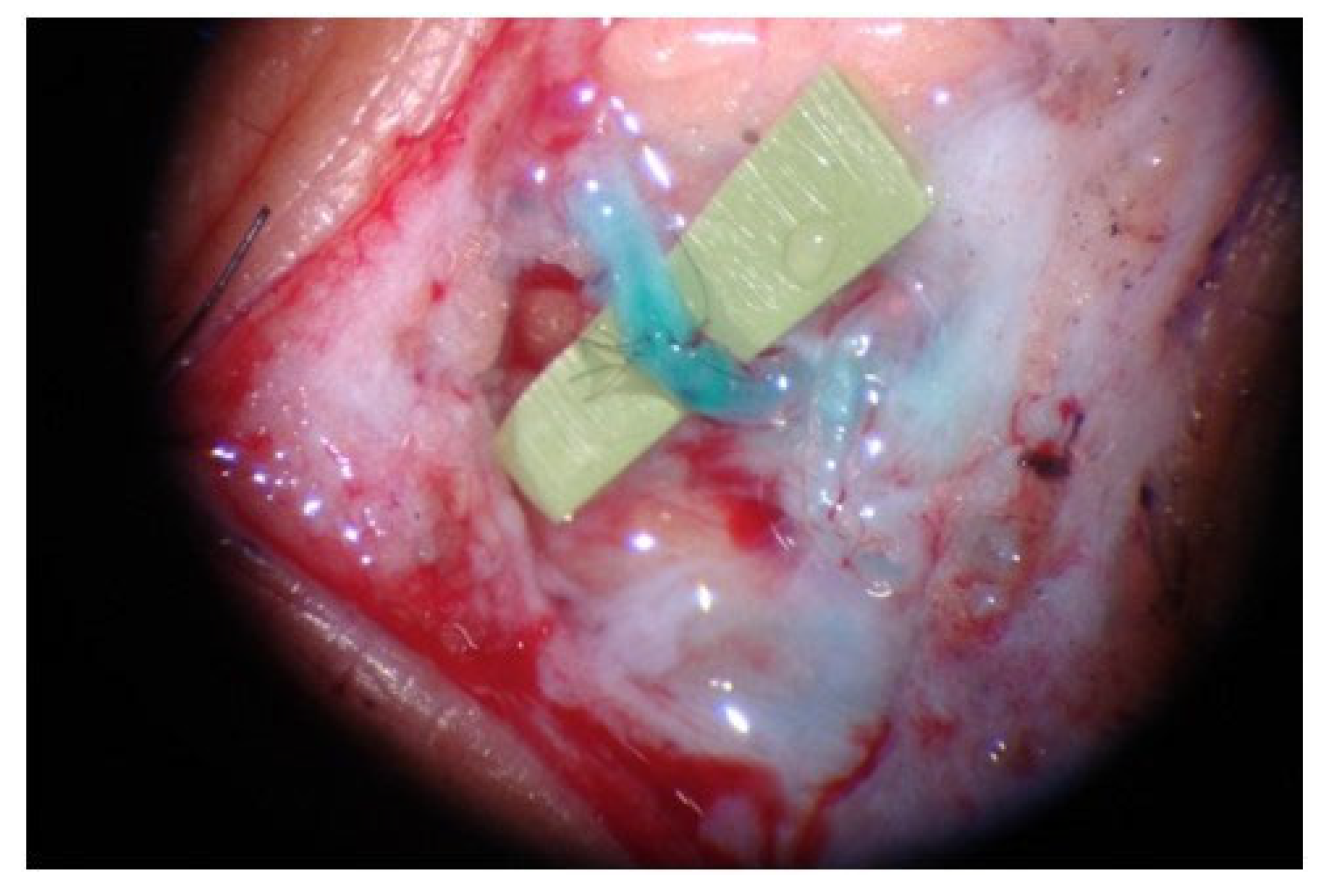
Figure 5. LVA was performed successfully in an end-to-end fashion. The patent blue on the subdermal venule shows the patency of LVA.
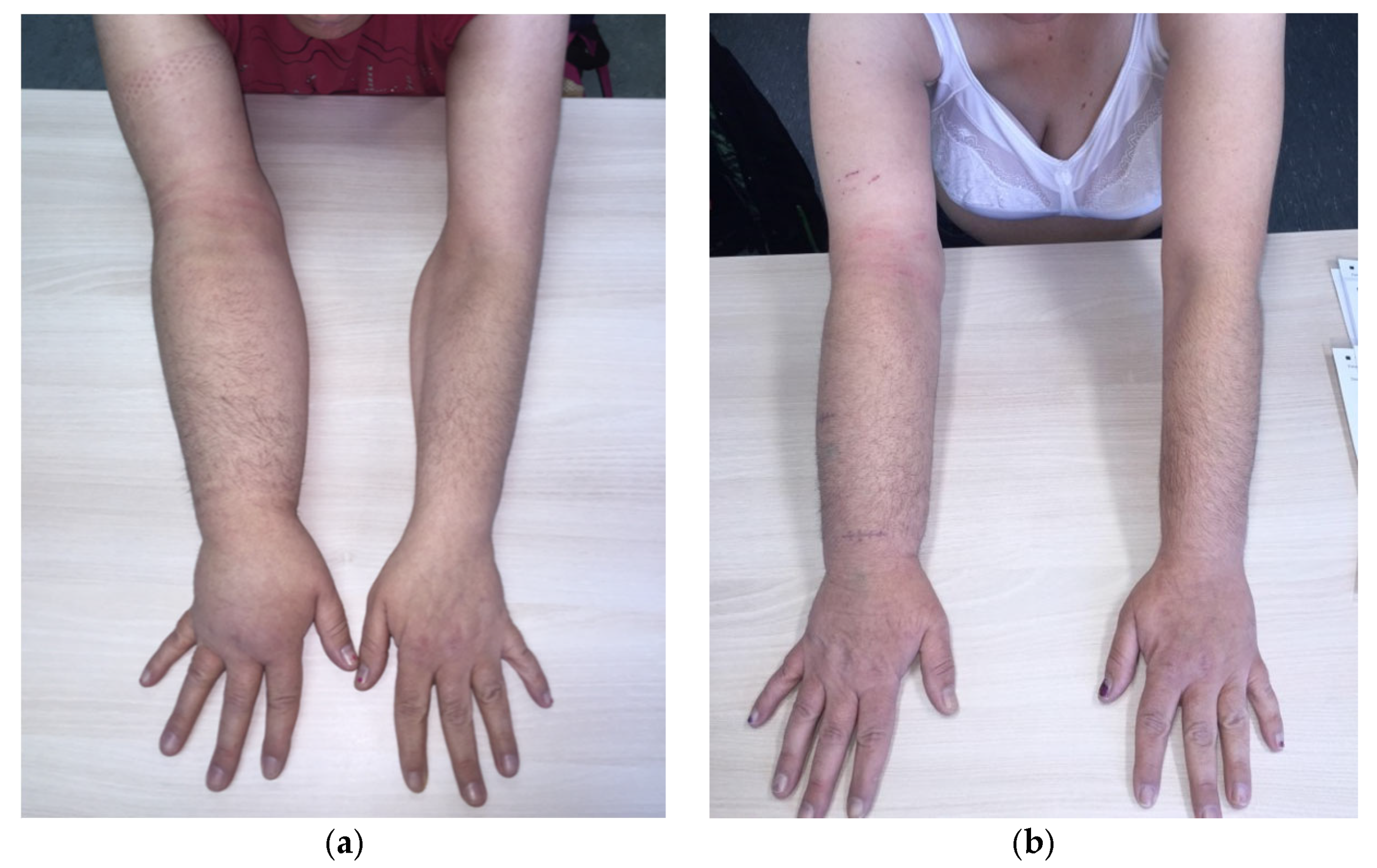
Figure 6. (a) Patient with right upper extremity lymphedema stage 1; (b) patient treated with 3 LVA. A reduction in the interlimb volume from 34% to 12% was achieved.
4.2.2. VLNT
For later stages of lymphedema, when the lymphatic channels are obliterated, alternative surgical interventions have been proposed, including vascularized lymph node transfer (VLNT). VLNT is a microsurgical technique that involves the transplantation of a vascularized lymph node and the surrounding tissue into the affected limb, anastomosing it to the arterial and venous systems in the recipient site. Typically, VLNT is performed for patients with moderate to advanced lymphedema, with damaged lymphatic vessels or decreased lymph node function. The precise physiological mechanisms behind the effects of VLNT on lymphedema are not yet fully understood. However, two primary hypotheses have been put forward to explain these effects. The first hypothesis proposes that VLNT induces lymphangiogenesis, which establishes connections between the lymph nodes and the recipient site’s lymphatic vessels. The second hypothesis suggests that the transferred lymph node functions as a “pump” by absorbing the interstitial fluid and transporting it into the systemic circulation via the intrinsic lymphovenous shunt within the nodes [38]. The proposed mechanisms provide evidence for the efficacy of both proximal anatomical (orthotopic) and distal non-anatomical (heterotopic) placement of lymph node flaps. Indeed, there is an ongoing debate regarding the ideal location of the recipient site [33]. Typical locations for VLNT include the axilla, elbow, wrist, groin, knee, and ankle. In selected patients who have planned for postmastectomy breast reconstruction and are suitable for autologous reconstruction, a chimeric flap of deep inferior epigastric artery perforator (DIEP) and a groin vascularized lymph node flap placed in the axilla may be suggested as an optimal solution for breast reconstruction and lymphedema. Since upper-extremity lymphedema often occurs after previous surgery with or without radiation to the axilla, scar tissue in the area and around the axillary vein may need to be released to provide a healthy bed for lymphangiogenesis. Similar to the axilla, the groin region may also need extensive removal or dissection of the scar tissue from past surgeries and radiotherapy [32]. In such situations, the orthotopic placement of VLNT is likely more reasonable as it can address both objectives. However, research suggests that the selection of the recipient sites does not have a significant impact on the outcomes, and hence, the choice is typically based on the availability of recipient vessels and surgeon preference. Although multiple studies have shown encouraging results of vascularized lymph node transfer (VLNT) in improving the symptoms and quality of life of patients with lymphedema, patients are still required to use compression garments after the surgery [39]. There are several potential donor sites for VLNT, including the groin, lateral thoracic, supraclavicular, submental, omental, and jejunal mesenteric node flaps (Table 2). Among these options, the most commonly used is the groin flap [40]. Its surgical anatomy and safety of harvesting has been clearly described [41]. Although VLNT has shown promising results in treating lymphedema, mild to severe secondary iatrogenic lymphedema at the donor site was reported in some cases [1][25][40][42]. Even in the absence of clinical lymphedema of the donor site, lymphatic function alterations were seen; thus, caution should be taken [43][44]. However, according to the literature, symptomatic iatrogenic donor-site lymphedema is a rare complication [25][33]. To minimize this risk, reverse lymphatic mapping was suggested as a mandatory test, involving the injection of ICG or patent blue dye in the distal part of the limbs and the avoidance of marked draining nodes during flap harvesting [45]. Other complications, such as seroma, lymphocele, infection, and delayed wound closure, have also been observed. Compared to LVA, VLNT requires longer hospital stay and surgical time [25].
Table 2. Characteristics of the vascularized lymph node flap options.
| VLNT | Advantages | Limitations |
|---|---|---|
| Groin flap |
|
|
| Lateral thoracic flap |
|
|
| Supraclavicular flap |
|
|
| Submental flap |
|
|
| Omental flap |
|
|
| Jejunal flap |
|
|
Groin Lymph Node Flap
The vascularized groin lymph node flap was the first to be described and remains the most commonly used donor site for VLNT [46]. This flap is based on the superficial circumflex iliac vessels and is favored due to well-described anatomic studies, a well-concealed scar, and feasibility to be combined with breast reconstruction [47][48]. The major complication associated with its harvest is the possibility of causing iatrogenic secondary lymphedema. To minimize this risk, the flap harvest should be limited to the area between the superficial inferior epigastric vein and the superficial circumflex iliac vein. Additionally, it is recommended to avoid harvesting lymphatic tissue caudal to the groin crease, belowto the deep fascia, and medial to a circle of a 2 cm diameter area centered half across the inguinal ligament [41] Reverse lymphatic mapping can be used intraoperatively, further reducing the risk of iatrogenic lymphedema, by identifying lymph nodes that preferentially drain the extremity related to the donor site. Limitations of the flap include a short pedicle length and small arterial caliber [26].
Lateral Thoracic Lymph Node Flap
The lateral thoracic lymph node flap is usually based on the lateral thoracic vessels and can also be harvested based on branches of the axillary artery or the thoracodorsal artery in cases where the artery is absent [49][50]. Since it includes axillary level I lymph nodes, it may not be suitable after an axillary dissection. The complication rate of this flap has been described as the highest among the five most frequently used lymph node donor sites [51]. Therefore, it is imperative to use reverse lymphatic mapping and avoid dissection over the drainage of the upper extremity, cephalad to the second intercostal brachial nerve, and medial to the lateral border of the pectoralis minor [52]. However, the flap presents some advantages that make it an attractive alternative to other VLNT donor sites, such as consistent anatomy, an inconspicuous donor site scar, and a long pedicle length [49]. It can be placed on an orthotopic or heterotopic position due to its versatility in flap design [26], and it includes abundant lymph nodes. An anatomical study revealed an average of 13.06 ± 3.42 lymph nodes within the flap [51], but it is unknown if all 13 lymph nodes can be safely harvested since the study did not use reverse lymphatic mapping.
Supraclavicular Lymph Node Flap
The supraclavicular lymph node flap is based on the supraclavicular branch of the transverse cervical vessels and encompasses cervical level Vb lymph nodes. The lymph nodes drain lymph fluid mainly from the breast, lung, esophagus, and oral cavity [53][54]. Although there is a risk of injuring the supraclavicular nerves during flap harvest, the risk of iatrogenic lymphedema is significantly lower compared to groin or lateral thoracic flaps. Only one case of secondary lymphedema following supraclavicular lymph node flap harvest has been reported in the literature so far [49]. Harvesting from the right side is usually preferred to avoid thoracic duct injury, even though it contains fewer lymph nodes than the left side [26]. Other advantages of the flap are its reliability, pliability, and a well-hidden scar that can be easily concealed by clothing [26][49][54].
Submental Lymph Node Flap
The submental lymph node flap is based on the submental artery, which is a branch of the facial artery. The flap can include submental (Ia) and submandibular (Ib) lymph nodes [26]. On average, the flap contains 3–5 lymph nodes and has a consistent anatomy [49][55][56][57]. M H Cheng’s group [58] suggested that the outcome of lymphedema surgery is dependent on the number of lymph nodes present in the flap; hence, some authors recommend preoperative neck imaging to identify the side with the greatest number of lymph nodes and their position [55][56]. There are no cases of iatrogenic lymphedema with a submental lymph node flap in the literature. Another advantage of the flap is its small volume, making it suitable for heterotopic placement. The disadvantages of the flap include a short pedicle length, a visible scar, and the possibility of injuring the marginal mandibular nerve during harvest [26][49][55].
Omental Lymph Node Flap
The omental lymph node flap can be utilized as either a pedicled or free flap. Complications such as iatrogenic hernia, small bowel obstruction, and donor site infection have been reported with the pedicled flap technique, which has limited its widespread use, and therefore, its transfer is mostly carried out via microsurgery [49][59]. While the entire omentum can be harvested, it is typically limited to the perivascular tissue and lymph nodes surrounding the gastroepiploic vascular arcade, using the right or left gastroepiploic vessels while sparing the rest [49][59]. Due to its high lymph node density and versatility in flap size, bilateral or dual-level transfer on both sides of the pedicle is feasible [26][49][59][60]. The flap is highly reliable, and unlike other VLNTs, its anatomy also permits a consistent efferent lymphatic vessel to be used for LVA [61]. There is no risk of donor-site lymphedema. The main complications are related to abdominal surgery, and previous abdominal operations, intra-abdominal adhesions, and scarring may restrict its use. The use of laparoscopic techniques, including robotic surgery, to harvest the flap reduces donor site morbidity while also allowing for minimal scarring that can be concealed under the patient’s clothing [26][49][59][61].
Jejunal Mesenteric Lymph Node Flaps
The use of the jejunal mesenteric lymph node flap as a pedicled flap was abandoned due to its associated complications, like the omental lymph node flap. As a free flap, it can be harvested from the periphery of the mesentery to incorporate a vascular arcade adjacent to the jejunum or from closer to the root of the mesentery, with each approach having some limitations. The former approach may be associated with ischemic bowel complications, while the latter may require a flow-through design to augment venous outflow. As an intra-abdominal procedure, harvesting the flap can lead to hernias and obstructions [49][59][62]. Nonetheless, the flap provides a reliable lymph node cluster with no risk of donor-site lymphedema. It can be harvested through a mini-laparotomy, abdominoplasty, or laparoscopic incisions, allowing for a well-concealed scar [26][49][59].
4.3. Preventive Lymphatic Surgery (LYMPHA Approach) [63]
In recent times, there has been a growing interest in preventive lymphatic surgery as a potential approach to reduce the risk of lymphedema resulting from cancer surgery. This surgical approach involves immediate lymphatic reconstruction at the time of node dissection. However, studies have yielded conflicting results, and its effectiveness remains controversial. A systematic review and meta-analysis conducted by Ciudad et al. [64] revealed that patients treated with prophylactic LVA (lymphaticovenular anastomosis) had a pooled lymphedema rate that was significantly lower than that reported in the literature as follows: 5.15% for axillary lymph nodes and 6.66% for ilio-inguinal nodes. Nevertheless, high-quality studies are necessary to establish clear recommendations regarding the use of preventive lymphatic surgery.
References
- Zeltzer, A.A.; Anzarut, A.; Hamdi, M. A Review of Lymphedema for the Hand and Upper-Extremity Surgeon. J. Hand Surg. 2018, 43, 1016–1025.
- Zeltzer, A.A.; Brussaard, C.; Koning, M.; De Baerdemaeker, R.; Hendrickx, B.; Hamdi, M.; de Mey, J. MR lymphography in patients with upper limb lymphedema: The GPS for feasibility and surgical planning for lympho-venous bypass. J. Surg. Oncol. 2018, 118, 407–415.
- Will, P.A.; Wan, Z.; Seide, S.E.; Berner, J.E.; Kneser, U.; Gazyakan, E.; Hirche, C. Supermicrosurgical treatment for lymphedema: A systematic review and network meta-analysis protocol. Syst. Rev. 2022, 11, 18.
- Winters, H.; Tielemans, H.J.; Paulus, V.; Hummelink, S.; Slater, N.J.; Ulrich, D.J. A systematic review and meta-analysis of vascularized lymph node transfer for breast cancer-related lymphedema. J. Vasc. Surg. Venous Lymphat. Disord. 2021, 10, 786–795.e1.
- Beederman, M.; Garza, R.M.; Agarwal, S.; Chang, D.W. Outcomes for Physiologic Microsurgical Treatment of Secondary Lymphedema Involving the Extremity. Ann. Surg. 2020, 276, e255–e263.
- Verhey, E.M.B.; Kandi, L.A.B.; Lee, Y.S.; Morris, B.E.; Casey, W.J.; Rebecca, A.M.M.; Marks, L.A.M.; Howard, M.A.; Teven, C.M. Outcomes of Lymphovenous Anastomosis for Lower Extremity Lymphedema: A Systematic Review. Plast. Reconstr. Surg.-Glob. Open 2022, 10, e4529.
- Knackstedt, R.; Chen, W.F. Current Concepts in Surgical Management of Lymphedema. Phys. Med. Rehabil. Clin. N. Am. 2022, 33, 885–899.
- Beederman, M.; Chang, D.W. Advances in surgical treatment of lymphedema. Arch. Plast. Surg. 2021, 48, 670–677.
- Pons, G.; Clavero, J.; Alomar, X.; Rodríguez-Bauza, E.; Tom, L.; Masia, J. Preoperative planning of lymphaticovenous anastomosis: The use of magnetic resonance lymphangiography as a complement to indocyanine green lymphography. J. Plast. Reconstr. Aesthetic Surg. 2019, 72, 884–891.
- Chang, E.I.; Chu, C.K.; Chang, E.I. Advancements in imaging technology for microvascular free tissue transfer. J. Surg. Oncol. 2018, 118, 729–735.
- Akita, S.; Unno, N.; Maegawa, J.; Kimata, Y.; Fukamizu, H.; Yabuki, Y.; Kitayama, S.; Shinaoka, A.; Yamada, K.; Sano, M.; et al. A phase III, multicenter, single-arm study to assess the utility of indocyanine green fluorescent lymphography in the treatment of secondary lymphedema. J. Vasc. Surg. Venous Lymphat. Disord. 2021, 10, 728–737.e3.
- Guerrini, S.; Gentili, F.; Mazzei, F.G.; Gennaro, P.; Volterrani, L.; Mazzei, M.A. Magnetic resonance lymphangiography: With or without contrast? Diagn. Interv. Radiol. 2020, 26, 587–595.
- Park, K.E.; Allam, O.; Chandler, L.; Mozzafari, M.A.; Ly, C.; Lu, X.; Persing, J.A. Surgical management of lymphedema: A review of current literature. Gland. Surg. 2020, 9, 503–511.
- Dayan, J.H.; Wiser, I.M.; Verma, R.; Shen, J.; Talati, N.; Goldman, D.M.; Mehrara, B.J.; Smith, M.L.; Dayan, E.; Coriddi, M.; et al. Regional Patterns of Fluid and Fat Accumulation in Patients with Lower Extremity Lymphedema Using Magnetic Resonance Angiography. Plast. Reconstr. Surg. 2020, 145, 555–563.
- Forte, A.J.; Boczar, D.; Huayllani, M.T.; Avila, F.R.; Guliyeva, G.; Lu, X.; Mash, W.R.; Kung, T.A. Use of magnetic resonance imaging lymphangiography for preoperative planning in lymphedema surgery: A systematic review. Microsurgery 2021, 41, 384–390.
- Gentileschi, S.; Albanese, R.; Pino, V.; Stefanizzi, G.; Fragomeni, S.; Zagaria, L.; Ieria, F.P.; Salgarello, M.; Scambia, G.; Garganese, G. SPECT/CT and fusion ultrasound to target the efferent groin lymph node for lymphatic surgery. Microsurgery 2019, 39, 605–612.
- Du, X.; Liu, C. Application of imaging in lymphedema surgical therapies. Gland. Surg. 2020, 9, 582–588.
- Iimura, T.; Fukushima, Y.; Kumita, S.; Ogawa, R.; Hyakusoku, H. Estimating Lymphodynamic Conditions and Lymphovenous Anastomosis Efficacy Using 99mTc-phytate Lymphoscintigraphy with SPECT-CT in Patients with Lower-limb Lymphedema. Plast. Reconstr. Surg.-Glob. Open 2015, 3, e404.
- Weiss, M.; Baumeister, R.G.; Frick, A.; Wallmichrath, J.; Bartenstein, P.; Rominger, A. Primary Lymphedema of the Lower Limb: The Clinical Utility of Single Photon Emission Computed Tomography/CT. Korean J. Radiol. 2015, 16, 188–195.
- Hayashi, A.; Giacalone, G.; Yamamoto, T.; Belva, F.; Visconti, G.; Hayashi, N.; Handa, M.; Yoshimatsu, H.; Salgarello, M. Ultra High-frequency Ultrasonographic Imaging with 70 MHz Scanner for Visualization of the Lymphatic Vessels. Plast. Reconstr. Surg.-Glob. Open 2019, 7, e2086.
- Van Heumen, S.; Riksen, J.J.M.; Bramer, W.M.; van Soest, G.; Vasilic, D. Imaging of the Lymphatic Vessels for Surgical Planning: A Systematic Review. Ann. Surg. Oncol. 2022, 30, 462–479.
- Bianchi, A.; Visconti, G.; Hayashi, A.; Santoro, A.; Longo, V.; Salgarello, M. Ultra-High frequency ultrasound imaging of lymphatic channels correlates with their histological features: A step forward in lymphatic surgery. J. Plast. Reconstr. Aesthetic Surg. 2020, 73, 1622–1629.
- Suzuki, Y.; Kajita, H.; Watanabe, S.; Okabe, K.; Sakuma, H.; Imanishi, N.; Aiso, S.; Kishi, K. Application of Photoacoustic Imaging for Lymphedema Treatment. J. Reconstr. Microsurg. 2021, 38, 254–262.
- Suzuki, Y.; Kajita, H.; Watanabe, S.; Otaki, M.; Okabe, K.; Sakuma, H.; Takatsume, Y.; Imanishi, N.; Aiso, S.; Kishi, K. Surgical Applications of Lymphatic Vessel Visualization Using Photoacoustic Imaging and Augmented Reality. J. Clin. Med. 2021, 11, 194.
- Gallagher, K.; Marulanda, K.; Gray, S. Surgical Intervention for Lymphedema. Surg. Oncol. Clin. N. Am. 2018, 27, 195–215.
- Schaverien, M.V.; Coroneos, C.J. Surgical Treatment of Lymphedema. Plast. Reconstr. Surg. 2019, 144, 738–758.
- Greene, A.K.M.; Maclellan, R.A.M. Operative Treatment of Lymphedema Using Suction-Assisted Lipectomy. Ann. Plast. Surg. 2016, 77, 337–340.
- Greene, A.K.M.; Maclellan, R.M. Management of Lymphedema with Suction-Assisted Lipectomy. Plast. Reconstr. Surg. 2014, 134, 36.
- Van der Walt, J.C.; Perks, T.J.; Zeeman, B.J.; Bruce-Chwatt, A.J.; Graewe, F.R. Modified Charles Procedure Using Negative Pressure Dressings for Primary Lymphedema: A functional assessment. Ann. Plast. Surg. 2009, 62, 669–675.
- Carl, H.M.; Walia, G.; Bello, R.; Clarke-Pearson, E.; Hassanein, A.H.; Cho, B.; Pedreira, R.; Sacks, J.M. Systematic Review of the Surgical Treatment of Extremity Lymphedema. J. Reconstr. Microsurg. 2017, 33, 412–425.
- Gupta, R.B.; Mathijs, E.; Hart, J.D.; Bates, J.D.; Powers, J.; Chaiyasate, K. May-Thurner Syndrome and Lymphedema Reconstruction. Plast. Reconstr. Surg.-Glob. Open 2022, 10, e4377.
- Chang, D.W.; Masia, J.; Garza, R.; Skoracki, R.; Neligan, P.C. Lymphedema: Surgical and Medical Therapy. Plast. Reconstr. Surg. 2016, 138 (Suppl. 3), 209s–218s.
- Hanson, S.E.; Chang, E.I.; Schaverien, M.V.M.; Chu, C.; Selber, J.C.; Hanasono, M.M. Controversies in Surgical Management of Lymphedema. Plast. Reconstr. Surg.-Glob. Open 2020, 8, e2671.
- Scaglioni, M.F.; Fontein, D.B.Y.; Arvanitakis, M.; Giovanoli, P. Systematic review of lymphovenous anastomosis (LVA) for the treatment of lymphedema. Microsurgery 2017, 37, 947–953.
- De Sire, A.; Losco, L.; Lippi, L.; Spadoni, D.; Kaciulyte, J.; Sert, G.; Ciamarra, P.; Marcasciano, M.; Cuomo, R.; Bolletta, A.; et al. Surgical Treatment and Rehabilitation Strategies for Upper and Lower Extremity Lymphedema: A Comprehensive Review. Medicina 2022, 58, 954.
- De Carvalho, F.M.; Almeida, A.; Silva, Á.; Marques, M. Treatment of Lymphorrhea Associated with an Amputation Stump with Lymphaticovenular Anastomosis. Acta Médica Port. 2022, 35, 384–387.
- Yang, J.C.-S.; Yen, Y.-H.; Wu, S.-C.; Lin, W.-C.; Chiang, M.-H.; Hsieh, C.-H. Supermicrosurgical Lymphaticovenous Anastomosis as an Alternative Treatment Option for Patients with Lymphorrhea. Plast. Reconstr. Surg. 2019, 144, 1214–1224.
- Cheng, M.-H.M.; Huang, J.-J.; Wu, C.-W.; Yang, C.-Y.M.; Lin, C.-Y.M.; Henry, S.L.; Kolios, L. The Mechanism of Vascularized Lymph Node Transfer for Lymphedema: Natural lym-phaticovenous drainage. Plast. Reconstr. Surg. 2014, 133, 192e–198e.
- De Brucker, B.; Zeltzer, A.; Seidenstuecker, K.; Hendrickx, B.; Adriaenssens, N.; Hamdi, M. Breast Cancer–Related Lymphedema: Quality of Life after Lymph Node Transfer. Plast. Reconstr. Surg. 2016, 137, 1673–1680.
- Scaglioni, M.F.; Arvanitakis, M.; Chen, Y.-C.; Giovanoli, P.; Yang, J.C.-S.; Chang, E.I. Comprehensive review of vascularized lymph node transfers for lymphedema: Outcomes and complications. Microsurgery 2016, 38, 222–229.
- Zeltzer, A.A.; Anzarut, A.; Braeckmans, D.; Seidenstuecker, K.; Hendrickx, B.; Van Hedent, E.; Hamdi, M. The vascularized groin lymph node flap (VGLN): Anatomical study and flap planning using multi-detector CT scanner. The golden triangle for flap harvesting. J. Surg. Oncol. 2017, 116, 378–383.
- Vignes, S.; Blanchard, M.; Yannoutsos, A.; Arrault, M. Complications of Autologous Lymph-node Transplantation for Limb Lymphoedema. Eur. J. Vasc. Endovasc. Surg. 2013, 45, 516–520.
- Sulo, E.; Hartiala, P.; Viitanen, T.; Mäki, M.; Seppänen, M.; Saarikko, A. Risk of donor-site lymphatic vessel dysfunction after microvascular lymph node transfer. J. Plast. Reconstr. Aesthetic Surg. 2014, 68, 551–558.
- Viitanen, T.P.; Mäki, M.T.; Seppänen, M.P.; Suominen, E.A.; Saaristo, A.M. Donor-Site Lymphatic Function after Microvascular Lymph Node Transfer. Plast. Reconstr. Surg. 2012, 130, 1246–1253.
- Dayan, J.H.; Dayan, E.; Smith, M.L. Reverse Lymphatic Mapping: A new technique for maximizing safety in vascularized lymph node transfer. Plast. Reconstr. Surg. 2015, 135, 277–285.
- Becker, C.; Assouad, J.; Riquet, M.; Hidden, G. Postmastectomy Lymphedema: Long-term results following microsurgical lymph node transplantation. Ann. Surg. 2006, 243, 313–315.
- Masià, J.; Smith, M.L.; Chang, E.I. Combining Autologous Breast Reconstruction and Vascularized Lymph Node Transfer. Semin. Plast. Surg. 2018, 32, 036–041.
- Chang, E.I. Optimizing Treatment of Breast Cancer Related Lymphedema Using Combined DIEP Flap and Lymphedema Surgery. Arch. Plast. Surg. 2022, 49, 150–157.
- Schaverien, M.V.; Badash, I.; Selber, J.C.; Cheng, M.-H.; Patel, K.M. Vascularized Lymph Node Transfer for Lymphedema. Semin. Plast. Surg. 2018, 32, 28–35.
- Baptista, R.R.; Kasai, K.E.; dos Anjos, D.M.; Busnardo, F.D.F.; Modolin, M.; Ferreira, M.C.; Barreiro, G.C. Lymph Fasciocutaneous Lateral Thoracic Artery Flap: Anatomical Study and Clinical Use. J. Reconstr. Microsurg. 2014, 30, 389–396.
- Tinhofer, I.E.; Meng, S.; Steinbacher, J.; Roka-Palkovits, J.; Györi, E.; Reissig, L.F.; Cheng, M.-H.; Weninger, W.J.; Tzou, C.H. The surgical anatomy of the vascularized lateral thoracic artery lymph node flap-A cadaver study. J. Surg. Oncol. 2017, 116, 1062–1068.
- Coroneos, C.J.; Asaad, M.; Wong, F.C.; Hall, M.S.; Chen, D.N.; Hanasono, M.M.; Schaverien, M.V. Outcomes and technical modifications of vascularized lymph node transplantation from the lateral thoracic region for treatment of lymphedema. J. Surg. Oncol. 2022, 125, 603–614.
- Visconti, G.; Bianchi, A.; Salgarello, M.; Di Leone, A.; Hayashi, A.; Masetti, R.; Franceschini, G. Lymphatic Tissue Transfer: Ultrasound-Guided Description and Preoperative Planning of Vascularised Lymph Nodes, Lymphatic Units, and Lymphatic Vessels Transfers. J. Pers. Med. 2022, 12, 1346.
- Mardonado, A.A.; Chen, R.; Chang, D.W. The use of supraclavicular free flap with vascularized lymph node transfer for treatment of lymphedema: A prospective study of 100 consecutive cases. J. Surg. Oncol. 2016, 115, 68–71.
- Mazerolle, P.; Meresse, T.; Gangloff, D.; Kolsi, K.; Dupret-Bories, A. Vascularized lymph node transfer with submental free flap. Eur. Ann. Otorhinolaryngol. Head Neck Dis. 2019, 137, 73–77.
- Paulus, V.A.A.; Winters, H.; Hummelink, S.; Schulten, S.; Ulrich, D.J.O.; Vasilic, D. Submental flap for vascularized lymph node transfer; a CTA-based study on lymph node distribution. J. Surg. Oncol. 2020, 122, 1226–1231.
- Tzou, C.H.; Meng, S.; Ines, T.; Reissig, L.; Pichler, U.; Steinbacher, J.; Pona, I.; Roka-Palkovits, J.; Rath, T.; Weninger, W.J.; et al. Surgical anatomy of the vascularized submental lymph node flap: Anatomic study of correlation of submental artery perforators and quantity of submental lymph node. J. Surg. Oncol. 2016, 115, 54–59.
- Gustafsson, J.; Chu, S.-Y.; Chan, W.-H.; Cheng, M.-H. Correlation between Quantity of Transferred Lymph Nodes and Outcome in Vascularized Submental Lymph Node Flap Transfer for Lower Limb Lymphedema. Plast. Reconstr. Surg. 2018, 142, 1056–1063.
- Danforth, R.; Skoracki, R. Intra-abdominal donors for vascularized lymph node transfer: An update and review. Plast. Aesthetic Res. 2021, 8, 48.
- Kenworthy, E.O.; Nelson, J.A.; Verma, R.; Mbabuike, J.; Mehrara, B.J.; Dayan, J.H. Double vascularized omentum lymphatic transplant (VOLT) for the treatment of lymphedema. J. Surg. Oncol. 2018, 117, 1413–1419.
- Nguyen, A.T.; Suami, H.; Hanasono, M.M.; Womack, V.A.; Wong, F.C.; Chang, E.I. Long-term outcomes of the minimally invasive free vascularized omental lymphatic flap for the treatment of lymphedema. J. Surg. Oncol. 2016, 115, 84–89.
- Kraft, C.T.; Eiferman, D.; Jordan, S.; Skoracki, R.J. Complications after vascularized jejunal mesenteric lymph node transfer: A 3-year experience. Microsurgery 2019, 39, 497–501.
- Boccardo, F.; Casabona, F.; De Cian, F.; Friedman, D.; Villa, G.; Bogliolo, S.; Ferrero, S.; Murelli, F.; Campisi, C. Lymphedema Microsurgical Preventive Healing Approach: A New Technique for Primary Prevention of Arm Lymphedema after Mastectomy. Ann. Surg. Oncol. 2009, 16, 703–708.
- Ciudad, P.; Escandón, J.M.; Bustos, V.P.; Manrique, O.J.; Kaciulyte, J. Primary Prevention of Cancer-Related Lymphedema Using Preventive Lymphatic Surgery: Systematic Review and Meta-analysis. Indian J. Plast. Surg. 2022, 55, 018–025.
More
Information
Subjects:
Surgery
Contributors
MDPI registered users' name will be linked to their SciProfiles pages. To register with us, please refer to https://encyclopedia.pub/register
:
View Times:
802
Revisions:
2 times
(View History)
Update Date:
04 Sep 2023
Table of Contents


Notice
You are not a member of the advisory board for this topic. If you want to update advisory board member profile, please contact office@encyclopedia.pub.
OK
Confirm
Only members of the Encyclopedia advisory board for this topic are allowed to note entries. Would you like to become an advisory board member of the Encyclopedia?
Yes
No
${ textCharacter }/${ maxCharacter }
Submit
Cancel
Back
Comments
${ item }
|
${ item.createdUser.fullName }
${ item.createdAt }
${ item.vote }
${ item.reply }
Delete
${ reply.createdUser.fullName }
${ reply.createdAt }
${ reply.vote }
Delete
There is no reply to this comment~
${ item.replyTextCharacter }/${ item.replyMaxCharacter }
Submit
Cancel
More
No more~
There is no comment~
${ textCharacter }/${ maxCharacter }
Submit
Cancel
${ selectedItem.replyTextCharacter }/${ selectedItem.replyMaxCharacter }
Submit
Cancel
Confirm
Are you sure to Delete?
Yes
No