Submitted Successfully!
To reward your contribution, here is a gift for you: A free trial for our video production service.
Thank you for your contribution! You can also upload a video entry or images related to this topic.
Version Summary Created by Modification Content Size Created at Operation
1 -- 3541 2023-08-30 05:24:27 |
2 only format change Meta information modification 3541 2023-08-30 06:03:59 |

Video Upload Options

Do you have a full video?

Confirm

Are you sure to Delete?
Cite
If you have any further questions, please contact Encyclopedia Editorial Office.
Álvarez-Zúñiga, C.D.; Garza-Veloz, I.; Martínez-Rendón, J.; Ureño-Segura, M.; Delgado-Enciso, I.; Martinez-Fierro, M.L. Circulating Biomarkers for B-Cell Progenitor Acute Lymphoblastic Leukemia. Encyclopedia. Available online: https://encyclopedia.pub/entry/48621 (accessed on 06 August 2024).
Álvarez-Zúñiga CD, Garza-Veloz I, Martínez-Rendón J, Ureño-Segura M, Delgado-Enciso I, Martinez-Fierro ML. Circulating Biomarkers for B-Cell Progenitor Acute Lymphoblastic Leukemia. Encyclopedia. Available at: https://encyclopedia.pub/entry/48621. Accessed August 06, 2024.
Álvarez-Zúñiga, Claudia Daniela, Idalia Garza-Veloz, Jacqueline Martínez-Rendón, Misael Ureño-Segura, Iván Delgado-Enciso, Margarita L. Martinez-Fierro. "Circulating Biomarkers for B-Cell Progenitor Acute Lymphoblastic Leukemia" Encyclopedia, https://encyclopedia.pub/entry/48621 (accessed August 06, 2024).
Álvarez-Zúñiga, C.D., Garza-Veloz, I., Martínez-Rendón, J., Ureño-Segura, M., Delgado-Enciso, I., & Martinez-Fierro, M.L. (2023, August 30). Circulating Biomarkers for B-Cell Progenitor Acute Lymphoblastic Leukemia. In Encyclopedia. https://encyclopedia.pub/entry/48621
Álvarez-Zúñiga, Claudia Daniela, et al. "Circulating Biomarkers for B-Cell Progenitor Acute Lymphoblastic Leukemia." Encyclopedia. Web. 30 August, 2023.
Circulating Biomarkers for B-Cell Progenitor Acute Lymphoblastic Leukemia
Edit

Acute lymphoblastic leukemia (ALL) is a hematological disease characterized by the dysfunction of the hematopoietic system that leads to arrest at a specific stage of stem cells development, suppressing the average production of cellular hematologic components. BCP (B-cell progenitor)-ALL is a neoplasm of the B-cell lineage progenitor. BCP-ALL is caused and perpetuated by several mechanisms that provide the disease with its tumor potential and genetic and cytological characteristics. These pathological features are used for diagnosis and the prognostication of BCP-ALL. The BCP-ALL diagnostic protocol is well established. Firstly, it is necessary to demonstrate ≥ 20% lymphoblasts in bone marrow (BM) based on a BPM. Second, a hematopathological review is performed; it comprises a morphological assessment, and flow cytometric and genetic characterization.

biomarkers circulating acute lymphoblastic leukemia BCP-ALL

1. Introduction

Leukemias are a group of hematological diseases characterized by an abnormal cell population suppressing the average production of cellular components of the hematopoietic system [1]. There are four main subtypes of leukemia that have been identified based on their evolution time and hematological lineage: acute lymphoblastic leukemia (ALL), chronic lymphocytic leukemia (CLL), acute myelogenous leukemia (AML), and chronic myelogenous leukemia (CML) [2]. ALL and CLL are the most common leukemia types in children and adults, respectively [3][4].
ALL is a malignant disorder that occurs when typical lymphoid cell development malfunctions due to arrest at a specific stage of development; the name and classification and based on the dysfunctional stage [5]. ALL can develop from primitive precursor cells with multilineage potential from the two types of lymphoblasts: B or T. These cells proliferate uncontrollably in bone marrow (BM) and peripheral blood (PB) [1][6][7][8] and interfere with the functions of normal blood cells [8][9]. Of the two kinds of lymphoblasts, it is more common for ALL to originate from B-cell progenitor (BCP-ALL), representing about 80% of cases; the remaining 20% originate from T-cell progenitors (TCP-ALL) [10].
According to the World Health Organization (WHO), BCP-ALL is defined as a neoplasm of precursor lymphoid cells committed to the B cell lineage, typically composed of small-to-medium-sized blast cells with scant cytoplasm, moderately condensed to dispersed chromatin, and inconspicuous nucleoli [5]. BCP-ALL diagnosis involves clinical and laboratory tests. BCP-ALL symptoms indicate blast infiltration in the bone marrow, lymphoid system, and extramedullary sites and may include fatigue or lethargy, constitutional symptoms, dyspnea, dizziness, infections, and easy bruising or bleeding [11]. A BM aspirate is mandatory for laboratory tests to confirm the diagnosis, demonstrating ≥ 20% bone marrow lymphoblasts [12]. In addition, clinicians can examine cell morphology, immunophenotyping, and genetics and integrate these measures according to the classification established by the WHO [5]. BCP-ALL has multiple subtypes characterized by immunophenotype B-I (pro-B), B-II (Common), B-III (pre-B), and B-IV (B—mature Burkitt type) [5]. Recently, the nomenclature of leukemia with genetic abnormalities has shifted from focusing on cytogenetic alterations to molecular events, and new genetic entities have been added. The WHO classification in 2022 is based on clinical–biological entities defined by cytogenetic alterations that confer different prognoses. The United Kingdom ALL copy-number alteration (UKALL-CNA) classification has been incorporated, which distinguishes three genetic risk groups [13].
The peak incidence of ALL is from 2 to 5 years of age. It is rare in adults, but it can be found after 50 years of age. BCP-ALL constitutes approximately 80–85% of total cases of ALL [14][15][16]. Many factors affect the prognosis of BCP-ALL, such as age, the white blood cell (WBC) count, clinical features at diagnosis, cytogenetic abnormalities, pharmacodynamic and pharmacogenetic characteristics of normal patient cells, early response to therapy, and the results of the measurements of minimal/measurable residual disease (MRD: the presence of leukemic cells at specific time points in bone marrow or even in peripheral blood circulation) [17][18][19][20][21][22]. The prognostic implication of cytogenic abnormalities relates to poorly regulated signaling pathways [5][23], which can affect leukemogenesis and directly influences ALL development [24][25].
The above-mentioned factors help determine a “risk stratification” classification for BCP-ALL. This classification determines the treatment regimen and intensity that will be settled to treat BCP-ALL. This classification has contributed to a marked improvement in the prognosis of patients with leukemia [19][26][27].
As mentioned before, a BM puncture (BMP)/aspirate is needed to diagnose BCP-ALL. However, because of its invasive nature, potential side effects, and expensive procedures, there is a real need for alternative measures and molecular markers for the effective diagnosis and prognosis of pediatric patients with BCP-ALL [14][28]. Suitable substitutes for BMP are molecular biomarkers found in peripheral blood; collecting blood produces minimal trauma, and biomarkers can be measured relatively easy and early in the disease [29][30][31]. Nevertheless, the mentioned replacement must fulfill the biomarker standards.
In oncology, a biomarker is any measurable indicator that demonstrates the presence of malignancy, tumor behavior, prognosis, or responses to treatments [32]. Among the myriad of biological materials circulating in the bloodstream, the most promising biomarkers include circulating tumor cells, cell-free DNA (cf-DNA) and RNA, proteins and metabolites, and extracellular vesicles (EVs) [33].

2. Diagnostic Biomarkers for BCP-ALL

The BCP-ALL diagnostic protocol is well established. Firstly, it is necessary to demonstrate ≥ 20% lymphoblasts in BM based on a BPM. Second, a hematopathological review is performed; it comprises a morphological assessment, and flow cytometric and genetic characterization. Once these studies are completed, a diagnosis according to the WHO classification can be made [12]. The BCP-ALL classification integrates morphology, immunophenotyping, and genetics/cytogenetics. There are no morphological features to distinguish between the BCP-ALL and TCP-ALL. Nevertheless, some lymphoblast characteristics are relevant: scant cytoplasm, size, and shape, wide relatively dispersed chromatin, and nuclear, and nucleolar peculiarities [34]. The immunophenotyping in BCP-ALL show some markers as almost always positive, namely CD19, cCD79a, cCD22, CD22, CD24, PAX5, and TdT; CD20, CD34, CD13, and CD33 expression is variable. Finally, among the genetic abnormalities BCR—ABL1, KMT2A-rearranged, ETV6-RUNX1, hyperdiploidy, hypodiploidy, IGH/IL3, TCF3-PBX1, BCR-ABL1-like, and iAMP21 can be mentioned [5].
The following circulating biomarkers have shown promise for either the diagnosis or prognosis of the illness.

2.1. Proteins Type BCP-ALL Biomarkers

Proteins are helpful biomarkers because they have multiple crucial functions, making them central in biological systems [35][36]. In an individual manner, tumor necrosis factor α (TNF-α) is among the most studied biomarkers for ALL. TNF-α is a cytokine that induces apoptosis by inhibiting the activity of caspases [37]. Ahmed et al. evaluated its levels in serum from adult patients with ALL, comparing patients who received chemotherapy to those who did not (Table 1). Based on receiver operating characteristic (ROC) curve analysis, TNF-α showed good ALL diagnostic utility, an area under the ROC curve (AUC) of 0.94, a sensitivity of 91.7%, and a specificity of 100% [3]. These reports are consistent with the findings reported by Aref et al., who found an increase in TNF-α levels among patients newly diagnosed and in remission compared with controls [38]. Ahmed et al., was able to confirm this using an ROC curve, even though Aref’s cohort of patients had both BCP-ALL and BCP-CLL. These findings indicate that TNF-α can be a valuable tool in diagnosing ALL [3][38][39]. Table 1 summarizes the markers whose diagnostic values for ALL and/or BCP-ALL have been determined.
Table 1. ROC values of diagnostic biomarkers for BCP-ALL.
With the aim of highlighting a functional role of Smad 7, transforming growth factor β1 (TGF-β1), and miR-181a in clinical diagnosis and prognosis of pediatric-ALL, Nabhan M et al. evaluated miR-181 expression and Smad 7 and TGF-β1 protein levels in the serum of 60 children with ALL [16]. The authors reported lower TGF-β and higher levels of Smad7 expression in controls compared to patients. Smad 7 showed a correlation with hepatomegaly, splenomegaly, and lymphadenopathy [16]. The authors reported high AUC values, especially when they were combined (sensitivity = 100%; specificity = 93.3%; cutoff values: miR-181a ≤ 0.97, Smad7 ≥ 400.1 pg/mL, and TGF-β1 ≤ 0.25 ng/mL). Regarding cell proliferation, the TGF-β1 signaling pathway is regulated at different levels and can act as tumor suppressor or promotor depending on the cell type [16]. So, TGF-β1 regulates cell proliferation, differentiation, apoptosis, adhesion, and migration. To activate Smad proteins, a family of receptors is required to undergo phosphorylation and activation. These receptors belong to the kinase domain type I TGF-β, which play a crucial role in controlling the TGF-β signaling pathway. The results showed resistance to the growth inhibitory and apoptotic effects of the TGF-β1 signaling pathway due to elevated Smad 7 levels, thus meaning that the TGF-β1 signaling pathway showed to be involved in ALL pathogenesis [16].
The presence of autoantibodies VDAC1 (voltage-dependent anion-selective channel 1) (p < 0.05) and α-enolase (p < 0.05) has also been reported as overexpressed in the serum of patients with BCP-ALL [14]. Researchers have postulated that the presence of these proteins could be immunogenic at the early stages of the disease (although their mechanism remains unclear). Because of their functions, α-enolase and VDAC1 may be used as auxiliary tools for the diagnosis and immunological surveillance, treatment, and prognosis of pediatric BCP-ALL [14].
A valuable method for evaluating multiple proteins is “proteomics”, which evaluates the proteome. The proteome is a group of proteins expressed in a cell, characterized by their distinct localization, post-translational modifications, interactions, and turnover [35]. In leukemias, aberrant protein expression patterns can identify the pathogenesis of the disease, post-translational modifications, origin, and the differentiation of leukemic cells [7][35][36][56]. If successful, the study of one or a small number of proteins contributes to the understanding of the pathogenesis of disease; understanding the dynamics of the human proteome is crucial for developing biomarkers to be used as measurable indicators for disease severity and progression, patient stratification, and drug development. To distinguish between hematological malignancies with a proteome profile, researchers evaluated plasma from 107 patients older than 18 years with AML, ALL, or APLL, and 144 patients with lymphomas by using a proximity extension assay (PEA) [56]. PEA is a technology that translates protein information into actionable knowledge by linking protein-specific antibodies to DNA-encoded tags [57]. Regarding AL, the authors reported von Willebrand factor (vWF), SYND1, TNF-RSF6B, MPO, VIM, TNF-R1, IL-6, CTSD, and FURIN levels were remarkably higher compared to controls (fold change [FC] > 2; p-values < 1.0 × 10−40); meanwhile, ADAM-TS15 (FC = 0.43; P = 3.34 × 10−40) levels were lower. Moreover, the most differentiating proteins between ALL, AML, APL, and controls were TCL1A, CD27, and CD48. They did not find significant protein expression differences between of AML and ALL, perhaps due to the small number of patients. Therefore, those proteins can distinguish between leukemia, healthy patients, and other hematological malignancies. The functions of altered proteins were associated with hemostasis, inflammation, and cell-to-matrix integration [56][58][59][60]. Speaking of hemostasis, vWF helps bind platelets to vascular injury sites after a proteolytic reaction regulated by FURIN [56][60]. These data are consistent with increased vWF and FURIN levels because thrombocytopenia and blood coagulation disturbances are crucial in acute leukemia. Similarly, the expression of the members of the TNF-R superfamily is involved in the inflammatory response and contributes to cell differentiation and survival [56][61].
In a study using high throughput technologies for proteins, Zhu et al. evaluated exosomal proteins in plasma from the blood samples of patients with BCP-ALL [45]. After isolating exosomes, they performed proteomic analysis by mass spectrometry and identified 342 differentially expressed proteins associated with Notch and the autophagy pathways, confirmed by Kyoto Encyclopedia of Genes and Genomes (KEGG) enrichment analysis. Because of this analysis and their potential role in BCP-ALL, the authors selected ATG3 and ADAM17 as candidate biomarkers for BCP-ALL progression. In the study, both proteins were overexpressed in patients with BCP-ALL, as verified by Western blotting. A high AUC value calculated for ADAM17 (AUC = 0.989, 95% confidence interval (CI): 0.960–1.018, p < 0.0001) and for ATG3 (AUC = 0.956, 95% CI: 0.885–1.026, p < 0.0001), indicating their potential as diagnostic biomarkers for BCP-ALL [45].
In another study, the authors evaluated the peripheral lymphocytes of 15 children diagnosed with ALL by using 2D electrophoresis, mass spectrometry, and identification in silico [4]. Among the 15 differentially expressed proteins, they could only identify eight. Glutathione S-transferase P (GSTP) (p < 0.01) and prohibitin (PHB) (p < 0.01) were upregulated. Peroxiredoxin 4 (PRDX4) (p < 0.01), the 60S acidic ribosomal protein P0 (p < 0.01), pyridoxine-5′ -phosphate oxidase (p < 0.01), triosephosphate isomerase 1 (p < 0.05), cytoplasmic actin (p < 0.01), and hypothetical protein FLJ26567 (p < 0.01), were downregulated; it is theorized that the expression levels of this last protein depends on the cell type. Although the authors were not able to speculate on the specific role of each protein in leukemogenesis, the whole panel of proteins demonstrated diagnostic value [4]. Similarly, in children with ALL, Shi et al. evaluated protein differentiation in serum blood samples. Mass spectrometry, bioinformatic analysis, and additional validation with immunoassays revealed that platelet factor 4 (PF4) (p = 1.54 × 10⁻⁷) and connective tissue activating peptide III (CTAP-III) (p = 7.19 × 10⁻⁸) were downregulated, and the fragments C3a-8137 (p = 5.35 × 10⁻⁵) and C3a-8937 (p = 5.13 × 10⁻⁵) were upregulated. Together, these proteins showed a sensitivity of 96% and a specificity of 98% for ALL diagnosis, implicating them as potential biomarkers for pediatric-ALL [7].

2.2. RNA Type BCP-ALL Biomarkers

RNA can generate numerous proteins through protein-coding regions. Most genomic sequences can be transcribed into protein-coding RNA. In contrast, the non-coding parts are transcribed to produce non-coding RNAs (ncRNAs) [62][63]. Based on their size, location, and interacting partners, ncRNAs are classified into several types: transfer RNAs (tRNAs), ribosome RNAs (rRNAs), small nucleolar RNAs (snoRNAs), small nuclear RNAs (snRNAs), microRNAs (miRNAs), small interfering RNAs (siRNAs), long non-coding RNAs (lncRNAs), circular RNA (circRNAs), Piwi-interacting RNAs (piRNAs), and enhancer RNAs (eRNAs) [62].
It has been discovered that numerous microRNAs are essential in the initiation, progression, and metastasis of cancer [64]. miRNAs are stable in serum and display distinct expression patterns in healthy individuals and those with cancer. Hence, they are suitable biomarkers for cancer detection and prognosis [32]. Furthermore, one miRNA can modulate the expression of several genes, and there are many miRNA expression patterns in patients with ALL [65]. In a study carried out in Mexico, the authors evaluated the diagnostic usefulness of the circulating miRNA expression profile in plasma samples from patients with BCP-ALL. miR-511 showed the highest mean overexpression (FC: 159.5, p = 0.002), while miR-199a-3p was the most under-expressed (FC: −13.48, p < 0.001). ROC curves analysis provided good values for miR-511 (cut-off = 9.458, specificity 1, sensitivity 1, AUC = 1), miR-34a (cut-off = 7.179, specificity 1, sensitivity 0.92, AUC = 0.98), miR-222 (cut-off = −0.1325, AUC = 0.91), miR-26a (cut-off = 2.073, AUC = 0.91), miR-221 (cut-off = −0.1861, AUC = 0.92), and miR-223 (cut-off −4.309, specificity 1, sensitivity 0.89, AUC = 0.93). Moreover, pathway analysis revealed that mainly Wnt, MAPK, TGF-beta, p53, Jak-STAT, NOTCH, and B- and T-cell receptor signaling pathways are activated by the evaluated miRNAs (miR-511, miR-19b, miR-195, miR-565, miR-34a, miR-222, miR-363, miR-181a, miR-181c, miR-199a-3p, miR-340, miR-335, miR-99b, miR-221, miR-744, miR-223, miR-26a, miR-224, and miR-151-3p). These miRNAs have value as markers for diagnosis and to identify therapeutic targets for BCP-ALL [50].
CircRNAs are a type of non-coding RNA and represent a recent research hotspot in the field of RNA. circRNAs form covalently closed loop structures with neither 5′–3′ polarities nor polyadenylated tails and lengths between 100 to thousands of nucleotides [66][67]. circRNAs can modulate miRNA-target expression, acting like miRNA axes. Moreover, they can interact with RNA-binding proteins and regulate cellular processes [68].
Circular RNA (circRNA) expression in PBMCs of pediatric patients with BCP-ALL has also been evaluated [68]. After the comparison of circRNA expression profiles in B- and T-cells, and monocyte populations, which was validated by reverse transcriptase quantitative polymerase chain reaction (RT-qPCR), four circRNAs were more highly expressed in healthy donors than in patients with BCP-ALL: circIL4R (p < 0.0001), circZCCH7 (p = 0.0307), and circX (intergenic) (p = 0.076). Furthermore, circPVT1 (p = 0.0002), circHIPK3 (p < 0.0001), circPAX5 (p < 0.0001), and circAFF3 (p = 0.0115) were overexpressed in patients with BCP-ALL. The expression of the target set of circRNAs according to BCP-ALL cytogenetic subtype showed circAFF2 was highly expressed in TCF3-PBX1 (p = 0.0252), BCP-ALL, and to a lesser extent in ETV6- RUNX1 BCP-ALL (p = 0.021). CircBCL2 (intronic) was upregulated in ETV6-RUNX1 fusions (p = 0.0166), circSETBP1 and circX (intergenic) were significantly reduced in MLL rearranged samples (p = 0.0274 and p = 0.0472, respectively), and circIKZF1 was lower in BCP-ABL and hyperdiploid leukemias than in the ETV6-RUNX1 subtype (p = 0.0154), in which the expression was conserved at levels comparable to B-cells. Overall, circRNAs have the potential to regulate specific cell functions, cell differentiation, maturation stages, and the growth of leukemic cells. As circRNA deregulation has been observed in patients with BCP-ALL, based on their cytogenetic subtype and previous cancers, circRNAs could potentially serve as markers for BCP-ALL. However, further research is necessary to determine the exact role that circRNAs play in leukemogenesis and their potential as markers for the disease [68][69]. Figure 1 shows a summary of the biomarkers associated with BCP-ALL grouped as protein markers (Figure 1A), nucleic acid molecules (Figure 1B,C), metabolites (Figure 1D), and according to the different stages of disease.
Figure 1. Biomarkers associated with BCP-ALL grouped according to their value in prognosis, risk stratification, treatment response, and evolution stages of the disease. The heat maps show the circulating biomarkers with positive association with BCP-ALL grouped as protein markers (A), nucleic acid molecules (B,C), and metabolites (D).

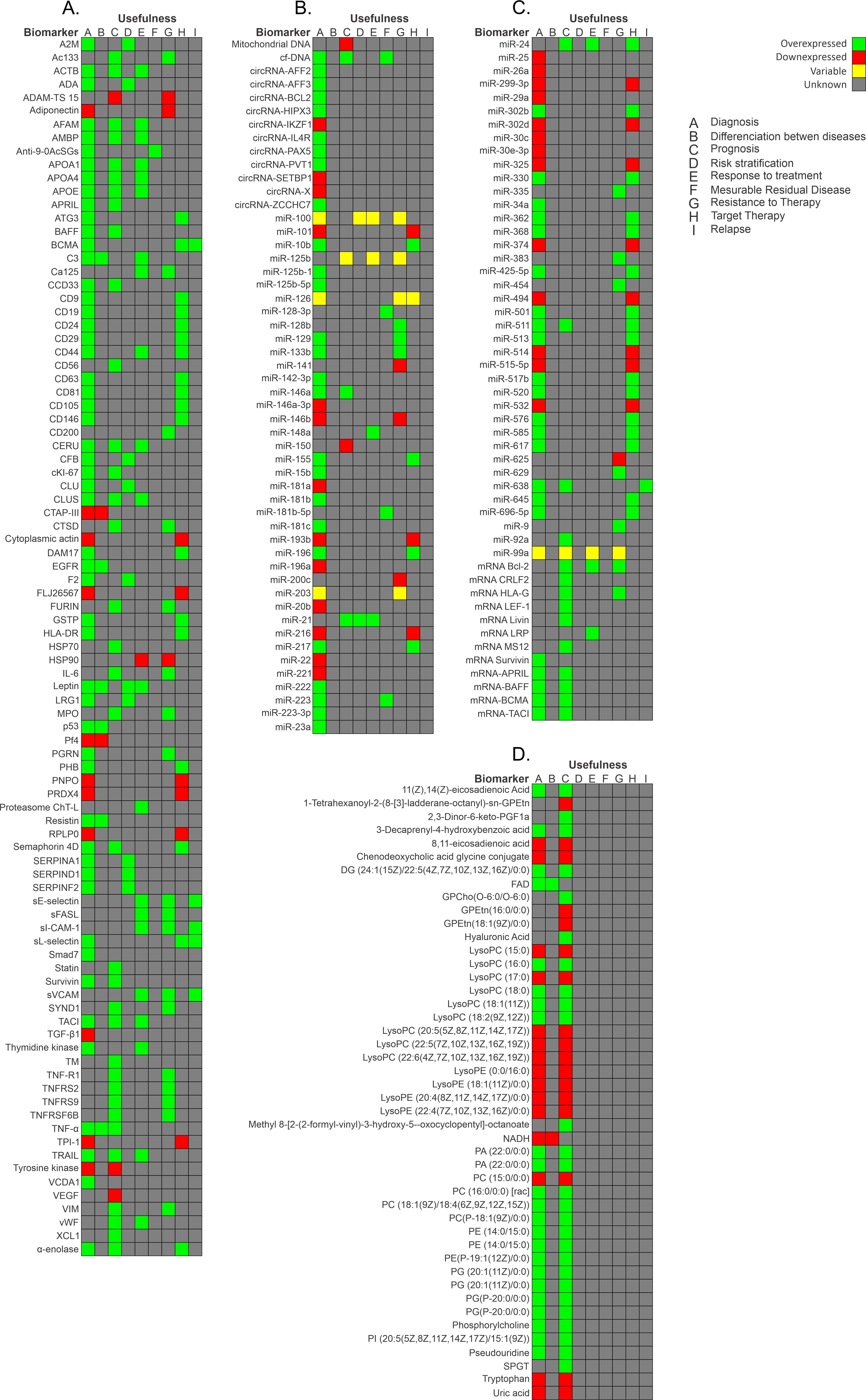
2.3. Metabolites Type BCP-ALL Biomarkers

Metabolites are the final products of gene expression and the direct outcome of enzymatic and protein activity. Metabolite profiles provide information about tumor microenvironments for ALL [30][70]. These metabolite profiles can be studied by metabolomics. “Metabolomics” involves the quantitative measurement of time of the metabolic responses of multicellular systems to pathophysiological stimuli or genetic modification [17]. Perturbation in the metabolism of patients at different disease stages could provide a unique metabolic signature to monitor treatment outcomes and disease progression [71]. Metabolism in cancer is a major research area in cancer biology. It examines how metabolic activities are changed in cancer cells compared to normal cells [30].
As shown in Table 1 and Figure 1D, researchers have reported metabolic differences in patients with ALL compared to healthy controls. Musharraf et al. [70] compared the metabolite profiles in the serum of 96 patients with ALL, AML, and APA, obtained by using the nuclear magnetic resonance spectroscopy technique. The authors reported high lactate levels and low levels of alanine, glutamine, histidine, lysine, valine, and proline. They theorized that patients adopt a secondary metabolic pathway to generate glucose because cancer cells require more glucose than can be provided by glycolysis. These findings are in contrast with those reported by Morad et al. [30], who evaluated the plasma of patients with ALL (n = 14), AML (n = 16), and breast cancer (n = 25). They reported high threonine, proline, glycine, alanine and lysine levels and low lactate levels in the ALL group. These changes could be due to tumor tissue competing for nitrogen compounds found in the amino acid structure [30][70]. Another group found that fatty acids were elevated as reservoirs, corresponding with an accumulation of carnitine, which plays a role in fatty acid metabolism [70]. Upon closer analysis, there seem to be notable variations in the approaches employed, which could explain the discrepancies in the outcomes. A crucial factor to consider is that the patient selection was comprised of diverse cancer types, like breast cancer and aplastic anemia, which may have influenced the results. Furthermore, though both studies utilized nuclear magnetic resonance spectroscopy, the validation techniques differed. One study used variable importance in projection values, while the other used principal component analysis. To gain a comprehensive understanding of the implications of these disparities, providing further context and specific examples would be highly beneficial. Overall, with a more thoughtful and informed examination, people can better appreciate the nuances of these findings.
In a recent study focused on developing a novel photo-diagnostic strategy using fluorescence emission spectra, the authors used plasma or red blood cell extract from 45 patients with AL. They found four metabolites that were altered and correlated: nicotinamide adenine dinucleotide (NAD) + hydrogen (H) (NADH), FAD, tyrosine kinase, and tryptophan (p < 0.05). NADH and tryptophan were decreased in patients with ALL, and there was a connection between tyrosine kinase and tryptophan. However, FAD was increased in patients with AL. Thus, the AL diagnosis can be made based on the FAD, NADH, tyrosine kinase, and tryptophan levels, but the ratios can help to discern between AML and ALL [8].

References

  1. Fujita, T.C.; Sousa-Pereira, N.; Amarante, M.K.; Watanabe, M.A.E. Acute lymphoid leukemia etiopathogenesis. Mol. Biol. Rep. 2021, 48, 817–822.
  2. Kaplan, J.A. Leukemia in Children. Pediatr. Rev. 2019, 40, 319–331.
  3. Ahmed, M.B.; Shehata, H.H.; Moussa, M.; Ibrahim, T.M. Prognostic significance of survivin and tumor necrosis factor-alpha in adult acute lymphoblastic leukemia. Clin. Biochem. 2012, 45, 112–116.
  4. Wang, D.; Lv, Y.Q.; Liu, Y.F.; Du, X.J.; Li, B. Differential protein analysis of lymphocytes between children with acute lymphoblastic leukemia and healthy children. Leuk. Lymphoma 2013, 54, 381–386.
  5. Swerdlow, S. WHO Classification of Haematopoietic and Lymphoid Tissues; International Agency for Research on Cancer: Lyon, France, 2017.
  6. Fayed, D.; Donia, T.; El-Shanshory, M.; Ali, E.M.M.; Mohamed, T.M. Evaluation of MicroRNA92, MicroRNA638 in Acute Lymphoblastic Leukemia of Egyptian Children. Asian Pac. J. Cancer Prev. 2021, 22, 1567–1572.
  7. Shi, L.; Zhang, J.; Wu, P.; Feng, K.; Li, J.; Xie, Z.; Xue, P.; Cai, T.; Cui, Z.; Chen, X.; et al. Discovery and identification of potential biomarkers of pediatric acute lymphoblastic leukemia. Proteome Sci. 2009, 7, 7.
  8. Masilamani, V.; Devanesan, S.; AlSalhi, M.S.; AlQahtany, F.S.; Farhat, K.H. Fluorescence spectral detection of acute lymphoblastic leukemia (ALL) and acute myeloid leukemia (AML): A novel photodiagnosis strategy. Photodiag. Photodyn. Ther. 2020, 29, 101634.
  9. Shahid, S.; Shahid, W.; Shaheen, J.; Akhtar, M.W.; Sadaf, S. Circulating miR-146a expression as a non-invasive predictive biomarker for acute lymphoblastic leukemia. Sci. Rep. 2021, 11, 22783.
  10. Hassan, N.M.; Refaat, L.A.; Ismail, G.N.; Abdellateif, M.; Fadel, S.A.; AbdelAziz, R.S. Diagnostic, prognostic and predictive values of miR-100 and miR-210 in pediatric acute lymphoblastic Leukemia. Hematology 2020, 25, 405–413.
  11. Brown, P.; Inaba, H.; Annesley, C.; Beck, J.; Colace, S.; Dallas, M.; DeSantes, K.; Kelly, K.; Kitko, C.; Lacayo, N.; et al. Pediatric Acute Lymphoblastic Leukemia, Version 2.2020, NCCN Clinical Practice Guidelines in Oncology. J. Natl. Compr. Canc. Netw. 2020, 18, 81–112.
  12. Brown, P.A.; Shah, B.; Advani, A.; Aoun, P.; Boyer, M.W.; Burke, P.W.; DeAngelo, D.J.; Dinner, S.; Fathi, A.T.; Gauthier, J.; et al. Acute Lymphoblastic Leukemia, Version 2.2021, NCCN Clinical Practice Guidelines in Oncology. J. Natl. Compr. Cancer Netw. 2021, 19, 1079–1109.
  13. Alaggio, R.; Amador, C.; Anagnostopoulos, I.; Attygalle, A.D.; Araujo, I.B.O.; Berti, E.; Bhagat, G.; Borges, A.M.; Boyer, D.; Calaminici, M.; et al. The 5th edition of the World Health Organization Classification of Haematolymphoid Tumours: Lymphoid Neoplasms. Leukemia 2022, 36, 1720–1748.
  14. Yu, R.; Yang, S.; Liu, Y.; Zhu, Z. Identification and validation of serum autoantibodies in children with B-cell acute lymphoblastic leukemia by serological proteome analysis. Proteome Sci. 2022, 20, 3.
  15. Swellam, M.; El-Khazragy, N. Clinical impact of circulating microRNAs as blood-based marker in childhood acute lymphoblastic leukemia. Tumour Biol. 2016, 37, 10571–10576.
  16. Nabhan, M.; Louka, M.L.; Khairy, E.; Tash, F.; Ali-Labib, R.; El-Habashy, S. MicroRNA-181a and its target Smad 7 as potential biomarkers for tracking child acute lymphoblastic leukemia. Gene 2017, 628, 253–258.
  17. Bai, Y.; Zhang, H.; Sun, X.; Sun, C.; Ren, L. Biomarker identification and pathway analysis by serum metabolomics of childhood acute lymphoblastic leukemia. Clin. Chim. Acta 2014, 436, 207–216.
  18. Carceles-Alvarez, A.; Ortega-Garcia, J.A.; Lopez-Hernandez, F.A.; Fuster-Soler, J.L.; Ramis, R.; Kloosterman, N.; Castillo, L.; Sanchez-Solis, M.; Claudio, L.; Ferris-Tortajada, J. Secondhand smoke: A new and modifiable prognostic factor in childhood acute lymphoblastic leukemias. Environ. Res. 2019, 178, 108689.
  19. Vrooman, L.M.; Silverman, L.B. Treatment of Childhood Acute Lymphoblastic Leukemia: Prognostic Factors and Clinical Advances. Curr. Hematol. Malig. Rep. 2016, 11, 385–394.
  20. Aly, R.M.; Yousef, A.B. Prognostic significance of lymphoid enhancer-binding factor-1 expression in egyptian adult B-acute lymphocytic leukemia patients. Turk. J. Haematol. 2015, 32, 15–20.
  21. Gutierrez-Aguirre, C.H.; Flores-Jimenez, J.A.; Alatorre-Ricardo, J.; Cantu-Rodriguez, O.G.; Rosas-Taraco, A.; Salazar-Riojas, R.; Jaime-Perez, J.C.; Sanchez-Cardenas, M.; Lopez-Silva, L.; Martinez-Castilla, A.M.; et al. The prognostic significance of serum XCL1 concentration in patients with acute lymphoblastic leukemia: A pilot study. Ann. Hematol. 2017, 96, 2015–2024.
  22. Kruse, A.; Abdel-Azim, N.; Kim, H.N.; Ruan, Y.; Phan, V.; Ogana, H.; Wang, W.; Lee, R.; Gang, E.J.; Khazal, S.; et al. Minimal Residual Disease Detection in Acute Lymphoblastic Leukemia. Int. J. Mol. Sci. 2020, 21, 1054.
  23. Zhang, Q.; Shi, C.; Han, L.; Jain, N.; Roberts, K.G.; Ma, H.; Cai, T.; Cavazos, A.; Tabe, Y.; Jacamo, R.O.; et al. Inhibition of mTORC1/C2 signaling improves anti-leukemia efficacy of JAK/STAT blockade in CRLF2 rearranged and/or JAK driven Philadelphia chromosome-like acute B-cell lymphoblastic leukemia. Oncotarget 2018, 9, 8027–8041.
  24. Cante-Barrett, K.; Spijkers-Hagelstein, J.A.; Buijs-Gladdines, J.G.; Uitdehaag, J.C.; Smits, W.K.; van der Zwet, J.; Buijsman, R.C.; Zaman, G.J.; Pieters, R.; Meijerink, J.P. MEK and PI3K-AKT inhibitors synergistically block activated IL7 receptor signaling in T-cell acute lymphoblastic leukemia. Leukemia 2016, 30, 1832–1843.
  25. Tovar, C.F.; Zeron, H.M.; Romero, M.D.; Sanchez, Y.V.; Romero, I.T. Glycogen Synthase Kinase-3beta (GSK-3beta) and Nuclear Factor Kappa-B (NFKB) in Childhood Acute Lymphoblastic Leukemia. Adv. Clin. Exp. Med. 2016, 25, 1139–1147.
  26. Vrooman, L.M.; Silverman, L.B. Childhood acute lymphoblastic leukemia: Update on prognostic factors. Curr. Opin. Pediatr. 2009, 21, 1–8.
  27. Aly, R.M.; Ghazy, H.F. Prognostic significance of MSI2 predicts unfavorable outcome in adult B-acute lymphoblastic leukemia. Int. J. Lab. Hematol. 2015, 37, 272–278.
  28. Aguirre-Guillen, W.A.; Angeles-Floriano, T.; Lopez-Martinez, B.; Reyes-Morales, H.; Zlotnik, A.; Valle-Rios, R. Omics techniques and biobanks to find new biomarkers for the early detection of acute lymphoblastic leukemia in middle-income countries: A perspective from Mexico. Bol. Med. Hosp. Infant. Mex. 2017, 74, 227–232.
  29. Yan, M.; Liu, H.; Xu, J.; Cen, X.; Wang, Q.; Xu, W.; Wang, W.; Qiu, Z.; Ou, J.; Dong, Y.; et al. Expression of human Krüppel-like factor 3 in peripheral blood as a promising biomarker for acute leukemia. Cancer Med. 2020, 9, 2803–2811.
  30. Morad, H.M.; Abou-Elzahab, M.M.; Aref, S.; El-Sokkary, A.M.A. Diagnostic Value of (1)H NMR-Based Metabolomics in Acute Lymphoblastic Leukemia, Acute Myeloid Leukemia, and Breast Cancer. ACS Omega 2022, 7, 8128–8140.
  31. Hussan, S.S.; Maqsood, N.; Wang, Q.; Tao, S.; Sadaf, S. A panel of epigenetically dysregulated Wnt signaling pathway genes for non-invasive diagnosis of pediatric acute lymphoblastic leukemia. Cancer Biomark. 2021, 32, 459–470.
  32. Li, G.; Hu, J.; Hu, G. Biomarker Studies in Early Detection and Prognosis of Breast Cancer. Adv. Exp. Med. Biol. 2017, 1026, 27–39.
  33. Damanti, C.C.; Gaffo, E.; Lovisa, F.; Garbin, A.; Di Battista, P.; Gallingani, I.; Tosato, A.; Pillon, M.; Carraro, E.; Mascarin, M.; et al. MiR-26a-5p as a Reference to Normalize MicroRNA qRT-PCR Levels in Plasma Exosomes of Pediatric Hematological Malignancies. Cells 2021, 10, 101.
  34. Swerdlow, S.H.; Campo, E.; Harris, N.L.; Jaffe, E.S.; Pileri, S.A.; Stein, H.; Thiele, J.; Vardiman, J.W. WHO Classification of Tumours of Haematopoietic and Lymphoid Tissues; International Agency for Research on Cancer: Lyon, France, 2008; Volume 2.
  35. Cristea, I.M.; Gaskell, S.J.; Whetton, A.D. Proteomics techniques and their application to hematology. Blood 2004, 103, 3624–3634.
  36. Cui, J.W.; Wang, J.; He, K.; Jin, B.F.; Wang, H.X.; Li, W.; Kang, L.H.; Hu, M.R.; Li, H.Y.; Yu, M.; et al. Proteomic analysis of human acute leukemia cells: Insight into their classification. Clin. Cancer Res. 2004, 10, 6887–6896.
  37. Malcles, M.H.; Wang, H.W.; Koumi, A.; Tsai, Y.H.; Yu, M.; Godfrey, A.; Boshoff, C. Characterisation of the anti-apoptotic function of survivin-DeltaEx3 during TNFalpha-mediated cell death. Br. J. Cancer 2007, 96, 1659–1666.
  38. Aref, S.; Salama, O.; Shamaa, S.; El-Refaie, M.; Mourkos, H. Angiogenesis factor pattern differs in acute lymphoblastic leukemia and chronic lymphocytic leukemia. Hematology 2007, 12, 319–324.
  39. Potapnev, M.P.; Petyovka, N.V.; Belevtsev, M.V.; Savitskiy, V.P.; Migal, N.V. Plasma level of tumor necrosis factor-alpha (TNF-alpha) correlates with leukocytosis and biological features of leukemic cells, but not treatment response of children with acute lymphoblastic leukemia. Leuk. Lymphoma 2003, 44, 1077–1079.
  40. Zakhary, N.I.; Boshra, S.A.; El-Sawalhi, M.M.; Fahim, A.T.; Ebeid, E.N. Insulin-like growth factor system in Egyptian children with acute lymphoblastic leukemia. Genet. Test. Mol. Biomark. 2012, 16, 1067–1072.
  41. Pal, S.; Bandyopadhyay, S.; Chatterjee, M.; Bhattacharya, D.K.; Minto, L.; Hall, A.G.; Mandal, C. Antibodies against 9-O-acetylated sialoglycans: A potent marker to monitor clinical status in childhood acute lymphoblastic leukemia. Clin. Biochem. 2004, 37, 395–403.
  42. Liang, T.; Wang, N.; Li, W.; Li, A.; Wang, J.; Cui, J.; Liu, N.; Li, Y.; Li, L.; Yang, G.; et al. Identification of complement C3f-desArg and its derivative for acute leukemia diagnosis and minimal residual disease assessment. Proteomics 2010, 10, 90–98.
  43. Abdel-Aziz, M.M. Clinical significance of serum p53 and epidermal growth factor receptor in patients with acute leukemia. Asian Pac. J. Cancer Prev. 2013, 14, 4295–4299.
  44. Pane, F.; Savoia, M.; Fortunato, G.; Camera, A.; Rotoli, B.; Salvatore, F.; Sacchetti, L. Serum pseudouridine in the diagnosis of acute leukaemias and as a novel prognostic indicator in acute lymphoblastic leukaemia. Clin. Biochem. 1993, 26, 513–520.
  45. Zhu, S.; Xing, C.; Li, R.; Cheng, Z.; Deng, M.; Luo, Y.; Li, H.; Zhang, G.; Sheng, Y.; Peng, H.; et al. Proteomic profiling of plasma exosomes from patients with B-cell acute lymphoblastic leukemia. Sci. Rep. 2022, 12, 11975.
  46. Elgendi, H.M.; Mekawy, M.A.; Abdel Wahab, S.E.; Tawfik, L.M.; Ismail, E.A.; Adly, A.A. AC133 expression in egyptian children with acute leukemia: Impact on treatment response and disease outcome. J. Pediatr. Hematol. Oncol. 2010, 32, 286–293.
  47. Mohammadi, M.; Amirmahani, F.; Goharrizi, K.J.; Pakzad, R.; Dolat, H. Evaluating the expression level of Survivin gene in different groups of B-cell acute lymphoblastic leukemia patients of Iran. Mol. Biol. Rep. 2019, 46, 2679–2684.
  48. Alkhouly, N.; Shehata, I.; Ahmed, M.B.; Shehata, H.; Hassan, S.; Ibrahim, T. HLA-G expression in acute lymphoblastic leukemia: A significant prognostic tumor biomarker. Med. Oncol. 2013, 30, 460.
  49. Swellam, M.; Hashim, M.; Mahmoud, M.S.; Ramadan, A.; Hassan, N.M. Aberrant Expression of Some Circulating miRNAs in Childhood Acute Lymphoblastic Leukemia. Biochem. Genet. 2018, 56, 283–294.
  50. Luna-Aguirre, C.M.; de la Luz Martinez-Fierro, M.; Mar-Aguilar, F.; Garza-Veloz, I.; Trevino-Alvarado, V.; Rojas-Martinez, A.; Jaime-Perez, J.C.; Malagon-Santiago, G.I.; Gutierrez-Aguirre, C.H.; Gonzalez-Llano, O.; et al. Circulating microRNA expression profile in B-cell acute lymphoblastic leukemia. Cancer Biomark. 2015, 15, 299–310.
  51. El-maadawy, E.A.; Bakry, R.M.; Moussa, M.M.; El-Naby, S.; Talaat, R.M.J.C. Alteration in miRNAs expression in paediatric acute lymphocyticleukaemia: Insight into patients’ therapeutic response. Pharmacol. Pharm. 2021, 48, 35–43.
  52. Ohyashiki, J.H.; Umezu, T.; Kobayashi, C.; Hamamura, R.S.; Tanaka, M.; Kuroda, M.; Ohyashiki, K. Impact on cell to plasma ratio of miR-92a in patients with acute leukemia: In vivo assessment of cell to plasma ratio of miR-92a. BMC Res. Notes 2010, 3, 347.
  53. El-Khazragy, N.; Elshimy, A.A.; Hassan, S.S.; Matbouly, S.; Safwat, G.; Zannoun, M.; Riad, R.A. Dysregulation of miR-125b predicts poor response to therapy in pediatric acute lymphoblastic leukemia. J. Cell Biochem. 2018, 120, 7428–7438.
  54. Nemes, K.; Csoka, M.; Nagy, N.; Mark, A.; Varadi, Z.; Danko, T.; Kovacs, G.; Kopper, L.; Sebestyen, A. Expression of certain leukemia/lymphoma related microRNAs and its correlation with prognosis in childhood acute lymphoblastic leukemia. Pathol. Oncol. Res. 2015, 21, 597–604.
  55. Gao, Y.J.; He, Y.J.; Yang, Z.L.; Shao, H.Y.; Zuo, Y.; Bai, Y.; Chen, H.; Chen, X.C.; Qin, F.X.; Tan, S.; et al. Increased integrity of circulating cell-free DNA in plasma of patients with acute leukemia. Clin. Chem. Lab. Med. 2010, 48, 1651–1656.
  56. Abu Sabaa, A.; Shen, Q.; Lennmyr, E.B.; Enblad, A.P.; Gammelgard, G.; Molin, D.; Hein, A.; Freyhult, E.; Kamali-Moghaddam, M.; Hoglund, M.; et al. Plasma protein biomarker profiling reveals major differences between acute leukaemia, lymphoma patients and controls. N Biotechnol. 2022, 71, 21–29.
  57. Wik, L.; Nordberg, N.; Broberg, J.; Bjorkesten, J.; Assarsson, E.; Henriksson, S.; Grundberg, I.; Pettersson, E.; Westerberg, C.; Liljeroth, E.; et al. Proximity Extension Assay in Combination with Next-Generation Sequencing for High-throughput Proteome-wide Analysis. Mol. Cell. Proteom. MCP 2021, 20, 100168.
  58. Limijadi, E.K.S.; Budiwijono, I.; Samsuria, I.K.; Adhipireno, P.; Devi, W.R. Coagulation and Fibrinolysis Profiles of Acute Myeloblastic Leukemia: Preliminary Assessment of Hypercoagulability. Eur. J. Mol. Clin. Med. 2021, 8, 607–615.
  59. Seftalioglu, A.; Karakus, S. Syndecan-1/CD138 expression in normal myeloid, acute lymphoblastic and myeloblastic leukemia cells. Acta Histochem. 2003, 105, 213–221.
  60. Lenting, P.J.; Christophe, O.D.; Denis, C.V. von Willebrand factor biosynthesis, secretion, and clearance: Connecting the far ends. Blood 2015, 125, 2019–2028.
  61. Aggarwal, B.B. Signalling pathways of the TNF superfamily: A double-edged sword. Nat. Rev. Immunol. 2003, 3, 745–756.
  62. Maimaitiyiming, Y.; Ye, L.; Yang, T.; Yu, W.; Naranmandura, H. Linear and Circular Long Non-Coding RNAs in Acute Lymphoblastic Leukemia: From Pathogenesis to Classification and Treatment. Int. J. Mol. Sci. 2022, 23, 4442.
  63. Li, Y.; Xu, J.; Shao, T.; Zhang, Y.; Chen, H.; Li, X. RNA Function Prediction. Methods Mol. Biol. 2017, 1654, 17–28.
  64. He, L.; He, X.; Lim, L.P.; de Stanchina, E.; Xuan, Z.; Liang, Y.; Xue, W.; Zender, L.; Magnus, J.; Ridzon, D.; et al. A microRNA component of the p53 tumour suppressor network. Nature 2007, 447, 1130–1134.
  65. Esquela-Kerscher, A.; Slack, F.J. Oncomirs—microRNAs with a role in cancer. Nat. Rev. Cancer 2006, 6, 259–269.
  66. Qu, S.; Yang, X.; Li, X.; Wang, J.; Gao, Y.; Shang, R.; Sun, W.; Dou, K.; Li, H. Circular RNA: A new star of noncoding RNAs. Cancer Lett. 2015, 365, 141–148.
  67. Perez de Acha, O.; Rossi, M.; Gorospe, M. Circular RNAs in Blood Malignancies. Front. Mol. Biosci. 2020, 7, 109.
  68. Gaffo, E.; Boldrin, E.; Dal Molin, A.; Bresolin, S.; Bonizzato, A.; Trentin, L.; Frasson, C.; Debatin, K.-M.; Meyer, L.H.; te Kronnie, G.; et al. Circular RNA differential expression in blood cell populations and exploration of circRNA deregulation in pediatric acute lymphoblastic leukemia. Sci. Rep. 2019, 9, 14670.
  69. Chen, J.; Li, Y.; Zheng, Q.; Bao, C.; He, J.; Chen, B.; Lyu, D.; Zheng, B.; Xu, Y.; Long, Z.; et al. Circular RNA profile identifies circPVT1 as a proliferative factor and prognostic marker in gastric cancer. Cancer Lett. 2017, 388, 208–219.
  70. Musharraf, S.G.; Siddiqui, A.J.; Shamsi, T.; Choudhary, M.I.; Rahman, A.U. Serum metabonomics of acute leukemia using nuclear magnetic resonance spectroscopy. Sci. Rep. 2016, 6, 30693.
  71. Bannur, Z.; Teh, L.K.; Hennesy, T.; Rosli, W.R.; Mohamad, N.; Nasir, A.; Ankathil, R.; Zakaria, Z.A.; Baba, A.; Salleh, M.Z. The differential metabolite profiles of acute lymphoblastic leukaemic patients treated with 6-mercaptopurine using untargeted metabolomics approach. Clin. Biochem. 2014, 47, 427–431.
More
Information
Subjects: Hematology
Contributors MDPI registered users' name will be linked to their SciProfiles pages. To register with us, please refer to https://encyclopedia.pub/register : , , , , ,
View Times: 376
Revisions: 2 times (View History)
Update Date: 30 Aug 2023
1000/1000
Video Production Service