Submitted Successfully!
To reward your contribution, here is a gift for you: A free trial for our video production service.
Thank you for your contribution! You can also upload a video entry or images related to this topic.
Version Summary Created by Modification Content Size Created at Operation
1 -- 5336 2023-08-24 18:10:51 |
2 format change + 4 word(s) 5340 2023-08-25 03:08:56 |

Video Upload Options

Do you have a full video?

Confirm

Are you sure to Delete?
Cite
If you have any further questions, please contact Encyclopedia Editorial Office.
Zhang, Z.; Bao, X.; Lin, C. Gene Editing in Pluripotent Stem Cells. Encyclopedia. Available online: https://encyclopedia.pub/entry/48461 (accessed on 09 September 2024).
Zhang Z, Bao X, Lin C. Gene Editing in Pluripotent Stem Cells. Encyclopedia. Available at: https://encyclopedia.pub/entry/48461. Accessed September 09, 2024.
Zhang, Zhenwu, Xinyu Bao, Chao-Po Lin. "Gene Editing in Pluripotent Stem Cells" Encyclopedia, https://encyclopedia.pub/entry/48461 (accessed September 09, 2024).
Zhang, Z., Bao, X., & Lin, C. (2023, August 24). Gene Editing in Pluripotent Stem Cells. In Encyclopedia. https://encyclopedia.pub/entry/48461
Zhang, Zhenwu, et al. "Gene Editing in Pluripotent Stem Cells." Encyclopedia. Web. 24 August, 2023.
Gene Editing in Pluripotent Stem Cells
Edit

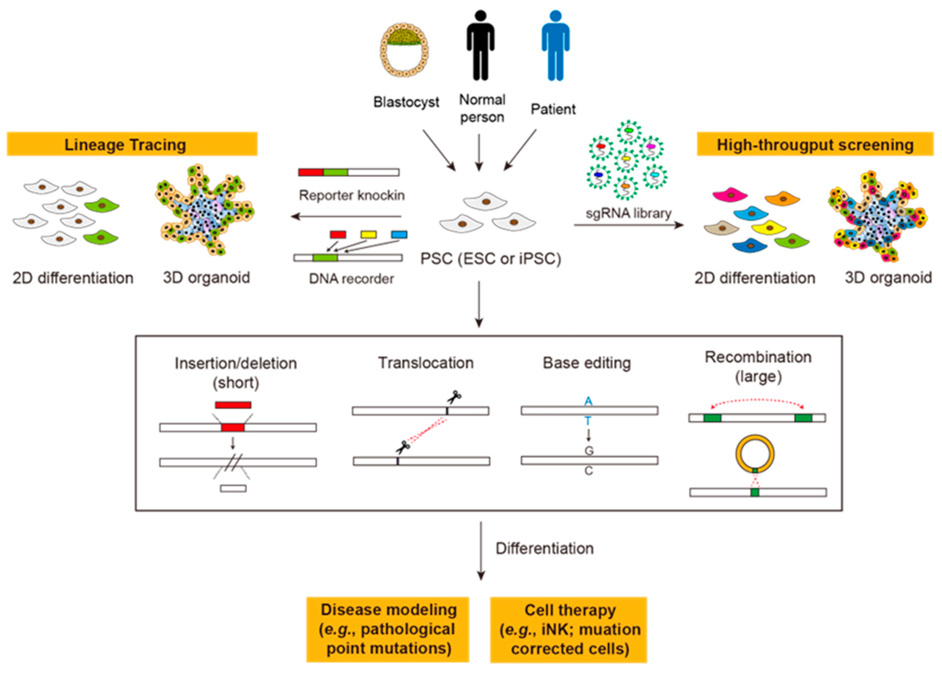
Gene editing in human pluripotent stem cells (PSCs), including embryonic stem cells (ESCs) and induced pluripotent stem cells (iPSCs), is highly relevant to clinical cell therapy and thus should be examined with particular caution. First, since all mutations in PSCs will be carried to all their progenies, off-target edits of editors will be amplified. Second, due to the hypersensitivity of PSCs to DNA damage, double-strand breaks (DSBs) made by gene editing could lead to low editing efficiency and the enrichment of cell populations with defective genomic safeguards. In this regard, DSB-independent gene editing tools, such as base editors and prime editors, are favored due to their nature to avoid these consequences. With more understanding of the microbial world, new systems, such as Cas-related nucleases, transposons, and recombinases, are also expanding the toolbox for gene editing.

pluripotent stem cell induced pluripotent stem cell CRISPR-Cas9 base editor prime editor gene editing

1. Introduction

Pluripotent stem cells (PSCs) possess two unique characteristics, indefinite self-renewal and the potential to differentiate into nearly all cell types of three germ layers, therefore holding great promise for regenerative medicine. The two major types of PSCs are embryonic stem cells (ESCs) derived from the inner cell mass of the preimplantation embryo [1] and induced pluripotent stem cells (iPSCs) generated by reprogramming of somatic cells [2][3]. With technical advances in the past decade, human iPSCs can now be generated by RNA viruses, episomal vectors, or chemical cocktails, avoiding genomic integrations [4][5][6][7][8][9]. Compared with human ESCs, iPSCs are generated from autologous cells and are easier to obtain, enabling iPSCs to be applied in cell-based therapies [10][11][12]. For example, mutations of patient-derived iPSCs can be corrected and differentiated towards specific cell types for therapeutic purposes [13][14]. For cancer immunotherapy, autologous or immune-compatible iPSCs can be modified (e.g., introducing chimeric antigen receptors) and serve as unlimited, “off-the-shelf” sources of engineered immune cells [15][16].
In addition to clinical purposes, PSCs also possess great value in basic research [17]. PSCs are long-standing models for investigating determinants or modulators of lineage specification or terminal differentiation. The advent of organoid cultures further expanded PSC applications [18][19]. Organoids are self-organized cell aggregates that recapitulate cellular compositions and organizations of corresponding tissues, and have been widely employed as in vitro models to study tissue development or diseases [20][21][22][23][24]. PSC-based in vitro differentiation models in 2D and 3D conditions are also platforms for high-throughput screening [25][26] or lineage tracing [27][28], serving as precious tools for studying human-specific development or diseases lacking appropriate mouse models (Figure 1).
Figure 1. Gene editing in PSC-based basic research and clinical applications. ESC, embryonic stem cell; iPSC, induced pluripotent stem cell; iNK, iPSC-derived NK cells. See the text for details.
Many of those aforementioned applications require gene editing in PSCs. A major merit of performing gene editing in PSCs is the high stability in both the genome and the cell fate potential. Thus, the integrity of the genome of engineered PSCs can be thoroughly examined before any application. Recently, the ability to edit the genome of PSCs has been greatly elevated with the development of gene editing tools, especially programmable nucleases. The ease of use and the high editing efficiency of programmable nucleases greatly facilitate the applications of PSCs in basic research, such as knock-out/knock-in, disease modeling, and correction of genetic mutations [29][30][31]. Nonetheless, gene editing in PSCs also requires careful evaluation due to their spectacular properties: comparing with adult (stem) cells which have limited longevity, PSCs can propagate almost indefinitely and pass all mutations to their progenies, making the preciseness of gene editing in PSCs of great concern. This concern is further exacerbated with recent studies demonstrating that gene editing mediated by CRISPR associated protein 9 (Cas9)-induced DNA cleavage in human PSCs produces genome-wide mutations and rearrangements [32][33]. Moreover, the selection of successfully edited PSC clones also favors the accumulation of p53 mutations, hampering further utilization of this powerful technology in human PSCs [33][34] (Table 1).
Table 1. Multifaceted comparison of gene editing tools.

2. DSB-Mediated Gene Editing by Programmable Nucleases

Since late 1980, homologous recombination (HR) has been widely employed for genome editing in mouse ESCs to create genetically modified mice, establishing a paradigm for studying gene functions, disease mechanisms, and lineage specifications [35]. In a general protocol, donor DNAs with homologous arms are electroporated to mouse ESCs, and the HR-mediated editing (knock-out or knock-in) occurs spontaneously at very low frequencies [36]. This laborious and time-consuming procedure was changed by groundbreaking works of the Haber and Jasin groups, demonstrating that the induction of double-strand breaks (DSBs) by endonucleases at sites aimed to be edited could trigger DNA repair pathways and dramatically increase the efficiencies of HR in yeasts and mammalian cells [37][38]. On the other hand, repair of DSBs by non-homologues end joining (NHEJ) can generate insertions/deletions (indels), ablating the protein expression or function. These findings inspired researchers to look for programmable nucleases that can induce DSBs at desired sites. To date, zinc finger nucleases (ZFNs), transcription activator-like effector nucleases (TALENs), and CRISPR-Cas systems are the most frequently employed methods.
Different cells exhibit distinct sensitivities to DSBs. PSCs, as derived from early embryos, have evolved at least two mechanisms to keep low DNA mutation rates compared to somatic cells, thus protecting the genome integrity from the accumulation of genetic mutations. First, PSCs possess a superior ability to repair DNA damages by expressing abundant mismatch repair proteins or avoiding error-prone repair pathways [39][40][41][42]. The high DNA repair capability also suggests the fast removal of the DSB marker γ-H2AX, thus antagonizing its association with apoptosis-inducing factor (AIF) for the formation of the “degradosome” that leads to chromatin remodeling and large-scale DNA fragmentation [43][44]. Second, once the level of DNA damage accumulates over a threshold, PSCs are highly prone to apoptosis, eliminating themselves from the whole population. Those self-protection mechanisms of PSCs are double-edged swords for applying those powerful DSB-dependent gene editing tools in PSCs, since the sensitivity of PSCs toward DNA damages could lead to the low editing efficiency or undesired loss/mutation of genomic safeguards [32][33]. Here, the researchers briefly review those three DSB-dependent programmable nucleases and their off-target effects in the context of PSCs, as their development and mechanisms have been extensively reviewed [30][45][46][47][48][49].

3. Base Editors

Since DSB-mediated editing raises concerns for undesired mutations in PSCs, base editors, which make base substitutions without introducing DSBs, are reasonably favored for gene editing in PSCs. Theoretically, six base editors will be needed for “any base to any base” substitutions (please refer to Chen et al. [50] for the illustration). Yet, pathogenic point mutations in humans are not evenly distributed (also Chen et al. [50] for the statistics), making it possible to cover most human diseases with fewer editors. The first two base editors reported, the cytidine base editor (CBE) and the adenine base editor (ABE), were realized by David Liu’s lab in 2016 and 2017, respectively [51][52]. CBE converts cytidines to thymines on one strand and thus can be used to create both C>T (C•T→T•A) and G>A (G•C→A•T) substitutions. Similarly, ABE substitutes adenines with guanines and thus can make both A>G (A•T→G•A) and T>C (T•A→C•G) substitutions. Those four types of editing cover ~60% of edits needed for correcting pathogenic mutations [50]. In 2020, two labs reported the glycosylase base editor that can make C>G and G>C conversions, which constitute ~10% of pathological mutations [53][54]. Recently, new editors were reported to be able to convert A to C or T [50][55], covering another ~25% of pathological mutations. In addition to these single-base editors, dual-base editors, which fuse two types of single-base editors, were also created to introduce multiple substitutions [56][57][58][59]. Together, these base editors constitute a tool collection for introducing or correcting point mutations in somatic or stem cells.

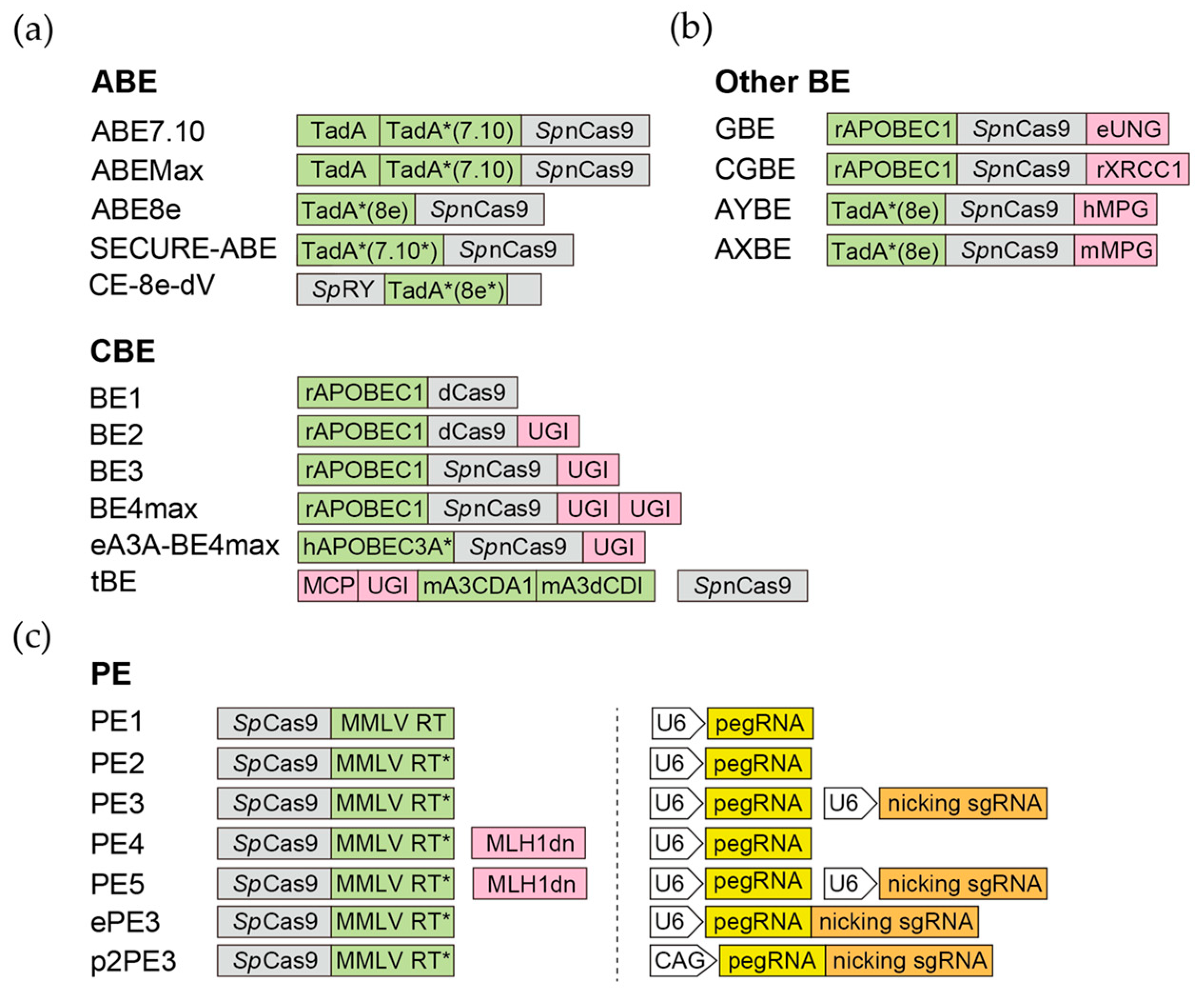
Figure 2. Base editors and prime editors. Representative versions of base editors and prime editors. The Cas9 derivatives are highlighted in grey, the key enzymatic components of BEs (a,b) and PEs (c) are highlighted in green, and the associated components are highlighted in pink. For PEs, the architectures of promoters, pegRNAs (yellow), and nicking sgRNAs (orange ) are also shown. The CAG promoter of P2PE3 can be replaced by other Pol II promoters.

3.1. Cytidine Base Editor (CBE)

CBEs take advantage of the cytidine deaminase activity, which converts cytidines to uracils (U), equivalent to T in base pairing, for base substitution. The rat APOBEC1 (apolipoprotein B mRNA editing enzyme, catalytic polypeptide-like 1, or rAPOBEC1) and the sea lamprey AID (activation-induced cytidine deaminase) were first employed in the BE series [60] and Target-AID [61], respectively. Cytidine deaminases from different species, including the CDA, AID, and APOBEC3 family, are also employed to build CBEs with different editing efficiencies and sequence preferences [62]. BE3 and BE4 are the third and fourth generation of BE, containing nCas9, rAPOBEC1, as well as one (BE3) or two (BE4) copies of uracil DNA glycosylase inhibitor (UGI) from the Bacillus subtilis bacteriophage [60]. UGI can suppress the repair of U by inhibiting the uracil DNA glycosylase (UNG), which excises uracil bases to form abasic sites for base excision repair (BER). To date, more than thirty CBE variants have been made by changing the editing window, expanding the PAM compatibility, or increasing/decreasing their on-target/off-target activities [63]. Notably, despite its successful application in multiple cell lines, BE3 exhibits lower editing efficiency in human PSCs [64] and may need further optimization for better efficiency [65].

3.2. Adenine Base Editor (ABE)

In 2017, David Liu’s lab reported the first ABE, which realized the A>G and T>C substitutions [52]. Theoretically, deamination of adenosines results in inosines (I), which could complement with cytidines and be eventually converted to guanines by the mismatch repair. However, there are no known DNA adenine deaminases. To solve this issue, Gaudelli et al. subjected the E. coli tRNA adenine deaminase TadA to extensive evolution to alter its activity towards DNA [52]. ABE7.10, which contains a wild-type TadA and an evolved TadA* (contains 14 amino acid substitutions in the catalytic domain), was finally retrieved with the highest efficiency of converting A•T to G•C [52]. This breakthrough opens the gate for the subsequent derivation of ABE variants with improved nuclear localization/expression (ABEmax) [66] or smaller size and faster editing kinetics (ABE8e) [67]. Recently, several groups, including us, modified those ABEs to further reduce their off-targeting activities [68][69][70] or expand the PAM compatibility [71][72]. Notably, compared with CBE, ABE exhibits high product purity and low rates of indels in mammalian cells, including human PSCs, possibly due to their lack of efficient glycosylase to initiate BER [52].

3.3. Glycosylase Base Editor (GBE) and C-to-G Base Editor (CGBE)

In 2020, three research groups reported base editors that can make C>G and G>C base transversions (purine > pyrimidine or pyrimidine > purine) in mammalian cells [53][54][64]. All three base editors employ nCas9 and rAPOBEC1 (the same one CBE uses), while two of them (Kurt et al. [GBE] and Zhao et al. [CGBE]) replace the UGI with UNG and Chen et al. use XRCC1 protein instead of UGI. The UNG excises the U base created by the deaminase and generates the apurinic/apyrimidinic (AP) site that, in combination with the nick made by nCas9, initiates the DNA repair process (translesion synthesis) which favors insertion of G at the AP site. The C-to-G BE designed by Chen et al. uses XRCC1 to recruit BER proteins to repair the AP site created by endogenous UNG [64]. GBE and CGBE are further modified or optimized to relieve the PAM restriction, as well as to be more predictable and purer [73][74]. Recently, the Li lab reported a TadA-derived C-to-G base editor, Td-CGBE, which is based on an N46L variant of TadA-8e [75].

3.4. Adenine Transversion Editors (AYBE and AXBE)

As mentioned above, the A>C substitution is required to reverse ~25% pathological point mutations. Recently, two breakthrough studies reported base editors that can transverse A to C or T in mammalian cells [50][55]. Both groups employ a strategy similar to GBE/CGBE, but aim at creating the AP site on A instead of C (in the GBE/CGBE case), which is expected to be mutagenized by the DNA repair pathway. To achieve this, Tong et al. constructed the adenine transverse base editor (AYBE, Y = C or T) by fusing the ABE8e with an engineered human hypoxanthine glycosylase enzyme, N-methylpurine DNA glycosylase (MPG, also called AAG). MPG excises the hypoxanthine group from the inosine produced by ABE, resulting in an AP site. The AP site will then be processed by the translesion synthesis pathway and replaced by C or T as the most common outcomes [55]. Chen et al. employed a similar strategy but used mouse AAG (MPG) instead of the human one, creating the AXBE. Importantly, by mutagenesis and Cas embedding strategies, Chen et al. further created the ACBE-Q editor, which exhibited high A>C activity and reduced A>G bystander substitutions [50]. Although the purity and efficiency of adenine transversion editors remain to be improved, these two editors have substantially expanded the potential of base editing.

3.5. Pros and Cons of Using Base Editors in PSCs

Although base editors cannot introduce indels or HR, they still possess wide applications in correcting point mutations, creating premature stop codons (knockout), and alternating splicing events. In contrast to DSB-dependent genome editing, base editors are considerably favored since most of them use nCas9, which only generates nicks. This advantage is particularly important for PSCs, in which the activation of p53-dependent DNA damage responses leads to detrimental consequences [33]. However, the deaminases may lead to undesired off-target edits on RNA or DNA in PSCs. Since off-target mutations on RNA could cause undesired phenotypes, and mutations on DNA will be transferred to all differentiated progenies, the off-target issue is one of the major concerns for applying base editors in PSCs.

4. Prime Editor (PE)

In 2019, a versatile gene-editing tool, prime editor (PE), was developed by the Liu lab [76][77]. The two essential components of the PE system are the editor, a nCas9 fused with reverse transcriptase (RT), and a single engineered prime editing guide RNA (pegRNA) which consists of the sgRNA and the intended sequence to edit. After being guided to the target site by the sgRNA component of the pegRNA, the nCas9 generates a nick in the single-stranded R-loop of the target site. The pegRNA then hybridizes with the nicked target DNA strand and serves as the template for RT to polymerize the desired sequence onto the nicked target DNA. After resolving the flap of the edited DNA by DNA repair machinery, the desired sequence will be incorporated into the genome. Decided by the templates, PE can make all types of base substitutions or insertions/deletions of small DNA fragments in mammalian cells [76][77].
PEs have evolved through multiple versions. The original version of prime editor (PE1) uses the wild-type RT from Moloney murine leukemia virus (MMLV) [76]. Although PE1 can make all gene edits in human cells, the gene editing efficiency is low, typically <5% [76]. In PE2, five mutations are introduced to MMLV-RT to enhance its thermostability, processivity, and binding affinity to the template. The gene editing efficiency of PE2 is increased 1.6- to 5.1-fold compared to PE1 in human cells [76]. On the basis of PE2, PE3 includes an additional sgRNA to direct the nCas9 component of the prime editor to also nick the non-edited strand, promoting the replacement of the non-edited strand with the sequence complementary to the edited DNA [76]. The gene editing efficiency of PE3 is further increased 1.5- to 4.2-fold compared to PE2 in HEK293T cells [76]. PE4 and PE5 prime editing systems are developed by the transient expression of an engineered mismatch repair (MMR)-inhibiting protein, MLH1dn, with PE2 and PE3, respectively. The rationale behind this design is that the MMR was found to strongly antagonize prime editing and promote the generation of undesired indel byproducts [78]. Compared with PE2 and PE3 systems, PE4 and PE5 prime editing systems enhance the editing efficiency by an average of 7.7- and 2.0-fold, respectively [78]. PEmax, which contains R221K/N394K mutations in Cas9, two NLS (nuclear localization signal) tags, and a codon-optimized MMLV-RT, exhibits further elevated editing efficacy [78]. Finally, replacing the nCas9 with the DSB-making Cas9 also significantly enhances the editing efficiency [79][80][81][82]. Notably, since DSBs are made by late versions of PE (since PE3), their side effects need to be determined, particularly in PSCs.
The PE system was also modified or optimized by researchers from the perspectives of editors or pegRNAs. The modification on editors is mostly by fusing with other proteins to enhance the editor’s performance. In the hyPE2 design, the Rad51 DNA-binding domain is inserted between nCas9 and RT to facilitate reverse transcription [83]. Fusion of the chromatin-modulating peptide to PE3 (CMP-PE3) or a DNA repair-related peptide to PE2 (IN-PE2) can significantly increase the editing efficiency in mammalian cells [84][85]. On the other hand, the pegRNA design is optimized by various rationales. The pegRNA contains a primer binding site (PBS) sequence to trigger the reverse transcription, whose length greatly affects the editing efficiency [86]. Several algorithms or approaches were developed to optimize the PBS length or the pegRNA sequence [87][88][89][90][91][92][93]. Modifying the pegRNA by stabilizing its secondary structure or preventing its circularization also enhances the editing efficiency [86][93][94][95][96].
The efficiencies of PEs vary widely, depending on the genomic context, the pegRNA design, and the cell type. In the researchers' work, PE editing efficiencies on the same sites are consistently lower in PSCs compared with immortalized cells, such as HEK293T [97]. The causes of such differences remain unclear. One possibility of the low efficiency could be simply the level of PE/pegRNA expressed in cells (see Conclusion and Future Prospects). The high MMR repair capacity of PSCs could also result in this low editing efficiency, as overexpressing MLH1dn (PE4/5) enhanced the editing efficiency of PE in PSCs [97]. Interestingly, inhibition of p53 by SV40 large T antigen (SV40LT) further increases the editing efficiency, suggesting p53 plays a role in modulating PE-mediated edits [97]. Despite the involvement of p53, the researchers' and other researchers’ results suggest that PE-editing, in the presence or absence of editing boosters (MLH1dn or SV40LT), does not lead to off-target mutations beyond the background level [97][98].
Although the efficiency remains to be improved, prime editing has been successfully applied for gene editing in human PSCs to induce nucleotide substitutions or small insertions/deletions. Habib et al. used PE to correct a liver disease-related mutation of SERPINA1 in patient-derived human iPSCs. PE was also used to precisely delete the intronic splicing silencer-N1 (ISS-N1) within survival motor neuron 2 (SMN2) to rescue full-length SMN expression in human iPSCs derived from spinal muscular atrophy (SMA) patients [99]. Finally, Li. et al. reported that delivering PE and pegRNA in the mRNA form greatly enhances the editing efficiency on multiple sites, which greatly facilitates applying PEs in human PSCs [100]. However, whether this approach can generally increase the efficiencies of PEs on different target sites remains to be investigated. Finally, compared with the base editors (CBEs or ABEs), PEs exhibit lower efficiency but fewer bystander edits. Importantly, the WGS confirmed that PE does not lead to off-target mutations in the genome in PSCs [98]. Together, these results suggest that PEs are promising editing tools to for PSCs, although their caveats remain to be solved.

5. New Gene Editing Tools

Although DSB-independent editing tools currently used are favored in PSCs, one major restriction of those tools is the inability or low efficiency to insert large DNA fragments. PEs can insert short DNA fragments (<20 nt), yet their efficiencies drop dramatically with the increased length of insertion [97]. This property does not meet the need of many clinical applications (e.g., CAR-iNK), which call for much larger insertions. Currently, several new gene editing tools have been developed for knocking in larger DNA fragments or to enrich cell populations containing these insertions. Of note, some of these tools are still under development and need improvements for applying in mammalian cells or PSCs.

5.1. CRISPR-Associated Transposon (CAST)

As the name suggests, the CRISPR-associated transposon (CAST) is the transposon containing a specific subtype of CRISPR-Cas systems [101]. Compared with RNA-guided endonucleases that function in the defense against MGE (mobile genetic elements), this specific CRISPR-Cas subtype is employed for RNA-guided transposition [102]. The mechanisms of two types of CASTs, CAST I-F and CAST V-K, were elucidated in prokaryotes [103]. As the recognition–integration process is independent of HDR, transposon-based CRISPR systems hold great expectation for inserting large DNA fragments into specific sites in eukaryotic cells. Recently, a system based on CAST, the HE-assisted large-sequence integrating CAST-complex (HELIX), has been able to insert DNA fragments into exogenous plasmids in human cells [104]. Furthermore, Lampe et al. reported that with the help of bacterial ClpX, Type I-F CAST could reach single-digit efficiencies in human endogenous genes [105]. Despite this success, CAST is still ineffective in editing human endogenous genes, possibly due to the unidentified regulatory factors, components, or features of eukaryotic chromatin. More understanding of structures and mechanisms of CASTs could facilitate their application in eukaryotic cells, even PSCs.

5.2. CRISPR-Associated Serine Recombinases (twinPE and PASTE)

Another strategy to integrate large DNA fragments is using recombinases, which integrate MGE into bacterial genomes on attachment sites, the specific sequences into which the payloads will be inserted. Recently, thousands of large serine recombinases (LSRs) and DNA attachment sites were predicted using computational approaches, and over 60 new LSRs were experimentally validated in human cells [106]. In combination with TwinPE, which exhibits superior ability to insert the landing pad sequence into the desired site, large DNA fragment insertion can be mediated by the site-specific serine recombinase/integrase, Bxb1 [107]. Another approach with a similar concept, PASTE (programmable addition via site-specific targeting elements), uses the PE-Bxb1 fusion protein and the pegRNA containing the attachment sequence (atgRNA for attachment site-containing guide RNA) [108]. Both twinPE and PASTE can insert large DNA fragments ranging from 5.6~36 kb into human immortalized cells or cancer cells, sufficient for most purposes. It will be valuable to test the efficiencies of those tools in PSCs.

5.3. Retron

Retrons are non-transposable retroelements firstly identified in prokaryotes [109]. A typical retron contains a reverse transcriptase (RT) and a template sequence, on which the RT acts to create the multi-copy single-stranded DNA (msDNA) [110][111]. The msDNAs are then joined to their template RNAs by a 2′–5′ phosphodiester bond, forming a special DNA–RNA hybrid structure [111]. Although the functions of retrons in their hosts remain poorly understood [112], the retron scaffold has been modified for the purpose of genome editing: a sgRNA can be added to the RNA component of the retron to guide it to the target site in the presence of Cas, while the desired donor sequence can be inserted into the retron scaffold and retro-transcribed into the msDNA [113]. As the consequence, those msDNAs containing desired sequences will be enriched in the proximity of the sgRNA-guided cleavage site and be used as the donor template for HR [113]. This system, named CRISPEY (Cas9 retron precise parallel editing via homology), has been employed in yeasts for massive parallel genome editing [113]. Recently, retrons have been successfully applied for gene editing in mammalian cells, although their efficiencies remain low [114][115]. Other concerns for the retron system are its DSB-dependent HDR mechanism and the limited fragment size (~700 bp) that can be inserted into the retron framework. Together, more mechanistic studies are required to apply retrons in mammalian cells.

5.4. SeLection by Essential-Gene Exon Knock-in (SLEEK)

Precise knock-in of genes at desired, endogenous sites is required for many clinical purposes. However, the desired knock-in mediated by CRISPR-Cas9-induced HDR is usually mixed or even overwhelmed by undesired indels generated by NHEJ. Recently, a simple but efficient approach to enrich cells with correct knock-in was developed. In SLEEK (selection by essential-gene exon knock-in), the donor DNA fragment is targeted to a site within an exon of an essential gene. The cargo template is designed in a way that the correct knock-in will retain the essential gene function, while all the cells containing undesired products get wiped out without the need for drug selection [116]. Importantly, this method has been applied in iPSCs to knock-in CD16 and mbIL-15, which enhance the anti-tumor activity and persistence of iNK cells [116]. Although the WGS of those iPSC clones is still needed to evaluate the consequences of DSB-dependent HDR, this method provides a great advantage in saving the cost of generating clinical level PSCs.

6. Conclusions and Future Prospects

6.1. Improving Editing Efficiencies in Human PSCs

Considering the laborious process to isolate and characterize single-cell derived colonies, improving the gene editing efficiencies is crucial to apply gene editing tools in human PSCs. The Doudna and Church groups demonstrated that Cas9-mediated gene editing in PSCs is less efficient than in other somatic cells [117][118]. Previous studies also reported decreased editing efficiencies of CBE and PE in human PSCs [119]. However, it is over-simplistic to directly compare the editing efficiencies between somatic/immortal cells and PSCs. Human PSCs are notoriously hard to deliver exogenous genes with high copies. Thus, the low editing efficiencies of editors in PSCs could be simply due to their low expression levels compared to cell lines that can be easily transfected. In agreement with this notion, recent studies suggested that PE delivered in the mRNA form greatly enhances the editing efficiency compared to other forms, such as plasmids or RNP complexes [100]. Recent studies also showed that delivering Cas RNPs (Cas9 or Cas12) by cell-penetrating peptides greatly enhances editing efficiencies in human T cells or hematopoietic progenitor cells [120][121]. It is anticipated that the editing efficiencies of base editors or PEs in human PSCs can also be improved with these delivery techniques.
In addition to modifications on editors or gRNAs, small molecules were also found to be able to manipulate editing efficiencies. Small molecules that can enhance the HDR activity, such as L755507 and Brefeldin A, also increase CRISPR-mediated HDR efficiencies in mouse ESCs and human non-pluripotent cells [122]. Inhibitors targeting key components of NHEJ also increase the HDR rate in human non-pluripotent cells and mouse embryos [123][124][125]. In addition, inhibition of ATM or ATR could enhance both knockout and knock-in efficiencies of Cas12a (Cpf1) in human PSCs, although whether it is applicable for Cas9 remains to be determined [126]. Finally, histone deacetylase (HDAC) inhibitors can enhance Cas9-, CBE-, and ABE-mediated editing by increasing both the expression level of proteins and target accessibility in human non-pluripotent cells [126]. Investigating these boosters and their influences on off-target effects as well as genome integrity in the context of human PSCs will benefit future applications.

6.2. Conditional Gene Editing

The major concern of performing gene editing in PSCs is that off-target edits will be carried to their differentiated progenies. One potential solution of that is to construct inactive editing components in PSCs which will be activated upon differentiation to perform editing in somatic (stem) cells that have limited longevity. This design can be achieved by putting editors and/or gRNAs under the control of specific promoters. However, most RNA polymerase III promoters used to drive gRNA expression are constitutively active. The researchers recently established a novel PE, p2PE3, using RNA polymerase II promoters to drive the expression of pegRNA and sgRNA [97] (Figure 2c). The p2PE3 displays 2.1-fold higher editing efficiency compared to PE3, and can be combined with SV40LT and/or MLH1dn to further increase its editing efficiency in human PSCs [97]. Using this system, the PE and pegRNA can be integrated as a cassette and put under the control of drug-inducible or lineage-specific promoters. This conditional editing strategy can also be employed by Cas9 or base editors to avoid undesired mutation in PSCs.

6.3. CRISPR-Cas as DNA Recorders of Cell Fates

PSCs are reliable in vitro models for differentiation, which involves progressive transitions of cellular states. General lineage tracing approaches only allow marking one or two states (e.g., the Cre or the Cre/Dre dual system). Recently, the CRISPR-Cas system has been employed to record signaling, cellular, or transcriptional events on DNA of eukaryotic cells (i.e., using DNA as the memory device) [127][128][129][130]. Among them, the “DNA typewriter” technique employs an elegant design, using sequential prime editing to capture different events in the happening order [130]. With this technique, different events, such as transcription activation or signal transduction, can be encoded by different pegRNAs driven by specific promoters (i.e., the p2PE3 system mentioned before) built in PSCs. Upon 2D or 3D differentiation, the order of happened events can be recorded on the “DNA tape” in each cell and decoded by single-cell sequencing. This system could be a unique tool to reveal complex event histories during cell fate specification.

6.4. New Systems to Be Explored

In nature, there are still broad varieties of RNA-guided nucleases, transposases, and recombinases that remain unexplored and can be potentially employed as gene editing tools. For example, the Doudna group identified the CIRSPR-CasΦ system from the Biggiephage [131], which is only half the size of Cas9 and has been employed for gene editing in plants [132]. A more thorough investigation identified ~6000 phage CRISPR-Cas systems covering all six known CRISPR-Cas types [133]. One of them, Casλ, was characterized and able to perform gene editing in HEK293T cells [133]. By tracing the ancestor of Cas proteins, the Zhang and Siksnys groups identified three IS200/IS605 transposon-encoded proteins, IscB, IsrB, and TnpB, which are also RNA-guided DNA nucleases [134][135]. Both IscB and TnpB exhibit gene editing activity and can be incorporated in base editors with high efficiencies in human cells [134][135][136][137]. Surprisingly, two very recent studies suggest that TnpB homologs are widespread in eukaryotes [138][139]. Saito et al. and Jiang et al. characterized the RNA-guided DNA nuclease activity of the eukaryotic transposon-encoded Fanzor proteins. Both studies demonstrated that Fanzor proteins from different species can be reprogrammed for human genome engineering in HEK293T cells [138][139]. Those eukaryotic RNA-guided endonucleases not only have hypercompact sizes but also exhibit low cleavage activity on collateral nucleic acids. Since they are originated from eukaryotic cells, Fanzor proteins are expected to have great application potential in the future.
In addition to novel nucleases, the Zhang group also elucidated the transposition mechanism of a non–long terminal repeat (non-LTR) retrotransposon, the R2 retrotransposon. Non-LTR transposons are inserted into genomes by a mechanism called target-primed reverse transcription (TPRT), during which the target DNA sequence is nicked, priming the reverse transcription of retrotransposon RNA. The Zhang group resolved the structure of the silk moth R2Bm (LINE type) TPRT complex and elucidated the mechanism of how R2Bm recognizes its native target to initiate TPRT [140]. Importantly, the Zhang group found that Cas9 can retarget R2 in vitro and initiate TPRT [140]. Although the integration events have not yet been observed in vitro or in vivo, this finding suggests its future use as a site-specific insertion tool.

6.5. Conclusions

With the optimization of current tools and the discovery of new tools, it is predictable that “safe” gene editing in PSCs will be easier to perform in the future. Notably, since the specificity of gene editing in PSCs has to meet the highest criteria, those gene editors validated in PSCs can also be potentially applied to other gene- or cell-therapies. Undeniably, current PSC-based therapies still face concerns, such as the removal of residual undifferentiated cells as well as the immunogenicity and under-performance of PSC-derived cells. Gene editing could also be employed to resolve those issues by engineering cells for lower immunogenicity (e.g., removal of HLA) or high efficacies/functions (e.g., expression of stimulatory or sustaining cytokines). Considering PSCs’ unique merits of stability and unlimited quantity, it is worthy to further develop and validate gene editing tools for PSCs to facilitate their applications in both basic and translational research.

References

  1. Thomson, J.A.; Itskovitz-Eldor, J.; Shapiro, S.S.; Waknitz, M.A.; Swiergiel, J.J.; Marshall, V.S.; Jones, J.M. Embryonic stem cell lines derived from human blastocysts. Science 1998, 282, 1145–1147.
  2. Takahashi, K.; Tanabe, K.; Ohnuki, M.; Narita, M.; Ichisaka, T.; Tomoda, K.; Yamanaka, S. Induction of pluripotent stem cells from adult human fibroblasts by defined factors. Cell 2007, 131, 861–872.
  3. Yu, J.; Vodyanik, M.A.; Smuga-Otto, K.; Antosiewicz-Bourget, J.; Frane, J.L.; Tian, S.; Nie, J.; Jonsdottir, G.A.; Ruotti, V.; Stewart, R.; et al. Induced pluripotent stem cell lines derived from human somatic cells. Science 2007, 318, 1917–1920.
  4. Chen, G.; Gulbranson, D.R.; Hou, Z.; Bolin, J.M.; Ruotti, V.; Probasco, M.D.; Smuga-Otto, K.; Howden, S.E.; Diol, N.R.; Propson, N.E.; et al. Chemically defined conditions for human iPSC derivation and culture. Nat. Methods 2011, 8, 424–429.
  5. Okita, K.; Matsumura, Y.; Sato, Y.; Okada, A.; Morizane, A.; Okamoto, S.; Hong, H.; Nakagawa, M.; Tanabe, K.; Tezuka, K.; et al. A more efficient method to generate integration-free human iPS cells. Nat. Methods 2011, 8, 409–412.
  6. Fusaki, N.; Ban, H.; Nishiyama, A.; Saeki, K.; Hasegawa, M. Efficient induction of transgene-free human pluripotent stem cells using a vector based on Sendai virus, an RNA virus that does not integrate into the host genome. Proc. Jpn. Acad. Ser. B Phys. Biol. Sci. 2009, 85, 348–362.
  7. Yu, J.; Hu, K.; Smuga-Otto, K.; Tian, S.; Stewart, R.; Slukvin, I.I.; Thomson, J.A. Human induced pluripotent stem cells free of vector and transgene sequences. Science 2009, 324, 797–801.
  8. Guan, J.; Wang, G.; Wang, J.; Zhang, Z.; Fu, Y.; Cheng, L.; Meng, G.; Lyu, Y.; Zhu, J.; Li, Y.; et al. Chemical reprogramming of human somatic cells to pluripotent stem cells. Nature 2022, 605, 325–331.
  9. Liuyang, S.; Wang, G.; Wang, Y.; He, H.; Lyu, Y.; Cheng, L.; Yang, Z.; Guan, J.; Fu, Y.; Zhu, J.; et al. Highly efficient and rapid generation of human pluripotent stem cells by chemical reprogramming. Cell Stem Cell 2023, 30, 450–459.e9.
  10. Yamanaka, S. Pluripotent Stem Cell-Based Cell Therapy-Promise and Challenges. Cell Stem Cell 2020, 27, 523–531.
  11. Kim, J.Y.; Nam, Y.; Rim, Y.A.; Ju, J.H. Review of the Current Trends in Clinical Trials Involving Induced Pluripotent Stem Cells. Stem Cell Rev. Rep. 2022, 18, 142–154.
  12. Mazzini, L.; De Marchi, F. iPSC-based research in ALS precision medicine. Cell Stem Cell 2023, 30, 748–749.
  13. Turan, S.; Farruggio, A.P.; Srifa, W.; Day, J.W.; Calos, M.P. Precise Correction of Disease Mutations in Induced Pluripotent Stem Cells Derived from Patients with Limb Girdle Muscular Dystrophy. Mol. Ther. 2016, 24, 685–696.
  14. Sumer, S.A.; Hoffmann, S.; Laue, S.; Campbell, B.; Raedecke, K.; Frajs, V.; Clauss, S.; Kaab, S.; Janssen, J.W.G.; Jauch, A.; et al. Precise Correction of Heterozygous SHOX2 Mutations in hiPSCs Derived from Patients with Atrial Fibrillation via Genome Editing and Sib Selection. Stem Cell Rep. 2020, 15, 999–1013.
  15. Li, Y.; Hermanson, D.L.; Moriarity, B.S.; Kaufman, D.S. Human iPSC-Derived Natural Killer Cells Engineered with Chimeric Antigen Receptors Enhance Anti-tumor Activity. Cell Stem Cell 2018, 23, 181–192.e5.
  16. Mattapally, S.; Pawlik, K.M.; Fast, V.G.; Zumaquero, E.; Lund, F.E.; Randall, T.D.; Townes, T.M.; Zhang, J. Human Leukocyte Antigen Class I and II Knockout Human Induced Pluripotent Stem Cell-Derived Cells: Universal Donor for Cell Therapy. J. Am. Heart Assoc. 2018, 7, e010239.
  17. Shi, Y.; Inoue, H.; Wu, J.C.; Yamanaka, S. Induced pluripotent stem cell technology: A decade of progress. Nat. Rev. Drug Discov. 2017, 16, 115–130.
  18. Aurora, M.; Spence, J.R. hPSC-derived lung and intestinal organoids as models of human fetal tissue. Dev. Biol. 2016, 420, 230–238.
  19. Frum, T.; Spence, J.R. hPSC-derived organoids: Models of human development and disease. J. Mol. Med. 2021, 99, 463–473.
  20. Huch, M.; Dorrell, C.; Boj, S.F.; van Es, J.H.; Li, V.S.; van de Wetering, M.; Sato, T.; Hamer, K.; Sasaki, N.; Finegold, M.J.; et al. In vitro expansion of single Lgr5+ liver stem cells induced by Wnt-driven regeneration. Nature 2013, 494, 247–250.
  21. Lancaster, M.A.; Renner, M.; Martin, C.A.; Wenzel, D.; Bicknell, L.S.; Hurles, M.E.; Homfray, T.; Penninger, J.M.; Jackson, A.P.; Knoblich, J.A. Cerebral organoids model human brain development and microcephaly. Nature 2013, 501, 373–379.
  22. Driehuis, E.; Kretzschmar, K.; Clevers, H. Establishment of patient-derived cancer organoids for drug-screening applications. Nat. Protoc. 2020, 15, 3380–3409.
  23. Bar-Ephraim, Y.E.; Kretzschmar, K.; Clevers, H. Organoids in immunological research. Nat. Rev. Immunol. 2020, 20, 279–293.
  24. Li, C.; Huang, J.; Yu, Y.; Wan, Z.; Chiu, M.C.; Liu, X.; Zhang, S.; Cai, J.P.; Chu, H.; Li, G.; et al. Human airway and nasal organoids reveal escalating replicative fitness of SARS-CoV-2 emerging variants. Proc. Natl. Acad. Sci. USA 2023, 120, e2300376120.
  25. Achberger, K.; Probst, C.; Haderspeck, J.; Bolz, S.; Rogal, J.; Chuchuy, J.; Nikolova, M.; Cora, V.; Antkowiak, L.; Haq, W.; et al. Merging organoid and organ-on-a-chip technology to generate complex multi-layer tissue models in a human retina-on-a-chip platform. eLife 2019, 8, e46188.
  26. Chang, C.Y.; Ting, H.C.; Liu, C.A.; Su, H.L.; Chiou, T.W.; Lin, S.Z.; Harn, H.J.; Ho, T.J. Induced Pluripotent Stem Cell (iPSC)-Based Neurodegenerative Disease Models for Phenotype Recapitulation and Drug Screening. Molecules 2020, 25, 2000.
  27. He, Z.; Maynard, A.; Jain, A.; Gerber, T.; Petri, R.; Lin, H.C.; Santel, M.; Ly, K.; Dupre, J.S.; Sidow, L.; et al. Lineage recording in human cerebral organoids. Nat. Methods 2022, 19, 90–99.
  28. Daoud, A.; Munera, J.O. Insights into Human Development and Disease from Human Pluripotent Stem Cell Derived Intestinal Organoids. Front. Med. 2019, 6, 297.
  29. Zhou, H.; Wang, Y.; Liu, L.P.; Li, Y.M.; Zheng, Y.W. Gene Editing in Pluripotent Stem Cells and Their Derived Organoids. Stem Cells Int. 2021, 2021, 8130828.
  30. Hendriks, D.; Clevers, H.; Artegiani, B. CRISPR-Cas Tools and Their Application in Genetic Engineering of Human Stem Cells and Organoids. Cell Stem Cell 2020, 27, 705–731.
  31. De Masi, C.; Spitalieri, P.; Murdocca, M.; Novelli, G.; Sangiuolo, F. Application of CRISPR/Cas9 to human-induced pluripotent stem cells: From gene editing to drug discovery. Hum. Genom. 2020, 14, 25.
  32. Haapaniemi, E.; Botla, S.; Persson, J.; Schmierer, B.; Taipale, J. CRISPR-Cas9 genome editing induces a p53-mediated DNA damage response. Nat. Med. 2018, 24, 927–930.
  33. Ihry, R.J.; Worringer, K.A.; Salick, M.R.; Frias, E.; Ho, D.; Theriault, K.; Kommineni, S.; Chen, J.; Sondey, M.; Ye, C.; et al. p53 inhibits CRISPR-Cas9 engineering in human pluripotent stem cells. Nat. Med. 2018, 24, 939–946.
  34. Merkle, F.T.; Ghosh, S.; Kamitaki, N.; Mitchell, J.; Avior, Y.; Mello, C.; Kashin, S.; Mekhoubad, S.; Ilic, D.; Charlton, M.; et al. Human pluripotent stem cells recurrently acquire and expand dominant negative P53 mutations. Nature 2017, 545, 229–233.
  35. Capecchi, M.R. Altering the genome by homologous recombination. Science 1989, 244, 1288–1292.
  36. Koller, B.H.; Smithies, O. Inactivating the beta 2-microglobulin locus in mouse embryonic stem cells by homologous recombination. Proc. Natl. Acad. Sci. USA 1989, 86, 8932–8935.
  37. Rudin, N.; Sugarman, E.; Haber, J.E. Genetic and physical analysis of double-strand break repair and recombination in Saccharomyces cerevisiae. Genetics 1989, 122, 519–534.
  38. Rouet, P.; Smih, F.; Jasin, M. Introduction of double-strand breaks into the genome of mouse cells by expression of a rare-cutting endonuclease. Mol. Cell. Biol. 1994, 14, 8096–8106.
  39. Saretzki, G.; Armstrong, L.; Leake, A.; Lako, M.; von Zglinicki, T. Stress defense in murine embryonic stem cells is superior to that of various differentiated murine cells. Stem Cells 2004, 22, 962–971.
  40. Maynard, S.; Swistowska, A.M.; Lee, J.W.; Liu, Y.; Liu, S.T.; Da Cruz, A.B.; Rao, M.; de Souza-Pinto, N.C.; Zeng, X.; Bohr, V.A. Human embryonic stem cells have enhanced repair of multiple forms of DNA damage. Stem Cells 2008, 26, 2266–2274.
  41. Fu, X.; Cui, K.; Yi, Q.; Yu, L.; Xu, Y. DNA repair mechanisms in embryonic stem cells. Cell. Mol. Life Sci. 2017, 74, 487–493.
  42. Tichy, E.D.; Pillai, R.; Deng, L.; Liang, L.; Tischfield, J.; Schwemberger, S.J.; Babcock, G.F.; Stambrook, P.J. Mouse embryonic stem cells, but not somatic cells, predominantly use homologous recombination to repair double-strand DNA breaks. Stem Cells Dev. 2010, 19, 1699–1711.
  43. Novo, N.; Romero-Tamayo, S.; Marcuello, C.; Boneta, S.; Blasco-Machin, I.; Velazquez-Campoy, A.; Villanueva, R.; Moreno-Loshuertos, R.; Lostao, A.; Medina, M.; et al. Beyond a platform protein for the degradosome assembly: The Apoptosis-Inducing Factor as an efficient nuclease involved in chromatinolysis. PNAS Nexus 2023, 2, pgac312.
  44. Artus, C.; Boujrad, H.; Bouharrour, A.; Brunelle, M.N.; Hoos, S.; Yuste, V.J.; Lenormand, P.; Rousselle, J.C.; Namane, A.; England, P.; et al. AIF promotes chromatinolysis and caspase-independent programmed necrosis by interacting with histone H2AX. EMBO J. 2010, 29, 1585–1599.
  45. Shivram, H.; Cress, B.F.; Knott, G.J.; Doudna, J.A. Controlling and enhancing CRISPR systems. Nat. Chem. Biol. 2021, 17, 10–19.
  46. Liu, G.; Lin, Q.; Jin, S.; Gao, C. The CRISPR-Cas toolbox and gene editing technologies. Mol. Cell 2022, 82, 333–347.
  47. Doudna, J.A. The promise and challenge of therapeutic genome editing. Nature 2020, 578, 229–236.
  48. Wang, J.Y.; Doudna, J.A. CRISPR technology: A decade of genome editing is only the beginning. Science 2023, 379, eadd8643.
  49. Katti, A.; Diaz, B.J.; Caragine, C.M.; Sanjana, N.E.; Dow, L.E. CRISPR in cancer biology and therapy. Nat. Rev. Cancer 2022, 22, 259–279.
  50. Chen, L.; Hong, M.; Luan, C.; Gao, H.; Ru, G.; Guo, X.; Zhang, D.; Zhang, S.; Li, C.; Wu, J.; et al. Adenine transversion editors enable precise, efficient A*T-to-C*G base editing in mammalian cells and embryos. Nat. Biotechnol. 2023, 1–13.
  51. Cui, Z.; Liu, H.; Zhang, H.; Huang, Z.; Tian, R.; Li, L.; Fan, W.; Chen, Y.; Chen, L.; Zhang, S.; et al. The comparison of ZFNs, TALENs, and SpCas9 by GUIDE-seq in HPV-targeted gene therapy. Mol. Ther. Nucleic Acids 2021, 26, 1466–1478.
  52. Gaudelli, N.M.; Komor, A.C.; Rees, H.A.; Packer, M.S.; Badran, A.H.; Bryson, D.I.; Liu, D.R. Programmable base editing of A*T to G*C in genomic DNA without DNA cleavage. Nature 2017, 551, 464–471.
  53. Kurt, I.C.; Zhou, R.; Iyer, S.; Garcia, S.P.; Miller, B.R.; Langner, L.M.; Grunewald, J.; Joung, J.K. CRISPR C-to-G base editors for inducing targeted DNA transversions in human cells. Nat. Biotechnol. 2021, 39, 41–46.
  54. Zhao, D.; Li, J.; Li, S.; Xin, X.; Hu, M.; Price, M.A.; Rosser, S.J.; Bi, C.; Zhang, X. Glycosylase base editors enable C-to-A and C-to-G base changes. Nat. Biotechnol. 2021, 39, 35–40.
  55. Tong, H.; Wang, X.; Liu, Y.; Liu, N.; Li, Y.; Luo, J.; Ma, Q.; Wu, D.; Li, J.; Xu, C.; et al. Programmable A-to-Y base editing by fusing an adenine base editor with an N-methylpurine DNA glycosylase. Nat. Biotechnol. 2023, 1–5.
  56. Grunewald, J.; Zhou, R.; Lareau, C.A.; Garcia, S.P.; Iyer, S.; Miller, B.R.; Langner, L.M.; Hsu, J.Y.; Aryee, M.J.; Joung, J.K. A dual-deaminase CRISPR base editor enables concurrent adenine and cytosine editing. Nat. Biotechnol. 2020, 38, 861–864.
  57. Sakata, R.C.; Ishiguro, S.; Mori, H.; Tanaka, M.; Tatsuno, K.; Ueda, H.; Yamamoto, S.; Seki, M.; Masuyama, N.; Nishida, K.; et al. Base editors for simultaneous introduction of C-to-T and A-to-G mutations. Nat. Biotechnol. 2020, 38, 865–869.
  58. Xie, J.; Huang, X.; Wang, X.; Gou, S.; Liang, Y.; Chen, F.; Li, N.; Ouyang, Z.; Zhang, Q.; Ge, W.; et al. ACBE, a new base editor for simultaneous C-to-T and A-to-G substitutions in mammalian systems. BMC Biol. 2020, 18, 131.
  59. Zhang, X.; Zhu, B.; Chen, L.; Xie, L.; Yu, W.; Wang, Y.; Li, L.; Yin, S.; Yang, L.; Hu, H.; et al. Dual base editor catalyzes both cytosine and adenine base conversions in human cells. Nat. Biotechnol. 2020, 38, 856–860.
  60. Komor, A.C.; Kim, Y.B.; Packer, M.S.; Zuris, J.A.; Liu, D.R. Programmable editing of a target base in genomic DNA without double-stranded DNA cleavage. Nature 2016, 533, 420–424.
  61. Nishida, K.; Arazoe, T.; Yachie, N.; Banno, S.; Kakimoto, M.; Tabata, M.; Mochizuki, M.; Miyabe, A.; Araki, M.; Hara, K.Y.; et al. Targeted nucleotide editing using hybrid prokaryotic and vertebrate adaptive immune systems. Science 2016, 353, aaf8729.
  62. Komor, A.C.; Zhao, K.T.; Packer, M.S.; Gaudelli, N.M.; Waterbury, A.L.; Koblan, L.W.; Kim, Y.B.; Badran, A.H.; Liu, D.R. Improved base excision repair inhibition and bacteriophage Mu Gam protein yields C:G-to-T:A base editors with higher efficiency and product purity. Sci. Adv. 2017, 3, eaao4774.
  63. Anzalone, A.V.; Koblan, L.W.; Liu, D.R. Genome editing with CRISPR-Cas nucleases, base editors, transposases and prime editors. Nat. Biotechnol. 2020, 38, 824–844.
  64. Chen, L.; Park, J.E.; Paa, P.; Rajakumar, P.D.; Prekop, H.T.; Chew, Y.T.; Manivannan, S.N.; Chew, W.L. Programmable C:G to G:C genome editing with CRISPR-Cas9-directed base excision repair proteins. Nat. Commun. 2021, 12, 1384.
  65. Zafra, M.P.; Schatoff, E.M.; Katti, A.; Foronda, M.; Breinig, M.; Schweitzer, A.Y.; Simon, A.; Han, T.; Goswami, S.; Montgomery, E.; et al. Optimized base editors enable efficient editing in cells, organoids and mice. Nat. Biotechnol. 2018, 36, 888–893.
  66. Koblan, L.W.; Doman, J.L.; Wilson, C.; Levy, J.M.; Tay, T.; Newby, G.A.; Maianti, J.P.; Raguram, A.; Liu, D.R. Improving cytidine and adenine base editors by expression optimization and ancestral reconstruction. Nat. Biotechnol. 2018, 36, 843–846.
  67. Gaudelli, N.M.; Lam, D.K.; Rees, H.A.; Sola-Esteves, N.M.; Barrera, L.A.; Born, D.A.; Edwards, A.; Gehrke, J.M.; Lee, S.J.; Liquori, A.J.; et al. Directed evolution of adenine base editors with increased activity and therapeutic application. Nat. Biotechnol. 2020, 38, 892–900.
  68. Liu, Y.; Zhou, C.; Huang, S.; Dang, L.; Wei, Y.; He, J.; Zhou, Y.; Mao, S.; Tao, W.; Zhang, Y.; et al. A Cas-embedding strategy for minimizing off-target effects of DNA base editors. Nat. Commun. 2020, 11, 6073.
  69. Rees, H.A.; Wilson, C.; Doman, J.L.; Liu, D.R. Analysis and minimization of cellular RNA editing by DNA adenine base editors. Sci. Adv. 2019, 5, eaax5717.
  70. Grunewald, J.; Zhou, R.; Iyer, S.; Lareau, C.A.; Garcia, S.P.; Aryee, M.J.; Joung, J.K. CRISPR DNA base editors with reduced RNA off-target and self-editing activities. Nat. Biotechnol. 2019, 37, 1041–1048.
  71. Jeong, Y.K.; Yu, J.; Bae, S. Construction of non-canonical PAM-targeting adenosine base editors by restriction enzyme-free DNA cloning using CRISPR-Cas9. Sci. Rep. 2019, 9, 4939.
  72. Zhang, Z.; Tao, W.; Huang, S.; Sun, W.; Wang, Y.; Jiang, W.; Huang, X.; Lin, C.P. Engineering an adenine base editor in human embryonic stem cells with minimal DNA and RNA off-target activities. Mol. Ther. Nucleic Acids 2022, 29, 502–510.
  73. Koblan, L.W.; Arbab, M.; Shen, M.W.; Hussmann, J.A.; Anzalone, A.V.; Doman, J.L.; Newby, G.A.; Yang, D.; Mok, B.; Replogle, J.M.; et al. Efficient C*G-to-G*C base editors developed using CRISPRi screens, target-library analysis, and machine learning. Nat. Biotechnol. 2021, 39, 1414–1425.
  74. Chen, S.; Liu, Z.; Lai, L.; Li, Z. Efficient C-to-G Base Editing with Improved Target Compatibility Using Engineered Deaminase-nCas9 Fusions. CRISPR J. 2022, 5, 389–396.
  75. Chen, L.; Zhu, B.; Ru, G.; Meng, H.; Yan, Y.; Hong, M.; Zhang, D.; Luan, C.; Zhang, S.; Wu, H.; et al. Re-engineering the adenine deaminase TadA-8e for efficient and specific CRISPR-based cytosine base editing. Nat. Biotechnol. 2023, 41, 663–672.
  76. Anzalone, A.V.; Randolph, P.B.; Davis, J.R.; Sousa, A.A.; Koblan, L.W.; Levy, J.M.; Chen, P.J.; Wilson, C.; Newby, G.A.; Raguram, A.; et al. Search-and-replace genome editing without double-strand breaks or donor DNA. Nature 2019, 576, 149–157.
  77. Chen, P.J.; Liu, D.R. Prime editing for precise and highly versatile genome manipulation. Nat. Rev. Genet. 2023, 24, 161–177.
  78. Chen, P.J.; Hussmann, J.A.; Yan, J.; Knipping, F.; Ravisankar, P.; Chen, P.F.; Chen, C.; Nelson, J.W.; Newby, G.A.; Sahin, M.; et al. Enhanced prime editing systems by manipulating cellular determinants of editing outcomes. Cell 2021, 184, 5635–5652.e29.
  79. Adikusuma, F.; Lushington, C.; Arudkumar, J.; Godahewa, G.I.; Chey, Y.C.J.; Gierus, L.; Piltz, S.; Geiger, A.; Jain, Y.; Reti, D.; et al. Optimized nickase- and nuclease-based prime editing in human and mouse cells. Nucleic Acids Res. 2021, 49, 10785–10795.
  80. Jiang, T.T.; Zhang, X.O.; Weng, Z.P.; Xue, W. Deletion and replacement of long genomic sequences using prime editing. Nat. Biotechnol. 2022, 40, 227–234.
  81. Tao, R.; Wang, Y.H.; Hu, Y.; Jiao, Y.G.; Zhou, L.F.; Jiang, L.R.; Li, L.; He, X.Y.; Li, M.; Yu, Y.M.; et al. WT-PE: Prime editing with nuclease wild-type Cas9 enables versatile large-scale genome editing. Signal Transduct. Tar. 2022, 7, 108.
  82. Li, X.; Zhang, G.; Huang, S.; Liu, Y.; Tang, J.; Zhong, M.; Wang, X.; Sun, W.; Yao, Y.; Ji, Q.; et al. Development of a versatile nuclease prime editor with upgraded precision. Nat. Commun. 2023, 14, 305.
  83. Song, M.; Lim, J.M.; Min, S.; Oh, J.S.; Kim, D.Y.; Woo, J.S.; Nishimasu, H.; Cho, S.R.; Yoon, S.; Kim, H.H. Generation of a more efficient prime editor 2 by addition of the Rad51 DNA-binding domain. Nat. Commun. 2021, 12, 5617.
  84. Park, S.J.; Jeong, T.Y.; Shin, S.K.; Yoon, D.E.; Lim, S.Y.; Kim, S.P.; Choi, J.; Lee, H.; Hong, J.I.; Ahn, J.; et al. Targeted mutagenesis in mouse cells and embryos using an enhanced prime editor. Genome Biol. 2021, 22, 170.
  85. Velimirovic, M.; Zanetti, L.C.; Shen, M.W.; Fife, J.D.; Lin, L.; Cha, M.; Akinci, E.; Barnum, D.; Yu, T.; Sherwood, R.I. Peptide fusion improves prime editing efficiency. Nat. Commun. 2022, 13, 3512.
  86. Nelson, J.W.; Randolph, P.B.; Shen, S.P.; Everette, K.A.; Chen, P.J.; Anzalone, A.V.; An, M.; Newby, G.A.; Chen, J.C.; Hsu, A.; et al. Engineered pegRNAs improve prime editing efficiency. Nat. Biotechnol. 2022, 40, 402–410.
  87. Chow, R.D.; Chen, J.S.; Shen, J.; Chen, S.D. A web tool for the design of prime-editing guide RNAs. Nat. Biomed. Eng. 2021, 5, 190–194.
  88. Hsu, J.Y.; Grunewald, J.; Szalay, R.; Shih, J.; Anzalone, A.V.; Lam, K.C.; Shen, M.W.; Petri, K.; Liu, D.R.; Joung, J.K.; et al. PrimeDesign software for rapid and simplified design of prime editing guide RNAs. Nat. Commun. 2021, 12, 1034.
  89. Hwang, G.H.; Jeong, Y.K.; Habib, O.; Hong, S.A.; Lim, K.; Kim, J.S.; Bae, S. PE-Designer and PE-Analyzer: Web-based design and analysis tools for CRISPR prime editing. Nucleic Acids Res. 2021, 49, W499–W504.
  90. Siegner, S.M.; Karasu, M.E.; Schroder, M.S.; Kontarakis, Z.; Corn, J.E. PnB Designer: A web application to design prime and base editor guide RNAs for animals and plants. BMC Bioinform. 2021, 22, 101.
  91. Anderson, M.V.; Haldrup, J.; Thomsen, E.A.; Wolff, J.H.; Mikkelsen, J.G. pegIT-a web-based design tool for prime editing. Nucleic Acids Res. 2021, 49, W505–W509.
  92. Yu, G.; Kim, H.K.; Park, J.; Kwak, H.; Cheong, Y.; Kim, D.; Kim, J.; Kim, J.; Kim, H.H. Prediction of efficiencies for diverse prime editing systems in multiple cell types. Cell 2023, 186, 2256–2272.e23.
  93. Li, X.; Zhou, L.; Gao, B.Q.; Li, G.; Wang, X.; Wang, Y.; Wei, J.; Han, W.; Wang, Z.; Li, J.; et al. Highly efficient prime editing by introducing same-sense mutations in pegRNA or stabilizing its structure. Nat. Commun. 2022, 13, 1669.
  94. Feng, Y.; Liu, S.; Mo, Q.; Liu, P.; Xiao, X.; Ma, H. Enhancing prime editing efficiency and flexibility with tethered and split pegRNAs. Protein Cell 2023, 14, 304–308.
  95. Liu, Y.; Yang, G.; Huang, S.; Li, X.; Wang, X.; Li, G.; Chi, T.; Chen, Y.; Huang, X.; Wang, X. Enhancing prime editing by Csy4-mediated processing of pegRNA. Cell Res. 2021, 31, 1134–1136.
  96. Zhang, G.; Liu, Y.; Huang, S.; Qu, S.; Cheng, D.; Yao, Y.; Ji, Q.; Wang, X.; Huang, X.; Liu, J. Enhancement of prime editing via xrRNA motif-joined pegRNA. Nat. Commun. 2022, 13, 1856.
  97. Huang, S.; Zhang, Z.; Tao, W.; Liu, Y.; Li, X.; Wang, X.; Harati, J.; Wang, P.Y.; Huang, X.; Lin, C.P. Broadening prime editing toolkits using RNA-Pol-II-driven engineered pegRNA. Mol. Ther. 2022, 30, 2923–2932.
  98. Habib, O.; Habib, G.; Hwang, G.H.; Bae, S. Comprehensive analysis of prime editing outcomes in human embryonic stem cells. Nucleic Acids Res. 2022, 50, 1187–1197.
  99. Zhou, M.; Tang, S.; Duan, N.; Xie, M.; Li, Z.; Feng, M.; Wu, L.; Hu, Z.; Liang, D. Targeted-Deletion of a Tiny Sequence via Prime Editing to Restore SMN Expression. Int. J. Mol. Sci. 2022, 23, 7941.
  100. Li, H.; Busquets, O.; Verma, Y.; Syed, K.M.; Kutnowski, N.; Pangilinan, G.R.; Gilbert, L.A.; Bateup, H.S.; Rio, D.C.; Hockemeyer, D.; et al. Highly efficient generation of isogenic pluripotent stem cell models using prime editing. Elife 2022, 11, e79208.
  101. Peters, J.E.; Makarova, K.S.; Shmakov, S.; Koonin, E.V. Recruitment of CRISPR-Cas systems by Tn7-like transposons. Proc. Natl. Acad. Sci. USA 2017, 114, E7358–E7366.
  102. Chavez, M.; Qi, L.S. Site-Programmable Transposition: Shifting the Paradigm for CRISPR-Cas Systems. Mol. Cell 2019, 75, 206–208.
  103. Strecker, J.; Ladha, A.; Gardner, Z.; Schmid-Burgk, J.L.; Makarova, K.S.; Koonin, E.V.; Zhang, F. RNA-guided DNA insertion with CRISPR-associated transposases. Science 2019, 365, 48–53.
  104. Tou, C.J.; Orr, B.; Kleinstiver, B.P. Precise cut-and-paste DNA insertion using engineered type V-K CRISPR-associated transposases. Nat. Biotechnol. 2023, 41, 968–979.
  105. Lampe, G.D.; King, R.T.; Halpin-Healy, T.S.; Klompe, S.E.; Hogan, M.I.; Vo, P.L.H.; Tang, S.; Chavez, A.; Sternberg, S.H. Targeted DNA integration in human cells without double-strand breaks using CRISPR-associated transposases. Nat. Biotechnol. 2023, 1–12.
  106. Durrant, M.G.; Fanton, A.; Tycko, J.; Hinks, M.; Chandrasekaran, S.S.; Perry, N.T.; Schaepe, J.; Du, P.P.; Lotfy, P.; Bassik, M.C.; et al. Systematic discovery of recombinases for efficient integration of large DNA sequences into the human genome. Nat. Biotechnol. 2023, 41, 488–499.
  107. Anzalone, A.V.; Gao, X.D.; Podracky, C.J.; Nelson, A.T.; Koblan, L.W.; Raguram, A.; Levy, J.M.; Mercer, J.A.M.; Liu, D.R. Programmable deletion, replacement, integration and inversion of large DNA sequences with twin prime editing. Nat. Biotechnol. 2022, 40, 731–740.
  108. Yarnall, M.T.N.; Ioannidi, E.I.; Schmitt-Ulms, C.; Krajeski, R.N.; Lim, J.; Villiger, L.; Zhou, W.; Jiang, K.; Garushyants, S.K.; Roberts, N.; et al. Drag-and-drop genome insertion of large sequences without double-strand DNA cleavage using CRISPR-directed integrases. Nat. Biotechnol. 2023, 41, 500–512.
  109. Lampson, B.C.; Sun, J.; Hsu, M.Y.; Vallejo-Ramirez, J.; Inouye, S.; Inouye, M. Reverse transcriptase in a clinical strain of Escherichia coli: Production of branched RNA-linked msDNA. Science 1989, 243, 1033–1038.
  110. Hsu, M.Y.; Inouye, M.; Inouye, S. Retron for the 67-base multicopy single-stranded DNA from Escherichia coli: A potential transposable element encoding both reverse transcriptase and Dam methylase functions. Proc. Natl. Acad. Sci. USA 1990, 87, 9454–9458.
  111. Hsu, M.Y.; Eagle, S.G.; Inouye, M.; Inouye, S. Cell-free synthesis of the branched RNA-linked msDNA from retron-Ec67 of Escherichia coli. J. Biol. Chem. 1992, 267, 13823–13829.
  112. Linquist, S.; Cottenie, K.; Elliott, T.A.; Saylor, B.; Kremer, S.C.; Gregory, T.R. Applying ecological models to communities of genetic elements: The case of neutral theory. Mol. Ecol. 2015, 24, 3232–3242.
  113. Sharon, E.; Chen, S.A.; Khosla, N.M.; Smith, J.D.; Pritchard, J.K.; Fraser, H.B. Functional Genetic Variants Revealed by Massively Parallel Precise Genome Editing. Cell 2018, 175, 544–557.e16.
  114. Lopez, S.C.; Crawford, K.D.; Lear, S.K.; Bhattarai-Kline, S.; Shipman, S.L. Precise genome editing across kingdoms of life using retron-derived DNA. Nat. Chem. Biol. 2022, 18, 199–206.
  115. Kong, X.; Wang, Z.; Zhang, R.; Wang, X.; Zhou, Y.; Shi, L.; Yang, H. Precise genome editing without exogenous donor DNA via retron editing system in human cells. Protein Cell 2021, 12, 899–902.
  116. Allen, A.G.; Khan, S.Q.; Margulies, C.M.; Viswanathan, R.; Lele, S.; Blaha, L.; Scott, S.N.; Izzo, K.M.; Gerew, A.; Pattali, R.; et al. A highly efficient transgene knock-in technology in clinically relevant cell types. Nat. Biotechnol. 2023, 1–12.
  117. Mali, P.; Yang, L.; Esvelt, K.M.; Aach, J.; Guell, M.; DiCarlo, J.E.; Norville, J.E.; Church, G.M. RNA-guided human genome engineering via Cas9. Science 2013, 339, 823–826.
  118. Lin, S.; Staahl, B.T.; Alla, R.K.; Doudna, J.A. Enhanced homology-directed human genome engineering by controlled timing of CRISPR/Cas9 delivery. eLife 2014, 3, e04766.
  119. Li, M.; Zhong, A.; Wu, Y.; Sidharta, M.; Beaury, M.; Zhao, X.; Studer, L.; Zhou, T. Transient inhibition of p53 enhances prime editing and cytosine base-editing efficiencies in human pluripotent stem cells. Nat. Commun. 2022, 13, 6354.
  120. Zhang, Z.; Baxter, A.E.; Ren, D.; Qin, K.; Chen, Z.; Collins, S.M.; Huang, H.; Komar, C.A.; Bailer, P.F.; Parker, J.B.; et al. Efficient engineering of human and mouse primary cells using peptide-assisted genome editing. Nat. Biotechnol. 2023, 1–11.
  121. Foss, D.V.; Muldoon, J.J.; Nguyen, D.N.; Carr, D.; Sahu, S.U.; Hunsinger, J.M.; Wyman, S.K.; Krishnappa, N.; Mendonsa, R.; Schanzer, E.V.; et al. Peptide-mediated delivery of CRISPR enzymes for the efficient editing of primary human lymphocytes. Nat. Biomed. Eng. 2023, 7, 647–660.
  122. Yu, C.; Liu, Y.; Ma, T.; Liu, K.; Xu, S.; Zhang, Y.; Liu, H.; La Russa, M.; Xie, M.; Ding, S.; et al. Small molecules enhance CRISPR genome editing in pluripotent stem cells. Cell Stem Cell 2015, 16, 142–147.
  123. Chu, V.T.; Weber, T.; Wefers, B.; Wurst, W.; Sander, S.; Rajewsky, K.; Kuhn, R. Increasing the efficiency of homology-directed repair for CRISPR-Cas9-induced precise gene editing in mammalian cells. Nat. Biotechnol. 2015, 33, 543–548.
  124. Maruyama, T.; Dougan, S.K.; Truttmann, M.C.; Bilate, A.M.; Ingram, J.R.; Ploegh, H.L. Increasing the efficiency of precise genome editing with CRISPR-Cas9 by inhibition of nonhomologous end joining. Nat. Biotechnol. 2015, 33, 538–542.
  125. Robert, F.; Barbeau, M.; Ethier, S.; Dostie, J.; Pelletier, J. Pharmacological inhibition of DNA-PK stimulates Cas9-mediated genome editing. Genome Med. 2015, 7, 93.
  126. Shin, H.R.; See, J.E.; Kweon, J.; Kim, H.S.; Sung, G.J.; Park, S.; Jang, A.H.; Jang, G.; Choi, K.C.; Kim, I.; et al. Small-molecule inhibitors of histone deacetylase improve CRISPR-based adenine base editing. Nucleic Acids Res. 2021, 49, 2390–2399.
  127. Perli, S.D.; Cui, C.H.; Lu, T.K. Continuous genetic recording with self-targeting CRISPR-Cas in human cells. Science 2016, 353, aag0511.
  128. Tang, W.; Liu, D.R. Rewritable multi-event analog recording in bacterial and mammalian cells. Science 2018, 360, aap8992.
  129. McKenna, A.; Findlay, G.M.; Gagnon, J.A.; Horwitz, M.S.; Schier, A.F.; Shendure, J. Whole-organism lineage tracing by combinatorial and cumulative genome editing. Science 2016, 353, aaf7907.
  130. Choi, J.; Chen, W.; Minkina, A.; Chardon, F.M.; Suiter, C.C.; Regalado, S.G.; Domcke, S.; Hamazaki, N.; Lee, C.; Martin, B.; et al. A time-resolved, multi-symbol molecular recorder via sequential genome editing. Nature 2022, 608, 98–107.
  131. Pausch, P.; Al-Shayeb, B.; Bisom-Rapp, E.; Tsuchida, C.A.; Li, Z.; Cress, B.F.; Knott, G.J.; Jacobsen, S.E.; Banfield, J.F.; Doudna, J.A. CRISPR-CasPhi from huge phages is a hypercompact genome editor. Science 2020, 369, 333–337.
  132. Li, Z.; Zhong, Z.; Wu, Z.; Pausch, P.; Al-Shayeb, B.; Amerasekera, J.; Doudna, J.A.; Jacobsen, S.E. Genome editing in plants using the compact editor CasPhi. Proc. Natl. Acad. Sci. USA 2023, 120, e2216822120.
  133. Al-Shayeb, B.; Skopintsev, P.; Soczek, K.M.; Stahl, E.C.; Li, Z.; Groover, E.; Smock, D.; Eggers, A.R.; Pausch, P.; Cress, B.F.; et al. Diverse virus-encoded CRISPR-Cas systems include streamlined genome editors. Cell 2022, 185, 4574–4586.e16.
  134. Karvelis, T.; Druteika, G.; Bigelyte, G.; Budre, K.; Zedaveinyte, R.; Silanskas, A.; Kazlauskas, D.; Venclovas, C.; Siksnys, V. Transposon-associated TnpB is a programmable RNA-guided DNA endonuclease. Nature 2021, 599, 692–696.
  135. Altae-Tran, H.; Kannan, S.; Demircioglu, F.E.; Oshiro, R.; Nety, S.P.; McKay, L.J.; Dlakic, M.; Inskeep, W.P.; Makarova, K.S.; Macrae, R.K.; et al. The widespread IS200/IS605 transposon family encodes diverse programmable RNA-guided endonucleases. Science 2021, 374, 57–65.
  136. Sasnauskas, G.; Tamulaitiene, G.; Druteika, G.; Carabias, A.; Silanskas, A.; Kazlauskas, D.; Venclovas, C.; Montoya, G.; Karvelis, T.; Siksnys, V. TnpB structure reveals minimal functional core of Cas12 nuclease family. Nature 2023, 616, 384–389.
  137. Han, D.; Xiao, Q.; Wang, Y.; Zhang, H.; Dong, X.; Li, G.; Kong, X.; Wang, S.; Song, J.; Zhang, W.; et al. Development of miniature base editors using engineered IscB nickase. Nat. Methods 2023, 20, 1029–1036.
  138. Saito, M.; Xu, P.; Faure, G.; Maguire, S.; Kannan, S.; Altae-Tran, H.; Vo, S.; Desimone, A.; Macrae, R.K.; Zhang, F. Fanzor is a eukaryotic programmable RNA-guided endonuclease. Nature 2023, 1–3.
  139. Jiang, K.; Lim, J.; Sgrizzi, S.; Trinh, M.; Kayabolen, A.; Yutin, N.; Koonin, E.V.; Abudayyeh, O.O.; Gootenberg, J.S. Programmable RNA-guided endonucleases are widespread in eukaryotes and their viruses. bioRxiv 2023.
  140. Wilkinson, M.E.; Frangieh, C.J.; Macrae, R.K.; Zhang, F. Structure of the R2 non-LTR retrotransposon initiating target-primed reverse transcription. Science 2023, 380, 301–308.
More
Information
Contributors MDPI registered users' name will be linked to their SciProfiles pages. To register with us, please refer to https://encyclopedia.pub/register : , ,
View Times: 473
Revisions: 2 times (View History)
Update Date: 25 Aug 2023
1000/1000
ScholarVision Creations