Submitted Successfully!
To reward your contribution, here is a gift for you: A free trial for our video production service.
Thank you for your contribution! You can also upload a video entry or images related to this topic.
Version Summary Created by Modification Content Size Created at Operation
1 -- 2323 2023-08-22 03:56:07

Video Upload Options

Do you have a full video?

Confirm

Are you sure to Delete?
Cite
If you have any further questions, please contact Encyclopedia Editorial Office.
Dube, P.; Deriso, A.; Patel, M.; Battepati, D.; Khatib-Shahidi, B.; Sharma, H.; Gupta, R.; Malhotra, D.; Dworkin, L.; Haller, S.; et al. Treatment for Vascular Calcification in Chronic Kidney Disease. Encyclopedia. Available online: https://encyclopedia.pub/entry/48296 (accessed on 06 July 2024).
Dube P, Deriso A, Patel M, Battepati D, Khatib-Shahidi B, Sharma H, et al. Treatment for Vascular Calcification in Chronic Kidney Disease. Encyclopedia. Available at: https://encyclopedia.pub/entry/48296. Accessed July 06, 2024.
Dube, Prabhatchandra, Armelle Deriso, Mitra Patel, Dhanushya Battepati, Bella Khatib-Shahidi, Himani Sharma, Rajesh Gupta, Deepak Malhotra, Lance Dworkin, Steven Haller, et al. "Treatment for Vascular Calcification in Chronic Kidney Disease" Encyclopedia, https://encyclopedia.pub/entry/48296 (accessed July 06, 2024).
Dube, P., Deriso, A., Patel, M., Battepati, D., Khatib-Shahidi, B., Sharma, H., Gupta, R., Malhotra, D., Dworkin, L., Haller, S., & Kennedy, D. (2023, August 22). Treatment for Vascular Calcification in Chronic Kidney Disease. In Encyclopedia. https://encyclopedia.pub/entry/48296
Dube, Prabhatchandra, et al. "Treatment for Vascular Calcification in Chronic Kidney Disease." Encyclopedia. Web. 22 August, 2023.
Treatment for Vascular Calcification in Chronic Kidney Disease
Edit

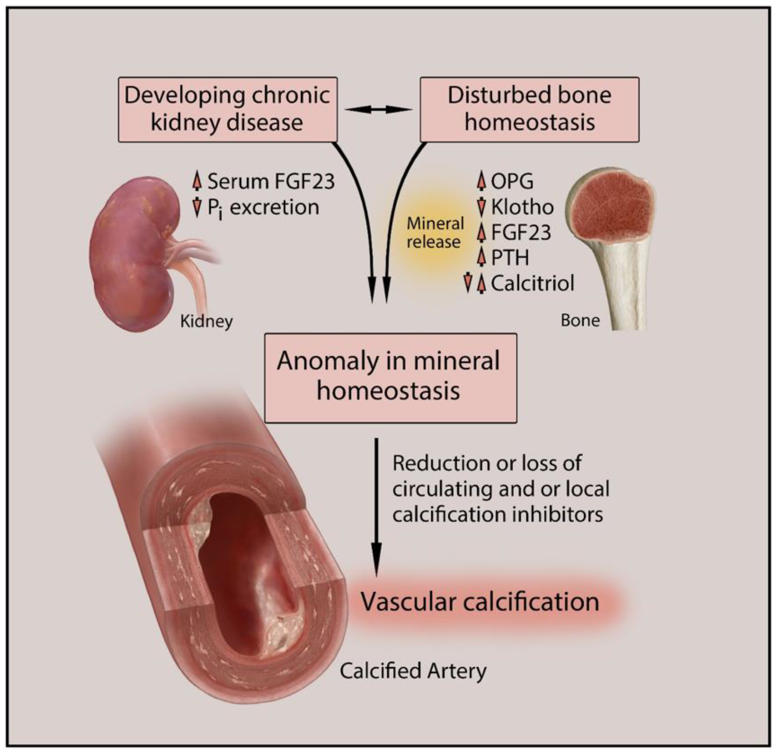
Vascular calcification (VC) is one of the major causes of cardiovascular morbidity and mortality in patients with chronic kidney disease (CKD). VC is a complex process expressing similarity to bone metabolism in onset and progression. VC in CKD is promoted by various factors not limited to hyperphosphatemia, Ca/Pi imbalance, uremic toxins, chronic inflammation, oxidative stress, and activation of multiple signaling pathways in different cell types, including vascular smooth muscle cells (VSMCs), macrophages, and endothelial cells.

vascular calcification chronic kidney disease CKD uremic toxins hyperphosphatemia vascular smooth muscle cells VSMCs macrophages endothelium

1. Introduction

Vascular calcification (VC) is a convoluted process that leads to pathological accumulation of calcium phosphate crystals in the intima and media layers of the vessel wall that worsens the course of atherosclerosis, diabetes, and chronic kidney disease (CKD) [1]. These mineral-enriched plaques induce arterial stiffening, putting patients at risk for fibrosis, inflammation, and oxidative stress on a cellular level. From a clinical perspective, VC is a major problem and is associated with worse outcomes in treatment of coronary artery disease (CAD) and peripheral artery disease (PAD). Coronary calcification is associated with worse outcomes after percutaneous coronary intervention (PCI), and VC is associated with higher risk of amputation after revascularization for lower extremity PAD [2][3]. These associations of CKD can directly increase risks of many clinical complications of VC such as worsening atherosclerosis and increased risk of vascular events such as myocardial infarction, stroke, and vascular occlusive events, which makes it a significant predictor of cardiovascular disease (CVD) and CKD [4][5][6][7][8]. We must give our attention to these patients in the clinical setting to detect early signs of VC in CKD and prepare for prevention and treatment.
VC is now considered as an active and finely regulated process similar to osteogenesis that involves cell-mediated processes and complex interaction between the inhibitor and promoter factors of the calcification process [9][10]. Table 1 lists some major promoter and inhibitor factors involved in VC.
Table 1. Major promoter and inhibitor factors involved in vascular calcification.
Moderate to severe calcification manifests in the aorta, cardiac valves, and peripheral vessels, including the tunica intima and tunica medial layers. Intimal calcification is linked to atherosclerosis, while both intimal and medial calcification has been observed in CKD patients [8][11][12].
The pathogenesis of this complication involves an excess build-up of calcium deposits by active and passive means, a surplus in the excretion of osteoid matrix when cells are triggered by toxic stimuli, or an integrated pathway of both processes [13]. These pathways may be attributed to underlying mechanisms at the cellular level, such as iron, calcium, and phosphate metabolism dysfunction. Multiple key processes that attack metabolism regulation include hyperphosphatemia, calcium phosphate imbalances, and a surge in reactive oxidation species (ROS) due to iron misdistribution [13]. Environmental stimuli can trigger these imbalances, transforming vascular smooth muscle cells (VSMCs) into osteoblast-like cells. This results in the build-up of hydroxyapatite in the various layers of the major and minor vessels, inducing calcification in vascular cells [13][14].
Other mechanisms contributing to VC are related to high glucose levels, lipids, and low-density lipoproteins circulating in the endothelial lining [15]. Research efforts demonstrate how a multitude of these combined actions link VC to CKD, a pathophysiological process that develops in response to irregular environmental stimuli [13].
Compared to the general population, patients with CKD are at an alarmingly higher risk of developing cardiovascular morbidity and mortality. About 50% of deaths from CKD are linked to CVD. Patients with chronic renal disease should be paid attention to for markers signaling cardiovascular complications [12][16]. Several studies show that cardiovascular calcifications in patients with renal disease are found to be more progressive and severe compared to non-CKD patients [6][13][17]. The pathogenesis of VC is enhanced in CKD through complex pathways. Modifications in iron, calcium, and phosphate levels due to kidney injury disturb the biochemical equilibrium, affecting bone remodeling in vascular cells [16]. Figure 1 depicts the pathogenesis of VC in CKD and its interconnection with altered bone and mineral homeostasis.
Figure 1. Pathogenesis of vascular calcification in chronic kidney disease (CKD). Altered bone metabolism and mineral homeostasis are commonly found and closely interconnected in CKD patients. The decline in kidney function also leads to elevated serum FGF23 levels and reduced inorganic phosphate excretion. The resulting pathological state is reflected by various altered biomarkers such as OPG, Klotho, PTH, and calcitriol. The disturbed mineral homeostasis also leads to altered serum and tissue levels of Ca, Pi, and Mg causing inflammation and other metabolic disorders. This leads to reduced or complete loss of circulating and/or local calcification inhibitors such as fetuin A, PPi, and MGP, causing vascular calcification. Ca, calcium; FGF23, fibroblast growth factor 23; Mg, magnesium; MGP, matrix Gla protein; OPG, osteoprotegerin; Pi, inorganic phosphate; PPi, inorganic pyrophosphate; PTH, parathyroid hormone. Upper arrow, increase; lower arrow, decrease.
In the presence of risk factors such as VC, CKD patients are more likely to develop pulmonary hypertension (PH), an overlapping complication in patients with renal disease [18]. Additionally, the damaging effects of oxidative stress exacerbate VC in patients with CKD. Antioxidant defenses and free radical generation are balanced under homeostasis. When balance is disturbed, oxidative stress becomes a trigger of the cessation of cellular division. This becomes a marker of chronic and progressive diseases, including CKD and VC [16][19]. The prominence of VC in CKD can be attributed mainly to the combination of CKD traditional risk factors and the underlying uremic-specific mechanisms that induce the cardiovascular condition.

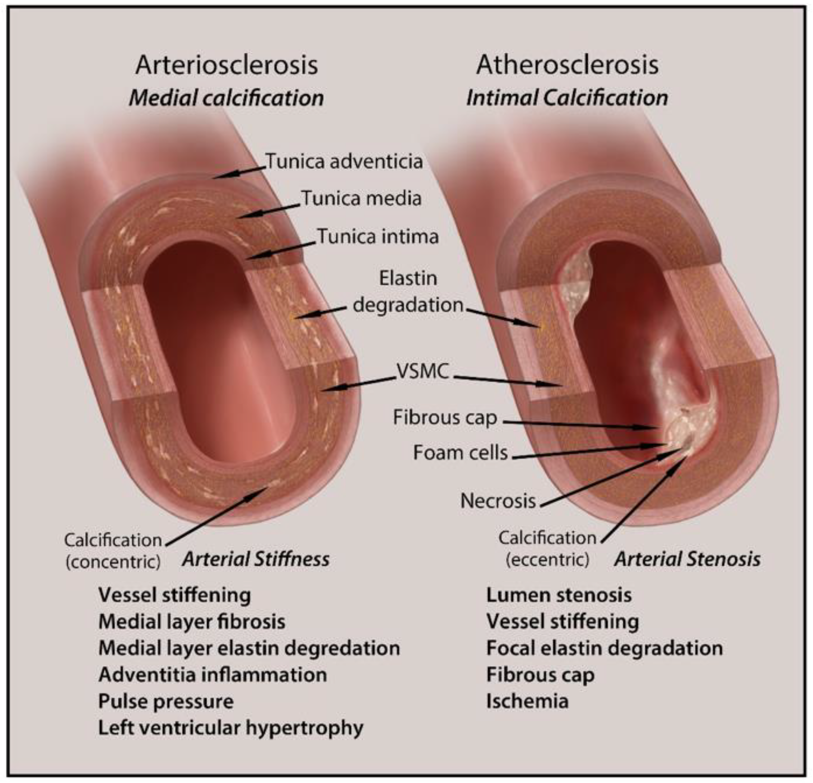
2. Types and Anatomical Presence of VC

VC pathogenesis shares similar mechanisms to that of bone formation. Both processes involve calcium deposition driven by bone matrix proteins, transcription factors, and stenosis. The two major classifications of VC are intimal and medial calcification. Figure 2 is a schematic representation of intimal and medial calcification and their associated pathologies.
Figure 2. Schematic representation of intimal and medial calcification and their related pathologies.
A less common type of calcification involving calcium accumulation in arterioles is termed calciphylaxis. Intimal calcification shares a significant association with atherosclerosis, chronic inflammation, and the transformation of vascular smooth muscle cells (VSMCs) into osteoblast-like cells [8][20]. Medial calcification is more closely linked to elastin degradation, extracellular matrix remodeling events, and hyperphosphatemia. Diseases associated with this type include CKD, hypertension, and type 2 diabetes mellitus [20][21][22]. There is little known about the true cause of calciphylaxis, though the rare multifactorial syndrome has been found to have a close relationship with end-stage renal disease [23].

3. Clinical Implications and Diagnosis of Vascular Calcification in CKD

Vascular disease is the most common cause of death in patients with CKD. In CKD, there are multiple contributing factors to increased VC, such as hyperphosphatemia, hypercalcemia, and increased levels of parathyroid hormone (PTH) [24][25]. High levels of these electrolytes and hormones cause increased cellular activity that increases matrix mineralization, deposition of hydroxyapatite, and inflammation in the vascular intimal and medial walls [26][27][28].
Amongst CKD patients, VC has been proven to increase cardiovascular mortality and morbidity; however, its predictive value is not clear. Additionally, the increased amount of VC increases one’s risk for heart failure, limb ischemia, stroke, and myocardial infarction. Because no data suggest that early detection benefits patients, we generally do not screen CKD patients for VC. Moreover, it is controversial whether interventions that delay the progression of calcification can clinically improve patient outcomes [29].
VC can be diagnosed, detected, and monitored through multiple modalities of imaging, including ultrasound, plain radiographs, and computed tomography (CT) scan. Ultrasound is usually limited to large superficial vessels such as the carotid and femoral arteries, but the information received is qualitative at best and not good for monitoring [30]. Plain radiographs have been used more often for the detection and quantitation of VC. This is normally seen when assessing larger arteries such as the aorta, femoral, and iliac arteries on both anteroposterior and lateral films. The limitation of this imaging modality is that it is difficult to measure VC progression over short periods of time and can be subjective based on the reader [30]. It is also semiquantitative and not as sensitive as a CT scan, which is a superior modality of assessment. VC is detected in >80% of patients undergoing dialysis, which are mostly late CKD and early end-stage renal disease (ESRD) patients. The prevalence amongst non-dialysis CKD patients is around 47% to 83% [29][31][32][33][34][35][36][37]. Multi-slice CT (MSCT) scans are often used for diagnosis and follow up for VC in CKD patients. The high image quality allows for precise, quantitative measurements of VC [29][30]. VC in CKD patients is known to be prominent in the media; however, none of these modalities can definitively differentiate intimal from medial calcification [30]. Currently, there are many controversies on the benefit of screening CKD patients for VC. Even if routine screening is performed, there is limited evidence that modifying risk factors and therapies have a significant impact on clinical outcomes [38]. The Kidney Disease: Improving Global Outcomes (KDIGO) guidelines do weakly recommend screening for VC in CKD patients with either lateral radiographs or CT scan [33][39].

4. Treatment Options for Vascular Calcification in CKD

Treatment options for VC in patients with chronic kidney disease include lifestyle modifications, pharmacologic agents, and surgical interventions. One of the most important lifestyle modifications that decreases mortality rates in these patients is smoking cessation [40].
Medical management in these patients is four-fold. Antiplatelet therapy to reduce platelet aggregation, blood pressure control, anti-lipid therapy for plaque-stabilization, and calcium/phosphate balance to prevent further calcification. Antiplatelet therapy, such as aspirin or clopidogrel, has been shown to reduce major cardiovascular events and overall mortality [41]. Blood pressure control is essential; however, there are no specific studies that show that aggressive blood pressure control has altered the course of peripheral artery disease in patients with CKD. Antilipid therapy such as statins significantly reduces the risk of major atherosclerotic events by stabilizing plaques from rupturing.
Though these three targets are important controls of peripheral vascular disease in CKD, calcium and phosphate balance is essential in preventing the progression of VC. Maintaining a calcium phosphate product of less than 60 is imperative in preventing the rapid progression of VC and complications such as calciphylaxis. Phosphate binders are recommended in all patients in CKD to lower phosphate levels [42]. There are two types of phosphate binders, calcium-based and non-calcium-based. Non-calcium-based phosphate binders such as sevelamer are more frequently used due to providing an all-cause mortality benefit in patients in CKD vs. calcium-based phosphate binders such as calcium acetate/magnesium carbonate [43]. Calcimimetics such as Cinacalcet act on calcium-sensing receptors providing a negative feedback loop to decrease PTH levels in an attempt to decrease serum calcium and phosphate levels. Though effective in reducing serum PTH levels, these medications have a questionable impact on all-cause mortality in patients with CKD [44]. Additional studies still need to be performed for assessment. Vitamin D supplements decrease PTH levels by a negative feedback mechanism. Vitamin D deficiency can cause endothelial dysfunction, which can worsen VC. Preventing this by supplementation can attenuate VC in CKD patients [45]. Currently, the KDIGO guidelines recommend vitamin D supplementation in all patients with CKD stage 4 and 5 [39].
Other options include osteoclast activity inhibitors such as bisphosphonates and denosumab. Bisphosphonates such as alendronate are antiresorptive drugs that inhibit osteoclast activity, preventing the release of calcium and phosphate into the bloodstream. They are usually well tolerated but bisphosphonates can cause worsening nephrotoxicity, focal segmental glomerulosclerosis, and osteonecrosis of the jaw [46][47]. Denosumab is a monoclonal antibody that inhibits NFkB-ligand (RANKL), which blocks its osteoclastic and resorptive properties. Few studies have been performed on the effect on VC in vivo and mortality benefits of both bisphosphonates and denosumab in CKD patients; therefore, their clinical benefit is still unclear, and they are not routinely recommended in these patients [42]. Other options also include magnesium supplementation, which has been shown to decrease phosphate-induced calcification in vitro; however, there are very few studies that have shown a statistically significant impact on reducing VC in CKD patients in vivo [42][48].
Interventions include revascularization or amputation. Revascularization via open surgical bypass or percutaneous angioplasty/stenting (also called endovascular revascularization) is usually used in patients experiencing acute limb ischemia or chronic claudication or critical limb ischemia with non-healing wounds. There are no clear data on which type of revascularization is more beneficial, but many physicians would prefer an endovascular first approach if the anatomy is amenable. Amputation is usually reserved for patients that fail revascularization or have non-viable limbs as suggested by symptoms such as paralysis, severe ulceration, or gangrene. Dialysis patients have an extremely high rate of nontraumatic lower extremity amputation compared with the general population [49]. As noted earlier, VC is associated with worse prognosis after coronary or peripheral vascular revascularization procedures [2][3].
There are few experimental therapies that are being studied to combat VC such as ethylenediaminetetraacetic acid (EDTA) chelation therapy, inducing a certain amount of metabolic acidosis, and the use of autologous osteoclasts [11]. EDTA is an amino acid that can bind calcium potentially decreasing free calcium in the blood. While a small human trial showed regression of VC in patients using EDTA, the study was confounded by the lack of an appropriate control group [50]. A state of metabolic acidosis can activate osteoclasts via RANKL and has been shown to decrease VC in vivo and in vitro; however, there are negative side effects of acidosis, and the risks vs. benefits have not been studied [51][52]. Lastly, localized osteoclast therapy from rat-derived bone marrow has been shown to decrease VC in vitro. This has promising potential as it can be used to treat or even prevent VC in certain areas; however, the clinical application of this approach is speculative [49][53]. Along these lines, there are many experimental treatments under active investigation, but more pre-clinical and clinical data will be necessary to determine the efficacy and safety of these treatments.

References

  1. Gao, J.; Zhang, K.; Chen, J.; Wang, M.-H.; Wang, J.; Liu, P.; Huang, H. Roles of aldosterone in vascular calcification: An update. Eur. J. Pharmacol. 2016, 786, 186–193.
  2. Généreux, P.; Redfors, B.; Witzenbichler, B.; Arsenault, M.-P.; Weisz, G.; Stuckey, T.D.; Rinaldi, M.J.; Neumann, F.-J.; Metzger, D.C.; Henry, T.D.; et al. Two-year outcomes after percutaneous coronary intervention of calcified lesions with drug-eluting stents. Int. J. Cardiol. 2017, 231, 61–67.
  3. Lee, H.Y.; Park, U.J.; Kim, H.T.; Roh, Y.-N. The Effect of Severe Femoropopliteal Arterial Calcification on the Treatment Outcome of Femoropopliteal Intervention in Patients with Ischemic Tissue Loss. Vasc. Spec. Int. 2020, 36, 96–104.
  4. Lacolley, P.; Regnault, V.; Laurent, S. Mechanisms of Arterial Stiffening. Arter. Thromb. Vasc. Biol. 2020, 40, 1055–1062.
  5. Lioufas, N.M.; Pedagogos, E.; Hawley, C.M.; Pascoe, E.M.; Elder, G.J.; Badve, S.V.; Valks, A.; Toussaint, N.D.; on behalf of the IMPROVE-CKD Investigators. Aortic Calcification and Arterial Stiffness Burden in a Chronic Kidney Disease Cohort with High Cardiovascular Risk: Baseline Characteristics of the Impact of Phosphate Reduction on Vascular End-Points in Chronic Kidney Disease Trial. Am. J. Nephrol. 2020, 51, 201–215.
  6. Jagieła, J.; Bartnicki, P.; Rysz, J. Selected cardiovascular risk factors in early stages of chronic kidney disease. Int. Urol. Nephrol. 2020, 52, 303–314.
  7. Demer, L.L.; Tintut, Y. Vascular Calcification. Circulation 2008, 117, 2938–2948.
  8. Chen, N.X.; Moe, S.M. Vascular calcification: Pathophysiology and risk factors. Curr. Hypertens. Rep. 2012, 14, 228–237.
  9. Hénaut, L.; Chillon, J.-M.; Kamel, S.; Massy, Z.A. Updates on the Mechanisms and the Care of Cardiovascular Calcification in Chronic Kidney Disease. Semin. Nephrol. 2018, 38, 233–250.
  10. Voelkl, J.; Lang, F.; Eckardt, K.-U.; Amann, K.; Kuro-O, M.; Pasch, A.; Pieske, B.; Alesutan, I. Signaling pathways involved in vascular smooth muscle cell calcification during hyperphosphatemia. Cell. Mol. Life Sci. 2019, 76, 2077–2091.
  11. Wu, M.; Rementer, C.; Giachelli, C.M. Vascular Calcification: An Update on Mechanisms and Challenges in Treatment. Calcif. Tissue Int. 2013, 93, 365–373.
  12. El Din, U.A.A.S.; Salem, M.M.; Azim, S.E.D.U.A. Vascular calcification: When should we interfere in chronic kidney disease patients and how? World J. Nephrol. 2016, 5, 398–417.
  13. Nakanishi, T.; Nanami, M.; Kuragano, T. The pathogenesis of CKD complications; Attack of dysregulated iron and phosphate metabolism. Free Radic. Biol. Med. 2020, 157, 55–62.
  14. Olapoju, S.O.; Adejobi, O.I.; Le Thi, X. Fibroblast growth factor 21; review on its participation in vascular calcification pathology. Vasc. Pharmacol. 2020, 125–126, 106636.
  15. Opdebeeck, B.; D’Haese, P.C.; Verhulst, A. Molecular and Cellular Mechanisms that Induce Arterial Calcification by Indoxyl Sulfate and P-Cresyl Sulfate. Toxins 2020, 12, 58.
  16. Huang, M.; Zheng, L.; Xu, H.; Tang, D.; Lin, L.; Zhang, J.; Li, C.; Wang, W.; Yuan, Q.; Tao, L.; et al. Oxidative stress contributes to vascular calcification in patients with chronic kidney disease. J. Mol. Cell. Cardiol. 2020, 138, 256–268.
  17. Sarnak, M.J. Cardiovascular complications in chronic kidney disease. Am. J. Kidney Dis. 2003, 41, 11–17.
  18. Sheng, B.; Zhu, T.; Li, J. End-stage renal disease and pulmonary hypertension. Zhong Nan Da Xue Xue Bao Yi Xue Ban = J. Cent. South Univ. Med. Sci. 2019, 44, 1419–1422.
  19. Arefin, S.; Buchanan, S.; Hobson, S.; Steinmetz, J.; Alsalhi, S.; Shiels, P.G.; Kublickiene, K.; Stenvinkel, P. Nrf2 in early vascular ageing: Calcification, senescence and therapy. Clin. Chim. Acta 2020, 505, 108–118.
  20. Lee, S.J.; Lee, I.-K.; Jeon, J.-H. Vascular Calcification—New Insights into Its Mechanism. Int. J. Mol. Sci. 2020, 21, 2685.
  21. Thompson, B.; Towler, D.A. Arterial calcification and bone physiology: Role of the bone–vascular axis. Nat. Rev. Endocrinol. 2012, 8, 529–543.
  22. Xu, J.; Shi, G.-P. Vascular wall extracellular matrix proteins and vascular diseases. Biochim. Biophys. Acta (BBA) Mol. Basis Dis. 2014, 1842, 2106–2119.
  23. Cucchiari, D.; Torregrosa, J.-V. Calcifilaxis en pacientes con enfermedad renal crónica: Una enfermedad todavía desconcertante y potencialmente mortal. Nefrología 2018, 38, 579–586.
  24. House, S.J.; Potier, M.; Bisaillon, J.; Singer, H.A.; Trebak, M. The non-excitable smooth muscle: Calcium signaling and phenotypic switching during vascular disease. Pflug. Arch. Eur. J. Physiol. 2008, 456, 769–785.
  25. Cianciolo, G.; Galassi, A.; Capelli, I.; Schillaci, R.; La Manna, G.; Cozzolino, M. Klotho-FGF23, Cardiovascular Disease, and Vascular Calcification: Black or White? Curr. Vasc. Pharmacol. 2018, 16, 143–156.
  26. Kapustin, A.N.; Davies, J.D.; Reynolds, J.L.; McNair, R.; Jones, G.T.; Sidibe, A.; Schurgers, L.J.; Skepper, J.N.; Proudfoot, D.; Mayr, M.; et al. Calcium Regulates Key Components of Vascular Smooth Muscle Cell–Derived Matrix Vesicles to Enhance Mineralization. Circ. Res. 2011, 109, e1–e12.
  27. Houben, E.; Neradova, A.; Schurgers, L.J.; Vervloet, M. The influence of phosphate, calcium and magnesium on matrix Gla-protein and vascular calcification: A systematic review. G. Ital. Nefrol. 2017, 33, 1724–5590.
  28. Kidney Disease: Improving Global Outcomes (KDIGO) CKD-MBD Work Group. KDIGO clinical practice guideline for the diagnosis, evaluation, prevention, and treatment of Chronic Kidney Disease-Mineral and Bone Disorder (CKD-MBD). Kidney Int. Suppl. 2009, 113, S1–S130.
  29. Reynolds, J.L.; Joannides, A.J.; Skepper, J.N.; McNair, R.; Schurgers, L.J.; Proudfoot, D.; Jahnen-Dechent, W.; Weissberg, P.L.; Shanahan, C.M. Human Vascular Smooth Muscle Cells Undergo Vesicle-Mediated Calcification in Response to Changes in Extracellular Calcium and Phosphate Concentrations: A Potential Mechanism for Accelerated Vascular Calcification in ESRD. J. Am. Soc. Nephrol. 2004, 15, 2857–2867.
  30. Disthabanchong, S. Vascular calcification in chronic kidney disease: Pathogenesis and clinical implication. World J. Nephrol. 2012, 1, 43–53.
  31. Merjanian, R.; Budoff, M.; Adler, S.; Berman, N.; Mehrotra, R. Coronary artery, aortic wall, and valvular calcification in nondialyzed individuals with type 2 diabetes and renal disease. Kidney Int. 2003, 64, 263–271.
  32. Lamprea-Montealegre, J.A.; McClelland, R.L.; Astor, B.C.; Matsushita, K.; Shlipak, M.; de Boer, I.H.; Szklo, M. Chronic kidney disease, plasma lipoproteins, and coronary artery calcium incidence: The Multi-Ethnic Study of Atherosclerosis. Arterioscler. Thromb. Vasc. Biol. 2013, 33, 652.
  33. Kramer, H.; Toto, R.; Peshock, R.; Cooper, R.; Victor, R. Association between Chronic Kidney Disease and Coronary Artery Calcification: The Dallas Heart Study. J. Am. Soc. Nephrol. 2004, 16, 507–513.
  34. Qunibi, W.Y.; AbouZahr, F.; Mizani, M.R.; Nolan, C.R.; Arya, R.; Hunt, K.J. Cardiovascular calcification in Hispanic Americans (HA) with chronic kidney disease (CKD) due to type 2 diabetes. Kidney Int. 2005, 68, 271–277.
  35. Russo, D.; Palmiero, G.; De Blasio, A.P.; Balletta, M.M.; Andreucci, V.E. Coronary artery calcification in patients with CRF not undergoing dialysis. Am. J. Kidney Dis. 2004, 44, 1024–1030.
  36. Chiu, Y.-W.; Adler, S.G.; Budoff, M.J.; Takasu, J.; Ashai, J.; Mehrotra, R. Coronary artery calcification and mortality in diabetic patients with proteinuria. Kidney Int. 2010, 77, 1107–1114.
  37. Bellasi, A.; Raggi, P. Vascular calcification in patients with kidney disease: Techniques and Technologies to Assess Vascular Calcification. Semin. Dial. 2007, 20, 129–133.
  38. Uhlig, K. There Is No Practical Utility in Routinely Screening Dialysis Patients for Vascular Calcification. Semin. Dial. 2010, 23, 277–279.
  39. Beto, J.; Bhatt, N.; Gerbeling, T.; Patel, C.; Drayer, D. Overview of the 2017 KDIGO CKD-MBD Update: Practice Implications for Adult Hemodialysis Patients. J. Ren. Nutr. 2019, 29, 2–15.
  40. Ix, J.H.; Katz, R.; Kestenbaum, B.; Fried, L.F.; Kramer, H.; Stehman-Breen, C.; Shlipak, M.G. Association of Mild to Moderate Kidney Dysfunction and Coronary Calcification. J. Am. Soc. Nephrol. 2008, 19, 579–585.
  41. Jun, M.; Lv, J.; Perkovic, V.; Jardine, M.J. Managing cardiovascular risk in people with chronic kidney disease: A review of the evidence from randomized controlled trials. Ther. Adv. Chronic Dis. 2011, 2, 265–278.
  42. Himmelsbach, A.; Ciliox, C.; Goettsch, C. Cardiovascular Calcification in Chronic Kidney Disease—Therapeutic Opportunities. Toxins 2020, 12, 181.
  43. Jamal, S.A.; Vandermeer, B.; Raggi, P.; Mendelssohn, D.C.; Chatterley, T.; Dorgan, M.; Lok, C.E.; Fitchett, D.; Tsuyuki, R.T. Effect of calcium-based versus non-calcium-based phosphate binders on mortality in patients with chronic kidney disease: An updated systematic review and meta-analysis. Lancet 2013, 382, 1268–1277.
  44. Block, G.A.; Bushinsky, D.A.; Cheng, S.; Cunningham, J.; Dehmel, B.; Drueke, T.B.; Ketteler, M.; KewalRamani, R.; Martin, K.J.; Moe, S.M.; et al. Effect of Etelcalcetide vs. Cinacalcet on Serum Parathyroid Hormone in Patients Receiving Hemodialysis with Secondary Hyperparathyroidism. JAMA 2017, 317, 156–164.
  45. Chitalia, N.; Recio-Mayoral, A.; Kaski, J.C.; Banerjee, D. Vitamin D deficiency and endothelial dysfunction in non-dialysis chronic kidney disease patients. Atherosclerosis 2012, 220, 265–268.
  46. Perazella, M.A.; Markowitz, G.S. Bisphosphonate nephrotoxicity. Kidney Int. 2008, 74, 1385–1393.
  47. Bergner, R.; Diel, I.J.; Henrich, D.; Hoffmann, M.; Uppenkamp, M. Differences in Nephrotoxicity of Intravenous Bisphosphonates for the Treatment of Malignancy Related Bone Disease. Oncol. Res. Treat. 2006, 29, 534–540.
  48. Louvet, L.; Büchel, J.; Steppan, S.; Passlick-Deetjen, J.; Massy, Z.A. Magnesium prevents phosphate-induced calcification in human aortic vascular smooth muscle cells. Nephrol. Dial. Transplant. 2012, 28, 869–878.
  49. Sharples, E.J.; Pereira, D.; Summers, S.; Cunningham, J.; Rubens, M.; Goldsmith, D.; Yaqoob, M.M. Coronary artery calcification measured with electron-beam computerized tomography correlates poorly with coronary artery angiography in dialysis patients. Am. J. Kidney Dis. 2004, 43, 313–319.
  50. Maniscalco, B.S.; Taylor, K.A. Calcification in coronary artery disease can be reversed by EDTA–tetracycline long-term chemotherapy. Pathophysiology 2004, 11, 95–101.
  51. Krieger, N.S.; Frick, K.K.; Bushinsky, D.A. Mechanism of acid-induced bone resorption. Curr. Opin. Nephrol. Hypertens. 2004, 13, 423–436.
  52. Mendoza, F.J.; Lopez, I.; Montes de Oca, A.; Perez, J.; Rodriguez, M.; Aguilera-Tejero, E. Metabolicacidosis inhibits soft tissue calcification in uremic rats. Kidney Int. 2008, 73, 407–414.
  53. Simpson, C.L.; Lindley, S.; Eisenberg, C.; Basalyga, D.M.; Starcher, B.C.; Simionescu, D.T.; Vyavahare, N.R. Toward cell therapy for vascular calcification: Osteoclast-mediated demineralization of calcifiedelastin. Cardiovasc. Pathol. 2007, 16, 29–37.
More
Information
Contributors MDPI registered users' name will be linked to their SciProfiles pages. To register with us, please refer to https://encyclopedia.pub/register : , , , , , , , , , ,
View Times: 346
Revision: 1 time (View History)
Update Date: 22 Aug 2023
1000/1000
Video Production Service