Submitted Successfully!
To reward your contribution, here is a gift for you: A free trial for our video production service.
Thank you for your contribution! You can also upload a video entry or images related to this topic.
Version Summary Created by Modification Content Size Created at Operation
1 -- 3046 2023-08-07 20:56:00 |
2 Reference format revised. Meta information modification 3046 2023-08-08 03:18:08 |

Video Upload Options

Do you have a full video?

Confirm

Are you sure to Delete?
Cite
If you have any further questions, please contact Encyclopedia Editorial Office.
Chai, Y.; Mieyal, J.J. Glutathione/Glutaredoxin in Cellular Redox Homeostasis and Signaling. Encyclopedia. Available online: https://encyclopedia.pub/entry/47758 (accessed on 07 July 2024).
Chai Y, Mieyal JJ. Glutathione/Glutaredoxin in Cellular Redox Homeostasis and Signaling. Encyclopedia. Available at: https://encyclopedia.pub/entry/47758. Accessed July 07, 2024.
Chai, Yuh-Cherng, John J. Mieyal. "Glutathione/Glutaredoxin in Cellular Redox Homeostasis and Signaling" Encyclopedia, https://encyclopedia.pub/entry/47758 (accessed July 07, 2024).
Chai, Y., & Mieyal, J.J. (2023, August 07). Glutathione/Glutaredoxin in Cellular Redox Homeostasis and Signaling. In Encyclopedia. https://encyclopedia.pub/entry/47758
Chai, Yuh-Cherng and John J. Mieyal. "Glutathione/Glutaredoxin in Cellular Redox Homeostasis and Signaling." Encyclopedia. Web. 07 August, 2023.
Glutathione/Glutaredoxin in Cellular Redox Homeostasis and Signaling
Edit

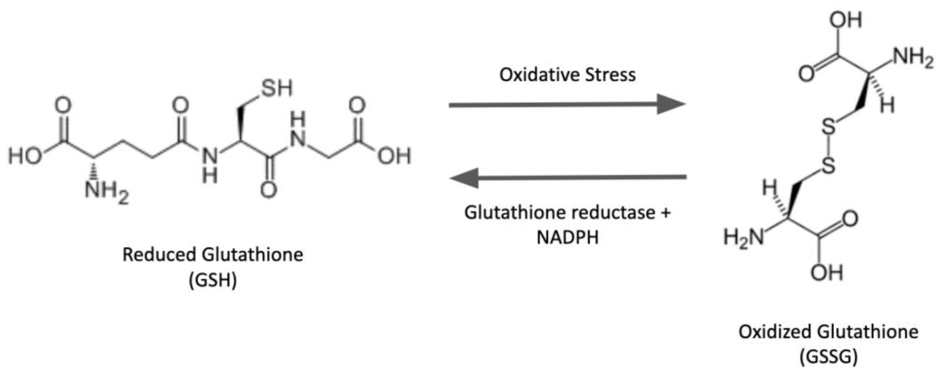
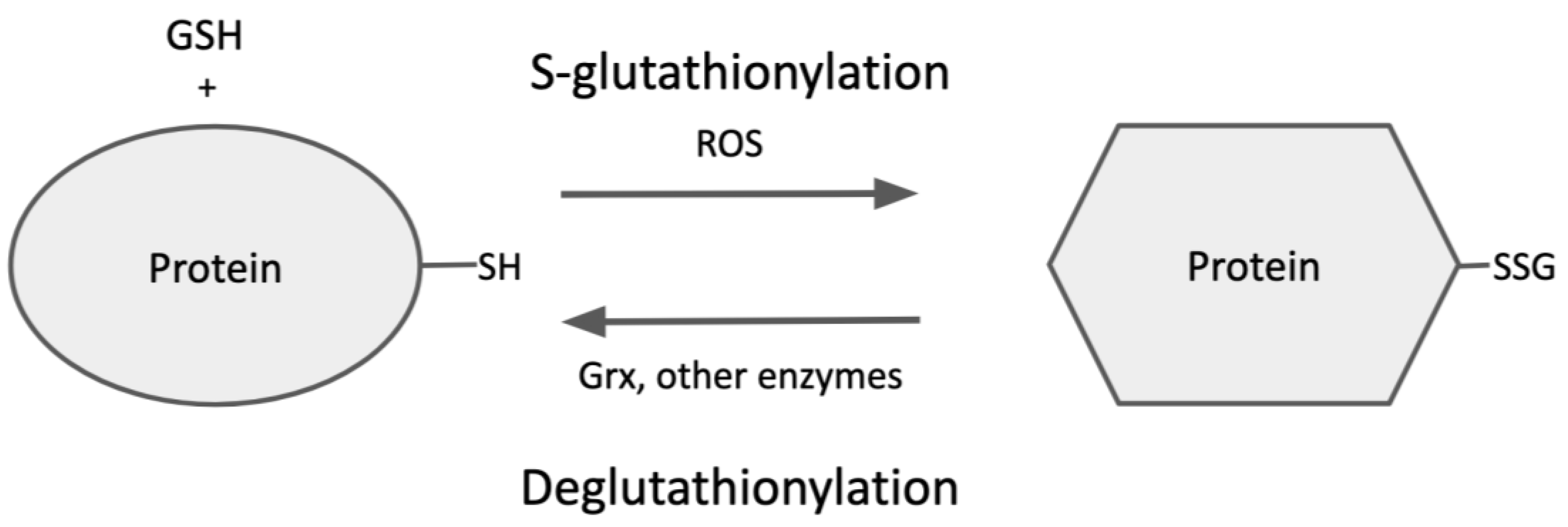
The tripeptide glutathione (GSH) is the most abundant non-enzymatic antioxidant/nucleophilic molecule in cells. In addition to various metabolic reactions involving GSH and its oxidized counterpart GSSG, oxidative post-translational modification (PTM) of proteins has been a focal point of keen interest in the redox field over the last few decades. In particular, the S-glutathionylation of proteins (protein-SSG formation), i.e., mixed disulfides between GSH and protein thiols, has been studied extensively. This reversible PTM can act as a regulatory switch to interconvert inactive and active forms of proteins, thereby mediating cell signaling and redox homeostasis. The unique architecture of the GSH molecule enhances its relative abundance in cells and contributes to the glutathionyl specificity of the primary catalytic activity of the glutaredoxin enzymes, which play central roles in redox homeostasis and signaling, and in iron metabolism in eukaryotes and prokaryotes under physiological and pathophysiological conditions. The class-1 glutaredoxins are characterized as cytosolic GSH-dependent oxidoreductases that catalyze reversible protein S-glutathionylation specifically, thereby contributing to the regulation of redox signal transduction and/or the protection of protein thiols from irreversible oxidation.

glutathione glutaredoxin glutathionylation redox homeostasis redox signaling oxidative stress

1. Overview

Glutathione (GSH) is a tripeptide composed of three amino acids, glycine, cysteine, and glutamate. It exists ubiquitously in abundance within the cells of a broad spectrum of species and plays key functional roles. The nucleophilic cysteine-thiol group is the reactive principle of the molecule, mediating its various biological activities. Among small-peptide molecules, the structure of GSH is unique because the N-terminal glutamic acid and cysteine residues are linked via an unusual peptide bond involving the γ-carboxyl group of the glutamyl moiety rather than the α-carboxyl group, as in conventional peptide bonds [1] (Figure 1). Consequently, glutathione is the most abundant non-protein cellular thiol because this unique γ-bond renders glutathione relatively stable and resistant to intracellular degradation by proteases. Moreover, the unique architecture of the GSH molecule contributes to the glutathionyl specificity of the primary catalytic activity of glutaredoxin enzymes (see below).
Figure 1. Structures of GSH and GSSG.
Glutathione is present at concentrations ranging from 1 mM to 10 mM in the cytosolic compartment of most cells [2], but it also resides at lower concentrations in the subcellular organelles (endoplasmic reticulum, mitochondria, and nucleus). The molecule in the cytosol primarily exists in its reduced form (GSH), with a minor amount in the oxidized form (glutathione disulfide, GSSG); diminution in the GSH/GSSG ratio serves as a biomarker of oxidative stress. Intracellular variations in the GSH/GSSG ratio also correspond to distinct redox environments in the different cellular compartments, influencing their functions. For example, the actively reducing nature of the cytoplasm results in a GSH/GSSG ratio approaching or exceeding 100/1, so the formation of intra- and intermolecular protein disulfides is difficult and rare. In contrast, the GSH/GSSG ratio approaches 1/1 in the endoplasmic reticulum, providing a more oxidizing environment conducive to intramolecular disulfide formation, facilitating protein folding [3].
GSH serves many critical homeostatic and regulatory functions in cells, including maintenance of a balanced redox environment (redox potential), defense against antioxidative stress, nucleophilic scavenging of reactive electrophiles, mediation of redox signaling, and regulation of cell growth versus cell death [4]

2. Glutaredoxin Enzymes

The glutaredoxin oxidoreductase enzymes play central roles in redox homeostasis and signaling, and iron metabolism in eukaryotes and prokaryotes, under physiological and pathophysiological conditions [5][6][7][8][9][10][11][12]. The glutaredoxins are members of the thioredoxin superfamily and are structurally quite similar to thioredoxins and the N-terminal domain of glutathione transferases [7]; however, glutaredoxins have distinct catalytic properties. Most glutaredoxins belong to two major classes. Class I (Grx) includes the glutathione-dependent thiol:disulfide oxidoreductases [5][7][12][13][14], and class II includes the Grx-like proteins (Glp), which are inactive in standard oxidoreductase assays but serve as iron sensors, playing a critical role in glutathione-dependent delivery of iron–sulfur (Fe-S) clusters [6][8][9][10][15]. There are other minor Grx subfamilies, including several plant-specific Grx isoforms [16], which exert additional functions, such as the transcriptional regulation of petal development in flowers [17].

3. Protein S-Glutathionylation

Although historically viewed as harmful byproducts of metabolism that are scavenged by antioxidants, reactive oxygen species (ROS) are also known to act as intracellular second messengers in redox-signaling pathways [18][19]. ROS serve this redox regulation function by modifying specific signaling proteins. In particular, ROS-mediated reversible oxidative modifications of the thiol moieties of protein-cysteine residues are characterized by their ability to modulate the protein activities, thereby propagating signal transduction and biological responses [20][21]. Such post-translational modifications of reactive protein-thiols include sulfenic acid formation (protein-SOH), nitrosylation (protein-SNO), S-glutathionylation (protein-SSG), and others. Considering the relative abundance of GSH in cells and the relative reactivity of various modified cysteine intermediates, it is likely that protein-SSG may represent the preponderant form of protein–cysteine modification [22].
Although cysteine is one of the least abundant amino acids, it stands out as functionally distinct [23]. Even though it has a rather low occurrence in proteins in general, cysteine is often found in the functional sites of proteins, where it plays key roles in catalysis, regulation, secondary structure, etc. [24]. Moreover, cysteine residues often constitute metal-binding sites on proteins, facilitating the action of metal ions as cofactors [25]. These characteristic properties of cysteine residues are dependent on the physical nature and chemistry of the sulfhydryl moiety. Thus, the nucleophilicity of the thiol group is responsible for cysteine’s role in catalysis by enzymes like kinases and phosphatases. The redox reactivity of the thiol group enables cysteine to participate in structural thiol-disulfide interchange reactions affecting protein stability, and in oxidative posttranslational modifications that regulate function and propagate signaling pathways [26].
About 214,000 cysteines are encoded in the human genome [27]. Proteins may have exposed cysteine residues on the surface within the aqueous environment [28], or may be embedded deep within the more hydrophobic globular domains. In an aqueous milieu, the thiol group of cysteine is prone to deprotonation, in equilibrium with the negatively charged thiolate moiety. Both the protonated and unprotonated forms of cysteine have non-bonded pairs of electrons, consistent with nucleophilicity, but the thiolate anions are much more reactive. The ratio (thiol/thiolate) at any pH condition is related to the thiol pKa, which for a typical cysteine residue is near 8.5 [23]; accordingly, only a small fraction of cysteine residues would be negatively charged at physiological pH (7.4). However, the local microenvironment (e.g., neighboring cations) can strongly affect the pKa values of protein thiols over a wide range [24]. Although a lower pKa value corresponds to a higher thiolate amount at neutral pH, it is important to note that the relative rates of cysteine-mediated reactions also depend on other factors [29].
Among the mechanisms of formation of protein-SSG adducts, thiol-disulfide exchange is one of the most extensively studied [5]; however, it relies primarily on the redox state of cellular glutathione. According to this mechanism, the intracellular GSH/GSSG ratio dictates the extent of protein S-glutathionylation ([Protein-SSG]/[Protein-SH]), and the equilibrium constant for the reaction (Kmix, Equation (1)) corresponds to the oxidation potential for the formation of the mixed disulfide (protein-SSG):
Protein-SH + GSSG ⇌ Protein-SSG + GSH,
K m i x = P r o t e i n S S G [ G S H ] P r o t e i n S H [ G S S G ]
The Kmix value for most cysteine residues is approximately 1.0, so the GSH/GSSG ratio would have to be decreased greatly in order to favor the formation of protein-SSG [5]. However, as described above, the cytosolic GSH/GSSG ratio usually remains very high, even under pronounced oxidative stress conditions [30], rendering the thiol-disulfide exchange mechanism with GSSG as the mediator thermodynamically unfavorable. 
Another important mechanism that may mediate protein-SSG formation in vivo involves another type of reactive thiol derivate, namely sulfenic acid. Several different oxidants, including hydrogen peroxide, alkyl hydroperoxides, peroxynitrite, hypochlorous acid, and chloramines, are implicated in mediating the conversion of protein-thiolates to sulfenic acids (protein-SOH) [31], but increased exposure to these oxidants can lead to further oxidation and irreversible modification (sulfinic and sulfonic acids), or promote reactions with neighboring thiols to form disulfides [31]. Sulfenic acid formation was found to be a regulatory mechanism for many proteins [23].

4. Deglutathionylation of Protein-SSG

As mentioned above, S-glutathionylation of proteins is dynamic and reversible, so the steady-state level of protein-SSG under various conditions depends on the relative rates of glutathionylation (formation) and deglutathionylation (breakdown) (Figure 2), providing another level of regulation for cellular processes [32]. Specific binding sites for glutathione have been characterized on glutaredoxin, which is understood to be the primary catalyst of deglutathionylation [33].
Figure 2. Protein S-glutathionylation and Deglutathionylation.
Glutaredoxin isoforms exist in various subcellular compartments in eukaryotes and also in prokaryotes. As described under Section 1, mammalian glutaredoxins are broadly classified into two subfamilies based on their active site sequences and relative activities/functions, with Grx1 and Grx2 representing the major forms of each family [34]. Human Grx1 is localized in the cytosol and mitochondrial intermembrane space, whereas Grx2 is primarily localized in mitochondria. Grx1 is better characterized and reported to catalyze most of the deglutathionylating activity in mammalian cells [35][36]. Although Grx1 is not an essential protein since knockout mice are viable with a life span similar to wild-type mice [33], it is implicated broadly in cellular functions and defense against disease. For example, the level of protein S-glutathionylation is linked to the development of diseases in Grx1-knockout models [33], and the high concentrations of protein-SSG could be reversed by the exogenous administration of recombinant Grx1. 
Grx1 and Grx2 have both been shown to selectivity catalyze dethiolation of S-glutathionylated substrates with GSH as co-substrate. The glutaredoxin substrate specificity was determined by a carefully designed experiment using various protein mixed-disulfide substrates with glutathione- and non-glutathione-containing thiols [37]. Grx effectively catalyzed only the glutathione-containing substrates but was ineffective for other substrates. These two isozymes share an analogous catalytic mechanism for deglutathionylation involving a nucleophilic, double-displacement (ping-pong) sequence, wherein only the N-terminal cysteine residue of the active-site CPYC motif participates in catalysis (monothiol mechanism) [38]. The main distinction between them is the decreased catalytic efficiency (kcat/KM) of Grx2, primarily due to a decreased kcat [38]. The lower Kcat of Grx2 is because of the catalytic cysteine’s higher pKa and a decreased nucleophilicity enhancement of the second substrate, GSH [38]. Like Grx1, Grx2 exhibits GS-thiyl radical (GS) scavenging activity, promoting the S-glutathionylation of various proteins [38].
Most glutaredoxin isoforms have analogous active-site motifs, displaying variations in the general 4-amino acid sequence Cys-X-X-Cys, which is redox reactive [39]. The primary activity of glutaredoxin involves nucleophilic displacement reactions corresponding to thiol-disulfide exchange. Monothiol and dithiol mechanisms have been described for different substrates; however, the monothiol mechanism is generally considered to be the preponderant mechanism for deglutathionylation [5]. The dithiol mechanism is used to explain the supporting role of Grx in the turnover of ribonucleotide reductase and DNA synthesis. 
Human glutaredoxin 2 (hGrx2) was the first member of the glutaredoxin family identified as an iron–sulfur protein. The Fe-S cofactor was shown to be bridged between two monomers via the N-terminal active site cysteine residues and two non-covalently bound GSH molecules [40]. The bound GSH was in exchange with the free GSH pool and played an essential role in stabilizing the Fe-S cluster. The dimeric form of hGrx2 was enzymatically inactive [40]. Under oxidative stress, an altered GSH/GSSG ratio limiting reduced GSH availability to maintain the Fe-S coordination resulted in the degradation of the cluster and formation of the enzymatically active Grx2 monomer. These results were interpreted to suggest that the Fe-S cluster functions as a redox sensor for Grx2 activity under oxidative stress. As explained earlier, other members of the broad glutaredoxin family have been characterized as participating in iron–sulfur homeostasis [8][9][15].

5. Highlights of the Special Issue on Glutathione and Glutaredoxin

Two articles [41][42] feature the specific roles of Grx1 in liver fibrosis and lung fibrosis. Importantly, the data presented in these papers suggest a potential therapeutic role for Grx1 as an anti-fibrotic agent. Thus, Reiko Matsui and her coworkers [41] showed that the overexpression of Grx1 inhibits age-induced hepatic apoptosis and liver fibrosis in mice. On the other hand, high-fat and high-fructose diet-induced non-alcoholic steatohepatitis (NASH) leads to the downregulation of Grx1 and higher levels of S-glutathionylated proteins in the liver; overexpression of Grx-1 significantly decreases the expression of Zbtb16 and leads to the reversal of NASH progression by attenuating inflammatory and fibrotic processes. Although the primary role of Zbtb16 in hepatocytes is unknown, the current study highlights it as an important redox-sensitive protein, whose expression is regulated by Grx1. Certainly, further study of Zbtb16 function is warranted.

Yvonne Janssen-Heininger and her coworkers [42] reported that Grx1 activity was directly correlated with lung function, whereas protein-SSG accumulation was inversely correlated with lung function in subjects with idiopathic pulmonary fibrosis. Epithelial cells lacking Grx1 were more susceptible to Fas-ligand-induced apoptosis and displayed elevated FAS-SSG compared to wild-type controls, whereas the overexpression of Grx1 attenuated epithelial cell apoptosis in association with diminished Fas-SSG. Several metabolites in the purine, creatine, and other metabolic pathways, including inosine monophosphate, spermidine, and others, were consistently released from multiple cell types subjected to various apoptotic stimuli, including Fas. These findings establish a link between Grx1 activity and the modulation of multiple pathways that regulate the synthesis and utilization of diverse metabolites released by apoptotic cells. 

David Davis, Robert Yarchoan, and their coworkers [43] reported that protein S-glutathionylation regulates retroviral protease activity, including human immunodeficiency virus type 1 (HIV-1), human T-cell leukemia virus (HTLV-1), and SARS-CoV-2 proteases. In general, particular proteases of each virus are required for viral maturation, and the protease activities are dependent on the dimeric forms of the enzymes, which can be altered by site-selective S-glutathionylation. For example, HIV-1 protease contains two cysteine residues, Cys67 and Cys95, with low pKa values. S-glutathionylation of Cys67 (C95A-mutant protease) increased the activity by two-fold. On the contrary, S-glutathionylation of Cys95 completely inhibits the activity by disrupting the dimerization of the protease. The oxidation of Cys95 in immature virions impaired viral maturation, and this effect can be reversed by disulfide reduction. Grx1 catalyzes the deglutathionylation of Cys95 and restores protease activity much more efficiently than it deglutathionylates Cys67. Likewise, HTLV-1 protease activity can be regulated by S-glutathionylation and activity restored by Grx1. The S-glutathionylation of Cys95 in HIV-1 protease and Cys109 in HLTV-1 protease sterically interfere with beta-sheet formation and dimerization, according to crystal structure studies. 

In analogous studies of Alzheimer’s disease (AD), increased S-glutathionylation of proteins has been observed in brain samples from AD patients, and actin-SSG is one of the target proteins. Importantly, the regulation of the dynamic polymerization of actin is vital to the function of neural synapses, affecting memory and learning, and it is noteworthy that overexpression of Grx1 in primary cortical culture leads to the restoration of F-actin nano-assembly and spine morphology. 

Continuing the focus on mitochondria as an engine of oxidative stress, Ryan Mailloux and coworkers in an original research contribution to the Special Issue [44] reported S-glutathionylation of the NDUFS1 subunit of Complex 1 of the electron transport chain in liver mitochondria. This modification caused inhibition of Complex I activity and increased the generation of hydrogen peroxide, using glycerol-3-phosphate or proline as fuel during reverse electron transfer. Adding a reducing agent to the reaction system was found to reverse both the inhibition and the ROS accumulation. Interestingly, two other mitochondria proteins, glycerol-3-phosphate dehydrogenase and proline dehydrogenase, were not S-glutathionylated under conditions similar to those that led to NDUFS1-SSG formation, even though these two proteins also produce ROS. Complex III also undergoes S-glutathionylation with an effect similar to that of Complex I, but whether this effect is due to the Complex III modification or glutathionylation of complexes upstream from ubquinone–cytochrome C oxidoreductase remains to be elucidated.
Both Grx and protein disulfide isomerase (PDI) belong to the thioredoxin superfamily, contain CXXC sequences at their respective active sites, and utilize glutathione as a co-substrate. Although these two proteins have a similar 3D structure and thiol-disulfide catalytic mechanism, their cellular environments are quite different; thus, Grx catalyzes the reduction of glutathionyl mixed disulfides, whereas PDI catalyzes the formation of intramolecular disulfides. Ruddock and coworkers have been studying these two enzymes, and they contributed an original research article to this compendium [45] in which they reported the mutation of the PDI CXXC motif, converting histidine to tyrosine or phenylalanine, thereby changing the signature sequence to be more similar to that of class I glutaredoxins. These substitutions for histidine were found to change the binding affinity of PDI for its protein substrates and glutathione.
A very important aspect of understanding the impact of post-translational modification of protein–cysteine residues on the functional consequences is the validity and accuracy of the analytical methods used to identify the PTMs. In this compendium, Wei-Jun Qian and coworkers [46] have contributed a review article focused on the current biochemical and analytical approaches to characterize protein S-glutathionylation at both the proteome level and with individual proteins, including a perspective on studies of the functional impacts. Importantly, they highlight the challenges of some of the methods and consider ways to overcome these difficulties in future work. For example, it is difficult to directly verify the S-glutathionylation of specific cysteinyl moieties in their native environment in situ using mass spectroscopic (MS) approaches. The glutathionyl moiety on a Cys residue can undergo fragmentation in the MS/MS mode of analysis, leading to neutral losses via cleavage of the peptide bond. Another challenge is interpreting the S-glutathionylation data due to the complexity of the oxidative modification of cysteine.
S-nitrosylation is a form of cysteine modification that has been broadly implicated in signal transduction, cellular homeostasis, and disease. Rajib Sengupta and coworkers have a special interest in S-nitrosylation as a regulatory mechanism. In their review that contributed to this Special Issue [47], they focused on the mechanisms of denitrosylation of protein-SNO and the enzymes that catalyze it, analogous to the catalysis of deglutathionylation by glutaredoxin. S-nitrosylated proteins can be denitrosylated by GSH within the range of the physiological concentrations of GSH (5–10 mM), except for a few proteins, such as caspase 3. It is conceivable that those proteins that are resistant to dentrosylation by GSH alone may be more likely to be regulated by reversible S-nitrosylation, requiring enzymatic reversal. GSH denitrosylates proteins in two ways: by displacement of the NO moiety, forming protein-SSG, or by transnitrosylation, forming GS-NO. A key element in this mechanism is GS-NO reductase (GSNOR), which is a significant regulator of the relative cellular levels of protein-SNO and GS-NO. Thus, GSNOR does not directly denitrosylate proteins; instead, it decreases protein-SNO levels by depleting GS-NO. Caspase3-SNO, which is not denitrosylated by GSH/GSNOR, is reduced by the thioredoxin (Trx) system. 

References

  1. Lu, S.C. Glutathione synthesis. Biochim. Biophys. Acta 2013, 1830, 3143–3153.
  2. Forman, H.J.; Zhang, H.; Rinna, A. Glutathione: Overview of its protective roles, measurement, and biosynthesis. Mol. Asp. Med. 2009, 30, 1–12.
  3. Hwang, C.; Sinskey, A.J.; Lodish, H.F. Oxidized redox state of glutathione in the endoplasmic reticulum. Science 1992, 257, 1496–1502.
  4. Pirie, N.W.; Pinhey, K.G. The Titration Curve of Glutathione. J. Biol. Chem. 1929, 184, 321–333.
  5. Mieyal, J.J.; Gallogly, M.M.; Qanungo, S.; Sabens, E.A.; Shelton, M.D. Molecular mechanisms and clinical implications of reversible protein S-glutathionylation. Antioxid. Redox Signal. 2008, 10, 1941–1988.
  6. Outten, C.E.; Albetel, A.N. Iron sensing and regulation in Saccharomyces cerevisiae: Ironing out the mechanistic details. Curr. Opin. Microbiol. 2013, 16, 662–668.
  7. Deponte, M. Glutathione catalysis and the reaction mechanisms of glutathione-dependent enzymes. Biochim. Biophys. Acta 2013, 1830, 3217–3266.
  8. Couturier, J.; Przybyla-Toscano, J.; Roret, T.; Didierjean, C.; Rouhier, N. The roles of glutaredoxins ligating Fe-S clusters: Sensing, transfer or repair functions? Biochim. Biophys. Acta 2015, 1853, 1513–1527.
  9. Berndt, C.; Lillig, C.H. Glutathione, Glutaredoxins, and Iron. Antioxid. Redox Signal. 2017, 27, 1235–1251.
  10. Talib, E.A.; Outten, C.E. Iron-sulfur cluster biogenesis, trafficking, and signaling: Roles for CGFS glutaredoxins and BolA proteins. Biochimica et biophysica acta. Mol. Cell Res. 2021, 1868, 118847.
  11. Berndt, C.; Christ, L.; Rouhier, N.; Mühlenhoff, U. Glutaredoxins with iron-sulphur clusters in eukaryotes—Structure, function and impact on disease. Biochimica et biophysica acta. Bioenergetics 2021, 1862, 148317.
  12. Liedgens, L.; Zimmermann, J.; Wäschenbach, L.; Geissel, F.; Laporte, H.; Gohlke, H.; Morgan, B.; Deponte, M. Quantitative assessment of the determinant structural differences between redox-active and inactive glutaredoxins. Nat. Commun. 2020, 11, 1725.
  13. Yang, Y.; Jao, S.C.; Nanduri, S.; Starke, D.W.; Mieyal, J.J.; Qin, J. Reactivity of the human thioltransferase (glutaredoxin) C7S, C25S, C78S, C82S mutant and NMR solution structure of its glutathionyl mixed disulfide intermediate reflect catalytic specificity. Biochemistry 1998, 37, 17145–17156.
  14. Saaranen, M.J.; Salo, K.E.; Latva-Ranta, M.K.; Kinnula, V.L.; Ruddock, L.W. The C-terminal active site cysteine of Escherichia coli glutaredoxin 1 determines the glutathione specificity of the second step of peptide deglutathionylation. Antioxid. Redox Signal. 2009, 11, 1819–1828.
  15. Trnka, D.; Engelke, A.D.; Gellert, M.; Moseler, A.; Hossain, M.F.; Lindenberg, T.T.; Pedroletti, L.; Odermatt, B.; de Souza, J.V.; Bronowska, A.K.; et al. Molecular basis for the distinct functions of redox-active and FeS-transfering glutaredoxins. Nat. Commun. 2020, 11, 3445.
  16. Couturier, J.; Jacquot, J.P.; Rouhier, N. Evolution and diversity of glutaredoxins in photosynthetic organisms. Cell. Mol. Life Sci. CMLS 2009, 66, 2539–2557.
  17. Gutsche, N.; Thurow, C.; Zachgo, S.; Gatz, C. Plant-specific CC-type glutaredoxins: Functions in developmental processes and stress responses. Biol. Chem. 2015, 396, 495–509.
  18. Finkel, T. Signal transduction by reactive oxygen species. J. Cell Biol. 2011, 194, 7–15.
  19. Sies, H. Hydrogen peroxide as a central redox signaling molecule in physiological oxidative stress: Oxidative eustress. Redox Biol. 2017, 11, 613–619.
  20. Moran, L.K.; Gutteridge, J.M.; Quinlan, G.J. Thiols in cellular redox signalling and control. Curr. Med. Chem. 2001, 8, 763–772.
  21. Leonberg, A.K.; Chai, Y.C. The functional role of cysteine residues for c-Abl kinase activity. Mol. Cell. Biochem. 2007, 304, 207–212.
  22. Gallogly, M.M.; Mieyal, J.J. Mechanisms of reversible protein glutathionylation in redox signaling and oxidative stress. Curr. Opin. Pharmacol. 2007, 7, 381–391.
  23. Poole, L.B. The basics of thiols and cysteines in redox biology and chemistry. Free Radic. Biol. Med. 2015, 80, 148–157.
  24. Marino, S.M.; Gladyshev, V.N. Analysis and functional prediction of reactive cysteine residues. J. Biol. Chem. 2012, 287, 4419–4425.
  25. Pace, N.J.; Weerapana, E. A competitive chemical-proteomic platform to identify zinc-binding cysteines. ACS Chem. Biol. 2014, 9, 258–265.
  26. Reddie, K.G.; Carroll, K.S. Expanding the functional diversity of proteins through cysteine oxidation. Curr. Opin. Chem. Biol. 2008, 12, 746–754.
  27. Go, Y.M.; Chandler, J.D.; Jones, D.P. The cysteine proteome. Free Radic. Biol. Med. 2015, 84, 227–245.
  28. Requejo, R.; Hurd, T.R.; Costa, N.J.; Murphy, M.P. Cysteine residues exposed on protein surfaces are the dominant intramitochondrial thiol and may protect against oxidative damage. FEBS J. 2010, 277, 1465–1480.
  29. Ferrer-Sueta, G.; Manta, B.; Botti, H.; Radi, R.; Trujillo, M.; Denicola, A. Factors affecting protein thiol reactivity and specificity in peroxide reduction. Chem. Res. Toxicol. 2011, 24, 434–450.
  30. Dalle-Donne, I.; Milzani, A.; Gagliano, N.; Colombo, R.; Giustarini, D.; Rossi, R. Molecular mechanisms and potential clinical significance of S-glutathionylation. Antioxid. Redox Signal. 2008, 10, 445–473.
  31. Gupta, V.; Carroll, K.S. Sulfenic acid chemistry, detection and cellular lifetime. Biochim. Biophys. Acta 2014, 1840, 847–875.
  32. Harmel, R.; Fiedler, D. Features and regulation of non-enzymatic post-translational modifications. Nat. Chem. Biol. 2018, 14, 244–252.
  33. Matsui, R.; Ferran, B.; Oh, A.; Croteau, D.; Shao, D.; Han, J.; Pimentel, D.R.; Bachschmid, M.M. Redox Regulation via Glutaredoxin-1 and Protein S-Glutathionylation. Antioxid. Redox Signal. 2020, 32, 677–700.
  34. Zimmermann, J.; Oestreicher, J.; Hess, S.; Herrmann, J.M.; Deponte, M.; Morgan, B. One cysteine is enough: A monothiol Grx can functionally replace all cytosolic Trx and dithiol Grx. Redox Biol. 2020, 36, 101598.
  35. Chrestensen, C.A.; Starke, D.W.; Mieyal, J.J. Acute cadmium exposure inactivates thioltransferase (Glutaredoxin), inhibits intracellular reduction of protein-glutathionyl-mixed disulfides, and initiates apoptosis. J. Biol. Chem. 2000, 275, 26556–26565.
  36. Jung, C.H.; Thomas, J.A. S-glutathiolated hepatocyte proteins and insulin disulfides as substrates for reduction by glutaredoxin, thioredoxin, protein disulfide isomerase, and glutathione. Arch. Biochem. Biophys. 1996, 335, 61–72.
  37. Gravina, S.A.; Mieyal, J.J. Thioltransferase is a specific glutathionyl mixed disulfide oxidoreductase. Biochemistry 1993, 32, 3368–3376.
  38. Gallogly, M.M.; Starke, D.W.; Leonberg, A.K.; Ospina, S.M.; Mieyal, J.J. Kinetic and mechanistic characterization and versatile catalytic properties of mammalian glutaredoxin 2: Implications for intracellular roles. Biochemistry 2008, 47, 11144–11157.
  39. Gallogly, M.M.; Starke, D.W.; Mieyal, J.J. Mechanistic and kinetic details of catalysis of thiol-disulfide exchange by glutaredoxins and potential mechanisms of regulation. Antioxid. Redox Signal. 2009, 11, 1059–1081.
  40. Lillig, C.H.; Berndt, C.; Vergnolle, O.; Lönn, M.E.; Hudemann, C.; Bill, E.; Holmgren, A. Characterization of human glutaredoxin 2 as iron-sulfur protein: A possible role as redox sensor. Proc. Natl. Acad. Sci. USA 2005, 102, 8168–8173.
  41. Tsukahara, Y.; Ferran, B.; Minetti, E.T.; Chong, B.S.H.; Gower, A.C.; Bachschmid, M.M.; Matsui, R. Administration of Glutaredoxin-1 Attenuates Liver Fibrosis Caused by Aging and Non-Alcoholic Steatohepatitis. Antioxidants 2022, 11, 867.
  42. Corteselli, E.; Aboushousha, R.; Janssen-Heininger, Y. S-Glutathionylation-Controlled Apoptosis of Lung Epithelial Cells; Potential Implications for Lung Fibrosis. Antioxidants 2022, 11, 1789.
  43. Davis, D.A.; Bulut, H.; Shrestha, P.; Mitsuya, H.; Yarchoan, R. Regulation of Retroviral and SARS-CoV-2 Protease Dimerization and Activity through Reversible Oxidation. Antioxidants 2022, 11, 2054.
  44. Wang, K.; Hirschenson, J.; Moore, A.; Mailloux, R.J. Conditions Conducive to the Glutathionylation of Complex I Subunit NDUFS1 Augment ROS Production following the Oxidation of Ubiquinone Linked Substrates, Glycerol-3-Phosphate and Proline. Antioxidants 2022, 11, 2043.
  45. Saaranen, M.J.; Alanen, H.I.; Salo, K.E.H.; Nji, E.; Kärkkäinen, P.; Schmotz, C.; Ruddock, L.W. Introduction of a More Glutaredoxin-like Active Site to PDI Results in Competition between Protein Substrate and Glutathione Binding. Antioxidants 2022, 11, 1920.
  46. Li, X.; Zhang, T.; Day, N.J.; Feng, S.; Gaffrey, M.J.; Qian, W.J. Defining the S-Glutathionylation Proteome by Biochemical and Mass Spectrometric Approaches. Antioxidants 2022, 11, 2272.
  47. Chakraborty, S.; Sircar, E.; Bhattacharyya, C.; Choudhuri, A.; Mishra, A.; Dutta, S.; Bhatta, S.; Sachin, K.; Sengupta, R. S-Denitrosylation: A Crosstalk between Glutathione and Redoxin Systems. Antioxidants 2022, 11, 1921.
More
Information
Subjects: Cell Biology
Contributors MDPI registered users' name will be linked to their SciProfiles pages. To register with us, please refer to https://encyclopedia.pub/register : ,
View Times: 254
Revisions: 2 times (View History)
Update Date: 08 Aug 2023
1000/1000
Video Production Service