Submitted Successfully!
To reward your contribution, here is a gift for you: A free trial for our video production service.
Thank you for your contribution! You can also upload a video entry or images related to this topic.
Version Summary Created by Modification Content Size Created at Operation
1 -- 1599 2023-08-01 16:48:15 |
2 layout + 2 word(s) 1601 2023-08-02 02:48:12 |

Video Upload Options

Do you have a full video?

Confirm

Are you sure to Delete?
Cite
If you have any further questions, please contact Encyclopedia Editorial Office.
Urzì, O.; Gasparro, R.; Costanzo, E.; De Luca, A.; Giavaresi, G.; Fontana, S.; Alessandro, R. Methods to Establish a 3D Cell Culture. Encyclopedia. Available online: https://encyclopedia.pub/entry/47499 (accessed on 05 July 2024).
Urzì O, Gasparro R, Costanzo E, De Luca A, Giavaresi G, Fontana S, et al. Methods to Establish a 3D Cell Culture. Encyclopedia. Available at: https://encyclopedia.pub/entry/47499. Accessed July 05, 2024.
Urzì, Ornella, Roberta Gasparro, Elisa Costanzo, Angela De Luca, Gianluca Giavaresi, Simona Fontana, Riccardo Alessandro. "Methods to Establish a 3D Cell Culture" Encyclopedia, https://encyclopedia.pub/entry/47499 (accessed July 05, 2024).
Urzì, O., Gasparro, R., Costanzo, E., De Luca, A., Giavaresi, G., Fontana, S., & Alessandro, R. (2023, August 01). Methods to Establish a 3D Cell Culture. In Encyclopedia. https://encyclopedia.pub/entry/47499
Urzì, Ornella, et al. "Methods to Establish a 3D Cell Culture." Encyclopedia. Web. 01 August, 2023.
Methods to Establish a 3D Cell Culture
Edit

The clear potential of three-dimensional (3D) systems to provide new models suitable for studying cell interactions in both basic and more specialized research, revolutionizing cell culture technology, and offering alternative methods for animal experimentation, has prompted the scientific community to develop different efficient methods to establish 3D cell cultures, all of which, in turn, affect 3D model characteristics. These techniques can be divided into two major categories: scaffold-free systems and scaffold-based systems. Scaffold-free systems are based on the self-aggregation capability of some cell types, which can be encouraged using specific cell plates and/or physical parameters that avoid cell attachment. On the other hand, in scaffold-based systems, cells are seeded in natural or synthetic materials, allowing cell proliferation, aggregation, and 3D organization.

3D cell culture bone brain heart liver lung skin cancer

1. Scaffold-Free Methods

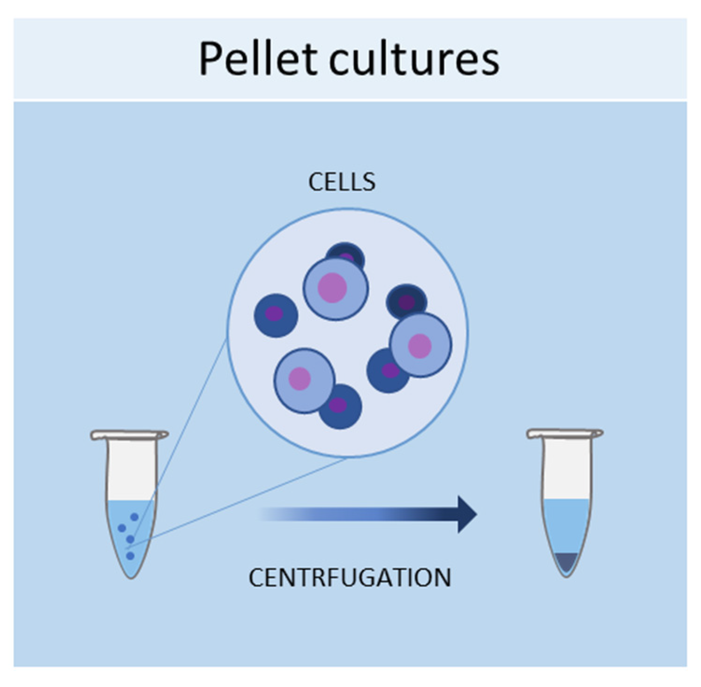
1.1. Pellet Cultures
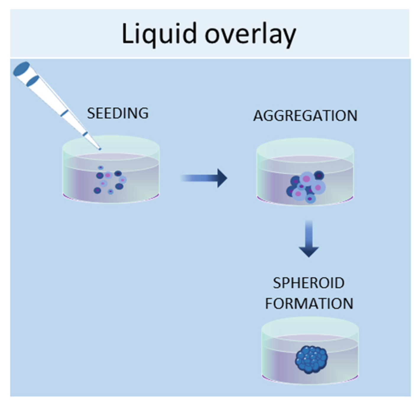
One of the simplest methods of culturing cells in three-dimensional (3D) is represented by pellet cultures, in which cells are pelleted to the bottom of the tube through centrifugal force (Figure 1). The supernatant is removed, while the pellet is suspended in a spheroid culture medium, and cells are seeded in multi-well plates with a cell-repellent surface [1]. It has been demonstrated that this technique can stimulate the chondrogenic differentiation of MSCs [2]. Using pellet cultures, dental pulp spheroids, bone marrow, and endothelial spheroids were also cultured [3][4].
Figure 1. Schematic representation of pellet culture method used to obtain 3D cell cultures.

1.2. Liquid Overlay

The liquid overlay technique allows the growth of cells in 3D by using non-adherent surfaces, typically coated with hydrophilic and neutrally charged polymers (agar or agarose gel). This coating prevents the attachment of cells to the plate or flask, thus encouraging the cells to interact with each other and produce their own ECMs (Figure 2). The formation of aggregates can be forced by continuous agitation and/or centrifugation. With this method, it is possible to establish 3D cell cultures containing one or more cell types, with a spherical morphology and a variable size (50–150 μm) [5]. Human iPSCs were seeded in 96-well ultra-low attachment plates to create a model of multicellular human liver organoids [6]. The liquid overlay technique was successfully employed to obtain other 3D cell cultures, such as neural organoids [7], fibroblast spheroids [8], cancer spheroids [9], and MSC spheroids [10].
Figure 2. Schematic representation of liquid overlay technique used to obtain 3D cell cultures.

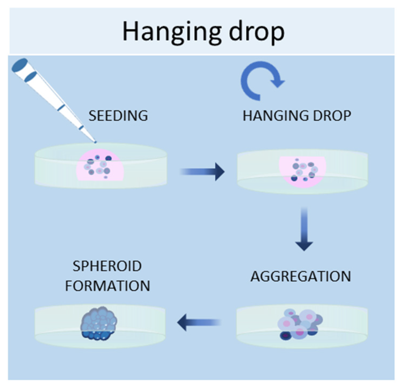
1.3. Hanging Drop Method

Another scaffold-free technique to culture cells in 3D is the hanging drop method, in which cells are placed in a suspended drop of medium, thus allowing them to aggregate and form spheroids at the bottom of the droplet (Figure 3) [11]. Once the cell suspension is prepared, a drop of the culture medium containing the desired number of cells is dispensed into the wells of a mini-tray, and then the mini-tray is inverted upside-down, while the drop containing the cell suspension remains attached to the mini-tray by surface tension. This method takes advantage of surface tension and gravitational force to form a 3D cell aggregate in the droplets, and it is possible to control the size of the spheroid since it depends on the size of the drop and the concentration of the cell suspension [12]. Bartosh et al. used hanging drop methods to culture MSCs in 3D, demonstrating that MSCs grown in 3D secreted more anti-inflammatory molecules than those grown in 2D [13]. Other groups established hepatic spheroids [14], cancer spheroids [15], a model of tumor angiogenesis [16], mammary fibroblasts spheroids [17], and pancreatic islet spheroids [18] through this technique.
Figure 3. Schematic representation of hanging drop method used to obtain 3D cell cultures.

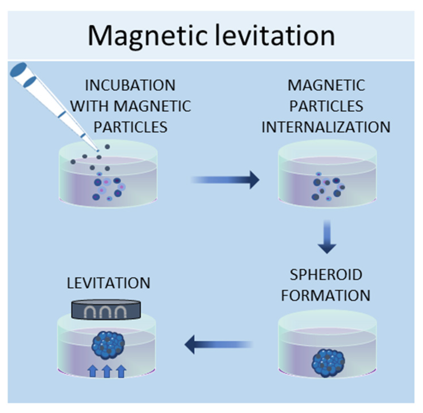
1.4. Magnetic Levitation Method

In the magnetic levitation method, cells are mixed with a solution of magnetic nanoparticles and subjected to a magnetic force [19]. The magnetic nanoparticles are incubated with the cells overnight to allow their internalization. Then, the cells are detached and seeded in low-adhesive plates. A magnet is placed on the top of the plate lid and produces a magnetic force, which causes the cells to levitate against gravity, promoting cell–cell contacts and, therefore, leading to cell aggregation (Figure 4). By using this system, it is possible to culture different cell types, such as [20], MSCs [19], hepatocytes [21], cancer cells [22][23], and osteoblasts [24].
Figure 4. Schematic representation of magnetic levitation method used to obtain 3D cell cultures.

2. Scaffold-Based Methods

The development of regenerative medicine based on engineered biomaterials offers a plethora of scaffolds that can be used as a template for tissue formation [25]. These scaffolds must possess specific characteristics, such as chemical composition, shape, structure, and porosity scaffolds, in order to promote cell migration, adhesion, and tissue production [26]. Additionally, for a biomaterial to be considered applicable in tissue engineering, it must meet the following requirements, which include high biocompatibility, reactivity to cell adhesion, biodegradability, elasticity, and minimal toxicity. The shape, porosity, and surface morphology are also important aspects to create a suitable scaffold that can represent the real architecture of the tissue to be repaired and replicate its vascularization and multicellularity of the tissues [27], ensuring better transport of mass of the nutrients and oxygen [28]. Hydrogel scaffolds, such as gelatin and Matrigel, are natural [29][30] or synthetic [31] crosslinked hydrophobic biomaterials that can absorb and retain water, thus reproducing the natural ECM in compositional and structural terms [25]. Hydrogels have been used for various applications, such as for unicellular or multicellular 3D cultures to regenerate or study healthy or diseases tissues [32][33], or for the development of tumor spheroids [34][35] to study the tumor microenvironment. Commonly used scaffolds for 3D culture include naturally derived matrices and synthetic materials, which are discussed below.

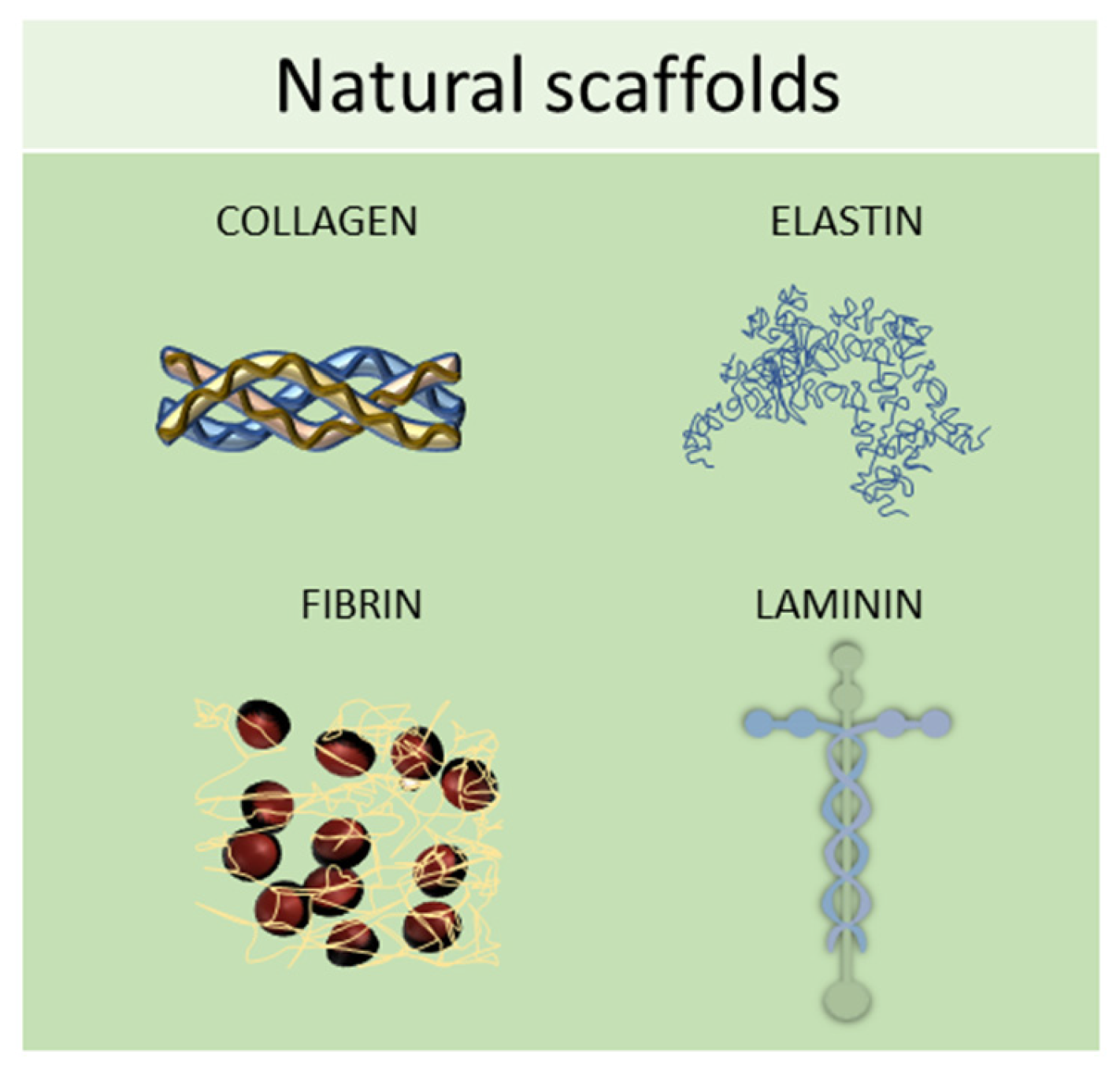
2.1. Natural Scaffolds

Natural scaffolds are represented mainly by decellularized scaffolds or composed of typical components of the ECM, such as collagen, elastin, laminin, or fibrin. Scaffolds made from natural compounds are biodegradable and biocompatible and can promote cellular interactions, adhesion, and signaling [36]. However, some of them showed major limitations, such as inconsistent purity resulting from batch-to-batch variability and difficulty in sterilization and purification. To find solutions to these limitations, tissue engineering continually searches for natural biomaterials from plant and animal sources, such as silk fibroin, chitosan, alginate, gelatin, or Matrigel, or from the glycosaminoglycan family, such as hyaluronic acid, heparin, derman sulfate, chondroitin sulfate, or heparan sulfate (Figure 5) [25].
Figure 5. Schematic representation of some of the most used natural scaffolds (collagen, elastin, fibrin, and laminin) employed in scaffold-based 3D cell cultures.

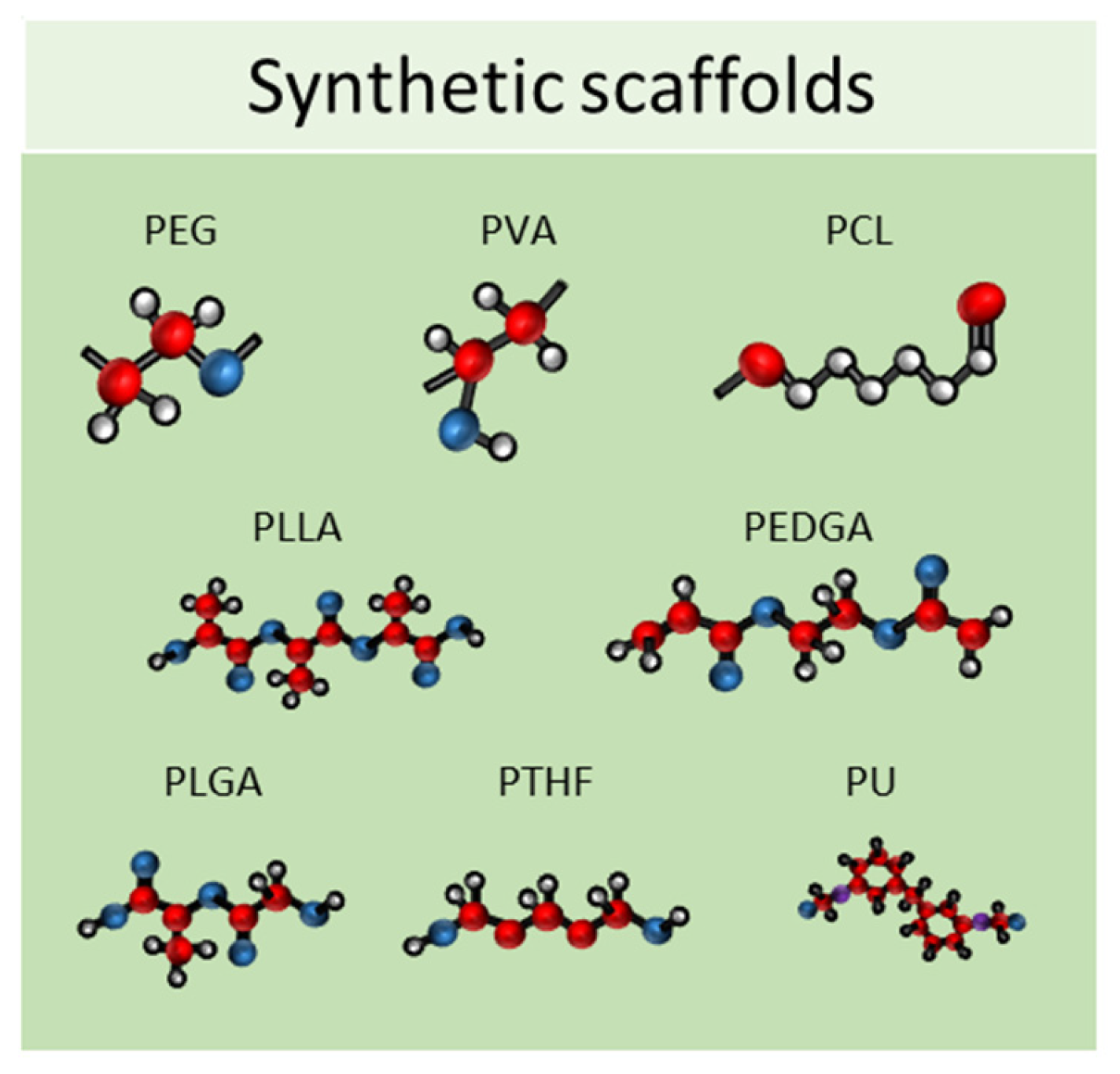
2.2. Synthetic Scaffolds

Synthetic scaffolds are represented mainly by polyethylene glycol (PEG), polyvinyl alcohol (PVA), polylactide-coglycolide (PLG), polycaprolactone (PCL), poly L-lactic acid (PLLA), poly (ethylene glycol) diacrylate (PEGDA), poly lactic-co-glycolic acid (PLGA), polytetrahydrofuran (PTHF), polyurethane (PU), and polyethylene terephthalate (PET); or by ceramics, such as calcium phosphate biomaterials, bioactive hydroxyapatite, and tricalcium phosphate or their associations (HA/TCP) and metals (Figure 6) [37][38]. All these materials meet the criteria needed for tissue engineering because they have higher biocompatibility, excellent biodegradability, and minimal toxicity.
Figure 6. Schematic representation of some of the most used synthetic scaffolds used in scaffold-based 3D cell cultures. PEG: polyethylene glycol, PVA: polyvinyl alcohol, PCL: polycaprolactone, PLLA: poly L-lactic acid, PEGDA: poly (ethylene glycol) diacrylate, PLGA: poly lactic-co-glycolic acid, PTHF: polytetrahydrofuran, PU: polyure-thane.

3. Three-Dimensional Culture in Dynamic Conditions

3.1. Bioreactors

One of the main limitations of using static cell culture conditions to obtain 3D models is the exchange of nutrients, as cell aggregates can reach 1–2 mm of thickness, making it difficult for the transfer of gases and waste products [39]. Bioreactors allow for the establishment of dynamic 3D cell cultures, by controlling several parameters of the extracellular microenvironment, such as pH, temperature, flow rate, oxygen, nutrients, and waste products [39]. Several designs for bioreactors are available, including rotating wall vessels, direct perfusion systems, hollow fibers, spinner flasks, and mechanical force systems. The spinner flask technique, for example, consists of seeding cells into spinner flask bioreactors, where the cell suspension is continuously mixed by stirring [40]. It is crucial to choose the right convectional force of the stirring for the spheroid formation—if the force is too slow the cells will lay on the bottom of the container and not aggregate; conversely, if the force is too high the spheroids will be damaged [1]. This system has been employed in dynamic cell cultures of MSCs, resulting in a high grade of adipogenesis and osteogenic differentiation [41]. Another type of bioreactor is the rotating wall vessel, in which cells are subjected to microgravity by constant circular rotation [42]. This constant rotation keeps the cells in suspension, thus modulating the differentiation capabilities of the MSCs. Sheyn et al. found that in microgravity conditions, adipogenic differentiation is favoured over osteogenic and chondrogenic differentiation [43]. Figure 7 shows a schematic example of a bioreactor used to obtain 3D cell cultures.
Figure 7. Schematic representation of a bioreactor which can be used to obtain 3D cell cultures.

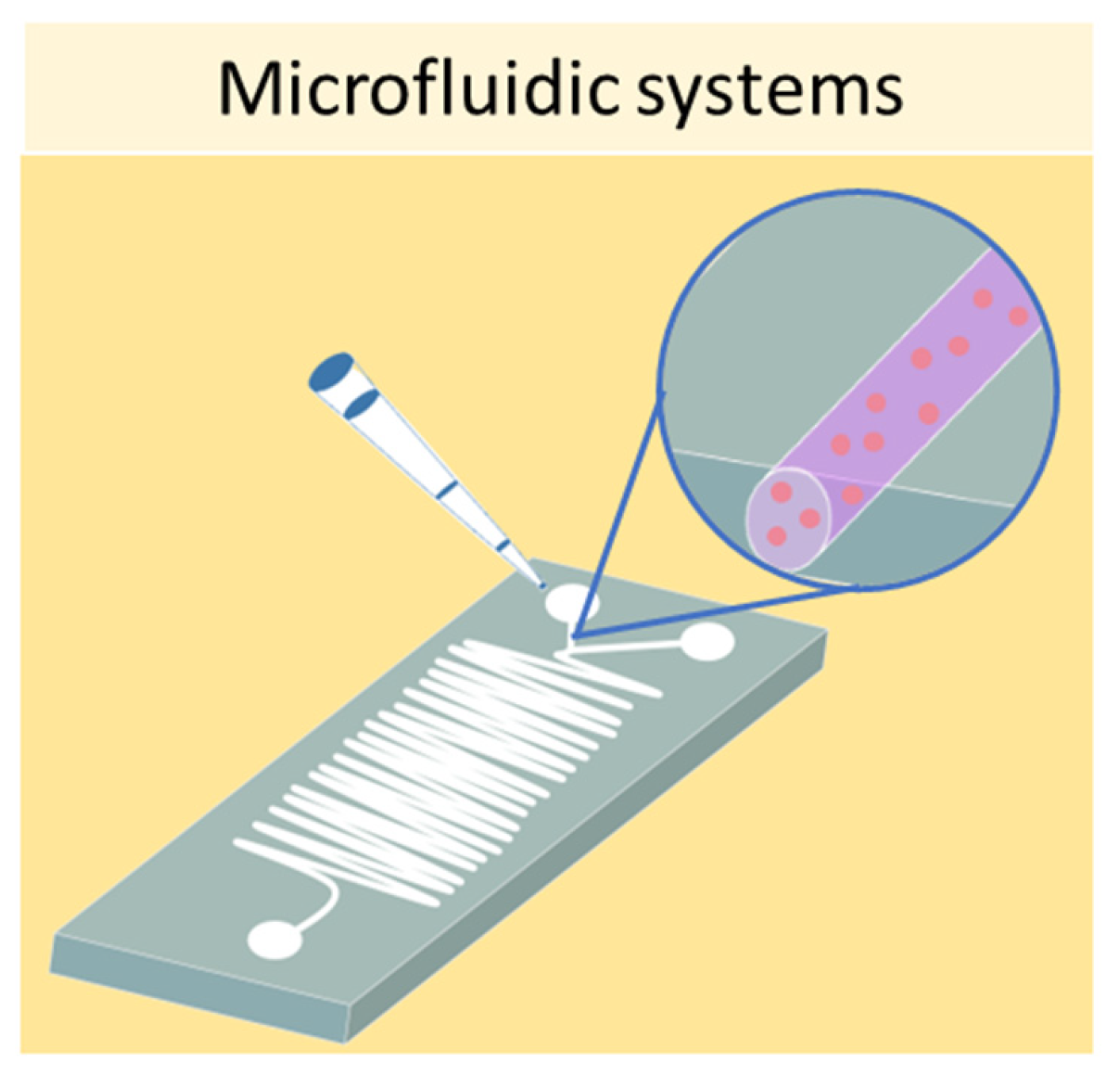
3.2. Microfluidic Systems

Finally, one of the most sophisticated methods to culture cells in 3D is represented by microfluidic devices, also known as organ-on-a-chip. Microfluidic systems are composed of microwells connected by microfluidic channels, thus allowing the continuous infusion of nutrients and growth factors (Figure 8) [44]. This is a key characteristic of microfluidic devices, as it addresses the main limitation of static 3D cell cultures, which is the inhomogeneous concentration of oxygen and nutrients in the cell aggregate. The resulting dynamic microenvironment reflects the in vivo conditions faithfully. This method has allowed the culture of hepatic spheroids with higher viability than those obtained with static culture conditions [45]. Moreover, tumor spheroids were found to have higher resistance to drug treatment when cultured in flow conditions compared with static ones [46]. Using microfluidic systems, it is also possible to co-culture different cell types, as demonstrated by Sun et al., who set up a model for drug screening by culturing spheroids composed of tumor cells and fibroblasts [47].
Figure 8. Schematic representation of a microfluidic device which can be used to obtain 3D cell cultures

References

  1. Achilli, T.M.; Meyer, J.; Morgan, J.R. Advances in the formation, use and understanding of multi-cellular spheroids. Expert. Opin. Biol. Ther. 2012, 12, 1347–1360.
  2. Grigull, N.P.; Redeker, J.I.; Schmitt, B.; Saller, M.M.; Schonitzer, V.; Mayer-Wagner, S. Chondrogenic Potential of Pellet Culture Compared to High-Density Culture on a Bacterial Cellulose Hydrogel. Int. J. Mol. Sci. 2020, 21, 2785.
  3. Zhang, S.; Buttler-Buecher, P.; Denecke, B.; Arana-Chavez, V.E.; Apel, C. A comprehensive analysis of human dental pulp cell spheroids in a three-dimensional pellet culture system. Arch. Oral. Biol. 2018, 91, 1–8.
  4. Inglis, S.; Kanczler, J.M.; Oreffo, R.O.C. 3D human bone marrow stromal and endothelial cell spheres promote bone healing in an osteogenic niche. FASEB J. 2019, 33, 3279–3290.
  5. LaBarbera, D.V.; Reid, B.G.; Yoo, B.H. The multicellular tumor spheroid model for high-throughput cancer drug discovery. Expert. Opin. Drug Discov. 2012, 7, 819–830.
  6. Thompson, W.L.; Takebe, T. Generation of multi-cellular human liver organoids from pluripotent stem cells. Methods Cell Biol. 2020, 159, 47–68.
  7. Choy Buentello, D.; Koch, L.S.; Trujillo-de Santiago, G.; Alvarez, M.M.; Broersen, K. Use of standard U-bottom and V-bottom well plates to generate neuroepithelial embryoid bodies. PLoS ONE 2022, 17, e0262062.
  8. Kubouchi, K.; Mukai, H. PKN2 is involved in aggregation and spheroid formation of fibroblasts in suspension culture by regulating cell motility and N-cadherin expression. Biochem. Biophys. Rep. 2021, 25, 100895.
  9. Sirenko, O.; Mitlo, T.; Hesley, J.; Luke, S.; Owens, W.; Cromwell, E.F. High-content assays for characterizing the viability and morphology of 3D cancer spheroid cultures. Assay. Drug Dev. Technol. 2015, 13, 402–414.
  10. Redondo-Castro, E.; Cunningham, C.J.; Miller, J.; Cain, S.A.; Allan, S.M.; Pinteaux, E. Generation of Human Mesenchymal Stem Cell 3D Spheroids Using Low-binding Plates. Bio. Protoc. 2018, 8, 2968.
  11. Foty, R. A simple hanging drop cell culture protocol for generation of 3D spheroids. J. Vis. Exp. 2011, 51, e2720.
  12. Ryu, N.E.; Lee, S.H.; Park, H. Spheroid Culture System Methods and Applications for Mesenchymal Stem Cells. Cells 2019, 8, 1620.
  13. Bartosh, T.J.; Ylostalo, J.H.; Mohammadipoor, A.; Bazhanov, N.; Coble, K.; Claypool, K.; Lee, R.H.; Choi, H.; Prockop, D.J. Aggregation of human mesenchymal stromal cells (MSCs) into 3D spheroids enhances their antiinflammatory properties. Proc. Natl. Acad. Sci. USA 2010, 107, 13724–13729.
  14. Hurrell, T.; Ellero, A.A.; Masso, Z.F.; Cromarty, A.D. Characterization and reproducibility of HepG2 hanging drop spheroids toxicology in vitro. Toxicol. Vitr. 2018, 50, 86–94.
  15. Gupta, P.; Kar, S.; Kumar, A.; Tseng, F.G.; Pradhan, S.; Mahapatra, P.S.; Santra, T.S. Pulsed laser assisted high-throughput intracellular delivery in hanging drop based three dimensional cancer spheroids. Analyst 2021, 146, 4756–4766.
  16. Timmins, N.E.; Dietmair, S.; Nielsen, L.K. Hanging-drop multicellular spheroids as a model of tumour angiogenesis. Angiogenesis 2004, 7, 97–103.
  17. Cavnar, S.P.; Salomonsson, E.; Luker, K.E.; Luker, G.D.; Takayama, S. Transfer, imaging, and analysis plate for facile handling of 384 hanging drop 3D tissue spheroids. J. Lab. Autom. 2014, 19, 208–214.
  18. Kim, H.J.; Alam, Z.; Hwang, J.W.; Hwang, Y.H.; Kim, M.J.; Yoon, S.; Byun, Y.; Lee, D.Y. Optimal formation of genetically modified and functional pancreatic islet spheroids by using hanging-drop strategy. Transplant. Proc. 2013, 45, 605–610.
  19. Lewis, N.S.; Lewis, E.E.; Mullin, M.; Wheadon, H.; Dalby, M.J.; Berry, C.C. Magnetically levitated mesenchymal stem cell spheroids cultured with a collagen gel maintain phenotype and quiescence. J. Tissue Eng. 2017, 8, 2041731417704428.
  20. Kim, J.A.; Choi, J.H.; Kim, M.; Rhee, W.J.; Son, B.; Jung, H.K.; Park, T.H. High-throughput generation of spheroids using magnetic nanoparticles for three-dimensional cell culture. Biomaterials 2013, 34, 8555–8563.
  21. Miyamoto, Y.; Koshidaka, Y.; Noguchi, H.; Oishi, K.; Saito, H.; Yukawa, H.; Kaji, N.; Ikeya, T.; Suzuki, S.; Iwata, H.; et al. Observation of Positively Charged Magnetic Nanoparticles Inside HepG2 Spheroids Using Electron Microscopy. Cell Med. 2013, 5, 89–96.
  22. Mary, G.; Malgras, B.; Perez, J.E.; Nagle, I.; Luciani, N.; Pimpie, C.; Asnacios, A.; Pocard, M.; Reffay, M.; Wilhelm, C. Magnetic Compression of Tumor Spheroids Increases Cell Proliferation In Vitro and Cancer Progression In Vivo. Cancers 2022, 14, 366.
  23. Palzer, J.; Mues, B.; Goerg, R.; Aberle, M.; Rensen, S.S.; Olde Damink, S.W.M.; Vaes, R.D.W.; Cramer, T.; Schmitz-Rode, T.; Neumann, U.P.; et al. Magnetic Fluid Hyperthermia as Treatment Option for Pancreatic Cancer Cells and Pancreatic Cancer Organoids. Int. J. Nanomed. 2021, 16, 2965–2981.
  24. Gaitan-Salvatella, I.; Lopez-Villegas, E.O.; Gonzalez-Alva, P.; Susate-Olmos, F.; Alvarez-Perez, M.A. Case Report: Formation of 3D Osteoblast Spheroid Under Magnetic Levitation for Bone Tissue Engineering. Front. Mol. Biosci. 2021, 8, 672518.
  25. Geevarghese, R.; Sajjadi, S.S.; Hudecki, A.; Sajjadi, S.; Jalal, N.R.; Madrakian, T.; Ahmadi, M.; Wlodarczyk-Biegun, M.K.; Ghavami, S.; Likus, W.; et al. Biodegradable and Non-Biodegradable Biomaterials and Their Effect on Cell Differentiation. Int. J. Mol. Sci. 2022, 23, 16185.
  26. Nyga, A.; Cheema, U.; Loizidou, M. 3D tumour models: Novel in vitro approaches to cancer studies. J. Cell Commun. Signal 2011, 5, 239–248.
  27. Pampaloni, F.; Reynaud, E.G.; Stelzer, E.H. The third dimension bridges the gap between cell culture and live tissue. Nat. Rev. Mol. Cell Biol. 2007, 8, 839–845.
  28. Lee, J.; Cuddihy, M.J.; Kotov, N.A. Three-dimensional cell culture matrices: State of the art. Tissue Eng. Part. B Rev. 2008, 14, 61–86.
  29. Lou, J.; Stowers, R.; Nam, S.; Xia, Y.; Chaudhuri, O. Stress relaxing hyaluronic acid-collagen hydrogels promote cell spreading, fiber remodeling, and focal adhesion formation in 3D cell culture. Biomaterials 2018, 154, 213–222.
  30. Chen, D.; Qu, Y.; Hua, X.; Zhang, L.; Liu, Z.; Pflugfelder, S.C.; Li, D.Q. A hyaluronan hydrogel scaffold-based xeno-free culture system for ex vivo expansion of human corneal epithelial stem cells. Eye 2017, 31, 962–971.
  31. Zhang, N.; Milleret, V.; Thompson-Steckel, G.; Huang, N.P.; Voros, J.; Simona, B.R.; Ehrbar, M. Soft Hydrogels Featuring In-Depth Surface Density Gradients for the Simple Establishment of 3D Tissue Models for Screening Applications. SLAS Discov. 2017, 22, 635–644.
  32. Sasai, Y. Next-generation regenerative medicine: Organogenesis from stem cells in 3D culture. Cell Stem Cell 2013, 12, 520–530.
  33. Xu, L.; Wang, S.; Sui, X.; Wang, Y.; Su, Y.; Huang, L.; Zhang, Y.; Chen, Z.; Chen, Q.; Du, H.; et al. Mesenchymal Stem Cell-Seeded Regenerated Silk Fibroin Complex Matrices for Liver Regeneration in an Animal Model of Acute Liver Failure. ACS Appl. Mater. Interfaces 2017, 9, 14716–14723.
  34. Li, Y.; Kumacheva, E. Hydrogel microenvironments for cancer spheroid growth and drug screening. Sci. Adv. 2018, 4, eaas8998.
  35. Pradhan, S.; Clary, J.M.; Seliktar, D.; Lipke, E.A. A three-dimensional spheroidal cancer model based on PEG-fibrinogen hydrogel microspheres. Biomaterials 2017, 115, 141–154.
  36. Brigo, L.; Urciuolo, A.; Giulitti, S.; Della Giustina, G.; Tromayer, M.; Liska, R.; Elvassore, N.; Brusatin, G. 3D high-resolution two-photon crosslinked hydrogel structures for biological studies. Acta Biomater. 2017, 55, 373–384.
  37. Edmondson, R.; Broglie, J.J.; Adcock, A.F.; Yang, L. Three-dimensional cell culture systems and their applications in drug discovery and cell-based biosensors. Assay. Drug Dev. Technol. 2014, 12, 207–218.
  38. Rubio, R.; Abarrategi, A.; Garcia-Castro, J.; Martinez-Cruzado, L.; Suarez, C.; Tornin, J.; Santos, L.; Astudillo, A.; Colmenero, I.; Mulero, F.; et al. Bone environment is essential for osteosarcoma development from transformed mesenchymal stem cells. Stem Cells 2014, 32, 1136–1148.
  39. Martin, I.; Wendt, D.; Heberer, M. The role of bioreactors in tissue engineering. Trends Biotechnol. 2004, 22, 80–86.
  40. Lin, R.Z.; Chang, H.Y. Recent advances in three-dimensional multicellular spheroid culture for biomedical research. Biotechnol. J. 2008, 3, 1172–1184.
  41. Frith, J.E.; Thomson, B.; Genever, P.G. Dynamic three-dimensional culture methods enhance mesenchymal stem cell properties and increase therapeutic potential. Tissue Eng. Part. C Methods 2010, 16, 735–749.
  42. Phelan, M.A.; Gianforcaro, A.L.; Gerstenhaber, J.A.; Lelkes, P.I. An Air Bubble-Isolating Rotating Wall Vessel Bioreactor for Improved Spheroid/Organoid Formation. Tissue Eng. Part. C Methods 2019, 25, 479–488.
  43. Sheyn, D.; Pelled, G.; Netanely, D.; Domany, E.; Gazit, D. The effect of simulated microgravity on human mesenchymal stem cells cultured in an osteogenic differentiation system: A bioinformatics study. Tissue Eng. Part. A 2010, 16, 3403–3412.
  44. Nath, S.; Devi, G.R. Three-dimensional culture systems in cancer research: Focus on tumor spheroid model. Pharmacol. Ther. 2016, 163, 94–108.
  45. Lee, S.A.; No, D.Y.; Kang, E.; Ju, J.; Kim, D.S.; Lee, S.H. Spheroid-based three-dimensional liver-on-a-chip to investigate hepatocyte-hepatic stellate cell interactions and flow effects. Lab Chip 2013, 13, 3529–3537.
  46. Ruppen, J.; Cortes-Dericks, L.; Marconi, E.; Karoubi, G.; Schmid, R.A.; Peng, R.; Marti, T.M.; Guenat, O.T. A microfluidic platform for chemoresistive testing of multicellular pleural cancer spheroids. Lab Chip 2014, 14, 1198–1205.
  47. Sun, Q.; Tan, S.H.; Chen, Q.; Ran, R.; Hui, Y.; Chen, D.; Zhao, C.X. Microfluidic Formation of Coculture Tumor Spheroids with Stromal Cells As a Novel 3D Tumor Model for Drug Testing. ACS Biomater. Sci. Eng. 2018, 4, 4425–4433.
More
Information
Subjects: Cell Biology
Contributors MDPI registered users' name will be linked to their SciProfiles pages. To register with us, please refer to https://encyclopedia.pub/register : , , , , , ,
View Times: 565
Revisions: 2 times (View History)
Update Date: 02 Aug 2023
1000/1000
Video Production Service