Submitted Successfully!
To reward your contribution, here is a gift for you: A free trial for our video production service.
Thank you for your contribution! You can also upload a video entry or images related to this topic.
Version Summary Created by Modification Content Size Created at Operation
1 -- 4706 2023-07-31 13:56:59 |
2 format change + 1 word(s) 4707 2023-08-01 05:16:47 |

Video Upload Options

Do you have a full video?

Confirm

Are you sure to Delete?
Cite
If you have any further questions, please contact Encyclopedia Editorial Office.
Chowdhury, I.; Dashi, G.; Keskitalo, S. CMGC Kinases in Health and Cancer. Encyclopedia. Available online: https://encyclopedia.pub/entry/47436 (accessed on 11 September 2024).
Chowdhury I, Dashi G, Keskitalo S. CMGC Kinases in Health and Cancer. Encyclopedia. Available at: https://encyclopedia.pub/entry/47436. Accessed September 11, 2024.
Chowdhury, Iftekhar, Giovanna Dashi, Salla Keskitalo. "CMGC Kinases in Health and Cancer" Encyclopedia, https://encyclopedia.pub/entry/47436 (accessed September 11, 2024).
Chowdhury, I., Dashi, G., & Keskitalo, S. (2023, July 31). CMGC Kinases in Health and Cancer. In Encyclopedia. https://encyclopedia.pub/entry/47436
Chowdhury, Iftekhar, et al. "CMGC Kinases in Health and Cancer." Encyclopedia. Web. 31 July, 2023.
CMGC Kinases in Health and Cancer
Edit

CMGC kinases, encompassing cyclin-dependent kinases (CDKs), mitogen-activated protein kinases (MAPKs), glycogen synthase kinases (GSKs), and CDC-like kinases (CLKs), play pivotal roles in cellular signaling pathways, including cell cycle regulation, proliferation, differentiation, apoptosis, and gene expression regulation. The dysregulation and aberrant activation of these kinases have been implicated in cancer development and progression, making them attractive therapeutic targets. Kinase inhibitors targeting CMGC kinases, such as CDK4/6 inhibitors and BRAF/MEK inhibitors, have demonstrated clinical success in treating specific cancer types.

CMGC kinases cancer protein–protein interaction therapy

1. Introduction

Eukaryotic cells are characterized by the simultaneous activity of multiple molecular networks. Central to these networks are the reversible reactions of protein phosphorylation, catalyzed by protein kinases, and dephosphorylation, catalyzed by protein phosphatases. Protein kinases play a crucial role in signal transduction by phosphorylating specific protein substrates. Changes in the phosphorylation status of numerous proteins are closely associated with their activity, cellular location, and interactions with other proteins. Consequently, alterations in protein phosphorylation can influence a wide range of cellular processes, including metabolism, transcription, cell cycle progression, cytoskeletal rearrangement, cell motility, apoptosis, and differentiation. Dysregulation of protein phosphorylation has been implicated in various human diseases, including cancer. Phosphorylation involves the transfer of a phosphate group from an ATP molecule to a target protein’s serine (S), threonine (T), or tyrosine (Y) residues by protein kinases. Based on their target amino acid selectivity, protein kinases can be classified into serine/threonine (Ser-/Thr-specific), tyrosine (tyrosine-specific), and dual-specificity protein kinases, which primarily phosphorylate serine/threonine residues but can also target tyrosine residues. Serine/threonine protein kinases are the predominant regulators of most cellular processes, with phosphorylation event rates yielding ratios of 1800(Ser):200(Thr):1(Tyr) [1].
All eukaryotic protein kinases share a conserved catalytic core, comprising N- and C-terminal lobes with an ATP-binding cleft between them. The N-lobe contains five β-strands and an α-helix, while the C-lobe is primarily composed of helices [2]. The N-lobe β-strands serve as ATP-binding sites, and the α-helix acts as a regulatory unit crucial for kinase activation, connecting the N- and C-lobes. The C-lobe houses the substrate binding groove and the activation segment, which encompasses the catalytic loop, DFG motif, activation loop, P + 1 loop, and APE region [3]. The activation segment’s catalytic and regulatory functions facilitate the accurate transfer of the phosphate residue to the substrate. Mammalian protein kinases are tightly regulated, with the DFG-motif-regulated phosphorylation of the activation loop functioning as an activation/deactivation switch [2]. Activation loop phosphorylation represents the most common mechanism for regulating kinase activity [4]. Phosphorylated protein kinases are typically in an active ‘on’ state, while dephosphorylated kinases assume a basal ‘off’ state. The three-dimensional structure of the activation segment in different active protein kinases is highly conserved, whereas inactive conformations exhibit significant variation, allowing for kinase-specific activation mechanisms and high specificity [2]. Protein kinase activation can also be dependent on the priming phosphorylation of the substrate [3].
Manning and colleagues identified 518 protein kinases in the human genome in 2002, constituting one of the largest gene families in eukaryotes and accounting for approximately 2% of all human genes [5]. This collection of over 500 protein kinases, which forms the human ‘kinome’, was further categorized into seven major families based on structural similarities within their catalytic domains. Cyclin-dependent kinases (CDKs), mitogen-activated protein kinases (MAPKs), glycogen synthase kinases (GSK3s), CDC-like kinases (CLKs), and related kinase families were grouped together to form the CMGC kinase family based on similarities in their kinase domains [5]. The CMGC family is highly conserved across various species, from nematodes to humans, suggesting that their functions are essential for the survival of these organisms.
CMGC kinases possess a conserved kinase core, similar to other protein kinases. However, they also share a unique CMGC-insert segment absent in other protein kinases [3]. Located in the C-lobe, the CMGC-insert is involved in binding co-proteins that participate in kinase function [6]. This domain exhibits low homology among family members, thereby enabling substrate specificity. CMGC kinases are primarily regulated through tyrosine phosphorylation in the activation loop [7] or pre-phosphorylation of the substrate, which prepares it for sequential recognition and phosphorylation by these kinases [8]. Another shared functional feature is their preference for phosphorylating substrates with proline in the P + 1 position.
In total, the CMGC family comprises 62 members (uniprot.org (accessed on 29 May 2023) [9]), which can be further subdivided into eight subfamilies [5] (Figure 1). CDKs and MAPKs constitute the two largest and most extensively studied families, with 21 and 14 members, respectively [10]. Other families include DYRKs (dual-specificity tyrosine (Y)-phosphorylation-regulated kinases; 10), CLKs (4), RCKs (tyrosine kinase gene v-ros cross-hybridizing kinase; 3), CDKL (cyclin-dependent kinase-like; 5), GSK (2), and SRPK (SR-specific protein kinase; 3). Most CMGC kinases are associated with specific biological functions, such as controlling the cell cycle (CDKs), determining cell fate (MAPKs), regulating multiple signaling pathways (GSKs), and facilitating RNA splicing (CLKs). The individual subfamilies and their unique features will be described in detail below.
Figure 1. (A) A schematic diagram illustrating the taxonomy of the eight subfamilies of CMGC kinases based on their kinase domain sequence similarity. (B) CMGC kinase subfamily dysregulation in most common cancer types with the prevalent deregulated mechanisms involved in cancer progression. Images created with BioRender.com (accessed on 11 July 2023).
Given the crucial roles of CMGC kinases in regulating various cellular processes, their dysregulation can contribute to cancer development and progression. Here, the researchers will delve into the importance of CMGC kinases in cancer biology, their involvement in cellular signaling pathways, protein–protein interactions, and the current state of kinase inhibitors targeting these kinases. Furthermore, the researchers will discuss the challenges and future perspectives in targeting CMGC kinases for cancer therapy, including potential strategies to overcome resistance, the development of more selective inhibitors, and novel therapeutic approaches.
CMGC kinases have gained considerable attention in the context of targeted cancer therapies, with several FDA-approved inhibitors already available and numerous others in various stages of clinical trials. While these inhibitors have demonstrated efficacy and safety to varying degrees, challenges persist, such as resistance development and off-target effects. The need for selective kinase inhibitors and novel therapeutic approaches, including combination therapies, underscores the importance of continued research in this area. Furthermore, the discovery and validation of specific biomarkers for patient stratification and personalized medicine approaches are crucial for selecting the most appropriate therapeutic strategies for individual patients, ultimately improving treatment outcomes and minimizing potential side effects.

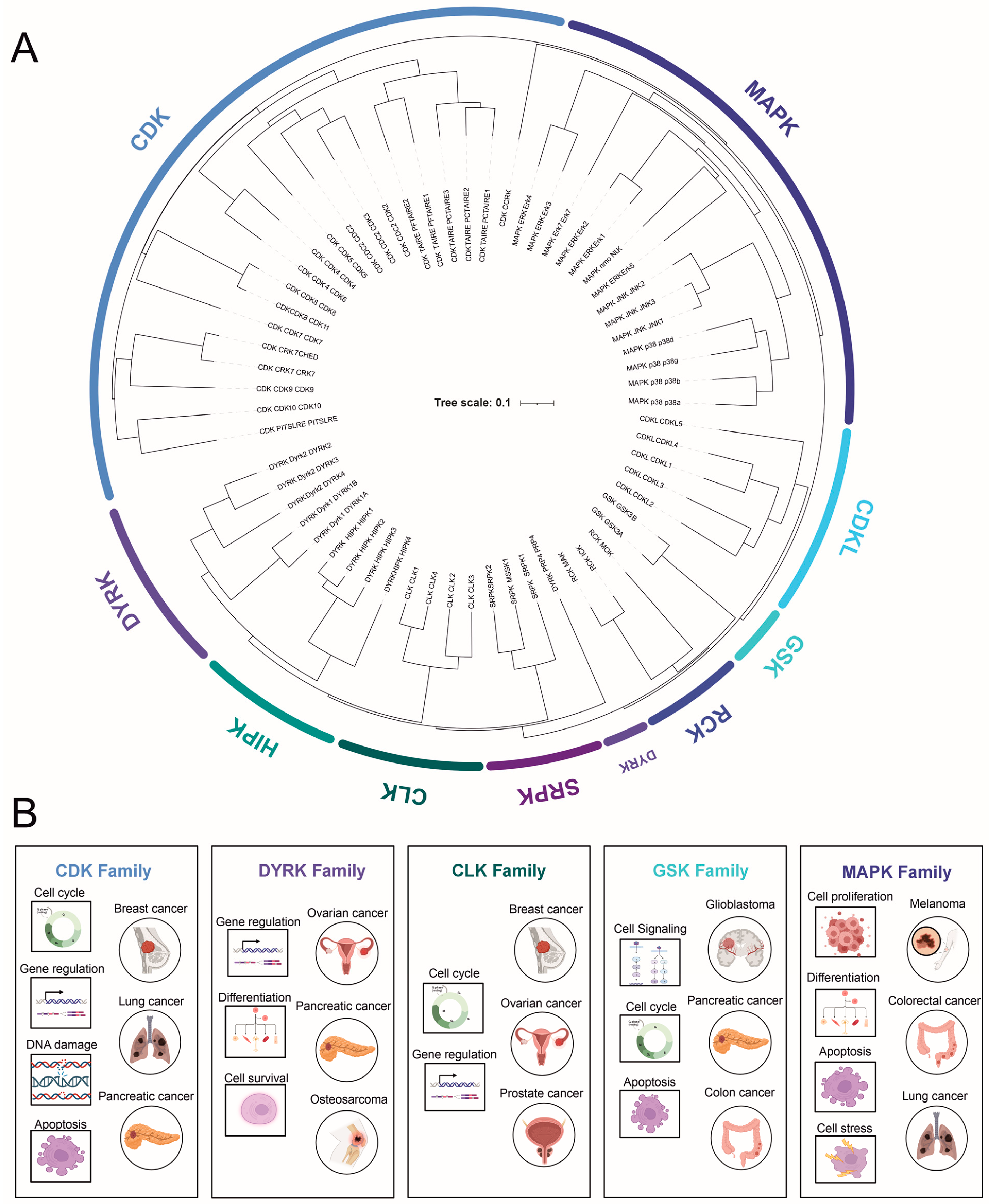
2. CMGC Kinase Subfamilies

2.1. Cyclin-Dependent Kinases (CDKs)

CDKs or cyclin-dependent kinases form a family of 21 constitutively expressed serine/threonine kinases that regulate the cell cycle, transcription, mRNA processing, and the differentiation of cells [11]. As their name suggests, CDKs depend on cyclins, proteins whose levels constantly fluctuate in a tightly coordinated manner during the cell cycle. In CDK/cyclin complexes, CDKs function as catalytic subunits, while cyclins serve as regulatory subunits required for CDKs to exhibit enzymatic activity. The primary mechanism for regulating CDK enzymatic activity is the production and degradation rate of cyclins. The CDK activation loop contains residues for both cyclin and subsequent ATP binding. Like most eukaryotic protein kinases, CDKs are also regulated by the phosphorylation of the activation segment. Phosphorylation can either inhibit by interfering with ATP binding at the catalytic cleft or activate when the phosphorylation site serves as a substrate for CDK-activating kinases, including other CDKs. Activating phosphorylation enhances substrate binding by promoting full kinase activity [12][13].
CDK activity is downregulated by two families of small proteins that function as CDK inhibitors during the cell cycle. The INK4 protein family acts during the G1 phase, specifically inhibiting CDK4/CDK6 binding with cyclin D [11][14]. The broader-spectrum inhibitors of the Cip/Kip family inhibit CDK/cyclin complex activity throughout the cell cycle [11][14]. Based on evolutionary clustering, CDKs form kinase subfamilies that regulate separate cellular functions. CDKs 1–4 and 6 primarily regulate cell cycle progression, whereas CDKs 7–9, 11–13, and 19 have established roles in transcription [11]. Other CDKs have varying roles that will be discussed later.

2.2. Mitogen-Activated Protein Kinases (MAPKs)

The mitogen-activated protein kinase (MAPK) family consists of 14 serine/threonine kinases that can be divided into atypical and conventional kinases.
The atypical mitogen-activated protein kinases (MAPKs) form a distinct, unique subgroup within the larger MAPK family, a major regulator of cellular processes. This group includes extracellular signal-regulated kinase 3 (ERK3), ERK4, ERK7, and Nemo-like kinase (NLK) [15]. Unlike their typical counterparts, atypical MAPKs feature a divergence from the canonical MAPK architecture in their kinase domain, deviating from the conserved TXY motif crucial for activation [16]. Moreover, they exhibit unique activation and regulatory mechanisms, such as autophosphorylation [17]. Their specific physiological roles and functions remain more undefined compared to classical MAPKs. However, emerging evidence links atypical MAPKs to several critical cellular processes, including cell differentiation, proliferation, motility, and apoptosis [18]. For instance, ERK3 and ERK4 are associated with cell migration and actin cytoskeleton reorganization, while ERK7/8 plays roles in cell cycle progression and response to stress [19]. In spite of this, the mechanisms by which atypical MAPKs exert their influence and their potential implications in human diseases, such as cancer, are areas of ongoing research and discovery.
The conventional kinases can be further subdivided into four subfamilies: (1) extracellular signal-regulated kinases 1/2 (ERK1/2), (2) c-Jun amino-terminal kinases 1, 2, and 3 (JNK1-3), (3) p38 MAPKs, and (4) extracellular signal-regulated kinase 5 (ERK5). These enzymes are characterized by a system in which three kinases act sequentially to phosphorylate the downstream kinase. Activators for ERKs are MEK1 and MEK2; for JNKs-, MKK4 and MKK7; for p38-, MKK3 and MKK6; and for ERK5-, MEK5. MAPKs are highly conserved, respond to extracellular signaling cues, and regulate numerous essential cellular processes such as proliferation, differentiation, stress responses, and the transcriptional control of CDKs in organisms ranging from yeast to humans. Over the past few decades, extensive research has been conducted on MAPKs, from their substrates and functions to their roles in health and cancer.
Extracellular signal-regulated kinases 1/2 respond to various stimuli, including growth factors, insulin, cytokines, and carcinogens [19][20]. They are components of the cell surface receptor (receptor tyrosine kinase)/RasGTP/Raf/MEK/ERK signaling cascade. Components of this signaling cascade are essential for proper cell proliferation. ERKs function both in the cytoplasm to promote cell proliferation (G1 to S phase transition) and in the nucleus to phosphorylate and activate numerous transcription factors (TFs) [19][21]. Upon TF activation, c-Jun is expressed and stabilized by direct phosphorylation by ERK1/2 [22], allowing association with c-Fos and the subsequent formation of the AP-1 complex. AP-1 is essential for cyclin D1 expression, which interacts with CDKs to drive G1 to S transition.
  • c-Jun N-terminal kinases are highly homologous (>85%) but have a distinct tissue distribution. JNK1 and 2 are ubiquitously expressed, while JNK3 expression is mainly limited to the brain. In contrast to ERKs, JNKs are primarily activated by stress signals such as oxidative stress, radiation, and DNA-damaging agents. JNKs are mainly localized in the cytoplasm, but their identified substrates are mostly TFs, including c-Jun, p53, STAT3, and c-Myc. The phosphorylation of c-Jun leads to AP-1 complex formation and thus the transcription of cyclin D1, promoting cell cycle progression, similar to ERK1/2 [23]. To date, only a few cytoplasmic interaction partners of JNKs have been identified [19][24].
  • The p38 subfamily consists of four members (α, β, γ, δ), which respond to various environmental stress stimuli and cytokines such as interleukin-1 and tumor necrosis factor α(TNF). Interestingly, p38 both regulates the production of cytokines and responds to them. Other targets of p38 regulation are TFs and other protein kinases. Based on observations of p38 activation, it plays a role in inflammation, cell cycle regulation, and apoptosis [19][25].
  • ERK5 (BMK1 or big MAP kinase 1) has a kinase domain similar to ERK1/2, sharing 51% similarity with ERK2. ERK5 is essential during normal embryogenesis [26]. An upstream activator of ERK5 is MEK5, whose expression is elevated in metastatic prostate cancer [27]. Similar to ERK1/2 and JNK, ERK5 also promotes cyclin D1 expression and cell cycle progression [28], as well as plays a crucial role in the maintenance of mitochondrial function and neuronal survival [29]. ERK5 is involved in various cellular processes, including cell survival, differentiation, and angiogenesis. Its activation has been linked to growth factors, oxidative stress, and other extracellular stimuli.
  • The MAPK pathway has a critical role in cancer biology, extending beyond the extensively studied BRAF mutation in melanoma. The MAPK/ERK pathway, for instance, has been implicated in colorectal cancer, with mutations in KRAS and NRAS genes leading to its persistent activation, promoting uncontrolled cell proliferation and tumor growth [30]. These mutations, unfortunately, render the tumors resistant to EGFR-targeted therapies, highlighting the need for novel therapeutic strategies [31].
  • Additionally, the JNK MAPK pathway, associated primarily with responses to stress signals and apoptosis, has shown links to cancer biology. Aberrations in JNK signaling can lead to an imbalance between cell proliferation and death, thereby contributing to oncogenesis. For instance, overactive JNK signaling has been found in several cancers, including breast and gastric cancer, often correlating with a worse prognosis [32]. Furthermore, the p38 MAPK pathway, typically associated with inflammation and cell differentiation, is also relevant in cancer research. Its complex, dual role in tumorigenesis is being unraveled; while its activation can suppress tumor growth by promoting cell cycle arrest and apoptosis, chronic activation can also enhance cancer cell survival, contributing to chemoresistance [33].
In summary, the MAPK family comprises a diverse group of kinases that play crucial roles in cellular signaling pathways. These kinases are involved in various cellular processes, including proliferation, differentiation, and stress responses, and are integral to the transcriptional control of CDKs. Alterations in the function of the components of the cellular signaling cascade can lead to cancer. Thus, understanding the mechanisms and functions of MAPKs is essential for advancing the knowledge of cellular processes and uncovering potential therapeutic targets in various diseases.

2.3. Glycogen Synthase Kinase-3 (GSK-3)

Glycogen synthase kinase-3 is a conserved, ubiquitously expressed serine/threonine kinase with unique characteristics, such as its high activity in resting cells. Humans have two forms of GSK-3s: GSK-3α and GSK-3β, which share an 85% overall sequence homology [34]. The kinase domain of these kinases is highly homologous (98%), while the N- and C-terminal domains differ [34]. GSK-3α and GSK-3β have distinct functions, as evidenced by the fact that homozygous GSK-3β knockout mice are embryonic-lethal, whereas GSK3α knockout mice are viable [35][36].
Unlike the CDK and MAPK families, GSK activity is regulated through multiple levels and mechanisms, ranging from phosphorylation by other kinases, autophosphorylation, and priming phosphorylation of the target substrate by another kinase [37][38][39]. In most cases, the phosphorylation of serine residues inhibits GSK’s kinase activity, while tyrosine phosphorylation increases its activity [40]. GSK-3 (mainly GSK-3α) has been extensively studied and is known to function in various signaling pathways, including Wnt, Notch, and Hedgehog (proliferation), as well as growth factors affecting differentiation/survival. It has been implicated in multiple roles in many human pathological conditions, ranging from neurodegenerative diseases to diabetes and several types of cancers [39][41].
GSK is a critical component of the canonical Wnt signaling pathway, which is essential during normal development and is often dysregulated in cancers. The main events during Wnt-β-catenin signaling involve a central component known as the destruction complex that regulates cytoplasmic β-catenin levels. This complex comprises tumor suppressors Axin and adenomatosis polyposis coli (APC), as well as casein kinase-1α (CK1α) and GSK-3 [42]. In the absence of the Wnt-ligand, β-catenin is assembled into this complex and phosphorylated by CK1 and GSK-3, marking it for proteasomal degradation and thus preventing the transcription of β-catenin-dependent genes. When Wnt binds to its receptor Frizzled, the destruction complex cannot phosphorylate and ubiquitinate β-catenin, allowing it to translocate to the nucleus, where it forms complexes with transcription factors and promotes transcription. Phosphorylation by GSK-3 is also essential for the targeting of multiple other proteins for proteasomal degradation [41][43]. In addition to its role in Wnt signaling, GSK-3 has been implicated in other critical signaling pathways. For example, GSK-3 plays a role in the insulin signaling pathway, which is essential for glucose homeostasis. Abnormalities in GSK-3 function have been linked to insulin resistance and the development of type 2 diabetes [44]. GSK-3 inhibitors have shown promise in preclinical studies as potential therapeutic agents for the treatment of type 2 diabetes [45].
GSK-3 has been found to be overactive in various types of cancers, including colon, breast, and prostate cancer. The overactivation of GSK-3 can lead to increased cell proliferation, migration, and invasion, as well as reduced apoptosis [39]. Targeting GSK-3 may represent a potential therapeutic strategy for cancer treatment. Preclinical studies have shown that GSK-3 inhibitors can suppress tumor growth and metastasis, as well as sensitize cancer cells to chemotherapy and radiation therapy [41].
In summary, GSK-3 is a multifunctional kinase that plays a pivotal role in numerous signaling pathways and cellular processes. The dysregulation of GSK-3 activity has been implicated in various pathological conditions. Consequently, GSK-3 has emerged as a potential therapeutic target, and the development of GSK-3 inhibitors may hold promise for the treatment of these diseases. However, due to the pleiotropic nature of GSK-3, further research is needed to understand the precise mechanisms of GSK-3 regulation and its involvement in different pathologies to develop safe and effective therapeutic strategies.

2.4. Dual-Specificity Tyrosine (Y)-Phosphorylation-Regulated Kinases (DYRKs)

DYRK is a relatively large, mammalian, dual-specificity protein kinase family that, based on homology analysis, consists of three subfamilies with a total of 10 members: DYRK (DYRK1A-B, 2–4), HIPKs (homeodomain-interacting protein kinase 1–4), and PRP4s (pre-mRNA processing protein 4 kinase).

2.5. Cdc2-like Kinase (CLK) and Other Less-Studied Kinases

The Cdc2-like kinase (CLK) family consists of four dual-specificity kinases (CLK1–4) that autophosphorylate on tyrosine residues and specifically phosphorylate serine/threonine residues on their substrates [46]. CLKs are found in both the cytoplasm and the nucleus, where they phosphorylate the arginine- and serine-rich (RS) domains of serine/arginine (SR) splicing factor proteins, thereby controlling their nuclear distribution [47]. SR splicing factor proteins are extensively phosphorylated by multiple kinases, and the proper phosphorylation status has been shown to be essential for SR protein activity [48]. These proteins play a crucial role in alternative mRNA splicing, a fundamental process responsible for generating the complex human proteome during normal development.
In a recent study, CLKs were identified as upstream regulators of Aurora B, acting as novel components in the final steps of cytokinesis, the division of the cytoplasm into two daughter cells [49]. However, as Petsalaki and Zachos concluded, the current understanding of CLKs’ cellular functions remains limited [49]. A 2017 study reported that CLK inhibition could modulate the alternative splicing of Mcl-1, an anti-apoptotic protein, and promote cancer cell death, suggesting a potential therapeutic approach for cancer treatment [50]. Given the importance of alternative mRNA splicing in the regulation of gene expression and the emerging roles of CLKs in cell division, further investigation into the functions and regulation of CLKs could provide valuable insights into their role in various cellular processes and their potential as therapeutic targets in human diseases.

2.6. SR-Specific Protein Kinase (SRPK)

Serine–arginine protein kinases (SRPKs) specifically phosphorylate the serine residues in serine/arginine (SR) dipeptides. They are primarily localized in the cytoplasm, where these constitutively active kinases regiospecifically phosphorylate SR splicing factors on multiple serines, inducing the translocation of these factors into the nucleus. In the nucleus, SR splicing factors are further phosphorylated by CLKs [51][52]. The change in the cellular localization of SR proteins is essential for phosphorylation by CLKs [47]. As mentioned earlier, SR protein phosphorylation is necessary for correct mRNA splicing and maturation.
Over 100 SR-domain-containing proteins have been identified in the human genome [53], indicating that there are many open questions regarding the functional roles of SRPKs in mammalian cells. SRPK1 expression is elevated in breast, colon, and pancreatic cancers [54], as well as in acute T-cell leukemia [55]. A 2020 study identified SRPK1 as a potential therapeutic target in glioblastoma, with the SRPK1 inhibitor SPHINX31 showing promising results in preclinical models [56]. SRPK2 has a specific role in phosphorylating apoptosis-promoting protein ACIN1 and increasing cyclin A1 expression in leukemia cells [57]. Given the importance of SRPKs in the regulation of alternative splicing and their association with various types of cancer, further studies on the functional roles of these kinases and their potential as therapeutic targets in human cancer are warranted.

2.7. Tyrosine Kinase Gene v-Ros Cross-Hybridizing Kinase (RCK)

The RCK serine/threonine-protein kinase family consists of three kinases: MAK, ICK, and MOK. The functions of these kinases are poorly understood. Structurally, the RCK family resembles both MAPKs and CDKs [5]. MAK and ICK autophosphorylate on a tyrosine residue, but they need a second phosphorylation on their MAPK-like motif in their activation loop by an upstream kinase for full enzymatic activity [58][59][60]. Different from MAPK, the activating kinase for MAK and ICK is cell-cycle-related kinase (CCRK), but for MOK, it remains unknown [60][61].
  • MAK (male germ cell-associated kinase) is mainly expressed in testicular germ cells during spermatogenesis and in the retina. In the retina, MAK localizes to connecting cilia in photoreceptor cells, negatively regulates the length of their cilia, and is essential for the survival of these cells [62]. Not surprisingly, MAK mutations are associated with retinitis pigmentosa, a photoreceptor degeneration disease in the retina [63][64].
  • ICK (intestinal cell kinase) is highly conserved, constitutively, and widely expressed. Similarly to MAK, it negatively regulates ciliary length and is identified as an essential component of sonic hedgehog signaling [65]. These two factors seem to be the underlying cause of human ECO syndrome, a multi-organ illness affecting the endocrine, cerebral, and skeletal systems, caused by a missense mutation in the ICK gene [66]. In 2017, a study reported the role of ICK in colorectal cancer progression and its potential as a therapeutic target for the treatment of colorectal cancer [67].
The cellular function of the third RCK, MOK (MAPK/MAK/MRK-overlapping kinase), is largely unknown. To date, only the expression of MOK in mouse wild-type and cancerous intestinal cells has been analyzed by Western blotting, showing the downregulation of MOK in adenomas [68]. This observation suggests that MOK might have a role in intestinal cancer development, but further studies are needed.

2.8. Cyclin-Dependent Kinase-like (CDKL)

This family of five members of serine/threonine kinases with homology to MAPK and CDK families is relatively uncharacterized. Studies have focused on CDKL5, a ubiquitous protein mainly expressed in the brain, testes, and thymus [69]. In the context of cancer, CDKL proteins are now emerging as potential players. Studies suggest that the dysregulation of CDKL5 may contribute to tumorigenesis in certain cancers. For instance, the aberrant expression of CDKL5 has been suggested in glioma and some evidence exists on its role in breast cancer, indicating its possible role in disease progression and patient prognosis [70].
Similarly, another family member, CDKL1, has been associated with colorectal cancer, with its overexpression correlating with poor survival outcomes [71]. These preliminary findings suggest that CDKL proteins could potentially act as novel markers for cancer diagnosis or targets for therapeutic intervention.

3. Protein Kinase Therapeutics—Kinase Inhibitors

The protein kinase domain is among the most commonly encountered domain in known cancer genes [72]. As described earlier, most of the CMGC kinases affect various cellular signaling pathways that control critical processes like cell cycle progression, proliferation, differentiation, apoptosis, or survival. Often, abnormal phosphorylation is either the cause or consequence in cancer [73]. Therefore, protein kinases are among the most studied druggable targets in pharmacological research [74][75]. In human cancers, kinases are often found overexpressed or overactive, due to a vast array of genetic and epigenetic events, including point mutations, chromosomal gene amplifications (copy number alterations), and chromosomal translocations giving rise to gene fusions [76] (Table 1).
Table 1. Prevalence and pattern of CMGC kinase subfamily dysregulation in cancer.

Kinase Family

Deregulation Mechanism

Examples of Cancer Types

CDKs

Mutations, amplifications, deletions, and altered expression levels

Breast cancer [77][78][79], lung cancer [80], pancreatic cancer [81]

MAPKs

Mutations in pathway components (e.g., RAS, RAF)

Melanoma [82][83][84], colorectal cancer [85], lung cancer [17][86]

DYRK

Overexpression

Pancreatic [87][88], ovarian cancer [89][90], osteosarcoma [91], rhabdomyosarcoma [92]

GSKs

Altered expression levels, post-translational modifications

Glioblastoma [93], pancreatic cancer [94], colon cancer [95]

CLKs

Overexpression, cancer-associated splicing alterations

Breast cancer [96], prostate cancer [97], ovarian cancer [98]

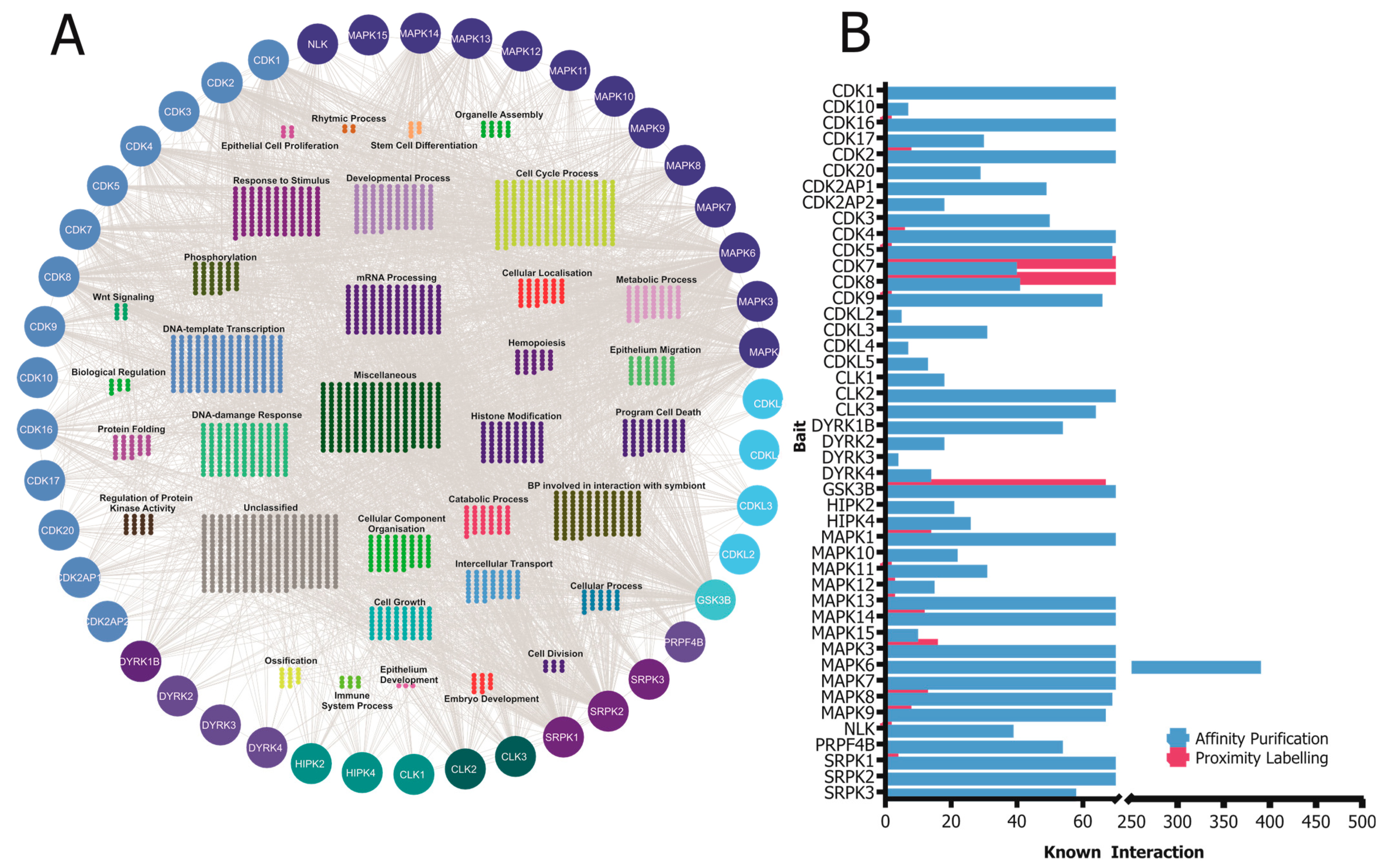
4. Protein–Protein Interactions of the CMGC Kinases

CMGC kinases participate in various protein–protein interactions that are crucial for their activity, functions, and signaling pathways. Understanding these interactions is essential to elucidate the molecular mechanisms governing CMGC-kinase-mediated cellular processes. In this section, the researchers will discuss some of the known interactions involving CMGC kinases and their importance (Figure 2).
Figure 2. (A) A network of known high-confidence protein–protein interactions of CMGC kinases obtained from the IntAct Molecular Interaction Database are grouped according to their simplified GO process annotation. (B) Distribution of the number of known interactors of the CMGC kinases based on the affinity purification and proximity labeling detection method. The visualization of the protein–protein interaction (PPI) network was generated by importing the publicly available interaction dataset from https://www.ebi.ac.uk/intact (accessed on 8 May 2023) protein interaction data into Cytoscape v3.9.
CMGC kinases contribute to the development and progression of various diseases. Therefore, understanding the intricate network of protein–protein interactions involving CMGC kinases can provide valuable insights into the molecular mechanisms underlying their functions and offer potential therapeutic targets for the treatment of diseases associated with their dysregulation. For instance, the development of small molecule inhibitors that specifically target the protein–protein interactions of CMGC kinases could represent a promising approach to modulate their activity and restore normal cellular functions in disease settings [99].
The development of small molecule inhibitors that target the protein–protein interactions (PPIs) of these kinases has emerged as a promising strategy in cancer therapeutics. Traditional kinase inhibitors target the ATP-binding site of kinases. However, this site’s high conservation across different kinases challenges the development of inhibitors with high specificity. In contrast, the PPI interfaces are often less conserved, presenting an opportunity for creating more specific inhibitors [100][101]. Small molecule inhibitors act by obstructing the critical protein interactions necessary for the activation or functioning of these kinases. For instance, inhibitors may preclude a kinase from interacting with its activator or substrate, consequently inhibiting the kinase’s activity and downstream signaling [102]. In cancer, these inhibitors can restore normal cellular functions by blocking the aberrant signaling engendered by dysregulated kinases. For example, if a kinase is overactive in a cancer cell, promoting uncontrolled cell proliferation, an inhibitor that obstructs this kinase’s interactions could impede or slow down the proliferation [103]. Considerable progress has been made in the development of such inhibitors. High-throughput screening techniques have enabled researchers to identify small molecules that can bind to the PPI interfaces of CMGC kinases [102]. Furthermore, advances in structural biology have provided intricate details of these kinases’ 3D structures and their complexes with interacting proteins, aiding in the design of more effective inhibitors [104]. Nonetheless, challenges persist in developing PPI inhibitors. The interfaces involved in PPIs are often expansive and flat, complicating the ability of small molecules to bind with high affinity. Also, these inhibitors must navigate cellular barriers to reach their intracellular targets [103].
Most of the studies regarding kinase interactions have focused on a single kinase and its function or dysfunction in a disease. Additionally, the names of the CDK kinases were unified as late as in 2009 [10], and that might have contributed to the slowness of studies of some these kinases. It is, after all, rather difficult to assign a kinase to this interesting family when the name is very different, such as PFTK1 or CRKRS.

5. Assessment of Kinase Activity

As the function of protein kinases is to transfer a phosphate group, Western blotting with antibodies against a substrate’s phosphoserine, -threonine or -tyrosine is the most common and widely used method to indirectly identify kinase activity. This approach is very labor-intensive, highly dependent on the specificity and sensitivity of the available antibodies, and not directly comparable with high-throughput approaches.
A more sensitive, direct approach to quantitatively measure kinase activity in vitro is to measure the incorporation of radiolabeled phosphate from γ-33P ATP into the substrate [105]. A prerequisite to the solution-based in vitro kinase assays is a purified kinase, which in most cases is compared to the kinase dead mutant that abolishes ATP binding. An extensive study of 84 pairs of wild-type kinases and their kinase-dead counterparts [9] confirmed, using in vitro kinase assays, that indeed the mutated “VAIK” or “HRD” motif is essential for kinase activity. The lysine in the “VAIK” motif is among the most conserved amino acids in all kinases as it is a central catalyzer of the phosphate transfer [5].
Functional protein microarrays are an efficient ‘omics’-scale tool to detect kinase-substrate interactions [106][107]. The commercially available protein microarrays have thousands of purified proteins spotted onto a glass slide, providing an efficient means to identify the phosphorylation of candidate substrates for the kinases of interest when incubated together with radiolabeled ATP [108]. To date, the only large-scale study utilizing protein microarrays in kinase substrate identification has been performed with yeast protein kinases [109], although human protein microarrays are nowadays commercially available.

References

  1. Mann, M.; Ong, S.-E.; Grønborg, M.; Steen, H.; Jensen, O.N.; Pandey, A. Analysis of protein phosphorylation using mass spectrometry: Deciphering the phosphoproteome. Trends Biotechnol. 2002, 20, 261–268.
  2. Taylor, S.S.; Kornev, A.P. Protein kinases: Evolution of dynamic regulatory proteins. Trends Biochem. Sci. 2011, 36, 65–77.
  3. Kannan, N.; Neuwald, A.F. Evolutionary constraints associated with functional specificity of the CMGC protein kinases MAPK, CDK, GSK, SRPK, DYRK, and CK2α. Protein Sci. 2004, 13, 2059–2077.
  4. Nolen, B.; Taylor, S.; Ghosh, G. Regulation of Protein Kinases: Controlling Activity through Activation Segment Conformation. Mol. Cell 2004, 15, 661–675.
  5. Manning, G.; Whyte, D.B.; Martinez, R.; Hunter, T.; Sudarsanam, S. The Protein Kinase Complement of the Human Genome. Science 2002, 298, 1912–1934.
  6. Pawson, T.; Nash, P. Assembly of Cell Regulatory Systems Through Protein Interaction Domains. Science 2003, 300, 445–452.
  7. Johnson, L.N.; Noble, M.E.; Owen, D.J. Active and Inactive Protein Kinases: Structural Basis for Regulation. Cell 1996, 85, 149–158.
  8. Haar, E. Structure of GSK3beta reveals a primed phosphorylation mechanism. Nat. Struct. Biol. 2001, 8, 593.
  9. Varjosalo, M.; Björklund, M.; Cheng, F.; Syvänen, H.; Kivioja, T.; Kilpinen, S.; Sun, Z.; Kallioniemi, O.; Stunnenberg, H.G.; He, W.-W.; et al. Application of Active and Kinase-Deficient Kinome Collection for Identification of Kinases Regulating Hedgehog Signaling. Cell 2008, 133, 537–548.
  10. Malumbres, M.; Harlow, E.; Hunt, T.; Hunter, T.; Lahti, J.M.; Manning, G.; Morgan, D.O.; Tsai, L.-H.; Wolgemuth, D.J. Cyclin-dependent kinases: A family portrait. Nat. Cell Biol. 2009, 11, 1275–1276.
  11. Malumbres, M. Cyclin-dependent kinases. Genome Biol. 2014, 15, 122.
  12. Jeffrey, P.D. Mechanism of CDK Activation Revealed by the Structure of a CyclinA-CDK2 Complex. Nature 1995, 376, 313–320.
  13. Russo, A.A.; Jeffrey, P.D.; Pavletich, N.P. Structural basis of cyclin-dependent kinase activation by phosphorylation. Nat. Struct. Biol. 1996, 3, 696–700.
  14. Harper, J.; Brooks, G. The Mammalian Cell Cycle—An Overwiew. From Methods in Molecular Biology. In Cell Cycle Control: Mechanisms and Protocols; Brooks, T.H., Ed.; Humana Press Inc.: Totowa, NJ, USA, 2005; Volume 296.
  15. Coulombe, P.; Meloche, S. Atypical mitogen-activated protein kinases: Structure, regulation and functions. Biochim. Biophys. Acta (BBA) Mol. Cell Res. 2007, 1773, 1376–1387.
  16. Deleris, A.; Stroud, H.; Bernatavichute, Y.; Johnson, E.; Klein, G.; Schubert, D.; Jacobsen, S.E. Loss of the DNA Methyltransferase MET1 Induces H3K9 Hypermethylation at PcG Target Genes and Redistribution of H3K27 Trimethylation to Transposons in Arabidopsis thaliana. PLoS Genet. 2012, 8, e1003062.
  17. Long, W.; Foulds, C.E.; Qin, J.; Liu, J.; Ding, C.; Lonard, D.M.; Solis, L.M.; Wistuba, I.I.; Qin, J.; Tsai, S.Y.; et al. ERK3 signals through SRC-3 coactivator to promote human lung cancer cell invasion. J. Clin. Investig. 2012, 122, 1869–1880.
  18. Kostenko, S.; Dumitriu, G.; Lægreid, K.J.; Moens, U. Physiological roles of mitogen-activated-protein-kinase-activated p38-regulated/activated protein kinase. World J. Biol. Chem. 2011, 2, 73–89.
  19. Cargnello, M.; Roux, P.P. Activation and Function of the MAPKs and Their Substrates, the MAPK-Activated Protein Kinases. Microbiol. Mol. Biol. Rev. 2011, 75, 50–83.
  20. Johnson, G.L.; Lapadat, R. Mitogen-Activated Protein Kinase Pathways Mediated by ERK, JNK, and p38 Protein Kinases. Science 2002, 298, 1911–1912.
  21. Burotto, M.; Chiou, V.L.; Lee, J.-M.; Kohn, E.C. The MAPK pathway across different malignancies: A new perspective. Cancer 2014, 120, 3446–3456.
  22. Murphy, L.O.; Smith, S.; Chen, R.-H.; Fingar, D.C.; Blenis, J. Molecular interpretation of ERK signal duration by immediate early gene products. Nat. Cell Biol. 2002, 4, 556–564.
  23. Sabapathy, K.; Hochedlinger, K.; Nam, S.Y.; Bauer, A.; Karin, M.; Wagner, E.F. Distinct Roles for JNK1 and JNK2 in Regulating JNK Activity and c-Jun-Dependent Cell Proliferation. Mol. Cell 2004, 15, 713–725.
  24. Bogoyevitch, M.A.; Ngoei, K.R.; Zhao, T.T.; Yeap, Y.Y.; Ng, D.C. c-Jun N-terminal kinase (JNK) signaling: Recent advances and challenges. Biochim. Biophys. Acta (BBA)—Proteins Proteom. 2010, 1804, 463–475.
  25. Cuadrado, A.; Nebreda, A.R. Mechanisms and functions of p38 MAPK signalling. Biochem. J. 2010, 429, 403–417.
  26. Regan, C.P.; Li, W.; Boucher, D.M.; Spatz, S.; Su, M.S.; Kuida, K. Erk5 null mice display multiple extraembryonic vascular and embryonic cardiovascular defects. Proc. Natl. Acad. Sci. USA 2002, 99, 9248–9253.
  27. Mehta, P.B.; Jenkins, B.L.; McCarthy, L.; Thilak, L.; Robson, C.N.; Neal, D.E.; Leung, H.Y. MEK5 overexpression is associated with metastatic prostate cancer, and stimulates proliferation, MMP-9 expression and invasion. Oncogene 2003, 22, 1381–1389.
  28. Mulloy, R.; Salinas, S.; Philips, A.; Hipskind, R.A. Activation of cyclin D1 expression by the ERK5 cascade. Oncogene 2003, 22, 5387–5398.
  29. Jo, M.; Lee, S.; Kim, K.; Lee, S.; Kim, S.R.; Kim, H.-J. Inhibition of MEK5 suppresses TDP-43 toxicity via the mTOR-independent activation of the autophagy-lysosome pathway. Biochem. Biophys. Res. Commun. 2019, 513, 925–932.
  30. Markowitz, S.D.; Bertagnolli, M.M. Molecular Origins of Cancer: Molecular Basis of Colorectal Cancer. N. Engl. J. Med. 2009, 361, 2449–2460.
  31. Van Cutsem, E.; Köhne, C.-H.; Hitre, E.; Zaluski, J.; Chien, C.-R.C.; Makhson, A.; D’Haens, G.; Pintér, T.; Lim, R.; Bodoky, G.; et al. Cetuximab and Chemotherapy as Initial Treatment for Metastatic Colorectal Cancer. N. Engl. J. Med. 2009, 360, 1408–1417.
  32. Min, L.; He, B.; Hui, L. Mitogen-activated protein kinases in hepatocellular carcinoma development. Semin. Cancer Biol. 2011, 21, 10–20.
  33. Wagner, E.F.; Nebreda, Á.R. Signal integration by JNK and p38 MAPK pathways in cancer development. Nat. Rev. Cancer 2009, 9, 537–549.
  34. Woodgett, J.R. Molecular Cloning and Expression of Glycogen Synthase Kinase-3/Factor A. EMBO J. 1990, 9, 2431.
  35. Hoeflich, K.P. Requirement for Glycogen Synthase Kinase-3beta in Cell Survival and NF-KappaB Activation. Nature 2000, 406, 86–90.
  36. MacAulay, K.; Doble, B.W.; Patel, S.; Hansotia, T.; Sinclair, E.M.; Drucker, D.J.; Nagy, A.; Woodgett, J.R. Glycogen Synthase Kinase 3α-Specific Regulation of Murine Hepatic Glycogen Metabolism. Cell Metab. 2007, 6, 329–337.
  37. Cole, A.R.; Knebel, A.; Morrice, N.A.; Robertson, L.A.; Irving, A.J.; Connolly, C.N.; Sutherland, C. GSK-3 Phosphorylation of the Alzheimer Epitope within Collapsin Response Mediator Proteins Regulates Axon Elongation in Primary Neurons. J. Biol. Chem. 2004, 279, 50176–50180.
  38. Frame, S.; Cohen, P. GSK3 Takes Centre Stage More than 20 Years after Its Discovery. Biochem. J. 2001, 359, 1–16.
  39. McCubrey, J.A. Effects of Mutations in Wnt/Beta-Catenin, Hedgehog, Notch and PI3K Pathways on GSK-3 Activity-Diverse Effects on Cell Growth, Metabolism and Cancer. Biochim. Biophys. Acta 2016, 1863, 2942.
  40. Medina, M.; Wandosell, F. Deconstructing GSK-3: The Fine Regulation of Its Activity. Int. J. Alzheimer’s Dis. 2011, 2011, 479249.
  41. Cormier, K.W.; Woodgett, J.R. Recent advances in understanding the cellular roles of GSK-3. F1000Research 2017, 6, 167.
  42. Niehrs, C. The complex world of WNT receptor signalling. Nat. Rev. Mol. Cell Biol. 2012, 13, 767–779.
  43. Kaidanovich-Beilin, O.; Woodgett, J.R. GSK-3: Functional Insights from Cell Biology and Animal Models. Front. Mol. Neurosci. 2011, 4, 40.
  44. Eldar-Finkelman, H.; Krebs, E.G. Phosphorylation of insulin receptor substrate 1 by glycogen synthase kinase 3 impairs insulin action. Proc. Natl. Acad. Sci. USA 1997, 94, 9660–9664.
  45. Ring, D.B.; Johnson, K.W.; Henriksen, E.J.; Nuss, J.M.; Goff, D.; Kinnick, T.R.; Ma, S.T.; Reeder, J.W.; Samuels, I.; Slabiak, T.; et al. Selective Glycogen Synthase Kinase 3 Inhibitors Potentiate Insulin Activation of Glucose Transport and Utilization In Vitro and In Vivo. Diabetes 2003, 52, 588–595.
  46. Nayler, O.; Stamm, S.; Ullrich, A. Characterization and comparison of four serine- and arginine-rich (SR) protein kinases. Biochem. J. 1997, 326, 693–700.
  47. Bullock, A.N.; Das, S.; Debreczeni, J.; Rellos, P.; Fedorov, O.; Niesen, F.H.; Guo, K.; Papagrigoriou, E.; Amos, A.L.; Cho, S.; et al. Kinase Domain Insertions Define Distinct Roles of CLK Kinases in SR Protein Phosphorylation. Structure 2009, 17, 352–362.
  48. Prasad, J.; Colwill, K.; Pawson, T.; Manley, J.L. The Protein Kinase Clk/Sty Directly Modulates SR Protein Activity: Both Hyper- and Hypophosphorylation Inhibit Splicing. Mol. Cell. Biol. 1999, 19, 6991–7000.
  49. Petsalaki, E.; Zachos, G. Clks 1, 2 and 4 prevent chromatin breakage by regulating the Aurora B-dependent abscission checkpoint. Nat. Commun. 2016, 7, 11451.
  50. Murai, A.; Ebara, S.; Sasaki, S.; Ohashi, T.; Miyazaki, T.; Nomura, T.; Araki, S. Synergistic apoptotic effects in cancer cells by the combination of CLK and Bcl-2 family inhibitors. PLoS ONE 2020, 15, e0240718.
  51. Ghosh, G.; Adams, J.A. Phosphorylation mechanism and structure of serine-arginine protein kinases. FEBS J. 2010, 278, 587–597.
  52. Giannakouros, T.; Nikolakaki, E.; Mylonis, I.; Georgatsou, E. Serine-arginine protein kinases: A small protein kinase family with a large cellular presence. FEBS J. 2011, 278, 570–586.
  53. Calarco, J.A.; Superina, S.; O’Hanlon, D.; Gabut, M.; Raj, B.; Pan, Q.; Skalska, U.; Clarke, L.; Gelinas, D.; van der Kooy, D.; et al. Regulation of Vertebrate Nervous System Alternative Splicing and Development by an SR-Related Protein. Cell 2009, 138, 898–910.
  54. Hayes, G.M.; Carrigan, P.E.; Miller, L.J. Serine-Arginine Protein Kinase 1 Overexpression Is Associated with Tumorigenic Imbalance in Mitogen-Activated Protein Kinase Pathways in Breast, Colonic, and Pancreatic Carcinomas. Cancer Res. 2007, 67, 2072–2080.
  55. Hishizawa, M.; Imada, K.; Sakai, T.; Ueda, M.; Hori, T.; Uchiyama, T. Serological identification of adult T-cell leukaemia-associated antigens. Br. J. Haematol. 2005, 130, 382–390.
  56. Chandra, A.; Ananda, H.; Singh, N.; Qamar, I. Identification of a novel and potent small molecule inhibitor of SRPK1: Mechanism of dual inhibition of SRPK1 for the inhibition of cancer progression. Aging 2020, 13, 163–180.
  57. Jang, S.-W.; Yang, S.-J.; Ehlén, A.; Dong, S.; Khoury, H.; Chen, J.; Persson, J.L.; Ye, K. Serine/Arginine Protein–Specific Kinase 2 Promotes Leukemia Cell Proliferation by Phosphorylating Acinus and Regulating Cyclin A1. Cancer Res. 2008, 68, 4559–4570.
  58. Fu, Z.; Schroeder, M.J.; Shabanowitz, J.; Kaldis, P.; Togawa, K.; Rustgi, A.K.; Hunt, D.F.; Sturgill, T.W. Activation of a Nuclear Cdc2-Related Kinase within a Mitogen-Activated Protein Kinase-Like TDY Motif by Autophosphorylation and Cyclin-Dependent Protein Kinase-Activating Kinase. Mol. Cell. Biol. 2005, 25, 6047–6064.
  59. Miyata, Y.; Akashi, M.; Nishida, E. Molecular cloning and characterization of a novel member of the MAP kinase superfamily. Genes Cells 1999, 4, 299–309.
  60. Wang, L.-Y.; Kung, H.-J. Male germ cell-associated kinase is overexpressed in prostate cancer cells and causes mitotic defects via deregulation of APC/CCDH1. Oncogene 2011, 31, 2907–2918.
  61. Fu, Z.; Larson, K.A.; Chitta, R.K.; Parker, S.A.; Turk, B.E.; Lawrence, M.W.; Kaldis, P.; Galaktionov, K.; Cohn, S.M.; Shabanowitz, J.; et al. Identification of Yin-Yang Regulators and a Phosphorylation Consensus for Male Germ Cell-Associated Kinase (MAK)-Related Kinase. Mol. Cell. Biol. 2006, 26, 8639–8654.
  62. Omori, Y.; Chaya, T.; Katoh, K.; Kajimura, N.; Sato, S.; Muraoka, K.; Ueno, S.; Koyasu, T.; Kondo, M.; Furukawa, T. Negative regulation of ciliary length by ciliary male germ cell-associated kinase (Mak) is required for retinal photoreceptor survival. Proc. Natl. Acad. Sci. USA 2010, 107, 22671–22676.
  63. Özgül, R.K.; Siemiatkowska, A.M.; Yücel, D.; Myers, C.A.; Collin, R.W.; Zonneveld, M.N.; Beryozkin, A.; Banin, E.; Hoyng, C.B.; Born, L.I.v.D.; et al. Exome Sequencing and cis-Regulatory Mapping Identify Mutations in MAK, a Gene Encoding a Regulator of Ciliary Length, as a Cause of Retinitis Pigmentosa. Am. J. Hum. Genet. 2011, 89, 253–264.
  64. Tucker, B.A.; Scheetz, T.E.; Mullins, R.F.; DeLuca, A.P.; Hoffmann, J.M.; Johnston, R.M.; Jacobson, S.G.; Sheffield, V.C.; Stone, E.M. Exome sequencing and analysis of induced pluripotent stem cells identify the cilia-related gene male germ cell-associated kinase (MAK) as a cause of retinitis pigmentosa. Proc. Natl. Acad. Sci. USA 2011, 108, E569–E576.
  65. Moon, H.; Song, J.; Shin, J.-O.; Lee, H.; Kim, H.-K.; Eggenschwiller, J.T.; Bok, J.; Ko, H.W. Intestinal cell kinase, a protein associated with endocrine-cerebro-osteodysplasia syndrome, is a key regulator of cilia length and Hedgehog signaling. Proc. Natl. Acad. Sci. USA 2014, 111, 8541–8546.
  66. Lahiry, P. A multiplex human syndrome implicates a key role for intestinal cell kinase in development of central nervous, skeletal, and endocrine systems. Am. J. Hum. Genet. 2009, 84, 134.
  67. Chen, T.-M.; Huang, Y.-T.; Wang, G.-C. Outcome of colon cancer initially presenting as colon perforation and obstruction. World J. Surg. Oncol. 2017, 15, 164.
  68. Chen, J.-Y.; Lin, J.-R.; Tsai, F.-C.; Meyer, T. Dosage of Dyrk1a Shifts Cells within a p21-Cyclin D1 Signaling Map to Control the Decision to Enter the Cell Cycle. Mol. Cell 2013, 52, 87–100.
  69. Lin, C.; Franco, B.; Rosner, M.R. CDKL5/Stk9 Kinase Inactivation Is Associated with Neuronal Developmental Disorders. Hum. Mol. Genet. 2005, 14, 3775–3786.
  70. Jiang, Z.; Gong, T.; Wei, H. CDKL5 promotes proliferation, migration, and chemotherapeutic drug resistance of glioma cells via activation of the PI3K/AKT signaling pathway. FEBS Open Bio 2020, 10, 268–277.
  71. Qin, C.; Ren, L.; Ji, M.; Lv, S.; Wei, Y.; Zhu, D.; Lin, Q.; Xu, P.; Chang, W.; Xu, J. CDKL1 promotes tumor proliferation and invasion in colorectal cancer. OncoTargets Ther. 2017, 10, 1613–1624.
  72. Futreal, P.A.; Coin, L.; Marshall, M.; Down, T.; Hubbard, T.; Wooster, R.; Rahman, N.; Stratton, M.R. A census of human cancer genes. Nat. Rev. Cancer 2004, 4, 177–183.
  73. Cohen, P. Protein kinases–the major drug targets of the twenty-first century? Nat. Rev. Drug Discov. 2002, 1, 309.
  74. Abbassi, R.; Johns, T.G.; Kassiou, M.; Munoz, L. DYRK1A in neurodegeneration and cancer: Molecular basis and clinical implications. Pharmacol. Ther. 2015, 151, 87–98.
  75. Knight, Z.A.; Lin, H.; Shokat, K.M. Targeting the cancer kinome through polypharmacology. Nat. Rev. Cancer 2010, 10, 130–137.
  76. Gross, S.; Rahal, R.; Stransky, N.; Lengauer, C.; Hoeflich, K.P. Targeting cancer with kinase inhibitors. J. Clin. Investig. 2015, 125, 1780–1789.
  77. Qian, J.-Y.; Gao, J.; Sun, X.; Cao, M.-D.; Shi, L.; Xia, T.-S.; Zhou, W.-B.; Wang, S.; Ding, Q.; Wei, J.-F. KIAA1429 acts as an oncogenic factor in breast cancer by regulating CDK1 in an N6-methyladenosine-independent manner. Oncogene 2019, 38, 6123–6141.
  78. Xing, Z.; Wang, X.; Liu, J.; Zhang, M.; Feng, K.; Wang, X. Expression and prognostic value of CDK1, CCNA2, and CCNB1 gene clusters in human breast cancer. J. Int. Med. Res. 2021, 49, 0300060520980647.
  79. Wang, N.; Zhang, H.; Li, D.; Jiang, C.; Zhao, H.; Teng, Y. Identification of novel biomarkers in breast cancer via integrated bioinformatics analysis and experimental validation. Bioengineered 2021, 12, 12431–12446.
  80. Tong, W.; Han, T.-C.; Wang, W.; Zhao, J. LncRNA CASC11 Promotes the Development of Lung Cancer through Targeting MicroRNA-302/CDK1 Axis. Eur. Rev. Med. Pharmacol. Sci. 2019, 23, 6539–6547.
  81. Piao, J.; Zhu, L.; Sun, J.; Li, N.; Dong, B.; Yang, Y.; Chen, L. High expression of CDK1 and BUB1 predicts poor prognosis of pancreatic ductal adenocarcinoma. Gene 2019, 701, 15–22.
  82. Flaherty, K.T.; Infante, J.R.; Daud, A.; Gonzalez, R.; Kefford, R.F.; Sosman, J.; Hamid, O.; Schuchter, L.; Cebon, J.; Ibrahim, N.; et al. Combined BRAF and MEK Inhibition in Melanoma with BRAF V600 Mutations. N. Engl. J. Med. 2012, 367, 1694–1703.
  83. Long, G.V.; Stroyakovskiy, D.; Gogas, H.; Levchenko, E.; de Braud, F.; Larkin, J.; Garbe, C.; Jouary, T.; Hauschild, A.; Grob, J.J.; et al. Combined BRAF and MEK Inhibition versus BRAF Inhibition Alone in Melanoma. N. Engl. J. Med. 2014, 371, 1877–1888.
  84. Ascierto, P.A.; Kirkwood, J.M.; Grob, J.-J.; Simeone, E.; Grimaldi, A.M.; Maio, M.; Palmieri, G.; Testori, A.; Marincola, F.M.; Mozzillo, N. The role of BRAF V600 mutation in melanoma. J. Transl. Med. 2012, 10, 85.
  85. Murugan, A.K.; Dong, J.; Xie, J.; Xing, M. MEK1 mutations, but not ERK2 mutations, occur in melanomas and colon carcinomas, but none in thyroid carcinomas. Cell Cycle 2009, 8, 2122–2124.
  86. Turke, A.B.; Zejnullahu, K.; Wu, Y.-L.; Song, Y.; Dias-Santagata, D.; Lifshits, E.; Toschi, L.; Rogers, A.; Mok, T.; Sequist, L.; et al. Preexistence and Clonal Selection of MET Amplification in EGFR Mutant NSCLC. Cancer Cell 2010, 17, 77–88.
  87. Luna, J.; Boni, J.; Cuatrecasas, M.; Bofill-De Ros, X.; Núñez-Manchón, E.; Gironella, M.; Vaquero, E.C.; Arbones, M.; de la Luna, S.; Fillat, C. DYRK1A modulates c-MET in pancreatic ductal adenocarcinoma to drive tumour growth. Gut 2018, 68, 1465–1476.
  88. Singh, R.; Dhanyamraju, P.K.; Lauth, M. DYRK1B blocks canonical and promotes non-canonical Hedgehog signaling through activation of the mTOR/AKT pathway. Oncotarget 2016, 8, 833–845.
  89. Hu, J.; Nakhla, H.; Friedman, E. Transient arrest in a quiescent state allows ovarian cancer cells to survive suboptimal growth conditions and is mediated by both Mirk/dyrk1b and p130/Rb2. Int. J. Cancer 2010, 129, 307–318.
  90. Thompson, F.H.; Nelson, M.A.; Trent, J.M.; Guan, X.-Y.; Liu, Y.; Yang, J.-M.; Emerson, J.; Adair, L.; Wymer, J.; Balfour, C.; et al. Amplification of 19q13.1–q13.2 sequences in ovarian cancer: G-band, FISH, and molecular studies. Cancer Genet. Cytogenet. 1996, 87, 55–62.
  91. Mercer, S.E.; Ewton, D.Z.; Shah, S.; Naqvi, A.; Friedman, E. Mirk/Dyrk1b Mediates Cell Survival in Rhabdomyosarcomas. Cancer Res. 2006, 66, 5143–5150.
  92. Yang, C.; Ji, D.; Weinstein, E.J.; Choy, E.; Hornicek, F.J.; Wood, K.B.; Liu, X.; Mankin, H.; Duan, Z. The kinase Mirk is a potential therapeutic target in osteosarcoma. Carcinogenesis 2009, 31, 552–558.
  93. Ugolkov, A.; Qiang, W.; Bondarenko, G.; Procissi, D.; Gaisina, I.; James, C.D.; Chandler, J.; Kozikowski, A.; Gunosewoyo, H.; O’Halloran, T.; et al. Combination Treatment with the GSK-3 Inhibitor 9-ING-41 and CCNU Cures Orthotopic Chemoresistant Glioblastoma in Patient-Derived Xenograft Models. Transl. Oncol. 2017, 10, 669–678.
  94. Ding, L.; Madamsetty, V.S.; Kiers, S.; Alekhina, O.; Ugolkov, A.; Dube, J.; Zhang, Y.; Zhang, J.-S.; Wang, E.; Dutta, S.K.; et al. Glycogen Synthase Kinase-3 Inhibition Sensitizes Pancreatic Cancer Cells to Chemotherapy by Abrogating the TopBP1/ATR-Mediated DNA Damage Response. Clin. Cancer Res. 2019, 25, 6452–6462.
  95. Kuroki, H.; Anraku, T.; Kazama, A.; Bilim, V.; Tasaki, M.; Schmitt, D.; Mazar, A.P.; Giles, F.J.; Ugolkov, A.; Tomita, Y. 9-ING-41, a small molecule inhibitor of GSK-3beta, potentiates the effects of anticancer therapeutics in bladder cancer. Sci. Rep. 2019, 9, 19977.
  96. Yoshida, T.; Kim, J.H.; Carver, K.; Su, Y.; Weremowicz, S.; Mulvey, L.; Yamamoto, S.; Brennan, C.; Mei, S.; Long, H.; et al. CLK2 Is an Oncogenic Kinase and Splicing Regulator in Breast Cancer. Cancer Res. 2015, 75, 1516–1526.
  97. Muraki, M.; Ohkawara, B.; Hosoya, T.; Onogi, H.; Koizumi, J.; Koizumi, T.; Sumi, K.; Yomoda, J.-I.; Murray, M.V.; Kimura, H.; et al. Manipulation of Alternative Splicing by a Newly Developed Inhibitor of Clks. J. Biol. Chem. 2004, 279, 24246–24254.
  98. Liu, Y.; Conaway, L.; Bethard, J.R.; Al-Ayoubi, A.M.; Bradley, A.T.; Zheng, H.; Weed, S.A.; Eblen, S.T. Phosphorylation of the alternative mRNA splicing factor 45 (SPF45) by Clk1 regulates its splice site utilization, cell migration and invasion. Nucleic Acids Res. 2013, 41, 4949–4962.
  99. Bogoyevitch, M.; Fairlie, D.P. A new paradigm for protein kinase inhibition: Blocking phosphorylation without directly targeting ATP binding. Drug Discov. Today 2007, 12, 622–633.
  100. Bosc, N.; Meyer, C.; Bonnet, P. The use of novel selectivity metrics in kinase research. BMC Bioinform. 2017, 18, 17.
  101. Ran, X.; Gestwicki, J.E. Inhibitors of protein–protein interactions (PPIs): An analysis of scaffold choices and buried surface area. Curr. Opin. Chem. Biol. 2018, 44, 75–86.
  102. Klaeger, S.; Heinzlmeir, S.; Wilhelm, M.; Polzer, H.; Vick, B.; Koenig, P.-A.; Reinecke, M.; Ruprecht, B.; Petzoldt, S.; Meng, C.; et al. The target landscape of clinical kinase drugs. Science 2017, 358, eaan4368.
  103. Arkin, M.R.; Tang, Y.; Wells, J.A. Small-Molecule Inhibitors of Protein-Protein Interactions: Progressing toward the Reality. Chem. Biol. 2014, 21, 1102–1114.
  104. Hu, L.; Yang, Y.; Zheng, S.; Xu, J.; Ran, T.; Chen, H. Kinase Inhibitor Scaffold Hopping with Deep Learning Approaches. J. Chem. Inf. Model. 2021, 61, 4900–4912.
  105. Hastie, C.J.; McLauchlan, H.J.; Cohen, P. Assay of protein kinases using radiolabeled ATP: A protocol. Nat. Protoc. 2006, 1, 968–971.
  106. Mok, J.; Im, H.; Snyder, M. Global identification of protein kinase substrates by protein microarray analysis. Nat. Protoc. 2009, 4, 1820–1827.
  107. Zhu, H.; Bilgin, M.; Bangham, R.; Hall, D.; Casamayor, A.; Bertone, P.; Lan, N.; Jansen, R.; Bidlingmaier, S.; Houfek, T.; et al. Global Analysis of Protein Activities Using Proteome Chips. Science 2001, 293, 2101–2105.
  108. Meng, L.; Michaud, G.A.; Merkel, J.S.; Zhou, F.; Huang, J.; Mattoon, D.R.; Schweitzer, B. Protein kinase substrate identification on functional protein arrays. BMC Biotechnol. 2008, 8, 22.
  109. Fasolo, J.; Sboner, A.; Sun, M.G.; Yu, H.; Chen, R.; Sharon, D.; Kim, P.M.; Gerstein, M.; Snyder, M. Diverse protein kinase interactions identified by protein microarrays reveal novel connections between cellular processes. Genes Dev. 2011, 25, 767–778.
More
Information
Contributors MDPI registered users' name will be linked to their SciProfiles pages. To register with us, please refer to https://encyclopedia.pub/register : , ,
View Times: 311
Revisions: 2 times (View History)
Update Date: 01 Aug 2023
1000/1000
ScholarVision Creations