Submitted Successfully!
To reward your contribution, here is a gift for you: A free trial for our video production service.
Thank you for your contribution! You can also upload a video entry or images related to this topic.
Version Summary Created by Modification Content Size Created at Operation
1 -- 2163 2023-07-13 13:34:05 |
2 layout Meta information modification 2163 2023-07-14 03:45:26 |

Video Upload Options

Do you have a full video?

Confirm

Are you sure to Delete?
Cite
If you have any further questions, please contact Encyclopedia Editorial Office.
Barrios, V.; Chowen, J.A.; Martín-Rivada, �.; Guerra-Cantera, S.; Pozo, J.; Yakar, S.; Rosenfeld, R.G.; Pérez-Jurado, L.A.; Suárez, J.; Argente, J. Pregnancy-Associated Plasma Protein (PAPP)-A2 in Physiology. Encyclopedia. Available online: https://encyclopedia.pub/entry/46764 (accessed on 27 July 2024).
Barrios V, Chowen JA, Martín-Rivada �, Guerra-Cantera S, Pozo J, Yakar S, et al. Pregnancy-Associated Plasma Protein (PAPP)-A2 in Physiology. Encyclopedia. Available at: https://encyclopedia.pub/entry/46764. Accessed July 27, 2024.
Barrios, Vicente, Julie A. Chowen, Álvaro Martín-Rivada, Santiago Guerra-Cantera, Jesús Pozo, Shoshana Yakar, Ron G. Rosenfeld, Luis A. Pérez-Jurado, Juan Suárez, Jesús Argente. "Pregnancy-Associated Plasma Protein (PAPP)-A2 in Physiology" Encyclopedia, https://encyclopedia.pub/entry/46764 (accessed July 27, 2024).
Barrios, V., Chowen, J.A., Martín-Rivada, �., Guerra-Cantera, S., Pozo, J., Yakar, S., Rosenfeld, R.G., Pérez-Jurado, L.A., Suárez, J., & Argente, J. (2023, July 13). Pregnancy-Associated Plasma Protein (PAPP)-A2 in Physiology. In Encyclopedia. https://encyclopedia.pub/entry/46764
Barrios, Vicente, et al. "Pregnancy-Associated Plasma Protein (PAPP)-A2 in Physiology." Encyclopedia. Web. 13 July, 2023.
Pregnancy-Associated Plasma Protein (PAPP)-A2 in Physiology
Edit

The growth hormone (GH)/insulin-like growth factor (IGF) axis plays fundamental roles during development, maturation, and aging. Members of this axis, composed of various ligands, receptors, and binding proteins, are regulated in a tissue- and time-specific manner that requires precise control that is not completely understood. Some of the most recent advances in understanding the implications of this axis in human growth are derived from the identifications of new mutations in the gene encoding the pregnancy-associated plasma protein PAPP-A2 protease that liberates IGFs from their carrier proteins in a selective manner to allow binding to the IGF receptor 1. The identification of three nonrelated families with mutations in the PAPP-A2 gene has shed light on how this protease affects human physiology. 

pregnancy-associated plasma protein PAPP-A2

1. Pregnancy-Associated Plasma Protein A2 Mouse Models: What Are We Learning?

While a few controversies exist, many of the findings obtained from studies using knock-out (KO), knock-in (KI) and knock-down (KD) mouse models of Pappa2 recapitulate the main features described in patients with pregnancy-associated plasma protein (PAPP-A2) mutations [1]. In this section, researchers summarize findings from mouse models of Pappa2 with a special focus on auxological, hormonal, metabolic and BMD alterations during postnatal growth and adulthood (Table 1). Finally, researchers propose studies of experimental treatments using recombinant murine Igf1 (rmIgf1) and recombinant murine Pappa2 (rmPappa2), in mouse models with Pappa2 deficiency, which could help to provide information regarding their ability to palliate growth retardation, as well as the effects on other physiological processes such as pubertal development, metabolism, and bone formation and quality, and possible long-term complications.
Table 1. Summary of the main effects of Pappa2 deficiency on auxological, hormonal, metabolic and bone mineral properties.
  Constitutive Pappa2 KO Constitutive Pappa2 KO Constitutive Induction of Pappa2 KO Conditional Pappa2 KO in Osteoblasts Constitutive Human PAPP-A2 KI
Main references Conover et al. 2011 Christians et al. 2013, 2015a, 2019; Rogowska et al. 2021; Rubio et al. 2021 Christians et al. 2015a Amiri and Christians, 2015 Fujimoto et al. 2019
Auxological parameters
Body weight (BW) Ns 1 in males
Reduction in females
Reduction ns Reduction Reduction
Body length Ns in males
Reduction in females
Reduction -- Reduction in tail Reduction
Organ size Ns in males
Increase in females
ns -- -- Increase in liver
Body composition -- -- -- -- Higher fat mass. Lower lean mass
Hormonal parameters
Free Igf1 Decrease Decrease -- -- Decrease
Total Igf1 Increase Increase -- -- Increase
Igfbp5 -- Increase in plasma
Ns in liver and kidney
Increase in ovaries
Ns in tibia
ns ns --
Igfbp3 -- Decrease in serum
Ns in liver and kidney
Increase in tibia
Decrease -- Increase
Igfals -- Ns in tibia -- -- Increase
Energy metabolism
Glucose tolerance -- ns -- -- Intolerant
Insulin sensitivity -- ns -- -- Resistant
Adiposity   ns -- -- --
BW loss -- Increase in fasting -- -- --
Caloric intake -- Increase in HCHD 2 -- -- --
Allometric parameters
Femur length ns Reduction -- Reduction Reduction
Femur weight -- Reduction -- -- --
Other bone dimensions -- Defects -- Defects --
Bone shape -- Defects (pelvic girdle and mandible) -- Defects (pelvic girdle and mandible) --
Bone mineral properties
Bone mineral content Decreases in trabecular and cortical femur Decrease in trabecular femur
Increase in cortical femur
--
--
--
--
Decrease in trabecular femur
Ns in cortical femur
Bone mineral composition -- Alterations in male femur -- -- --
Collagen maturity   Decrease in female femur -- -- --
Bone remodeling
Bone formation and resorption -- Decreases in female serum
Increases in female tibia
-- -- --
1 Ns, not significant; 2 HCHD, high carbohydrate diet.

2. Expression Pattern of Pappa2 in Mice

In mammals [2][3][4], Pappa2 is highly expressed in the feto-maternal interface of the placenta at all gestational stages, in addition to being expressed in specific locations in other tissues throughout the body of the embryo and adult mouse [5][6][7]. Christians et al. [6] reported high expression of Pappa2 in the stomach and skin of adult mice, as well as in murine embryos, and lower expression in the kidney, brain, heart, lung, testis, pancreas and prostate gland. Later, Conover et al. [5] indicated that, in addition to the elevated expression of Pappa2 in mouse placenta and embryo, it is also highly expressed in the prostate, colon, lung, ovaries, tibia, brain, spinal cord, testes and kidneys, while being undetectable in the spleen, soleus, adipose tissue, thymus, uterus, liver, skin and heart [5]. The importance of Pappa2 in bone and joint development is emphasized by the demonstration of its expression in the epiphyseal cartilage of the acetabulum and femoral head of the hip joints of rats at different perinatal stages [8], as well as in the epiphysis and metaphysis, including osteoblasts, of the femur and tibia of 19-day-old mice [9]. Analysis of Pappa2 expression has also been performed in zebrafish embryos, larvae and adults, with adult tissues such as bone, muscle, eye, brain, gill, kidney, gastrointestinal tract, ovary and testis shown to have higher expression levels than other tissues [10]. Together, these findings confirm the physiological importance of PAPP-A2 in the promotion of placental, fetal and postnatal growth, and support the hypothesis that PAPP-A2 may act as a local tissue-specific modulator of IGF1 bioavailability by cleavage of ternary complexes containing IGFBP3 or IGFBP5 and IGFALS.

3. Available Models and Their Differences in Growth and Body Weight

Two knock-out and one knock-in mouse models with targeted disruption of Pappa2 have been studied to date [5][11][12]. The most striking phenotype of these genetically modified mice is postnatal growth retardation with low levels of free Igf1 and high levels of total Igf1 [1], similar to that seen in PAPP-A2 deficient patients. The first homozygous mice with a constitutive Pappa2 deletion were developed and described by Conover and colleagues [5]. In this research, 16-week-old Pappa2 KO males weighed 10% less and were modestly smaller on average, but this was not significantly different from male wild-type (WT) littermates. In contrast, Pappa2 KO females had significantly lower body weight (30%) and length (10%), which was associated with disproportionally larger organs, expressed as wet weight relative to total body weight, including heart, spleen, liver, kidneys and brain. Two years later, the second constitutive Pappa2 KO mouse model was reported [12] resulting in further insights into postnatal growth retardation. These authors did not detect any effect of constitutive Pappa2 deletion on placental or embryonic mass or on the dry weights of stomach, spleen, pancreas, kidneys, liver, lungs or heart of Pappa2 KO mice at 10 weeks of age. However, they described reductions in both body weight and tail length at 3, 6 and 10 weeks of age [12]. Subsequently, these authors developed genetically modified mice by disrupting Pappa2 in SP7-expressing osteoblasts (conditional Pappa2 deletion) and reported a sex-independent decrease in body weight and tail lengths at all ages [9]. These results indicated that the effect of whole body Pappa2 deletion was greater than the effect of osteoblast-specific Pappa2 deletion. In contrast, when specific Pappa2 deletion was triggered at 20 weeks of age by tamoxifen-induced Cre-mediated recombination (resulting in whole body Pappa2 deletion), no difference between genotypes in body weight was detected [13]. Recently, using the second mouse model for constitutive Pappa2 deletion, the research group also reported a sex-independent reduction in body length of Pappa2 KO mice at 8 months of age [14]. A constitutive knock-in mouse model employing the human p.Ala1033Val mutation in PAPP-A2 was generated using the CRISPR/Cas9 system [11]. These human PAPP-A2 KI mice, expressing PAPP-A2 proteins that lack protease activity for IGFBP3, confirmed the growth retardation associated with the p.Ala1033Val missense mutation previously described in humans [15]. These mice showed reductions in body length and weight in both males and females (89.6% and 77.7%, respectively, of mean body weight with respect to WT littermates) when they were measured from 4 to 16 weeks of age. These authors also report a proportionally higher fat mass and lower lean mass in PAPP-A2 KI mice of both sexes. However, liver weight relative to total body weight was only elevated in female human-PAPP-A2 KI mice at 16 weeks of age.

4. Differences in Hormonal Alterations in Available Models of Pappa2 Deficiency
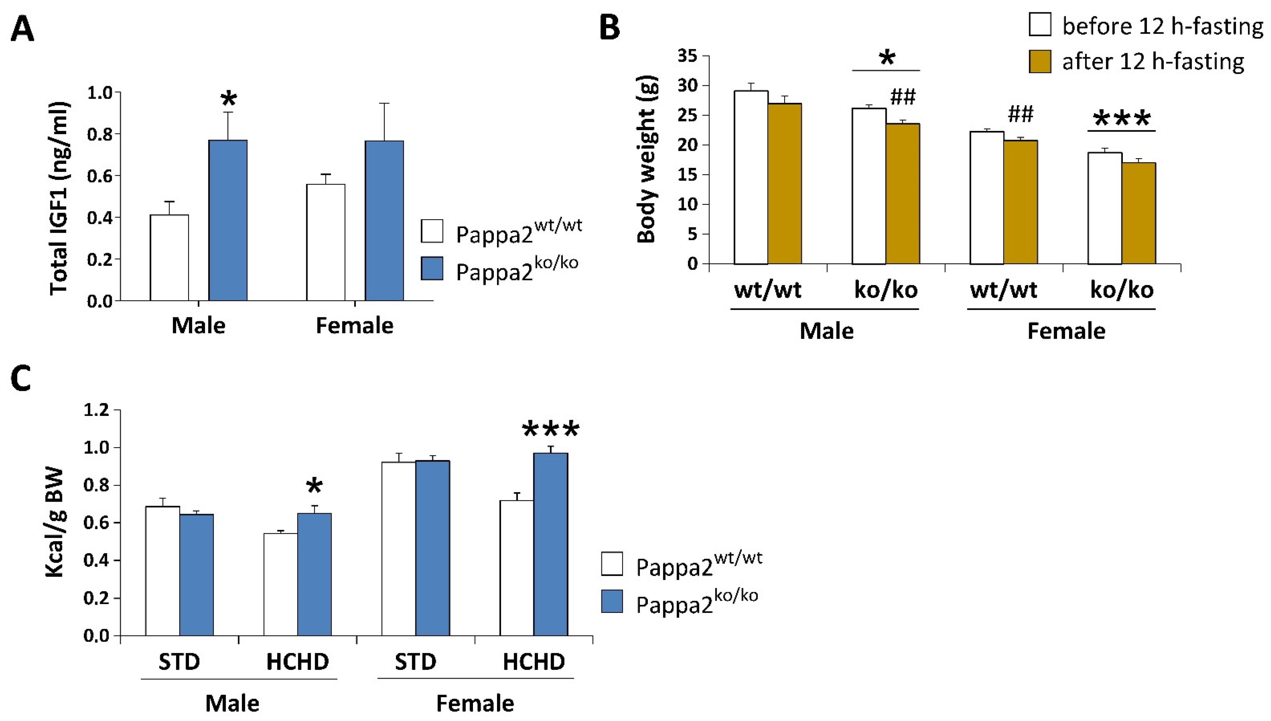
In the model presented by Conover and colleagues [5], no changes in Pappa2 mRNA levels or Igfbp5 proteolysis in fibroblasts of Pappa2 KO embryos were found. However, at 4 months of age, these Pappa2 KO mice had a dramatic reduction (95%) in free Igf1 concentrations and elevated concentrations (50%) of total Igf1, supporting functional PAPP-A2 defects in the release of IGF1 from its ternary complex, as observed in affected patients [15]. Christians and colleagues [13] also reported an increase (60%) in circulating levels of total Igf1 at 1.5 months of age in their Pappa2-KO model. Recently, these authors described a reduction in the circulating levels of a putative free bioactive Igf1 in 2-month-old Pappa2 KO mice [16]. Circulating free Igf1 is also decreased and total Igf1 increased in the Pappa2 KI mice at 4 months of age [11]. Using the second Pappa2 KO mouse model developed by Christians et al. [12], researchers have observed higher circulating levels of total Igf1, specifically in males, but not in Pappa2 KO females at 8 months of age (Figure 1A). At 1.5 months of age constitutive Pappa2 KO mice also had 2-fold higher levels of Igfbp5 and 15-fold lower levels of Igfbp3 [13]. Increased circulating levels of Igfbp5, recently confirmed in 2-month-old Pappa2 KO mice, were not influenced by gestation and recovery after lactation [16], with these variations also continuing to be observed in the plasma of older mice with a mean age of 7 months [13]. However, at 7 months of age, no effect of Pappa2 deletion was observed on mRNA levels of Igfbp3 or Igfbp5 in the liver or kidney, but Igfbp5 protein levels were increased in ovaries of female Pappa2 KO mice [17]. In contrast, osteoblast-specific deletion of Pappa2 did not alter circulating levels of Igfbp5 in mice at 18–19 days of age [9], suggesting a local Pappa2-Igfbp5 mechanism for Igf1 function in bone physiology not yet addressed. When whole body Pappa2 deletion was induced at 5 months of age, no difference in circulating levels of Igfbp5 was found between genotypes, but lower levels of circulating Igfbp3 were detected in a body size and sex-independent manner [13]. In contrast, increased circulating levels of Igfbp3 and Igfals were found in Pappa2 KI mice at 4 months of age [11]. Researchers found a genotype effect on Igfbp3 mRNA levels in the bone of 8-month-old Pappa2 KO mice with female Pappa2 KO mice having higher Igfbp3 mRNA levels in the tibia than female WT littermates [14].
Figure 1. Analysis of plasma concentrations of total IGF1 (A), body weight (BW) before and after 12 h-fasting (B), and caloric intake relative to BW after 3 week-feeding of a standard (STD: 2.9 kcal/g) or a high carbohydrate (HCHD: 3.85 kcal/g) diet (C) in constitutive Pappa2 KO mice of both sexes at 8 months old. Data are represented as mean ± S.E.M. (n = 7–8/group). Tukey-corrected tests: */*** p < 0.05/0.001 versus respective Pappa2wt/wt mice; ## p < 0.01 versus mice before 12 h-fasting.
Analysis of a morpholino-mediated Pappa2 KD zebrafish embryo model (50% and 43% overall sequence identity to human PAPP-A2 and PAPP-A, respectively, and conserved proteolytic activity in human IGFBP3 and IGFBP5) suggested that Pappa2 actions in normal growth and development may also be exerted through non-proteolytic modulation of Notch signaling [10]. While IGFBP-independent mechanisms of PAPP-A2 cannot be excluded, overall results to date indicate that the modulation of IGF1 bioavailability through its release from ternary and binary complexes should be further investigated in a tissue, sex and age-specific manner.

5. Metabolic Disturbance in Available Models of Pappa2 Deficiency

Despite the smaller size and lower weight of Pappa2 KO mice at 3, 6, 10 and 14 weeks of age in the model described by Christians and colleagues [13], disruption of circulating Igf1 levels did not affect glucose tolerance, body weight gain or adiposity at 6 weeks of age when constitutive Pappa2 KO mice were fed a high-fat diet (45% fat) from 17 to 25 weeks of age. Likewise, using the mouse model developed by Christians et al. [12], researchers found no modifications in glucose tolerance in constitutive Pappa2 KO mice at 2 months of age, but researchers found that these Pappa2 KO mice lost more body weight than their WT littermates when fasted for 12 h (Figure 1B). In contrast, Pappa2 KI mice are reported to be glucose intolerant and insulin resistant [11]. These discrepancies in glucose handling may be due to differences in the dietary supply of glucose, amino acids and lipids to the liver, in addition to metabolic compensation, such as an increase in insulin concentrations and/or decrease in the insulin-induced autophosphorylation of its receptor and insulin receptor substrate, observed in constitutive Pappa2 KO mice that may not be produced in human Pappa2 KI mice due to the presence of a Pappa2 protein, at least in some patients, despite its lack of protease activity. In the preliminary studies, we did not observe differences in body weight gain compared to WT when Pappa2 KO mice were fed a high-carbohydrate diet (HCHD, 70% sucrose and fructose) for 3 weeks, but they increased their caloric intake (relative to body weight), suggesting higher energy expenditure associated with lower Igf1 availability when exposed to a high carbohydrate diet (Figure 1C). Because diabetes and obesity induced by hypercaloric diets have been associated with impairments in bone mineral density [18][19][20], future studies should evaluate whether deficiency in Igf1 bioactivity impacts energy homeostasis, regulating bone mass in a sex and diet-specific manner [21].

References

  1. Fujimoto, M.; Andrew, M.; Dauber, A. Disorders caused by genetic defects associated with GH-dependent genes: PAPPA2 defects. Mol. Cell. Endocrinol. 2020, 518, 110967.
  2. Farr, M.; Strübe, J.; Geppert, H.-G.; Kocourek, A.; Mahne, M.; Tschesche, H. Pregnancy-associated plasma protein-E (PAPP-E). Biochim. Biophys. Acta BBA-Gene Struct. Expr. 2000, 1493, 356–362.
  3. Page, N.; Butlin, D.; Lomthaisong, K.; Lowry, P. The Characterization of Pregnancy Associated Plasma Protein-E and the Identification of an Alternative Splice Variant. Placenta 2001, 22, 681–687.
  4. Wagner, P.K.; Otomo, A.; Christians, J.K. Regulation of pregnancy-associated plasma protein A2 (PAPPA2) in a human placental trophoblast cell line (BeWo). Reprod. Biol. Endocrinol. 2011, 9, 48.
  5. Conover, C.A.; Boldt, H.B.; Bale, L.K.; Clifton, K.B.; Grell, J.A.; Mader, J.R.; Mason, E.J.; Powell, D.R. Pregnancy-Associated Plasma Protein-A2 (PAPP-A2): Tissue Expression and Biological Consequences of Gene Knockout in Mice. Endocrinology 2011, 152, 2837–2844.
  6. Christians, J.K.; Hoeflich, A.; Keightley, P.D. PAPPA2, an Enzyme That Cleaves an Insulin-Like Growth-Factor-Binding Protein, Is a Candidate Gene for a Quantitative Trait Locus Affecting Body Size in Mice. Genetics 2006, 173, 1547–1553.
  7. Wang, J.; Qiu, Q.; Haider, M.; Bell, M.; Gruslin, A.; Christians, J.K. Expression of pregnancy-associated plasma protein A2 during pregnancy in human and mouse. J. Endocrinol. 2009, 202, 337–345.
  8. Chen, Y.; Lv, H.; Li, L.; Wang, E.; Zhang, L.; Zhao, Q. Expression of PAPP-A2 and IGF Pathway-Related Proteins in the Hip Joint of Normal Rat and Those with Developmental Dysplasia of the Hip. Int. J. Endocrinol. 2019, 2019, 7691531.
  9. Amiri, N.; Christians, J.K. PAPP-A2 expression by osteoblasts is required for normal postnatal growth in mice. Growth Horm. IGF Res. 2015, 25, 274–280.
  10. Kjaer-Sorensen, K.; Engholm, D.H.; Jepsen, M.R.; Morch, M.G.; Weyer, K.; Hefting, L.L.; Skov, L.L.; Laursen, L.S.; Oxvig, C. Papp-a2 modulates development of cranial cartilage and angiogenesis in zebrafish embryos. J. Cell Sci. 2014, 127 Pt 23, 5027–5037.
  11. Fujimoto, M.; Andrew, M.; Liao, L.; Zhang, D.; Yildirim, G.; Sluss, P.; Kalra, B.; Kumar, A.; Yakar, S.; Hwa, V.; et al. Low IGF-I Bioavailability Impairs Growth and Glucose Metabolism in a Mouse Model of Human PAPPA2 p.Ala1033Val Mutation. Endocrinology 2019, 160, 1363–1376.
  12. Christians, J.K.; de Zwaan, D.; Fung, S.H.Y. Pregnancy Associated Plasma Protein A2 (PAPP-A2) Affects Bone Size and Shape and Contributes to Natural Variation in Postnatal Growth in Mice. PLoS ONE 2013, 8, e56260.
  13. Christians, J.K.; Bath, A.K.; Amiri, N. Pappa2 deletion alters IGFBPs but has little effect on glucose disposal or adiposity. Growth Horm. IGF Res. 2015, 25, 232–239.
  14. Rubio, L.; Vargas, A.; Rivera, P.; López-Gambero, A.; Tovar, R.; Christians, J.; Martín-De-Las-Heras, S.; de Fonseca, F.R.; Chowen, J.; Argente, J.; et al. Recombinant IGF-1 Induces Sex-Specific Changes in Bone Composition and Remodeling in Adult Mice with Pappa2 Deficiency. Int. J. Mol. Sci. 2021, 22, 4048.
  15. Dauber, A.; Muñoz-Calvo, M.T.; Barrios, V.; Domené, H.M.; Kloverpris, S.; Serra-Juhé, C.; Desikan, V.; Pozo, J.; Muzumdar, R.; Martos-Moreno, G.Á.; et al. Mutations in pregnancy-associated plasma protein A2 cause short stature due to low IGF-I availability. EMBO Mol. Med. 2016, 8, 363–374.
  16. Rogowska, M.D.; Pena, U.N.V.; Binning, N.; Christians, J.K. Recovery of the maternal skeleton after lactation is impaired by advanced maternal age but not by reduced IGF availability in the mouse. PLoS ONE 2021, 16, e0256906.
  17. Christians, J.K.; King, A.Y.; Rogowska, M.D.; Hessels, S.M. Pappa2 deletion in mice affects male but not female fertility. Reprod. Biol. Endocrinol. 2015, 13, 109.
  18. Carvalho, A.L.; DeMambro, V.E.; Guntur, A.R.; Le, P.; Nagano, K.; Baron, R.; de Paula, F.J.A.; Motyl, K.J. High fat diet attenuates hyperglycemia, body composition changes, and bone loss in male streptozotocin-induced type 1 diabetic mice. J. Cell. Physiol. 2018, 233, 1585–1600.
  19. Yanagihara, G.R.; Shimano, R.C.; Tida, J.A.; Yamanaka, J.S.; Fukada, S.Y.; Issa, J.P.M.; Tavares, J.M.R.S. Influence of High-Fat Diet on Bone Tissue: An Experimental Study in Growing Rats. J. Nutr. Health Aging 2017, 21, 1337–1343.
  20. Qiao, J.; Wu, Y.-W.; Ren, Y.-Z. The impact of a high fat diet on bones: Potential mechanisms. Food Funct. 2021, 12, 963–975.
  21. Kawai, M.; Rosen, C.J. The IGF-I regulatory system and its impact on skeletal and energy homeostasis. J. Cell. Biochem. 2010, 111, 14–19.
More
Information
Contributors MDPI registered users' name will be linked to their SciProfiles pages. To register with us, please refer to https://encyclopedia.pub/register : , , , , , , , , ,
View Times: 240
Revisions: 2 times (View History)
Update Date: 14 Jul 2023
1000/1000
Video Production Service