Submitted Successfully!
To reward your contribution, here is a gift for you: A free trial for our video production service.
Thank you for your contribution! You can also upload a video entry or images related to this topic.
Version Summary Created by Modification Content Size Created at Operation
1 -- 2263 2023-07-13 10:17:38 |
2 format correction -6 word(s) 2257 2023-07-14 02:32:09 | |
3 format correction + 4 word(s) 2261 2023-07-31 08:26:04 |

Video Upload Options

Do you have a full video?

Confirm

Are you sure to Delete?
Cite
If you have any further questions, please contact Encyclopedia Editorial Office.
Romano, I.R.; D’angeli, F.; Vicario, N.; Russo, C.; Genovese, C.; Lo Furno, D.; Mannino, G.; Tamburino, S.; Parenti, R.; Giuffrida, R. Adipose-Derived Mesenchymal Stromal Cells. Encyclopedia. Available online: https://encyclopedia.pub/entry/46738 (accessed on 03 May 2024).
Romano IR, D’angeli F, Vicario N, Russo C, Genovese C, Lo Furno D, et al. Adipose-Derived Mesenchymal Stromal Cells. Encyclopedia. Available at: https://encyclopedia.pub/entry/46738. Accessed May 03, 2024.
Romano, Ivana Roberta, Floriana D’angeli, Nunzio Vicario, Cristina Russo, Carlo Genovese, Debora Lo Furno, Giuliana Mannino, Serena Tamburino, Rosalba Parenti, Rosario Giuffrida. "Adipose-Derived Mesenchymal Stromal Cells" Encyclopedia, https://encyclopedia.pub/entry/46738 (accessed May 03, 2024).
Romano, I.R., D’angeli, F., Vicario, N., Russo, C., Genovese, C., Lo Furno, D., Mannino, G., Tamburino, S., Parenti, R., & Giuffrida, R. (2023, July 13). Adipose-Derived Mesenchymal Stromal Cells. In Encyclopedia. https://encyclopedia.pub/entry/46738
Romano, Ivana Roberta, et al. "Adipose-Derived Mesenchymal Stromal Cells." Encyclopedia. Web. 13 July, 2023.
Adipose-Derived Mesenchymal Stromal Cells
Edit

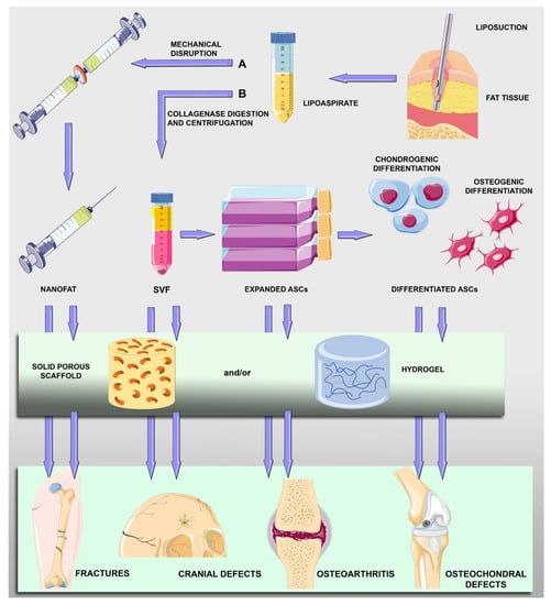
Being of mesodermal origin, ASCs (adipose-derived mesenchymal stromal cells) can be easily induced to differentiate into chondrocyte-like and osteocyte-like elements and used to repair damaged tissues. Moreover, they can be easily harvested and used for autologous implantation. A plethora of ASC-based strategies are being developed worldwide: they include the transplantation of freshly harvested cells, in vitro expanded cells or predifferentiated cells. Moreover, improving their positive effects, ASCs can be implanted in combination with several types of scaffolds that ensure the correct cell positioning; support cell viability, proliferation and migration; and may contribute to their osteogenic or chondrogenic differentiation. Examples of these strategies are described here, showing the enormous therapeutic potential of ASCs in this field. For safety and regulatory issues, most investigations are still at the experimental stage and carried out in vitro and in animal models. Clinical applications have, however, been reported with promising results and no serious adverse effects.

adipose-derived mesenchymal stromal cells stem cells bone cartilage osteochondral defects scaffolds osteogenic differentiation

1. Introduction

Regenerative medicine and tissue engineering have emerged with the purpose of rebuilding or repairing tissues and organs damaged by diseases, traumas or aging. In this context, a growing body of studies has been focused on stem-cell-based treatments for numerous human pathologies, including bone and cartilage disorders [1][2]. Stem cells are unspecialized cells capable of self-renewal and differentiation into various types of functional cells. There are different types of stem cells, such as embryonic stem cells, fetal stem cells, induced pluripotent stem cells and adult stem cells. The first three types are extensively studied for their marked proliferative potential and for their ability to differentiate into multiple cell lines; however, their use for clinical treatments is limited by considerable safety, ethical and regulatory issues [3]. For these reasons, much attention has been focused on adult stem or stromal cells that, although featuring lower differentiation capabilities, are more suitable for treatments in humans [4]. Adult stem cells are located in specialized niches of most tissues, where they continuously produce tissue-specific differentiated elements as well as other daughter stem cells that ensure a constant pool [5]. Hematopoietic stem cells in the bone marrow were the first population to be identified [6]. A few years later, a second population with different characteristics was discovered [7]. Defined as “bone marrow stromal cells” (BMSCs), they are a heterogeneous population of cells able to differentiate into mesodermal elements [8] and are thus considered mesenchymal stem cells (MSCs). Among these cells, marrow adipogenic lineage precursors seem to play important regulatory roles in bone remodeling, hematopoiesis regulation and marrow vasculature maintenance [9]. Under appropriate conditions, MSCs can differentiate not only into cells of the mesodermal lineage but also into elements of ectodermal or endodermal origin [10][11]. Other than bone marrow, MSCs can be isolated from numerous other tissues, such as the umbilical cord, dental pulp, skin, salivary glands [12][13] and from adipose tissue [14][15][16]. Comparative studies were carried out investigating the immunophenotype, proliferative potential, multilineage differentiation and immunomodulatory capacity of MSCs from different tissues (bone marrow, adipose tissue, the placenta and umbilical cord blood). On the basis of the gene expression profiles of stemness-related genes and lineage differentiation stage-related genes, it was found that no significant differences were observed in terms of the growth rate, colony-forming efficiency and immunophenotype. It was also found that BMSCs and ASCs shared both in vitro trilineage differentiation potential and gene expression profiles and represent the optimal stem cell source for tissue engineering and regenerative medicine [17]. In particular, adipose-derived MSCs (ASCs) will be considered, as they have been widely studied for their numerous advantages. Unlike BMSCs, which require general anesthesia because of the painful harvesting procedure, ASCs can be obtained in large quantities with less invasive methods, and moreover, they feature even more proliferative activity and are more easily available for autologous administration [18][19].

2. Adipose-Derived Mesenchymal Stromal Cells (ASCs)

ASCs are commonly isolated from the stromal vascular fraction (SVF) of adipose tissue, which is diffusely located in the human body at either the subcutaneous or visceral level. Depending on the anatomical region from which the adipose sample is taken, some differences have been reported. For example, a greater number of cells can be isolated from the adipose tissue of the arm, whereas cells with excellent plasticity can be obtained from the groin area [20]. Many studies have been carried out on adipose tissue contained in the lipoaspirate that is available after liposuction and would be otherwise discarded. Although the harvesting procedure is generally accurate, contaminations may occur. Specifically, microorganisms (bacteria, viruses and fungi) can infect samples during the various preparation steps so that infections in the host tissues and immune reactions might be generated after implantation [21]. For this reason, microbiological control is fundamental at each stage to ensure the absence of contamination and the safety of the final product. Once isolated, ASCs can be expanded and identified by their plastic adherence, colony forming capacity and rapid proliferation. They show a fibroblast-like morphology and are positive for typical MSC markers (CD44, CD73, CD90 and CD105) but not for typical hematopoietic markers (CD14, CD34 and CD45). According to their MSC nature, they are able to differentiate toward osteogenic, adipogenic, myogenic and chondrogenic lineages [22][23][24][25]. However, with specific treatment, ASCs can also differentiate not only into other cells of mesodermal derivation [26][27][28] but also into cells originating from endodermal or ectodermal embryonic layers, such as neural cells [29]. This can usually be obtained by supplementing the growth medium with bioactive molecules, although satisfactory results can be achieved by using conditioned media from other cell cultures [30][31][32][33][34][35]. It is important to underline that ASCs have a low immunological reactivity, thanks to the absence or low expression of immunogenic surface antigens (CD40, CD40L, CD80 and CD86) and major histocompatibility complex II. This low immunological reactivity also makes them suitable for allogeneic use [20]. In addition, ASCs are able to modulate T and B cell activity and exert anti-inflammatory effects [36]. Finally, ASC beneficial effects can also be due to their paracrine production of numerous cytokines and growth factors, such as vascular endothelial growth factor (VEGF), fibroblast growth factor (FGF) and insulin-like growth factor 1 (IGF-1) [37][38]. As a result, the use of an ASC-conditioned medium is also investigated for potential cell-free therapeutic applications [39].

2.1. ASC Osteogenic Differentiation

The osteogenic induction of ASCs is usually obtained in about 21 days by replacing the basal growth medium with osteogenic media containing dexamethasone, β-glycerophosphate and ascorbic acid [40]. To verify the osteogenic phenotype, specific histological staining is commonly used (Von Kossa or Alizarin Red), which reveals calcium deposits and the mineralized matrix [1]. During differentiation, ASCs produce a mineralized extracellular matrix (ECM) with an increased expression of bone markers, such as runt-related transcription factor 2 (RUNX2; a marker of osteoblast differentiation), osteonectin and osteocalcin, as well as an increased synthesis of alkaline phosphatase (ALP) and collagen type I [41][42]. A better degree of osteogenic differentiation has recently been obtained using ASCs transfected with osteo-specific genes, such as the insertion of encoding bone morphogenetic protein (BMP)-2 and Runx2 genes [43]. Controversial results were reported by comparing ASC and BMSC osteogenic potential. According to Brennan et al. [44], BMSCs show better osteogenesis, whereas ASCs feature enhanced angiogenesis. In contrast, in another comparative study, it was found that ASCs showed a higher proliferation rate and an increased ability to differentiate into osteocytes and chondrocytes, as evaluated using Alizarin Red and Alcian Blue staining, respectively [18]. In particular, these properties were more pronounced for cells expressing CD271, a neurotrophin receptor that is a member of the tumor necrosis factor (TNF) receptor superfamily. However, the differences might be simply due to different experimental procedures.

2.2. ASC Chondrogenic Differentiation

Various protocols have been adopted to obtain ASC chondrogenic differentiation, normally requiring 21–28 days [45]. Widely used differentiation media usually contain high glucose levels, fetal calf or bovine serum, penicillin and streptomycin, ascorbate-2-phosphate, dexamethasone, transforming growth factor beta (TGF-β1 or TGF-β3), insulin, transferrin, selenium, sodium pyruvate and L-glutamine. Additional supplements and growth factors can be included, such as BMP-2, 4 or 6, sex-determining region Y box 9 (SOX 9) and basic FGF [46][47][48]. The chondrogenic phenotype can be evaluated using various tests, including histological staining using Alcian blue, toluidine blue or safranin O, detecting the presence of proteoglycan in the cartilage-like matrix. In addition, real-time PCR, Western blot analysis, ELISA, RNA microarray analysis and immunohistochemistry reveal the expression of chondrocyte-specific genes or proteins, such as different collagen types, keratin sulfate, chondroitin sulfate, aggrecan, decorin and biglycan [41][49]. In monolayer cultures, ASCs hardly maintain a chondrogenic phenotype, showing a decrease in collagen type II expression, while collagen type X production increases. A three-dimensional (3D) culture of chondrocyte-like ASCs overcomes this issue. In a study by Musumeci et al. [50], ASCs treated with a specific chondrogenic medium for 28 days showed a high expression of collagen type I and II, as well as lubricin, a key molecule in cartilage wear prevention. This glycoprotein, which assures joint lubrication and synovial homeostasis, may represent an additional differentiation biomarker.

3. ASC-Based Repair Strategies

Different approaches have been explored for the treatment of cartilage and bone tissue disorders: the transplantation of uncultured ASCs; the transplantation of ASCs expanded in vitro; the transplantation of ASCs after their osteogenic or chondrogenic differentiation; and variously treated ASCs in combination with scaffolds (Figure 1).
Figure 1. Schematic drawing showing main steps of harvesting and treatments of adipose-derived mesenchymal stromal cells (ASCs) for bone and cartilage repair. After liposuction procedures, fat tissue in the lipoaspirate can be mechanically disrupted (A) to obtain micro fat particles (nanofat) containing ASCs and other cell types (lymphocytes, pericytes, vascular endothelial cells and vascular smooth muscle cells). Lipoaspirate can be digested and centrifuged (B) to obtain a pellet (stromal vascular fraction, SVF) containing ASCs that can be expanded and undergo osteogenic or chondrogenic differentiation. Nanofat particles, SVF cells, expanded ASCs or predifferentiated ASCs can be directly implanted or included in scaffold-assisted treatments.
For cell-based applications, freshly isolated ASCs present in adipose SVF are preferred in some instances, not only to shorten the interval from harvesting to the transplant procedure but also to avoid restrictions and risks related to cell culturing, such as the possibility of contamination, tumorigenesis or unexpected cell differentiation. A collagenase enzyme is normally added to the adipose tissue, producing a mixture that, after filtration and centrifugation, can be separated into a superficial adipocyte fraction and precipitated cellular components. To overcome restrictions associated with the enzymatic procedure, alternative methods to obtain an SVF-like compound have been developed. Although with a lower cell yield, SVF cells featuring virtually the same regenerative potential can be obtained using mechanical disruption of the adipose tissue giving rise to micro fat particles that are also called nanofat [51][52]. Nanofat grafting is widely used in plastic surgery [53] and for bone and cartilage repair, often combined with platelet-rich plasma (PRP) [16]. After centrifugation, ASCs contained in the SVF can be successively expanded for further uses. Based on cell–cell and cell–matrix interactions, 3D cultures of ASCs would offer a favorable context for their multilineage vascular and osteogenic differentiation, providing in the resulting spheroids an in vitro model of the interactive elements [54]. This strategy is particularly advantageous to improve the thickness of a scaffold-free implant. Figure 1 illustrates the main steps of ASC treatment for bone and cartilage repair.

Scaffold-Assisted Strategies

Scaffold-based approaches have been widely investigated as cell implantation alone often raises substantial issues related to postimplantation cell fate, cellular loss and dispersion. A variety of scaffolds have been designed, improving not only the correct positioning of stem cells but also their attachment, viability, proliferation, migration and differentiation [55][56][57]. Scaffolds can be subdivided into different categories (i.e., metallic or nonmetallic, natural or synthetic), and many of them are available for applications to skeletal and other tissues. Due to their properties, metallic implants using titanium and titanium dioxide are particularly suitable for bone defects. On the other hand, many more nonmetallic scaffolds have been designed, each of them characterized by different advantages and disadvantages. Examples of these scaffolds include the acellular matrix, coralline scaffolds, natural or synthetic polymers and hybrid scaffolds [20][58][59]. Ideal scaffolds should be degradable and biocompatible and mimic as closely as possible the physiological microenvironment of the target tissue. In addition, they can be loaded not only with stem cells but also with bioactive molecules, such as growth factors and anti-inflammatory or antibacterial agents [60]. Optimal pore size is a crucial characteristic of a scaffold and largely depends on the type of tissue for which the scaffold is planned [61]. For example, smaller pore sizes are suitable for the formation of fibrous tissues, while larger pore sizes are more appropriate for the formation of bone tissue. Too wide pores hamper cell attachment and the stiffness of the scaffold, whereas too small pores reduce cell viability and migration, nutrient diffusion and waste removal. After the initial adhesion and tissue formation at the external surfaces, stem cells should be able to penetrate inside the scaffold giving rise to an inward gradient of tissue formation [62]. The newly formed tissue would hopefully restore the proper shape and function.
Natural ECMs can be advantageously prepared in the form of hydrogels [58], which are considered a promising cell-supporting alternative material [63]. Hydrogels are characterized by a 3D polymeric network that, retaining a large amount of water, more closely resembles soft tissues, thus providing a wider range of scaffold applications. Hydrogels possess good biocompatibility, tunable swelling and mechanical properties and a biodegradation rate that is more predictable and adjustable than other scaffolds. The most used natural biomaterials include collagen, gelatin, hyaluronic acid, chitosan and alginate, noncollagenous proteins and proteoglycans [64][65].
For in vivo experiments, rodents are mostly investigated, but larger animals (goats, dogs or minipigs) are also often chosen because a large animal requires implantation procedures that are more similar to those for human treatments, thus improving the translational potential.

References

  1. Barba, M.; Cicione, C.; Bernardini, C.; Michetti, F.; Lattanzi, W. Adipose-derived mesenchymal cells for bone regereneration: State of the art. BioMed Res. Int. 2013, 2013, 416391.
  2. Perdisa, F.; Gostynska, N.; Roffi, A.; Filardo, G.; Marcacci, M.; Kon, E. Adipose-Derived Mesenchymal Stem Cells for the Treatment of Articular Cartilage: A Systematic Review on Preclinical and Clinical Evidence. Stem Cells Int. 2015, 2015, 597652.
  3. Zakrzewski, W.; Dobrzynski, M.; Szymonowicz, M.; Rybak, Z. Stem cells: Past, present, and future. Stem Cell Res. Ther. 2019, 10, 68.
  4. Kolios, G.; Moodley, Y. Introduction to stem cells and regenerative medicine. Respiration 2013, 85, 3–10.
  5. Mannino, G.; Russo, C.; Maugeri, G.; Musumeci, G.; Vicario, N.; Tibullo, D.; Giuffrida, R.; Parenti, R.; Lo Furno, D. Adult stem cell niches for tissue homeostasis. J. Cell Physiol. 2022, 237, 239–257.
  6. Friedenstein, A.J.; Petrakova, K.V.; Kurolesova, A.I.; Frolova, G.P. Heterotopic of bone marrow. Analysis of precursor cells for osteogenic and hematopoietic tissues. Transplantation 1968, 6, 230–247.
  7. Friedenstein, A.J.; Chailakhjan, R.K.; Lalykina, K.S. The development of fibroblast colonies in monolayer cultures of guinea-pig bone marrow and spleen cells. Cell Tissue Kinet. 1970, 3, 393–403.
  8. Kumar, R.; Godavarthy, P.S.; Krause, D.S. The bone marrow microenvironment in health and disease at a glance. J. Cell Sci. 2018, 131, jcs201707.
  9. Zhong, L.; Yao, L.; Seale, P.; Qin, L. Marrow adipogenic lineage precursor: A new cellular component of marrow adipose tissue. Best Pract. Res. Clin. Endocrinol. Metab. 2021, 35, 101518.
  10. Safford, K.M.; Hicok, K.C.; Safford, S.D.; Halvorsen, Y.D.; Wilkison, W.O.; Gimble, J.M.; Rice, H.E. Neurogenic differentiation of murine and human adipose-derived stromal cells. Biochem. Biophys. Res. Commun. 2002, 294, 371–379.
  11. Li, M.; Ikehara, S. Bone-marrow-derived mesenchymal stem cells for organ repair. Stem Cells Int. 2013, 2013, 132642.
  12. Arutyunyan, I.; Elchaninov, A.; Makarov, A.; Fatkhudinov, T. Umbilical Cord as Prospective Source for Mesenchymal Stem Cell-Based Therapy. Stem Cells Int. 2016, 2016, 6901286.
  13. Mushahary, D.; Spittler, A.; Kasper, C.; Weber, V.; Charwat, V. Isolation, cultivation, and characterization of human mesenchymal stem cells. Cytom. A 2018, 93, 19–31.
  14. Zuk, P.A.; Zhu, M.; Mizuno, H.; Huang, J.; Futrell, J.W.; Katz, A.J.; Benhaim, P.; Lorenz, H.P.; Hedrick, M.H. Multilineage cells from human adipose tissue: Implications for cell-based therapies. Tissue Eng. 2001, 7, 211–228.
  15. Dicker, A.; Le Blanc, K.; Astrom, G.; van Harmelen, V.; Gotherstrom, C.; Blomqvist, L.; Arner, P.; Ryden, M. Functional studies of mesenchymal stem cells derived from adult human adipose tissue. Exp. Cell Res. 2005, 308, 283–290.
  16. Sharma, S.; Muthu, S.; Jeyaraman, M.; Ranjan, R.; Jha, S.K. Translational products of adipose tissue-derived mesenchymal stem cells: Bench to bedside applications. World J. Stem Cells 2021, 13, 1360–1381.
  17. Heo, J.S.; Choi, Y.; Kim, H.S.; Kim, H.O. Comparison of molecular profiles of human mesenchymal stem cells derived from bone marrow, umbilical cord blood, placenta and adipose tissue. Int. J. Mol. Med. 2016, 37, 115–125.
  18. Calabrese, G.; Giuffrida, R.; Lo Furno, D.; Parrinello, N.L.; Forte, S.; Gulino, R.; Colarossi, C.; Schinocca, L.R.; Giuffrida, R.; Cardile, V.; et al. Potential Effect of CD271 on Human Mesenchymal Stromal Cell Proliferation and Differentiation. Int. J. Mol. Sci. 2015, 16, 15609–15624.
  19. Bacakova, L.; Zarubova, J.; Travnickova, M.; Musilkova, J.; Pajorova, J.; Slepicka, P.; Kasalkova, N.S.; Svorcik, V.; Kolska, Z.; Motarjemi, H.; et al. Stem cells: Their source, potency and use in regenerative therapies with focus on adipose-derived stem cells—A review. Biotechnol. Adv. 2018, 36, 1111–1126.
  20. Rahman, G.; Frazier, T.P.; Gimble, J.M.; Mohiuddin, O.A. The Emerging Use of ASC/Scaffold Composites for the Regeneration of Osteochondral Defects. Front. Bioeng. Biotechnol. 2022, 10, 893992.
  21. Mazini, L.; Ezzoubi, M.; Malka, G. Overview of current adipose-derived stem cell (ADSCs) processing involved in therapeutic advancements: Flow chart and regulation updates before and after COVID-19. Stem Cell Res. Ther. 2021, 12, 1.
  22. Zuk, P.A.; Zhu, M.; Ashjian, P.; De Ugarte, D.A.; Huang, J.I.; Mizuno, H.; Alfonso, Z.C.; Fraser, J.K.; Benhaim, P.; Hedrick, M.H. Human adipose tissue is a source of multipotent stem cells. Mol. Biol. Cell 2002, 13, 4279–4295.
  23. Lo Furno, D.; Graziano, A.C.; Avola, R.; Giuffrida, R.; Perciavalle, V.; Bonina, F.; Mannino, G.; Cardile, V. A Citrus bergamia Extract Decreases Adipogenesis and Increases Lipolysis by Modulating PPAR Levels in Mesenchymal Stem Cells from Human Adipose Tissue. PPAR Res. 2016, 2016, 4563815.
  24. Si, Z.; Wang, X.; Sun, C.; Kang, Y.; Xu, J.; Wang, X.; Hui, Y. Adipose-derived stem cells: Sources, potency, and implications for regenerative therapies. Biomed. Pharmacother. 2019, 114, 108765.
  25. Mannino, G.; Vicario, N.; Parenti, R.; Giuffrida, R.; Lo Furno, D. Connexin expression decreases during adipogenic differentiation of human adipose-derived mesenchymal stem cells. Mol. Biol. Rep. 2020, 47, 9951–9958.
  26. Mannino, G.; Gennuso, F.; Giurdanella, G.; Conti, F.; Drago, F.; Salomone, S.; Furno, D.L.; Bucolo, C.; Giuffrida, R. Pericyte-like differentiation of human adipose-derived mesenchymal stem cells: An in vitro study. World J. Stem Cells 2020, 12, 1152–1170.
  27. Mannino, G.; Longo, A.; Gennuso, F.; Anfuso, C.D.; Lupo, G.; Giurdanella, G.; Giuffrida, R.; Lo Furno, D. Effects of High Glucose Concentration on Pericyte-Like Differentiated Human Adipose-Derived Mesenchymal Stem Cells. Int. J. Mol. Sci. 2021, 22, 4604.
  28. Lupo, G.; Agafonova, A.; Cosentino, A.; Giurdanella, G.; Mannino, G.; Lo Furno, D.; Romano, I.R.; Giuffrida, R.; D’Angeli, F.; Anfuso, C.D. Protective Effects of Human Pericyte-like Adipose-Derived Mesenchymal Stem Cells on Human Retinal Endothelial Cells in an In Vitro Model of Diabetic Retinopathy: Evidence for Autologous Cell Therapy. Int. J. Mol. Sci. 2023, 24, 913.
  29. Guasti, L.; New, S.E.; Hadjidemetriou, I.; Palmiero, M.; Ferretti, P. Plasticity of human adipose-derived stem cells—Relevance to tissue repair. Int. J. Dev. Biol. 2018, 62, 431–439.
  30. Lo Furno, D.; Pellitteri, R.; Graziano, A.C.; Giuffrida, R.; Vancheri, C.; Gili, E.; Cardile, V. Differentiation of human adipose stem cells into neural phenotype by neuroblastoma- or olfactory ensheathing cells-conditioned medium. J. Cell Physiol. 2013, 228, 2109–2118.
  31. Lo Furno, D.; Mannino, G.; Giuffrida, R.; Gili, E.; Vancheri, C.; Tarico, M.S.; Perrotta, R.E.; Pellitteri, R. Neural differentiation of human adipose-derived mesenchymal stem cells induced by glial cell conditioned media. J. Cell Physiol. 2018, 233, 7091–7100.
  32. Russo, C.; Patane, M.; Vicario, N.; Di Bella, V.; Cosentini, I.; Barresi, V.; Gulino, R.; Pellitteri, R.; Russo, A.; Stanzani, S. Olfactory Ensheathing Cells express both Ghrelin and Ghrelin Receptor in vitro: A new hypothesis in favor of a neurotrophic effect. Neuropeptides 2020, 79, 101997.
  33. Russo, C.; Mannino, G.; Patane, M.; Parrinello, N.L.; Pellitteri, R.; Stanzani, S.; Giuffrida, R.; Lo Furno, D.; Russo, A. Ghrelin peptide improves glial conditioned medium effects on neuronal differentiation of human adipose mesenchymal stem cells. Histochem. Cell Biol. 2021, 156, 35–46.
  34. Lo Furno, D.; Mannino, G.; Pellitteri, R.; Zappala, A.; Parenti, R.; Gili, E.; Vancheri, C.; Giuffrida, R. Conditioned Media From Glial Cells Promote a Neural-Like Connexin Expression in Human Adipose-Derived Mesenchymal Stem Cells. Front. Physiol. 2018, 9, 1742.
  35. Mannino, G.; Cristaldi, M.; Giurdanella, G.; Perrotta, R.E.; Lo Furno, D.; Giuffrida, R.; Rusciano, D. ARPE-19 conditioned medium promotes neural differentiation of adipose-derived mesenchymal stem cells. World J. Stem Cells 2021, 13, 1783–1796.
  36. Mazini, L.; Rochette, L.; Admou, B.; Amal, S.; Malka, G. Hopes and Limits of Adipose-Derived Stem Cells (ADSCs) and Mesenchymal Stem Cells (MSCs) in Wound Healing. Int. J. Mol. Sci. 2020, 21, 1306.
  37. Mannino, G.; Russo, C.; Longo, A.; Anfuso, C.D.; Lupo, G.; Lo Furno, D.; Giuffrida, R.; Giurdanella, G. Potential therapeutic applications of mesenchymal stem cells for the treatment of eye diseases. World J. Stem Cells 2021, 13, 632–644.
  38. Mende, W.; Gotzl, R.; Kubo, Y.; Pufe, T.; Ruhl, T.; Beier, J.P. The Role of Adipose Stem Cells in Bone Regeneration and Bone Tissue Engineering. Cells 2021, 10, 975.
  39. Marolt Presen, D.; Traweger, A.; Gimona, M.; Redl, H. Mesenchymal Stromal Cell-Based Bone Regeneration Therapies: From Cell Transplantation and Tissue Engineering to Therapeutic Secretomes and Extracellular Vesicles. Front. Bioeng. Biotechnol. 2019, 7, 352.
  40. Wang, L.; Huang, C.; Li, Q.; Xu, X.; Liu, L.; Huang, K.; Cai, X.; Xiao, J. Osteogenic differentiation potential of adipose-derived stem cells from ovariectomized mice. Cell. Prolif. 2017, 50, e12328.
  41. Guasti, L.; Prasongchean, W.; Kleftouris, G.; Mukherjee, S.; Thrasher, A.J.; Bulstrode, N.W.; Ferretti, P. High plasticity of pediatric adipose tissue-derived stem cells: Too much for selective skeletogenic differentiation? Stem Cells Transl. Med. 2012, 1, 384–395.
  42. Calabrese, G.; Giuffrida, R.; Fabbi, C.; Figallo, E.; Lo Furno, D.; Gulino, R.; Colarossi, C.; Fullone, F.; Giuffrida, R.; Parenti, R.; et al. Collagen-Hydroxyapatite Scaffolds Induce Human Adipose Derived Stem Cells Osteogenic Differentiation In Vitro. PLoS ONE 2016, 11, e0151181.
  43. Lee, S.J.; Kang, S.W.; Do, H.J.; Han, I.; Shin, D.A.; Kim, J.H.; Lee, S.H. Enhancement of bone regeneration by gene delivery of BMP2/Runx2 bicistronic vector into adipose-derived stromal cells. Biomaterials 2010, 31, 5652–5659.
  44. Brennan, M.A.; Renaud, A.; Guilloton, F.; Mebarki, M.; Trichet, V.; Sensebe, L.; Deschaseaux, F.; Chevallier, N.; Layrolle, P. Inferior In Vivo Osteogenesis and Superior Angiogenesis of Human Adipose-Derived Stem Cells Compared with Bone Marrow-Derived Stem Cells Cultured in Xeno-Free Conditions. Stem Cells Transl. Med. 2017, 6, 2160–2172.
  45. Stromps, J.P.; Paul, N.E.; Rath, B.; Nourbakhsh, M.; Bernhagen, J.; Pallua, N. Chondrogenic differentiation of human adipose-derived stem cells: A new path in articular cartilage defect management? BioMed Res. Int. 2014, 2014, 740926.
  46. Wei, Y.; Sun, X.; Wang, W.; Hu, Y. Adipose-derived stem cells and chondrogenesis. Cytotherapy 2007, 9, 712–716.
  47. Veronesi, F.; Maglio, M.; Tschon, M.; Aldini, N.N.; Fini, M. Adipose-derived mesenchymal stem cells for cartilage tissue engineering: State-of-the-art in in vivo studies. J. Biomed. Mater. Res. A 2014, 102, 2448–2466.
  48. Gutierrez, R.A.; Fonseca, V.C.; Darling, E.M. Chondrogenesis of Adipose-Derived Stem Cells Using an Arrayed Spheroid Format. Cell Mol. Bioeng. 2022, 15, 587–597.
  49. Latief, N.; Raza, F.A.; Bhatti, F.U.; Tarar, M.N.; Khan, S.N.; Riazuddin, S. Adipose stem cells differentiated chondrocytes regenerate damaged cartilage in rat model of osteoarthritis. Cell Biol. Int. 2016, 40, 579–588.
  50. Musumeci, G.; Mobasheri, A.; Trovato, F.M.; Szychlinska, M.A.; Graziano, A.C.; Lo Furno, D.; Avola, R.; Mangano, S.; Giuffrida, R.; Cardile, V. Biosynthesis of collagen I, II, RUNX2 and lubricin at different time points of chondrogenic differentiation in a 3D in vitro model of human mesenchymal stem cells derived from adipose tissue. Acta Histochem. 2014, 116, 1407–1417.
  51. Tonnard, P.; Verpaele, A.; Peeters, G.; Hamdi, M.; Cornelissen, M.; Declercq, H. Nanofat grafting: Basic research and clinical applications. Plast. Reconstr. Surg. 2013, 132, 1017–1026.
  52. Lo Furno, D.; Tamburino, S.; Mannino, G.; Gili, E.; Lombardo, G.; Tarico, M.S.; Vancheri, C.; Giuffrida, R.; Perrotta, R.E. Nanofat 2.0: Experimental evidence for a fat grafting rich in mesenchymal stem cells. Physiol. Res. 2017, 66, 663–671.
  53. Tamburino, S.; Lombardo, G.A.; Tarico, M.S.; Perrotta, R.E. The Role of Nanofat Grafting in Vulvar Lichen Sclerosus: A Preliminary Report. Arch. Plast. Surg. 2016, 43, 93–95.
  54. Gorkun, A.A.; Revokatova, D.P.; Zurina, I.M.; Nikishin, D.A.; Bikmulina, P.Y.; Timashev, P.S.; Shpichka, A.I.; Kosheleva, N.V.; Kolokoltsova, T.D.; Saburina, I.N. The Duo of Osteogenic and Angiogenic Differentiation in ADSC-Derived Spheroids. Front. Cell Dev. Biol. 2021, 9, 572727.
  55. Marolt, D.; Knezevic, M.; Novakovic, G.V. Bone tissue engineering with human stem cells. Stem Cell Res. Ther. 2010, 1, 10.
  56. Litowczenko, J.; Wozniak-Budych, M.J.; Staszak, K.; Wieszczycka, K.; Jurga, S.; Tylkowski, B. Milestones and current achievements in development of multifunctional bioscaffolds for medical application. Bioact. Mater. 2021, 6, 2412–2438.
  57. Lee, E.J.; Kasper, F.K.; Mikos, A.G. Biomaterials for tissue engineering. Ann. Biomed. Eng. 2014, 42, 323–337.
  58. Zhang, Z.; Yang, X.; Cao, X.; Qin, A.; Zhao, J. Current applications of adipose-derived mesenchymal stem cells in bone repair and regeneration: A review of cell experiments, animal models, and clinical trials. Front. Bioeng. Biotechnol. 2022, 10, 942128.
  59. Carvalho, D.N.; Lobo, F.C.M.; Rodrigues, L.C.; Fernandes, E.M.; Williams, D.S.; Mearns-Spragg, A.; Sotelo, C.G.; Perez-Martin, R.I.; Reis, R.L.; Gelinsky, M.; et al. Advanced Polymeric Membranes as Biomaterials Based on Marine Sources Envisaging the Regeneration of Human Tissues. Gels 2023, 9, 247.
  60. Cao, S.J.; Zhao, Y.; Hu, Y.M.; Zou, L.; Chen, J.D. New perspectives: In-situ tissue engineering for bone repair scaffold. Compos. Part. B Eng. 2020, 202, 108445.
  61. Karageorgiou, V.; Kaplan, D. Porosity of 3D biomaterial scaffolds and osteogenesis. Biomaterials 2005, 26, 5474–5491.
  62. Wubneh, A.; Tsekoura, E.K.; Ayranci, C.; Uludag, H. Current state of fabrication technologies and materials for bone tissue engineering. Acta Biomater. 2018, 80, 1–30.
  63. Huang, Q.; Zou, Y.; Arno, M.C.; Chen, S.; Wang, T.; Gao, J.; Dove, A.P.; Du, J. Hydrogel scaffolds for differentiation of adipose-derived stem cells. Chem. Soc. Rev. 2017, 46, 6255–6275.
  64. Haeri, S.M.; Sadeghi, Y.; Salehi, M.; Farahani, R.M.; Mohsen, N. Osteogenic differentiation of human adipose-derived mesenchymal stem cells on gum tragacanth hydrogel. Biologicals 2016, 44, 123–128.
  65. Ye, J.; Yang, G.; Zhang, J.; Xiao, Z.; He, L.; Zhang, H.; Liu, Q. Preparation and characterization of gelatin-polysaccharide composite hydrogels for tissue engineering. PeerJ 2021, 9, e11022.
More
Information
Subjects: Physiology
Contributors MDPI registered users' name will be linked to their SciProfiles pages. To register with us, please refer to https://encyclopedia.pub/register : , , , , , , , , ,
View Times: 259
Revisions: 3 times (View History)
Update Date: 31 Jul 2023
1000/1000