Application of Hydrogels for Bone Regeneration
Edit

Hydrogels are versatile biomaterials characterized by three-dimensional, cross-linked, highly hydrated polymeric networks. These polymers exhibit a great variety of biochemical and biophysical properties, which allow for the diffusion of diverse molecules, such as drugs, active ingredients, growth factors, and nanoparticles. Meanwhile, these polymers can control chemical and molecular interactions at the cellular level. The polymeric network can be molded into different structures, imitating the structural characteristics of surrounding tissues and bone defects. Interestingly, the application of hydrogels in bone tissue engineering (BTE) has been gathering significant attention due to the beneficial bone improvement results that have been achieved.

bone regeneration hydrogels polymers natural hydrogels bone tissue engineering

1. Introduction

Bones consist of highly vascularized tissue, capable of auto-regenerating as part of a reparation process in response to injury, as well as during skeletal development and continuous remodeling throughout adulthood [1][2]. Contrasting with other tissues, most bone injuries (such as fractures) heal without forming scar tissue and are indistinguishable from the adjacent non-injured bones [2][3]. Currently, millions of patients suffer from bone defects due to trauma, bone disease, congenital malformations, and cancer [4]. The repair of significant bone defects is a great orthopedic challenge worldwide due to the difficulty of conducting and restoring new bone. [5]. Moreover, medical factors such as age, gender, lifestyle, and preexisting conditions influence the risk of fracture and complications arising during the recuperation process [6][7][8]. A recent study on the global burden of morbidity suggested that approximately 178 million people (53% male and 47% female) worldwide suffered from bone fractures in 2019, exemplifying an increase of roughly 34% since 1990 [9]. In fact, approximately 2.2 million bone grafting surgeries have been performed globally at the expense of about USD 2.5 billion per year, and that number is gradually increasing due to the aging population [10][11].
Despite the advanced methods available to treat bone damage and fractures, autografts are considered the “gold standard” since they provide optimal tissue acceptance and controlled osteogenesis. Nevertheless, autografts have significant downfalls; they can result in postoperative complications, such as hernias; blood loss; nerve damage; necrosis; and, more critically, systemic infections [3][12][13][14][15][16]. Surgeons have the alternative option of using allografts, although there is a risk of immunogenic reactions and viral transmissions [13][17][18][19]. Nonetheless, several options have been tested: for instance, the generation of synthetic prostheses capable of offering the same mechanical properties as bone. However, in the long term, prostheses can present the same complications as autologous and allogenic implants [20]. On the other hand, the application of natural hydrogels in BTE has been gathering more attention due to the advantages of designing matrix polymeric biomaterials loaded with osteogenic-inducing molecules. By controlling hydrogel synthesis and matrix properties, we can regulate the release profile and the mechanical parameters, making them ideal for BTE scaffold design [21]. Many significant advances have been achieved using natural polymers to construct hydrogels due to the precise control over chemical structures, low batch variability, and facile sourcing [22]. Recent trends have included the fabrication of hydrogels from biological macromolecules to introduce a specific biofunctionality, and this has promoted cell–material interactions inherent to the given hydrogels [23].

2. Hydrogels

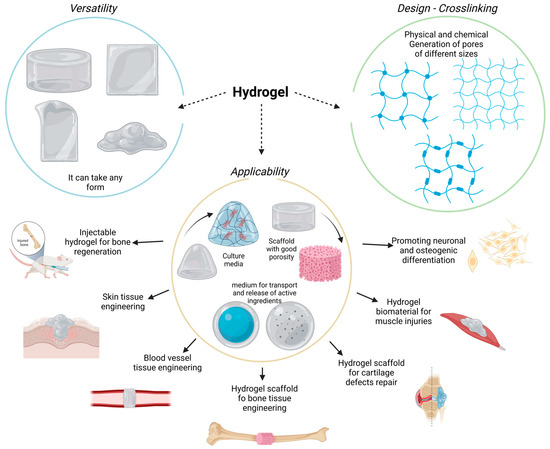
Hydrogels are 3D cross-linked polymeric networks capable of imbibing large amounts of water (Figure 1). Moreover, the hydrogel polymeric matrix structure allows for the diffusion of diverse molecules, such as drugs, active molecules, growth factors, nanoparticles, and more. On the other hand, we can control the chemical and molecular interactions of the polymeric chains guiding the biological behavior from the cellular level. Furthermore, the polymeric network can be molded into different arrangements and sizes, following the structural characteristics of repairing tissue defects. Therefore, they can provide constructive microenvironments suitable for controlled cell growth [20][21][24][25][26]. It is interesting to emphasize that hydrogels can show versatile control of physical properties according to the exposed environmental conditions. For instance, hydrogels work as soft materials that can form solid structures (after dryness) that diverge in terms of mechanical properties, providing the capacity to generate solid scaffolds. In contrast, hydrogels can absorb significant amounts of water and preserve humid environments without necessarily decomposing or degrading their structural architecture [27]. Their high water content makes hydrogel materials highly permeable and porous, allowing oxygen and nutrients to diffuse quickly and resulting in a balanced interconnected microenvironment that can stimulate a guided cellular fate [28].
Figure 1. Hydrogels are versatile and malleable biomaterials with several medical applications.

3. Hydrogel Classification

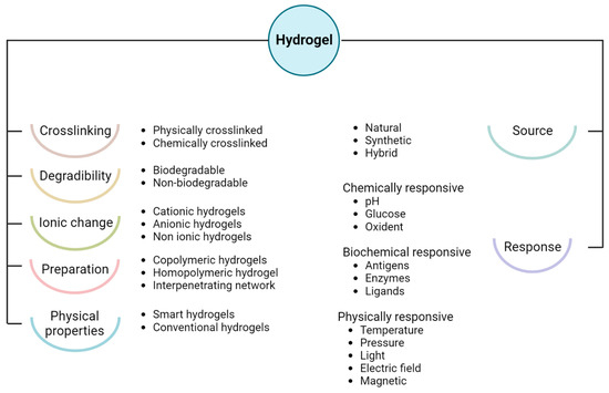
The classification of hydrogels (Figure 2) follows physicochemical properties, such as biochemical and biophysical response, synthetic methods, precursors, ionic charge, degradability, and cross-linking degree [29][30]. Inherently, the physically synthesized hydrogels show reversible cross-linking matrixes mainly characterized by coordinated electrostatic interaction, which forms Van der Waals forces and hydrogen bonds. Meanwhile, the chemically developed hydrogels are characterized by permanent and irreversible cross-linking bonds that require high energy to alter the matrix configuration [29]. On the other hand, the type of precursor material can also classify hydrogels as either natural or synthetic. It is generally considered that natural hydrogels are more biocompatible and bioactive than their synthetic counterparts. However, synthetic hydrogels promote controllable, mechanical, and degradable properties over naturally sourced polymers [21].
Figure 2. Hydrogel classification according to the physicochemical properties, and applications.
Natural hydrogels are composed of biopolymeric sources derived from animals and plants, which can be classified into two categories: polysaccharides and polypeptides. In the polysaccharides group, the most commonly used polymers are chitosan and alginate-based. On the other hand, polypeptides such as collagen and gelatin are mainly applied as supportive and guiding scaffolds for BTE. Therefore, biopolymers can incorporate attractive characteristics mandatory for functional biomaterials, including their chemical composition for cellular interaction and controlled degradation. Interestingly, the cross-linking degree of biopolymeric hydrogels plays an essential role in assembling (e.g., charge) due to the specific functional groups of certain biopolymers, or in interpolymer cross-linking due to physical and chemical modifications [31].
Biopolymers share similar components to the extracellular matrix (ECM), showing good biocompatibility, low immune response, and nearly null cytotoxicity compared to different synthetic polymers. Additionally, biopolymers can promote cellular adhesion, proliferation, and regeneration of bone-forming cells (osteoblasts) [32][33][34][35]. Therefore, from a biophysical point of view, hydrogels resemble many virtual properties of natural tissues. The morphological characteristics of the polymers allow for the exchange of substances, conducting cell adhesion in the initial stage and bone growth in the follow-up stage. It has been substantially demonstrated that cells are readily suspended in hydrogels and that the viability of encapsulated cells in the biopolymeric matrix can be largely preserved [36][37]. Thus, natural hydrogels are also biodegradable, providing initial support for the promotion of cellular adhesion. They degrade as cell populations grow and mature, changing the microenvironment and substituting with newly regenerated tissue [25].

4. Hydrogels for Bone Regeneration

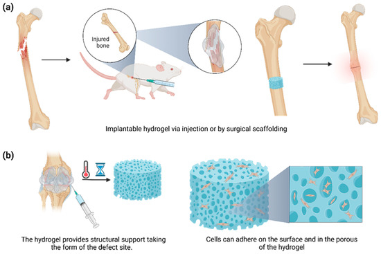
The application of hydrogels in tissue engineering and regenerative medicine, particularly BTE, has attracted increasing attention due to the osteogenic drug delivery benefits that involve the polymeric matrix [21]. Important advances have been achieved using hydrogels based on synthetic polymers due to the inherent biocompatible properties of their natural source [22]. Recent trends have included the fabrication of hydrogels from biomacromolecules to introduce specific and inherent biofunctionality to hydrogels [23]. Furthermore, these hydrogels have demonstrated excellent integration with the surrounding tissues, avoiding the complex process of surgical removal due to failing response and reducing the possibility of inflammatory side effects [38]. These polymers can be tailored to obtain the desired geometry for implantation or injection. Moreover, we can easily control the degradation, porosity, and release profile by altering the method and degree of cross-linking [32]. Considering these parameters, hydrogels successfully provide structural support by simulating the natural tissue environment while offering a conductive regenerating scaffold for defective or imperfect sites. Thus far, allowing the bone to carry out its healing mechanism is imperative, as osteoblasts can adhere both on the surface of the hydrogel and within the hydrogel’s pores, ultimately leading to differentiation and maturing of the proliferating cells (Figure 3) [39].
Figure 3. (a) Hydrogels can be implanted by injection or surgical scaffolding in the injured area. (b) The implantable hydrogel stimuli responsiveness (e.g., temperature and time) can follow the defect’s shape, acting as a fine-tuning platform for promoting bone adhesion and proliferation among the surface matrix and the new tissue.
Considering the above-stated information, we can postulate that hydrogel formulations, when used for bone regeneration, must meet specific standards when used as an implanted scaffold, or, in some circumstances, as an injectable system [40]:
  • No cytotoxic and no immunogenic response, in order to avoid a chronic and non-regulable inflammatory reaction;
  • Osteoinductive, osteoconductive, osteogenic, and osteocompatible qualities for better bone anchorage and regeneration;
  • Mimicking the natural ECM at the implant site;
  • Degradable by different enzymes or environmental molecules, leaving sufficient space for new bone formation;
  • Resistant and stable during sterilization;
  • Controlling the size and interconnection of the pores to optimize the characteristics of drug release, cell growth, oxygen diffusion, and nutrient exchange;
  • Patient-friendly injectable form to reduce pain and simplify the administration process.
The structures of natural polymers should be similar to the ECM, providing comparable mechanical stability and bone integrity to prevent chronic inflammatory or immune responses. Additionally, the physical and chemical characteristics can increase the materials’ bioactivity, strength, and toughness in hydrogel applications [41]. However, the challenges related to controlled release or drug encapsulation still require further investigation [32]. Expanding the application of hydrogels in bone regeneration demands continuous formulation and improvement of the methods of preparation, as well as the development of in vitro and in vivo tests to enhance biocompatibility and osteoconductive capabilities.

References

  1. Bates, P.; Yeo, A.; Ramachandran, M. Bone Injury, Healing and Grafting. In Basic Orthopaedic Sciences; CRC Press: Boca Raton, FL, USA, 2017; pp. 205–222.
  2. Einhorn, T.A. The Cell and Molecular Biology of Fracture Healing. Clin. Orthop. Relat. Res. 1998, 355S, S7–S21.
  3. Dimitriou, R.; Jones, E.; McGonagle, D.; Giannoudis, P.V. Bone regeneration: Current concepts and future directions. BMC Med. 2011, 9, 66.
  4. Cui, W.; Liu, Q.; Yang, L.; Wang, K.; Sun, T.; Ji, Y.; Liu, L.; Yu, W.; Qu, Y.; Wang, J.; et al. Sustained Delivery of BMP-2-Related Peptide from the True Bone Ceramics/Hollow Mesoporous Silica Nanoparticles Scaffold for Bone Tissue Regeneration. ACS Biomater. Sci. Eng. 2018, 4, 211–221.
  5. Mahanta, A.K.; Patel, D.K.; Maiti, P. Nanohybrid Scaffold of Chitosan and Functionalized Graphene Oxide for Controlled Drug Delivery and Bone Regeneration. ACS Biomater. Sci. Eng. 2019, 5, 5139–5149.
  6. Tzagiollari, A.; McCarthy, H.O.; Levingstone, T.J.; Dunne, N.J. Biodegradable and Biocompatible Adhesives for the Effective Stabilisation, Repair and Regeneration of Bone. Bioengineering 2022, 9, 250.
  7. Gupta, H.; Zioupos, P. Fracture of bone tissue: The ‘hows’ and the ‘whys’. Med. Eng. Phys. 2008, 30, 1209–1226.
  8. Nellans, K.W.; Kowalski, E.; Chung, K.C. The Epidemiology of Distal Radius Fractures. Hand Clin. 2012, 28, 113–125.
  9. Wu, A.-M.; Bisignano, C.; James, S.L.; Abady, G.G.; Abedi, A.; Abu-Gharbieh, E.; Alhassan, R.K.; Alipour, V.; Arabloo, J.; Asaad, M.; et al. Global, regional, and national burden of bone fractures in 204 countries and territories, 1990–2019: A systematic analysis from the Global Burden of Disease Study 2019. Lancet Healthy Longev. 2021, 2, e580–e592.
  10. Kerativitayanan, P.; Tatullo, M.; Khariton, M.; Joshi, P.; Perniconi, B.; Gaharwar, A.K. Nanoengineered Osteoinductive and Elastomeric Scaffolds for Bone Tissue Engineering. ACS Biomater. Sci. Eng. 2017, 3, 590–600.
  11. Giannoudis, P.V.; Dinopoulos, H.; Tsiridis, E. Bone substitutes: An update. Injury 2005, 36 (Suppl. S3), S20–S27.
  12. Audigé, L.; Griffin, D.; Bhandari, M.; Kellam, J.; Rüedi, T.P. Path Analysis of Factors for Delayed Healing and Nonunion in 416 Operatively Treated Tibial Shaft Fractures. Clin. Orthop. Relat. Res. 2005, 438, 221–232.
  13. Petite, H.; Viateau, V.; Bensaïd, W.; Meunier, A.; de Pollak, C.; Bourguignon, M.; Oudina, K.; Sedel, L.; Guillemin, G. Tissue-engineered bone regeneration. Nat. Biotechnol. 2000, 18, 959–963.
  14. Shrivats, A.R.; McDermott, M.C.; Hollinger, J.O. Bone tissue engineering: State of the union. Drug Discov. Today 2014, 19, 781–786.
  15. Chen, C.-Y.; Chen, C.-C.; Wang, C.Y.; Lee, A.K.-X.; Yeh, C.-L.; Lin, C.-P. Assessment of the Release of Vascular Endothelial Growth Factor from 3D-Printed Poly-ε-Caprolactone/Hydroxyapatite/Calcium Sulfate Scaffold with Enhanced Osteogenic Capacity. Polymers 2020, 12, 1455.
  16. Zyuzkov, G. Targeted Regulation of Intracellular Signal Transduction in Regeneration-Competent Cells: A new Direction for Therapy in Regenerative Medicine. Biointerface Res. Appl. Chem. 2021, 11, 12238–12251.
  17. Ansari, M. Bone tissue regeneration: Biology, strategies and interface studies. Prog. Biomater. 2019, 8, 223–237.
  18. Fibbe, W.E.; Dazzi, F.; LeBlanc, K. MSCs: Science and trials. Nat. Med. 2013, 19, 812–813.
  19. Ansari, M.; Eshghanmalek, M. Biomaterials for repair and regeneration of the cartilage tissue. Bio-Design Manuf. 2019, 2, 41–49.
  20. Muir, V.G.; Burdick, J.A. Chemically Modified Biopolymers for the Formation of Biomedical Hydrogels. Chem. Rev. 2021, 121, 10908–10949.
  21. Zhang, Y.; Yu, T.; Peng, L.; Sun, Q.; Wei, Y.; Han, B. Advancements in Hydrogel-Based Drug Sustained Release Systems for Bone Tissue Engineering. Front. Pharmacol. 2020, 11, 622.
  22. Janoušková, O. Synthetic Polymer Scaffolds for Soft Tissue Engineering. Physiol. Res. 2018, 67, S335–S348.
  23. Van Vlierberghe, S.; Dubruel, P.; Schacht, E. Biopolymer-Based Hydrogels As Scaffolds for Tissue Engineering Applications: A Review. Biomacromolecules 2011, 12, 1387–1408.
  24. Oliva, N.; Shin, M.; Burdick, J.A. Editorial: Special Issue on Advanced Biomedical Hydrogels. ACS Biomater. Sci. Eng. 2021, 7, 3993–3996.
  25. Zhang, Y.; Li, Z.; Guan, J.; Mao, Y.; Zhou, P. Hydrogel: A potential therapeutic material for bone tissue engineering. AIP Adv. 2021, 11, 010701.
  26. Ruedinger, F.; Lavrentieva, A.; Blume, C.; Pepelanova, I.; Scheper, T. Hydrogels for 3D mammalian cell culture: A starting guide for laboratory practice. Appl. Microbiol. Biotechnol. 2015, 99, 623–636.
  27. Liao, H.T.; Tsai, M.-J.; Brahmayya, M.; Chen, J.-P. Bone Regeneration Using Adipose-Derived Stem Cells in Injectable Thermo-Gelling Hydrogel Scaffold Containing Platelet-Rich Plasma and Biphasic Calcium Phosphate. Int. J. Mol. Sci. 2018, 19, 2537.
  28. Chimene, D.; Lennox, K.K.; Kaunas, R.R.; Gaharwar, A.K. Advanced Bioinks for 3D Printing: A Materials Science Perspective. Ann. Biomed. Eng. 2016, 44, 2090–2102.
  29. Ullah, F.; Othman, M.B.H.; Javed, F.; Ahmad, Z.; Akil, H.M. Classification, processing and application of hydrogels: A review. Mater. Sci. Eng. C 2015, 57, 414–433.
  30. Qiu, Y.; Park, K. Environment-sensitive hydrogels for drug delivery. Adv. Drug Deliv. Rev. 2001, 53, 321–339.
  31. Short, A.R.; Koralla, D.; Deshmukh, A.; Wissel, B.; Stocker, B.; Calhoun, M.; Dean, D.; Winter, J.O. Hydrogels that allow and facilitate bone repair, remodeling, and regeneration. J. Mater. Chem. B 2015, 3, 7818–7830.
  32. Bai, X.; Gao, M.; Syed, S.; Zhuang, J.; Xu, X.; Zhang, X.-Q. Bioactive hydrogels for bone regeneration. Bioact. Mater. 2018, 3, 401–417.
  33. Zhao, W.; Jin, X.; Cong, Y.; Liu, Y.; Fu, J. Degradable natural polymer hydrogels for articular cartilage tissue engineering. J. Chem. Technol. Biotechnol. 2013, 88, 327–339.
  34. Nabavi, M.H.; Salehi, M.; Ehterami, A.; Bastami, F.; Semyari, H.; Tehranchi, M.; Semyari, H. A collagen-based hydrogel containing tacrolimus for bone tissue engineering. Drug Deliv. Transl. Res. 2020, 10, 108–121.
  35. Salehi, M.; Naseri-Nosar, M.; Ebrahimi-Barough, S.; Nourani, M.; Vaez, A.; Farzamfar, S.; Ai, J. Regeneration of sciatic nerve crush injury by a hydroxyapatite nanoparticle-containing collagen type I hydrogel. J. Physiol. Sci. 2018, 68, 579–587.
  36. Gao, F.; Li, J.; Wang, L.; Zhang, D.; Zhang, J.; Guan, F.; Yao, M.-H. Dual-enzymatically crosslinked hyaluronic acid hydrogel as a long-time 3D stem cell culture system. Biomed. Mater. 2020, 15, 045013.
  37. Paez, J.I.; Farrukh, A.; Valbuena-Mendoza, R.; Włodarczyk-Biegun, M.K.; del Campo, A. Thiol-Methylsulfone-Based Hydrogels for 3D Cell Encapsulation. ACS Appl. Mater. Interfaces 2020, 12, 8062–8072.
  38. Silva, R.; Fabry, B.; Boccaccini, A.R. Fibrous protein-based hydrogels for cell encapsulation. Biomaterials 2014, 35, 6727–6738.
  39. Naahidi, S.; Jafari, M.; Logan, M.; Wang, Y.; Yuan, Y.; Bae, H.; Dixon, B.; Chen, P. Biocompatibility of hydrogel-based scaffolds for tissue engineering applications. Biotechnol. Adv. 2017, 35, 530–544.
  40. Lee, S.-H.; Shin, H. Matrices and scaffolds for delivery of bioactive molecules in bone and cartilage tissue engineering. Adv. Drug Deliv. Rev. 2007, 59, 339–359.
  41. Guarino, V.; Caputo, T.; Altobelli, R.; Ambrosio, L. Degradation properties and metabolic activity of alginate and chitosan polyelectrolytes for drug delivery and tissue engineering applications. AIMS Mater. Sci. 2015, 2, 497–502.
More
Information
Contributors MDPI registered users' name will be linked to their SciProfiles pages. To register with us, please refer to https://encyclopedia.pub/register : , , , ,
View Times: 1.1K
Revisions: 2 times (View History)
Update Date: 29 Jun 2023
Video Production Service