Submitted Successfully!
To reward your contribution, here is a gift for you: A free trial for our video production service.
Thank you for your contribution! You can also upload a video entry or images related to this topic.
Version Summary Created by Modification Content Size Created at Operation
1 -- 3306 2023-06-27 23:28:02 |
2 update references and layout -13 word(s) 3293 2023-06-28 05:06:20 | |
3 update Figure 1 + 60 word(s) 3353 2023-06-29 03:26:04 | |
4 update figure caption Meta information modification 3353 2023-06-30 07:33:52 | |
5 Corrections made to Figure 1 Caption -58 word(s) 3295 2023-06-30 20:59:27 |

Video Upload Options

Do you have a full video?

Confirm

Are you sure to Delete?
Cite
If you have any further questions, please contact Encyclopedia Editorial Office.
Patel, J.C.; Carr, K.D.; Rice, M.E. Insulin in the Striatum. Encyclopedia. Available online: https://encyclopedia.pub/entry/46147 (accessed on 27 July 2024).
Patel JC, Carr KD, Rice ME. Insulin in the Striatum. Encyclopedia. Available at: https://encyclopedia.pub/entry/46147. Accessed July 27, 2024.
Patel, Jyoti C., Kenneth D. Carr, Margaret E. Rice. "Insulin in the Striatum" Encyclopedia, https://encyclopedia.pub/entry/46147 (accessed July 27, 2024).
Patel, J.C., Carr, K.D., & Rice, M.E. (2023, June 27). Insulin in the Striatum. In Encyclopedia. https://encyclopedia.pub/entry/46147
Patel, Jyoti C., et al. "Insulin in the Striatum." Encyclopedia. Web. 27 June, 2023.
Insulin in the Striatum
Edit

Insulin crosses the blood–brain barrier to enter the brain from the periphery. In the brain, insulin has well-established actions as a satiation signal in the hypothalamus, as well as effects on feeding at the level of mesolimbic dopamine neurons in the midbrain. However, insulin also acts in the striatum, a forebrain region that is crucial for movement, mood and motivated behavior. The striatum shows abundant expression of insulin receptors (InsRs) throughout. These receptors are found on interneurons and striatal projections neurons, as well as on glial cells and dopamine axons.

dopamine acetylcholine insulin receptors brain striatum nucleus accumbens

1. Introduction

Since the discovery of insulin 100 years ago, a primary focus of insulin research has been to understand how this metabolic hormone regulates glucose homeostasis. The body of evidence gained has been fundamental for developing treatment strategies for conditions involving insulin resistance, including obesity-sensitive type 2 diabetes [1]. However, the actions of insulin in the body are not just metabolic in nature, and are not restricted to the periphery. Pancreatic insulin can enter the brain slowly at the level of the choroid plexus, which produces cerebrospinal fluid (CSF) in the ventricles of the brain. Ventricular CSF is contiguous with brain interstitial fluid, allowing a gradual increase in brain insulin levels following insulin release in the periphery. A more direct and rapid source for brain entry, however, is by trans-endothelial transport at the level of brain microvasculature, i.e., by crossing the blood–brain barrier (BBB) [2][3][4][5]. In addition, it has been suggested that insulin may be produced and released at the choroid plexus [6], and even by some neurons [7][8] and astrocytes [9] within the brain, providing an even more local and rapid source of insulin.
Physiological concentrations of insulin in CSF and the brain are estimated to reach 10 to 30 nM after food intake [2][10][11][12][13]. Within the brain, mRNA transcripts and protein for insulin receptors (InsRs) are distributed widely with high levels in the olfactory bulb, hypothalamus, hippocampus, cortex, cerebellum, midbrain and striatum [14][15][16][17][18][19][20]. Thus, insulin has the potential to impact multiple brain regions and their functions through InsR signaling. Insulin also acts at insulin-like growth factor 1 receptors (IGF-1Rs), albeit at higher concentrations than those required to activate InsRs [17][21], as well as to hybrid receptors containing InsR subunits with IGF-1R subunits [22]. Both receptor types, however, use similar downstream signaling machinery [23][24][25][26]. Consequently, discerning actions of insulin in the brain through InsRs vs. IGF-1Rs is vital for understanding insulin’s effect on brain function and for specific targeting of appropriate receptor-mediated signaling components to combat brain dysfunction.
The most studied function of insulin in the brain is its role as a satiation signal acting in the hypothalamus to limit food intake during a meal, as well as to indicate satiety between meals [27][28]. However, this is far from the only role of insulin in the brain, with increasing evidence for its influence on diverse brain functions, ranging from non-homeostatic hedonic feeding and food choices to cognition and regulation of emotional tone [29][30][31][32][33][34][35][36]. Importantly, development of insulin resistance in the periphery also extends to the brain, with mechanisms and consequences of dysregulated insulin signaling in specific regions including in the striatum largely under explored.

2. Insulin Promotes Striatal Dopamine Release through ChIs and nAChRs

Striatal dopamine release is governed by the activity of midbrain dopamine neurons, but can also be triggered from within the striatum by activation of nicotinic acetylcholine receptors (nAChRs) that are enriched on dopamine axons [37][38][39][40]. The source of acetylcholine (ACh) for this process are cholinergic interneurons (ChIs), which make up only a small percentage of the neuronal population of the striatum, yet exert a powerful influence on dopamine release and striatal function through extensive axonal processes that enable non-synaptic, as well as synaptic-like, transmission with dopamine axons [40].
Early studies using autoradiography show abundant InsR binding sites throughout the striatum (Figure 1a) [15][16][17] but lack the resolution to discern which striatal cells express these InsRs. Analysis of RNA-seq data in DropViz (http://dropviz.org, accessed on 16 September 2022) [41], shows the highest levels of mRNA for InsRs in ChIs compared to other striatal cells including striatal projection neurons (SPNs), that are the most prevalent neuronal population in the striatum (Figure 1b). Consistent with abundant mRNA in ChIs, immunohistochemical examination of InsRs in rat striatum indicates dense labeling for InsR proteins in ChIs, identified by co-immunostaining for the primary enzyme for ACh synthesis, choline acetyltransferase (ChAT) (Figure 1c) [19]. Thus, ChIs provide a significant target by which insulin can influence dopamine transmission.
Figure 1. InsR localization in striatum and insulin-dependent regulation of striatal dopamine release via ChIs, InsRs and nAChRs. (a) Autoradiographic image of insulin receptor (InsR) binding sites throughout the rat striatum, including in the dorsal striatum (dStr), nucleus accumbens (NAc) core and shell subregions marked by red asterisks; modified with permission from [16]. Abbreviations: cc, corpus callosum; FrPa, frontal parietal cortex; LS, lateral septum; MS, medial septum; PO, preoptic area; oTu, olfactory tubercle. (b) Comparison of relative mRNA levels for InsRs in striatal cells shows high abundance in cholinergic interneurons (ChIs) versus D1 or D2 spiny projection neurons (SPNs), parvalbumin positive interneurons (Pvalb-IN), somatostatin positive interneurons (Sst-IN) or astrocytes; RNA-seq data was obtained using DropViz. (c) Immunohistochemical staining of InsRs in a biocytin-filled, ChAT-positive ChI; scale = 10 μm; images from [19]. (d) Response of current-clamped rat ChI to depolarizing current pulses; insulin (30 nM) increases ChI excitability by attenuating action potential (AP) accommodation; from [19]. (e) Insulin (30 nM) enhances single-pulse evoked increases in extracellular dopamine concentration ([DA]o) in dStr but has a greater effect in NAc core and NAc shell subregions of rat striatal slices; from [19].

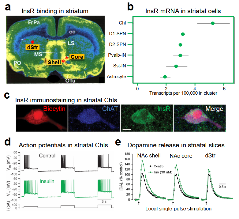
2.1. Insulin Increases the Excitability of Striatal ChIs

Electrophysiological recordings in ex vivo striatal slices have demonstrated functional consequences of InsR activation in ChIs. The method used is whole-cell recording, in which a micropipette containing a solution resembling the cytoplasm forms a continuum with an individual cell, enabling the electrical activity of the cell to be monitored. In experiments using current-clamp recording in which the current across a cell membrane is controlled and membrane potential is monitored, injecting positive current to depolarize a recorded neuron causes an initial burst of action potentials (APs) that shows a progressive decrease in frequency with continued depolarization. This is referred to as AP accommodation [42]. Addition of a physiological concentration of insulin (30 nM) to the artificial cerebrospinal fluid (aCSF) that superfuses the slice increases ChI excitability, seen as an increase in AP number during prolonged depolarization, i.e., attenuated AP accommodation (Figure 1d) [19]. This increase in excitability is InsR dependent, as the effect of insulin is prevented by an InsR inhibitor, whereas blocking IGF-1Rs has no effect [19]. The mechanism by which insulin increases ChI excitability is unresolved, but is likely to involve one or more ion channels governing the membrane dynamics of these neurons [42][43].

2.2. Insulin Boosts Dopamine Release via InsRs, and Requires PI3K and nAChRs

The action of insulin on ChI excitability is sufficient to have a profound impact on axonal dopamine release in striatal slices as monitored using fast-scan cyclic voltammetry (FSCV). FSCV is an electrochemical method used to monitor the profile of evoked increases in extracellular dopamine concentration ([DA]o) by detecting the current generated during the oxidation of dopamine at the surface of a carbon fiber microelectrode when a triangular voltage waveform is applied. The voltage waveform is typically applied every 100 ms and the carbon fiber microelectrode is typically 5 to 10 μm in diameter thereby providing a dopamine release profile with high temporal (subsecond) and high spatial (few micrometer) resolution [44][45]. Previous studies using FSCV show that up to 70% of dopamine release evoked by a single electrical pulse in striatal slices can be attributed to ChI-induced ACh release and consequent activation of nAChRs on dopamine axons [46][47][48][49]. Strikingly, application of low nM insulin via the aCSF amplifies single pulse-evoked [DA]o further, with a greater enhancement in the reward-related nucleus accumbens (NAc) core and shell regions of the ventral striatum than in the motor-related dorsal striatum (dStr) (Figure 1e). This regional heterogeneity in the response to insulin mirrors the regional density of InsRs in the striatum (Figure 1a) [15][16][17]. Showing that the influence of insulin on dopamine release involves dynamic regulation of the exocytotic process, tissue dopamine content of striatal slices is unaltered when exposed to 30 nM insulin, eliminating potential contributions from changes in dopamine synthesis or storage [19].
Like the changes seen in ChI excitability, amplification of evoked [DA]o by insulin is mediated by InsRs rather than IGF-1Rs; insulin-induced increases in dopamine release are prevented by blocking InsRs, but not by blocking IGF-1Rs [19]. Interestingly, a higher concentration of insulin, 100 nM, does not produce a significant enhancement of evoked dopamine release which could reflect competing activation of IGF-1Rs, as seen in SPNs, although this has not been investigated.
InsRs are members of the receptor tyrosine kinase superfamily which activate two major signaling pathways: the phosphatidylinositol 3-kinase (PI3K) pathway and the mitogen-activated protein kinase (MAPK) pathway [50]. Insulin-induced changes in evoked [DA]o are prevented in the presence of a PI3K inhibitor in all striatal subregions, revealing the involvement of known downstream signaling mechanisms for insulin activation of InsRs in this process [19]. Demonstrating a pivotal ACh-DA interaction, the effect of insulin on evoked [DA]o is prevented by nAChR antagonists [19]. Insulin amplifies evoked striatal [DA]o in slices from mice, as well as from rats; however, the effect of insulin is absent throughout the striatum of mice with genetic deletion of ChAT (ChAT KO) [19]. Together these observations provide a model by which insulin activation of InsRs on ChIs induces enhancement of ChI activity which then boosts ACh-mediated dopamine release via nAChRs on dopamine axons (Figure 2).
Figure 2. Summary of the multiple ways by which insulin can regulate striatal dopamine (DA) transmission. Activation of insulin receptors (InsRs) on cholinergic interneurons (ChIs) increases ChI excitability, which increases acetylcholine (ACh) release and activation of nicotinic ACh receptors (nAChRs) on dopamine axons to boost dopamine release and increase extracellular DA concentration ([DA]o). Activation of InsRs on dopamine axons increases dopamine transporter (DAT) surface expression via the PI3K/Akt pathway to increase the maximum velocity (Vmax) of dopamine uptake, thereby lowering [DA]o. Activation of InsRs on astrocytes increases exocytotic ATP release, which activates purinergic P2Y receptors (P2YR) on dopamine axons and enhances dopamine release. Insulin also decreases monoamine oxidase (MAO) activity in dopamine axons and glial cells, thereby decreasing dopamine metabolism and prolonging the time course of dopamine actions.

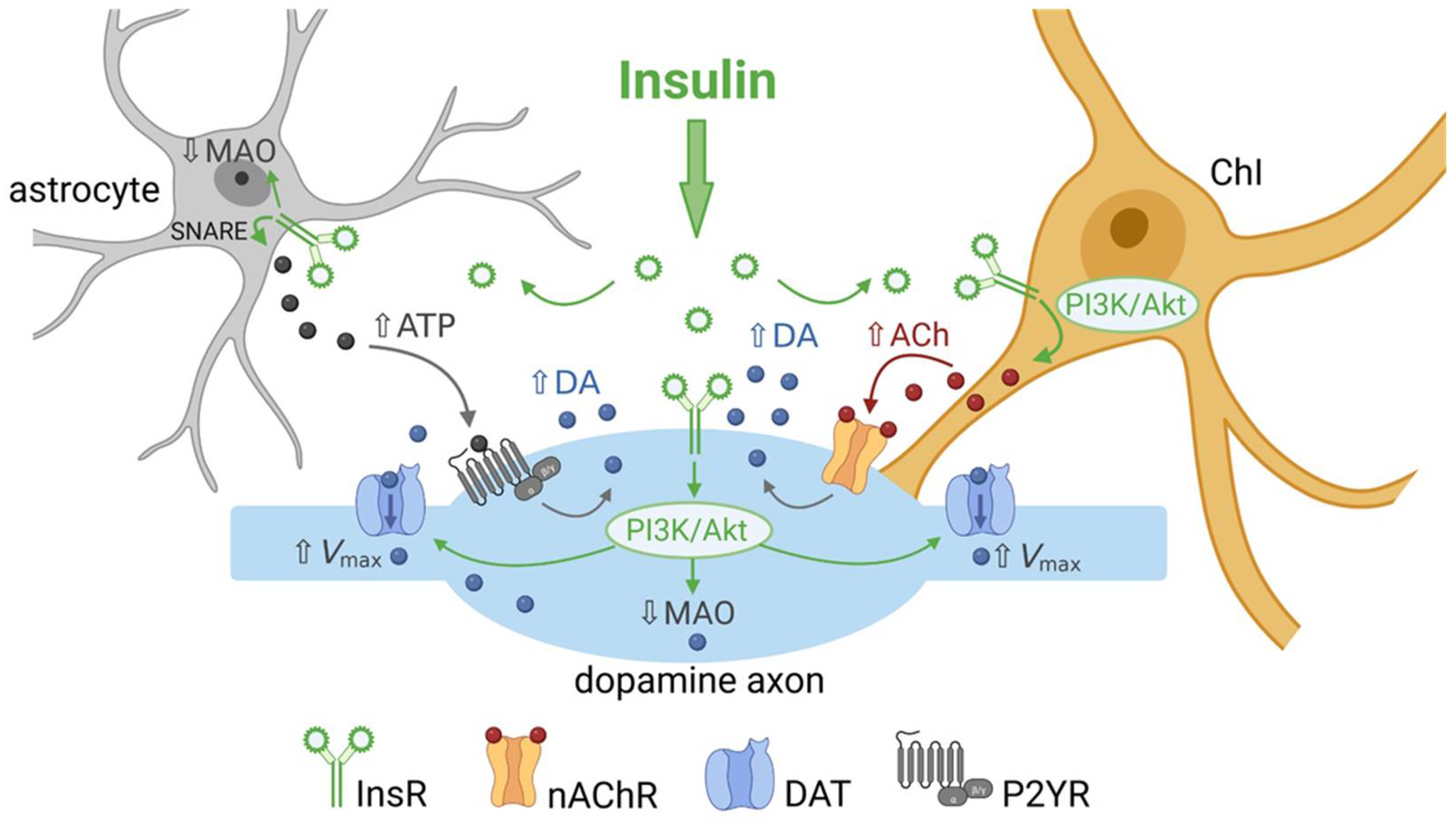
3. Insulin Enhances Striatal Dopamine Uptake via DAT on Dopamine Axons

The dopamine transporter (DAT) is expressed exclusively in dopamine neurons in both the midbrain cell body region and forebrain dopamine axons in the striatum [51][52][53] and is crucial for curtailing the amplitude and duration of dopamine transients, as well as maintaining homeostatic levels of [DA]o through transporter-mediated uptake [54][55]. Thus, [DA]o at a given location and time reflects a balance between the opposing processes of release and uptake [48][49]. In addition, DAT-dependent dopamine uptake provides a major source of dopamine for vesicular uptake and storage for subsequent release [56].
In addition to the localization of InsRs in striatal cells, immunohistochemical analysis of striatal InsRs show expression on dopamine axons, identified by the presence of tyrosine hydroxylase, the primary enzyme required for dopamine synthesis [19]. Downstream effects of InsR activation include the regulation of dopamine uptake. Insulin increases expression of mRNA for DAT in midbrain dopamine neurons [57], which could theoretically increase total DAT protein levels. However, the most profound effect that insulin has on the DAT is its ability to redistribute DAT localization within a dopamine neuron. Although an integral part of the plasma membrane, DAT surface expression is dynamically regulated by endocytic trafficking mediated by a variety of intracellular signaling pathways, including PKC, which promotes DAT internalization, and the PI3K-Akt signaling pathway which has the opposite effect [58][59][60]. Given that both InsRs and IGF-1Rs belong to the receptor tyrosine kinase superfamily that couple to PI3K via phosphorylation of InsR substrates [23][24][25][26], insulin can influence DAT surface expression and function via these pathways. In striatum, it has been shown that in addition to enhancing dopamine release, insulin promotes DAT trafficking to the plasma membrane through PI3K-Akt signaling, thereby increasing overall DAT activity and dopamine uptake [58][59][61][62][63]. Dopamine uptake in the striatum is governed by Michaelis–Menten kinetics [64] and has been assessed using several different methods and experimental preparations. Although the values for the uptake kinetic parameters obtained are not directly comparable across studies [65], the general consensus is that insulin increases the maximal uptake velocity, Vmax, for dopamine with little or no effect on Km, which is inversely related to the affinity of the DAT for dopamine [58][61][62][63][66]. Moreover, although dopamine uptake is less efficient in the ventral striatum than in the dorsal striatum [67][68][69], enhanced dopamine uptake by insulin is seen throughout the striatal complex and in both rats and mice [61][62][63][66].
Confirming activation of the PI3K-Akt pathway, insulin-induced increases in striatal DAT activity are prevented by PI3K inhibitors [58][61][63][66]. In addition, it has been shown that inhibiting PI3K alone can decrease Vmax for dopamine uptake into synaptosomes (isolated membrane bound axonal segments containing vesicles and mitochondria) implying that this pathway tonically regulates DAT activity [58]. However, in studies using striatal slices, PI3K inhibition prevents insulin-mediated increases in dopamine uptake without altering tonic regulation of DAT function [63]. Notably, although PI3K can be activated by insulin acting at either InsRs or IGF-1Rs [50], blocking InsRs occludes the effect of 30 nM insulin on dopamine uptake, whereas blocking IGF-1Rs does not [63]. Thus, insulin-induced enhancement of dopamine uptake in the striatum is mediated by InsRs and the PI3K pathway in dopamine axons (Figure 2).
The enhancing effect of insulin on dopamine uptake would be expected to decrease evoked [DA]o. However, as described in Section 2, studies using FSCV detect elevated increases in [DA]o in the presence of insulin [19][66], which reflects the dominant effect of insulin on increasing ACh-induced dopamine release. When striatal ACh is absent, as in ChAT-KO mice [47], insulin’s action on dopamine uptake prevails, revealing a net decrease in evoked [DA]o with insulin application [63]. Given that DAT is a mediator of the short-term plasticity of dopamine release [70], insulin’s actions on DAT expression and dopamine uptake will have implications for overall dopamine transmission, independent of the influence of ChIs on dopamine release.

4. Insulin Inhibits Dopamine Metabolism

In addition to enhancing dopamine release through nAChRs and increasing dopamine uptake through the DAT, there is evidence that insulin can alter dopamine metabolism [31][71][72]. Key metabolizing enzymes for dopamine are monoamine oxidase enzymes (MAOs), which may be located in dopamine axons or in astrocytes (see Figure 2). In rodents, MAO mainly catalyzes the formation of the dopamine metabolite dihydroxyphenylacetic acid (DOPAC). Samples of striatal extracellular fluid taken using microdialysis in freely moving rats show a profound and long-lasting decrease in DOPAC levels following a peripheral insulin injection [71]. Moreover, mice with brain-wide deletion of InsRs have increased MAO expression in the striatum [72]. Consequently, total area under the curve for single-pulse evoked [DA]o in the dStr and NAc is lower in striatal slices from mice lacking brain InsRs than from controls. Although peak amplitude was the same between genotypes, the overall duration of the release response was shortened [72]. This suggests another route by which endogenous insulin could enhance [DA]o, through inhibition of dopamine metabolism via MAO suppression (Figure 2).

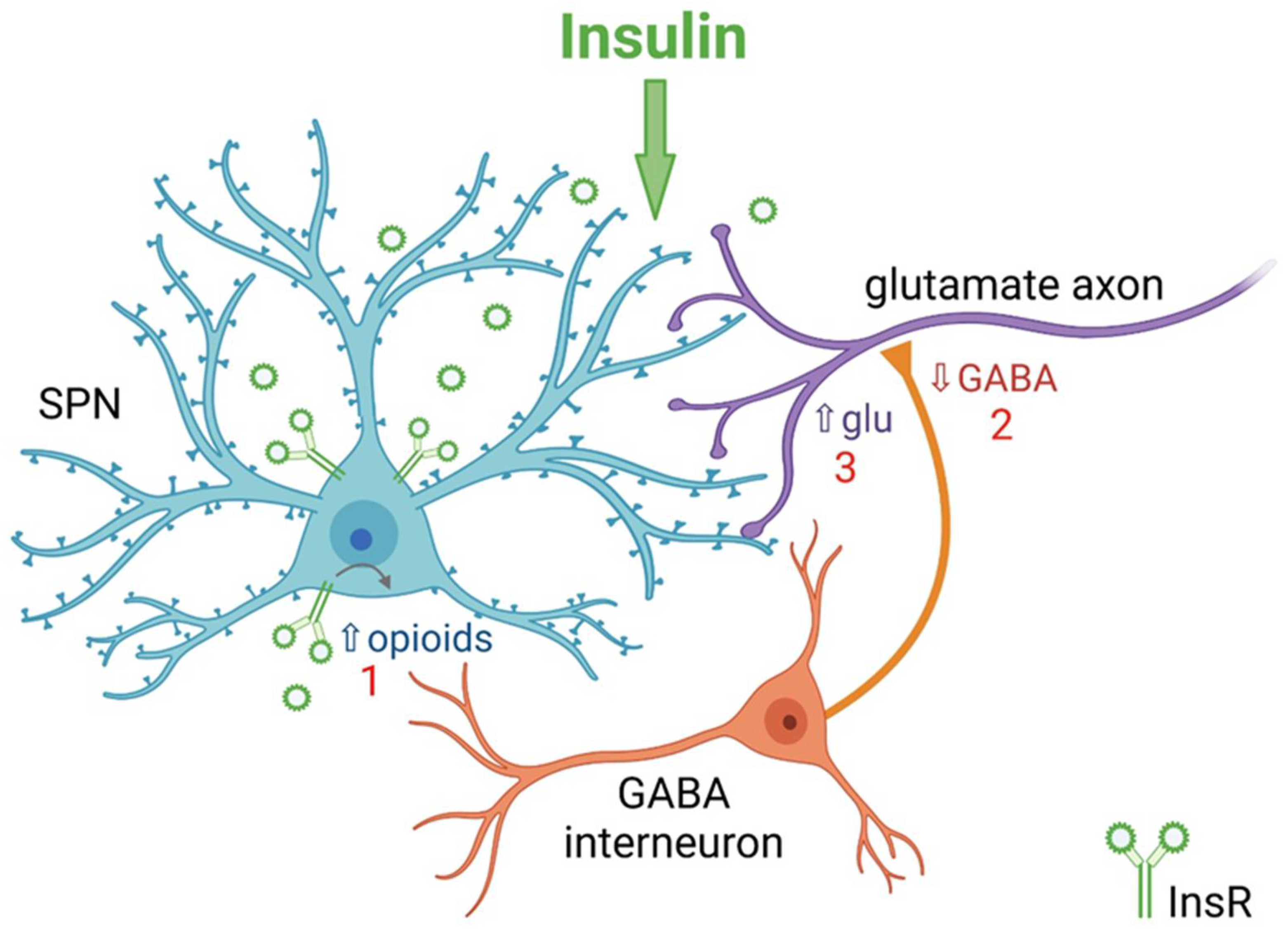
5. Insulin Bidirectionally Alters the Excitatory Regulation of Spiny Projection Neurons (SPNs)

In addition to its effect on ChIs and dopamine axons, insulin also acts at other striatal neurons including SPNs, thereby influencing overall striatal output. These projection neurons represent over 95% of the total neuronal population of the striatum, and can be subdivided into two classes based on expression of dopamine D1 vs. dopamine D2 receptor subtypes, as well as differential expression of endogenous opioid peptides, and different projection targets [73][74]. However, levels of InsR-mRNA appear to be similar in the two classes of SPNs (Figure 1b). Whole-cell voltage-clamp recordings of electrically evoked excitatory post-synaptic currents (eEPSCs) from individual SPNs in striatal slices show that 30 nM insulin increases the amplitude of eEPSCs via activation of InsRs on both D1- and D2-SPNs within the NAc core. By contrast, insulin concentrations ≥100 nM have the opposite effect of decreasing eEPSC amplitude through activation of IGF-1Rs [75]. Moreover, IGF-1Rs appear to provide tonic inhibition of SPNs because blocking IGF-1Rs with PPP alone increases eEPSC amplitude and also enhances the effect of 30 nM insulin. Analysis of the effect of 30 nM insulin on spontaneous miniature EPSCs (mEPSCs) show an increase in the frequency of these events, but not the amplitude, as well as a decrease in paired-pulse-ratio; all are consistent with enhanced excitatory transmission mediated by a boosting of presynaptic glutamate release. Again, a higher concentration of insulin, 100 nM, has the opposite effect through activation of IGF-1Rs, suggesting that IGF-1R-mediated reductions in excitatory transmission are also due to effects on presynaptic glutamate release [75]. Thus, insulin causes bidirectional effects on SPN excitability through activation of InsRs and IGF-1Rs. The mechanisms by which insulin modulates SPN activity appears to involve a feedback microcircuit in which insulin enhances SPN excitability, through activation of InsRs on SPNs and subsequent release of endogenous opioids, which then inhibit local GABAergic interneurons leading to disinhibition of presynaptic glutamate release that ultimately influences striatal output (Figure 3) [75].
Figure 3. Summary of mechanism by which insulin increases excitatory regulation of striatal projection neurons (SPNs) in nucleus accumbens (NAc) core. 1. Activation of insulin receptors (InsRs) on GABA/opioid containing SPNs in rat NAc core increases the release of endogenous opioids. 2. Opioid activation of GABAergic interneurons decreases GABA release onto glutamatergic inputs from cortex and thalamus. 3. Decreased GABA release from GABAergic interneurons disinhibits glutamatergic inputs resulting in increased presynaptic glutamate (Glu) release onto dendritic spines of SPNs. Higher concentrations of insulin act at insulin-like growth factor-1 receptors (IGF-1Rs) to oppose this process (not shown) [78].

6. Insulin Actions on Striatal Glial Cells

Astrocytes, like other glial cells in the brain (microglia and oligodendrocytes), play an important role in maintaining homeostasis of the brain microenvironment during all stages of life. This is achieved through maintaining the BBB, supplying trophic support to neurons, regulating local ionic concentrations, mediating synapse formation and function, pruning synapses during development and signaling across brain regions to modulate neuronal activity [76][77][78]. Moreover, astrocytes are instrumental in controlling levels of a variety of neurotransmitters/neuromodulators, including the key transmitters glutamate and GABA, as well as releasing these transmitters and other gliotransmitters [79][80][81][82].
Brain insulin has been shown to play an important role in regulating systemic glucose metabolism through astrocytic InsRs in the hypothalamus [82][83][84][85]. Moreover, insulin can act on astrocytes and other glial cells to regulate local cellular metabolism [86]. DropViz data show that striatal astrocytes express InsR mRNA (Figure 1b). Mice with astrocyte-specific InsR deletion exhibit a decrease in the peak amplitude of evoked [DA]o in both dStr and NAc with no change in response duration, or in MAO levels [87]. The mechanism by which InsRs in astrocytes regulate dopamine release has been proposed to be through targeting tyrosine phosphorylation of SNARE proteins that enable exocytotic ATP release, which in turn enhances axonal dopamine release via G-protein coupled P2Y purinergic receptors (Figure 2) [35][87]. These findings add to the range of mechanisms through which insulin can alter striatal dopamine signaling.

7. Conclusions

It is clear that insulin’s actions in the striatum are multifaceted in terms of the striatal elements involved and which insulin-sensitive receptors mediate the effects. Insulin can boost dopamine release indirectly through actions at InsRs on ChIs and astrocytes, with slower regulation by inhibiting dopamine metabolism by MAO. In addition, insulin actions on SPNs that engage local striatal microcircuits can have a direct influence on striatal and basal ganglia output. Information about the behavioral impact of striatal insulin signaling is limited. However, the few studies available have provided valuable evidence indicating an important role for striatal insulin beyond its role as a satiation/satiety signal. 

References

  1. Dagen, M.M. History of Insulin. In Encyclopedia of Life Sciences (eLS); John Wiley & Sons, Ltd.: Chichester, UK, 2016.
  2. Banks, W.A. The source of cerebral insulin. Eur. J. Pharmacol. 2004, 490, 5–12.
  3. Banks, W.A.; Owen, J.B.; Erickson, M.A. Insulin in the brain: There and back again. Pharmacol. Ther. 2012, 136, 82–93.
  4. Gray, S.M.; Barrett, E.J. Insulin transport into the brain. Am. J. Physiol. Cell. Physiol. 2018, 315, C125–C136.
  5. Nakabeppu, Y. Origins of brain insulin and its function. In Advances in Experimental Medicine and Biology; Springer: New York, NY, USA, 2019; pp. 1–11.
  6. Mazucanti, C.H.; Liu, Q.R.; Lang, D.; Huang, N.; O’Connell, J.F.; Camandola, S.; Egan, J.M. Release of insulin produced by the choroid plexis is regulated by serotonergic signaling. JCI Insight 2019, 4, e131682.
  7. Molnár, G.; Faragó, N.; Kocsis, Á.K.; Rózsa, M.; Lovas, S.; Boldog, E.; Báldi, R.; Csajbók, É.; Gardi, J.; Puskás, L.G.; et al. GABAergic neurogliaform cells represent local sources of insulin in the cerebral cortex. J. Neurosci. 2014, 34, 1133–1137.
  8. Csajbók, É.A.; Kocsis, Á.K.; Faragó, N.; Furdan, S.; Kovács, B.; Lovas, S.; Molnár, G.; Likó, I.; Zvara, Á.; Puskás, L.G.; et al. Expression of GLP-1 receptors in insulin-containing interneurons of rat cerebral cortex. Diabetologia 2019, 62, 717–725.
  9. Pitt, J.; Wilcox, K.C.; Tortelli, V.; Diniz, L.P.; Oliveira, M.S.; Dobbins, C.; Yu, X.W.; Nandamuri, S.; Gomes, F.C.A.; DiNunno, N.; et al. Neuroprotective astrocyte-derived insulin/insulin-like growth factor 1 stimulates endocytic processing and extracellular release of neuron-bound Aβ oligomers. Mol. Biol. Cell 2017, 28, 2623–2636.
  10. Strubbe, J.H.; Porte, D., Jr.; Woods, S.C. Insulin responses and glucose levels in plasma and cerebrospinal fluid during fasting and refeeding in the rat. Physiol. Behav. 1988, 44, 205–208.
  11. Banks, W.A.; Kastin, A.J. Differential permeability of the blood-brain barrier to two pancreatic peptides: Insulin and amylin. Peptides 1998, 19, 883–889.
  12. Havrankova, J.; Schmechel, D.; Roth, J.; Brownstein, M. Identification of insulin in rat brain. Proc. Natl. Acad. Sci. USA 1978, 75, 5737–5741.
  13. Woods, S.C.; Seeley, R.J.; Baskin, D.G.; Schwartz, M.W. Insulin and the blood brain barrier. Curr. Pharm. Des. 2003, 9, 795–800.
  14. Havrankova, J.; Roth, J.; Brownstein, M. Insulin receptors are widely distributed in the central nervous system of the rat. Nature 1978, 272, 827–829.
  15. Hill, J.M.; Lesniak, M.A.; Pert, C.B.; Roth, J. Autoradiographic localization of insulin receptors in rat brain: Prominence in olfactory and limbic areas. Neuroscience 1986, 17, 1127–1138.
  16. Werther, G.A.; Hogg, A.; Oldfield, B.J.; McKinley, M.J.; Figdor, R.; Allen, A.M.; Mendelsohn, F.A. Localization and characterization of insulin receptors in rat brain and pituitary gland using in vitro autoradiography and computerized densitometry. Endocrinology 1987, 121, 1562–1570.
  17. Schulingkamp, R.J.; Pagano, T.C.; Hung, D.; Raffa, R.B. Insulin receptors and insulin action in the brain: Review and clinical implications, Neurosci. Biobehav. Rev. 2000, 24, 855–872.
  18. Figlewicz, D.P.; Evans, S.B.; Murphy, J.; Hoen, M.; Baskin, D.G. Expression of receptors for insulin and leptin in ventral tegmental area/substantia nigra (VTA/SN) of the rat. Brain Res. 2003, 964, 107–115.
  19. Stouffer, M.A.; Woods, C.A.; Patel, J.C.; Lee, C.R.; Witkovsky, P.; Bao, L.; Machold, R.P.; Jones, K.T.; de Vaca, S.C.; Reith, M.E.; et al. Insulin enhances striatal dopamine release by activating cholinergic interneurons and thereby signals reward. Nat. Commun. 2015, 6, 8543.
  20. Figlewicz, D.P. Expression of receptors for insulin and leptin in the ventral tegmental area/substantia nigra (VTA/SN) of the rat: Historical perspective. Brain Res. 2016, 1645, 68–70.
  21. Bondy, C.A.; Cheng, C.M. Signaling by insulin-like growth factor 1 in brain. Eur. J. Pharmacol. 2004, 490, 25–31.
  22. Vigneri, R.; Squatrito, S.; Sciacca, L. Insulin and its analogs: Actions via insulin and IGF receptors. Acta. Diabetol. 2010, 47, 271–278.
  23. Bevan, P. Insulin signalling. J. Cell Sci. 2001, 114, 1429–1430.
  24. Taniguchi, C.M.; Emanuelli, B.; Kahn, C.R. Critical nodes in signalling pathways: Insights into insulin action. Nat. Rev. Mol. Cell Biol. 2006, 7, 85–96.
  25. Boucher, J.; Tseng, Y.H.; Kahn, C.R. Insulin and insulin-like growth factor-1 receptors act as ligand-specific amplitude modulators of a common pathway regulating gene transcription. J. Biol. Chem. 2010, 285, 17235–17245.
  26. Siddle, K. Signalling by insulin and IGF receptors: Supporting acts and new players. J. Mol. Endocrinol. 2011, 47, R1–R10.
  27. Schwartz, M.W.; Woods, S.C.; Porte, D., Jr.; Seeley, R.J.; Baskin, D. Central nervous system control of food intake. Nature 2000, 404, 661–671.
  28. Bellisle, F.; Drewnowski, A.; Anderson, G.H.; Westerterp-Plantenga, M.; Martin, C.K. Sweetness, satiation, and satiety. J. Nutr. 2012, 142, 1149S–1154S.
  29. Tiedemann, L.J.; Schmid, S.M.; Hettel, J.; Giesen, K.; Francke, P.; Büchel, C.; Brassen, S. Central insulin modulates food valuation via mesolimbic pathways. Nat. Commun. 2017, 8, 16052.
  30. Ferrario, C.R.; Reagan, L.P. Insulin-mediated synaptic plasticity in the CNS: Anatomical, functional and temporal contexts. Neuropharmacology 2018, 136, 182–191.
  31. Kleinridders, A.; Pothos, E.N. Impact of brain insulin signaling on dopamine function, food intake, reward, and emotional behavior. Curr. Nutr. Rep. 2019, 8, 83–91.
  32. Kullmann, S.; Heni, M.; Hallschmid, M.; Fritsche, A.; Preissl, H.; Häring, H.U. Brain insulin resistance at the crossroads of metabolic and cognitive disorders in humans. Physiol. Rev. 2016, 96, 1169–1209.
  33. Kullmann, S.; Kleinridders, A.; Small, D.M.; Fritsche, A.; Häring, H.U.; Preissl, H.; Heni, M. Central nervous pathways of insulin action in the control of metabolism and food intake. Lancet Diabetes Endocrinol. 2020, 8, 524–534.
  34. Sallam, N.A.; Borgland, S.L. Insulin and endocannabinoids in the mesolimbic system. J. Neuroendocrinol. 2021, 33, e12965.
  35. Chen, W.; Cai, W.; Hoover, B.; Kahn, C.R. Insulin action in the brain: Cell types, circuits and diseases. Trends Neurosci. 2022, 45, 384–400.
  36. Ferrario, C.R.; Finnell, J.E. Beyond the hypothalamus: Roles for insulin as a regulator of neurotransmission, motivation, and feeding. Neuropsychopharmacology 2023, 48, 232–233.
  37. Cachope, R.; Mateo, Y.; Mathur, B.N.; Irving, J.; Wang, H.L.; Morales, M.; Lovinger, D.M.; Cheer, J.F. Selective activation of cholinergic interneurons enhances accumbal phasic dopamine release: Setting the tone for reward processing. Cell Rep. 2012, 2, 33–41.
  38. Threlfell, S.; Lalic, T.; Platt, N.J.; Jennings, K.A.; Deisseroth, K.; Cragg, S.J. Striatal dopamine release is triggered by synchronized activity in cholinergic interneurons. Neuron 2012, 75, 58–64.
  39. Liu, C.; Cai, X.; Ritzau-Jost, A.; Kramer, P.; Li, Y.; Khaliq, Z.M.; Hallermann, S.; Kaeser, P.S. An action potential intiation mechanism in distal axons for the control of dopamine release. Science 2022, 375, 1378–1385.
  40. Kramer, P.F.; Brill-Weil, S.G.; Cummins, A.C.; Zhang, R.; Camacho-Hernandez, G.A.; Newman, A.H.; Eldridge, M.A.G.; Averbeck, B.B.; Khaliq, Z.M. Synaptic-like axo-axonal transmission from striatal cholinergic interneurons onto dopaminergic fibers. Neuron 2022, 18, 2949–2960.
  41. Saunders, A.; Macosko, E.Z.; Wysoker, A.; Goldman, M.; Krienen, F.M.; de Rivera, H.; Bein, E.; Baum, M.; Bortolin, L.; Wang, S.; et al. Molecular diversity and specializations among the cells of the adult mouse brain. Cell 2018, 174, 1015–1030.
  42. Sanchez, G.; Rodriguez, M.J.; Pomata, P.; Rela, L.; Murer, M.G. Reduction of an afterhyperpolarization current increases excitability in striatal cholinergic interneurons in rat parkinsonism. J. Neurosci. 2011, 31, 6553–6564.
  43. Bennett, B.D.; Callaway, J.C.; Wilson, C.J. Intrinsic membrane properties underlying spontaneous tonic firing in neostriatal cholinergic interneurons. J. Neurosci. 2000, 20, 8493–8503.
  44. Patel, J.C.; Rice, M.E. Monitoring axonal and somatodendritic dopamine release using fast-scan cyclic voltammetry in brain slices. Methods Mol. Biol. 2013, 96, 243–273.
  45. Patel, J.C. Voltammetry: Electrochemical detection of neurotransmitters in the brain. In Encyclopedia of Life Sciences (eLS); John Wiley & Sons, Ltd.: Chichester, UK, 2016.
  46. Rice, M.E.; Cragg, S.J. Nicotine amplifies reward-related dopamine signals in striatum. Nat. Neurosci. 2004, 7, 583–584.
  47. Patel, J.C.; Rossignol, E.; Rice, M.E.; Machold, R.P. Opposing regulation of striatal dopamine release and exploratory motor behavior by forebrain and brainstem cholinergic inputs. Nat. Commun. 2012, 3, 1172.
  48. Rice, M.E.; Patel, J.C.; Cragg, S.J. Dopamine release in the basal ganglia. Neuroscience 2011, 198, 112–137.
  49. Sulzer, D.; Cragg, S.J.; Rice, M.E. Striatal dopamine neurotransmission: Regulation of release and uptake. Basal Ganglia 2016, 6, 123–148.
  50. De Meyts, P. The Insulin Receptor and Its Signal Transduction Network. In Endotext ; Feingold, K.R., Anawalt, B., Boyce, A., Chrousos, G., de Herder, W.W., Dhatariya, K., Dungan, K., Hershman, J.M., Hofland, J., Kalra, S., et al., Eds.; MDText.com, Inc.: South Dartmouth, MA, USA, 2016. Available online: https://www.ncbi.nlm.nih.gov/books/NBK378978/ (accessed on 24 May 2022).
  51. Nirenberg, M.J.; Vaughan, R.A.; Uhl, G.R.; Kuhar, M.J.; Pickel, V.M. The dopamine transporter is localized to dendritic and axonal plasma membranes of nigrostriatal dopaminergic neurons. J. Neurosci. 1996, 16, 436–447.
  52. Nirenberg, M.J.; Chan, J.; Vaughan, R.A.; Uhl, G.R.; Kuhar, M.J.; Pickel, V.M. Immunogold localization of the dopamine transporter: An ultrastructural study of the rat ventral tegmental area. J. Neurosci. 1997, 17, 5255–5262.
  53. Hersch, S.M.; Yi, H.; Heilman, C.J.; Edwards, R.H.; Levey, A.I. Subcellular localization and molecular topology of the dopamine transporter in the striatum and substantia nigra. J. Comp. Neurol. 1997, 388, 211–227.
  54. Bu, M.; Farrer, M.J.; Khoshbouei, H. Dynamic control of the dopamine transporter in neurotransmission and homeostasis. NPJ Parkinsons Dis. 2021, 7, 22.
  55. Ryan, R.M.; Ingram, S.L.; Scimemi, A. Regulation of Glutamate, GABA and Dopamine Transporter Uptake, Surface Mobility and Expression. Front. Cell. Neurosci. 2021, 15, 670346.
  56. Jones, S.R.; Gainetdinov, R.R.; Jaber, M.; Giros, B.; Wightman, R.M.; Caron, M.G. Profound neuronal plasticity in response to inactivation of the dopamine transporter. Proc. Natl. Acad. Sci. USA 1998, 95, 4029–4034.
  57. Figlewicz, D.P.; Szot, P.; Chavez, M.; Woods, S.C.; Veith, R.C. Intraventricular insulin increases dopamine transporter mRNA in rat VTA/substantia nigra. Brain Res. 1994, 644, 331–334.
  58. Carvelli, L.; Moron, J.A.; Kahlig, K.M.; Ferrer, J.V.; Sen, N.; Lechleiter, J.D.; Leeb-Lundberg, L.M.F.; Merrill, G.; Lafer, E.M.; Ballou, L.M.; et al. PI3-kinase regulation of dopamine uptake. J. Neurochem. 2002, 81, 859–869.
  59. Garcia, B.G.; Wei, Y.; Moron, J.A.; Lin, R.Z.; Javitch, J.A.; Galli, A. Akt is essential for insulin modulation of amphetamine-induced human dopamine transporter cell-surface redistribution. Mol. Pharmacol. 2005, 68, 102–109.
  60. Fagan, R.R.; Kearney, P.J.; Melikian, H.E. In Situ regulated dopamine transporter trafficking: There’s no place like home. Neurochem. Res. 2020, 45, 1335–1343.
  61. Schoffelmeer, A.N.; Drukarch, B.; De Vries, T.J.; Hoenboom, F.; Schetters, D.; Pattij, T. Insulin modulates cocaine-sensitive monoamine transporter function and impulsive behavior. J. Neurosci. 2011, 31, 1284–1291.
  62. Jones, K.T.; Woods, C.; Zhen, J.; Antonio, T.; Carr, K.D.; Reith, M.E.A. Effects of diet and insulin on dopamine-transporter activity and expression in rat caudate-putamen, nucleus accumbens, and midbrain. J. Neurochem. 2017, 140, 728–740.
  63. Patel, J.C.; Stouffer, M.A.; Mancini, M.; Nicholson, C.; Carr, K.D.; Rice, M.E. Interactions between insulin and diet on striatal dopamine uptake kinetics in rodent brain slices. Eur. J. Neurosci. 2019, 49, 794–804.
  64. Wightman, R.M.; Amatore, C.; Engstrom, R.C.; Hale, P.D.; Kristensen, E.W.; Kuhr, W.G.; May, L.J. Real-time characterization of dopamine overflow and uptake in the rat striatum. Neuroscience 1988, 25, 513–523.
  65. Nicholson, C. Interaction between diffusion and Michaelis-Menten uptake of dopamine after iontophoresis in striatum. Biophys. J. 1995, 68, 1699–1715.
  66. Fordahl, S.C.; Jones, S.R. High-fat-diet-induced deficits in dopamine terminal function are reversed by restoring insulin signaling. ACS Chem. Neurosci. 2017, 8, 290–299.
  67. Marshall, J.F.; O’Dell, S.J.; Navarrete, R.; Rosenstein, A.J. Dopamine high-affinity transport site topography in rat brain: Major differences between dorsal and ventral striatum. Neuroscience 1990, 37, 11–21.
  68. Ciliax, B.J.; Heilman, C.; Demchyshyn, L.L.; Pristupa, Z.B.; Ince, E.; Hersch, S.M.; Niznik, H.B.; Levey, A.I. The dopamine transporter: Immunochemical characterization and localization in brain. J. Neurosci. 1995, 15, 1714–1723.
  69. Gonzalez-Hernandez, T.; Barroso-Chinea, P.; De La Cruz Muros, I.; Del Mar Perez-Delgado, M.; Rodriguez, M. Expression of dopamine and vesicular monoamine transporters and differential vulnerability of mesostriatal dopaminergic neurons. J. Comp. Neurol. 2004, 479, 198–215.
  70. Condon, M.D.; Platt, N.J.; Zhang, Y.F.; Roberts, B.M.; Clements, M.A.; Vietti-Michelina, S.; Tseu, M.Y.; Brimblecombe, K.R.; Threlfell, S.; Mann, E.O.; et al. Plasticity in striatal dopamine release is governed by release-independent depression and the dopamine transporter. Nat. Commun. 2019, 10, 1–15.
  71. Orosco, M.; Rouch, C.; Gripois, D.; Blouquit, M.F.; Roffi, J.; Jacquot, C.; Cohen, Y. Striatal dopamine metabolism is differentially affected by insulin according to the genotype in Zucker rats: A microdialysis study. Psychoneuroendocrinology 1992, 17, 443–452.
  72. Kleinridders, A.; Cai, W.; Cappellucci, L.; Ghazarian, A.; Collins, W.R.; Vienberg, S.G.; Pothos, E.N.; Kahn, C.R. Insulin resistance in brain alters dopamine turnover and causes behavioral disorders. Proc. Natl. Acad. Sci. USA 2015, 112, 3463–3468.
  73. Gerfen, C.R.; Engber, T.M.; Mahan, L.C.; Susel, Z.; Chase, T.N.; Monsma, F.J., Jr.; Sibley, D.R. D1 and D2 dopamine receptor-regulated gene expression of striatonigral and striatopallidal neurons. Science 1990, 250, 1429–1432.
  74. Gerfen, C.R.; Surmeier, D.J. Modulation of striatal projection systems by dopamine. Annu. Rev. Neurosci. 2011, 34, 441–466.
  75. Fetterly, T.L.; Oginsky, M.F.; Nieto, A.M.; Alonso-Caraballo, Y.; Santana-Rodriguez, Z.; Ferrario, C.R. Insulin bidirectionally alters NAc glutamatergic transmission: Interactions between insulin receptor activation, endogenous opioids, and glutamate release. J. Neurosci. 2021, 41, 2360–2372.
  76. Verkhratsky, A.; Nedergaard, M. Physiology of astroglia. Physiol. Rev. 2018, 98, 239–389.
  77. Hasel, P.; Liddelow, S.A. Astrocytes. Curr. Biol. 2021, 31, R326–R327.
  78. Verkhratsky, A.; Parpura, V.; Li, B.; Scuderi, C. Astrocytes: The housekeepers and guardians of the CNS. Adv. Neurobiol. 2021, 26, 21–53.
  79. Goubard, V.; Fino, E.; Venance, L. Contribution of astrocytic glutamate and GABA uptake to corticostriatal information processing. J. Physiol. 2011, 589, 2301–2319.
  80. Harada, K.; Kamiya, T.; Tsuboi, T. Gliotransmitter release from astrocytes: Functional, developmental, and pathological implications in the brain. Front. Neurosci. 2015, 9, 499.
  81. Liu, J.; Feng, X.; Wang, Y.; Xia, X.; Zheng, J.C. Astrocytes: GABAceptive and GABAergic cells in the brain. Front. Cell. Neurosci. 2022, 16, 892497.
  82. Sa, M.; Park, M.G.; Lee, C.J. Role of hypothalamic reactive astrocytes in diet-induced obesity. Mol. Cell 2022, 45, 65–75.
  83. Kleinridders, A.; Ferris, H.A.; Cai, W.; Kahn, C.R. Insulin action in brain regulates systemic metabolism and brain function. Diabetes 2014, 63, 2232–2243.
  84. García-Cáceres, C.; Quarta, C.; Varela, L.; Gao, Y.; Gruber, T.; Legutko, B.; Jastroch, M.; Johansson, P.; Ninkovic, J.; Yi, C.X.; et al. Astrocytic insulin signaling couples brain glucose uptake with nutrient availability. Cell 2016, 166, 867–880.
  85. González-García, I.; Gruber, T.; García-Cáceres, C. Insulin action on astrocytes: From energy homeostasis to behaviour. J. Neuroendocrinol. 2021, 33, e12953.
  86. Heni, M.; Hennige, A.M.; Peter, A.; Siegel-Axel, D.; Ordelheide, A.M.; Krebs, N.; Machicao, F.; Fritsche, A.; Häring, H.U.; Staiger, H. Insulin promotes glycogen storage and cell proliferation in primary human astrocytes. PLoS ONE 2011, 6, e21594.
  87. Cai, W.; Xue, C.; Sakaguchi, M.; Konishi, M.; Shirazian, A.; Ferris, H.A.; Li, M.E.; Yu, R.; Kleinridders, A.; Pothos, E.N.; et al. Insulin regulates astrocyte gliotransmission and modulates behavior. J. Clin. Investig. 2018, 128, 2914–2926.
More
Information
Contributors MDPI registered users' name will be linked to their SciProfiles pages. To register with us, please refer to https://encyclopedia.pub/register : , ,
View Times: 318
Revisions: 5 times (View History)
Update Date: 30 Jun 2023
1000/1000
Video Production Service