Submitted Successfully!
To reward your contribution, here is a gift for you: A free trial for our video production service.
Thank you for your contribution! You can also upload a video entry or images related to this topic.
Version Summary Created by Modification Content Size Created at Operation
1 -- 2629 2023-06-07 15:36:00 |
2 Reference format revised. Meta information modification 2629 2023-06-08 06:15:34 |

Video Upload Options

Do you have a full video?

Confirm

Are you sure to Delete?
Cite
If you have any further questions, please contact Encyclopedia Editorial Office.
Tyagi, P.; Moon, C.; Connell, M.; Ganguly, A.; Cho, K.J.; Tarin, T.; Dhir, R.; Sholosh, B.; Maranchie, J. Intravesical Contrast-Enhanced MRI. Encyclopedia. Available online: https://encyclopedia.pub/entry/45298 (accessed on 27 July 2024).
Tyagi P, Moon C, Connell M, Ganguly A, Cho KJ, Tarin T, et al. Intravesical Contrast-Enhanced MRI. Encyclopedia. Available at: https://encyclopedia.pub/entry/45298. Accessed July 27, 2024.
Tyagi, Pradeep, Chan-Hong Moon, Marc Connell, Anirban Ganguly, Kang Jun Cho, Tatum Tarin, Rajiv Dhir, Biatta Sholosh, Jodi Maranchie. "Intravesical Contrast-Enhanced MRI" Encyclopedia, https://encyclopedia.pub/entry/45298 (accessed July 27, 2024).
Tyagi, P., Moon, C., Connell, M., Ganguly, A., Cho, K.J., Tarin, T., Dhir, R., Sholosh, B., & Maranchie, J. (2023, June 07). Intravesical Contrast-Enhanced MRI. In Encyclopedia. https://encyclopedia.pub/entry/45298
Tyagi, Pradeep, et al. "Intravesical Contrast-Enhanced MRI." Encyclopedia. Web. 07 June, 2023.
Intravesical Contrast-Enhanced MRI
Edit

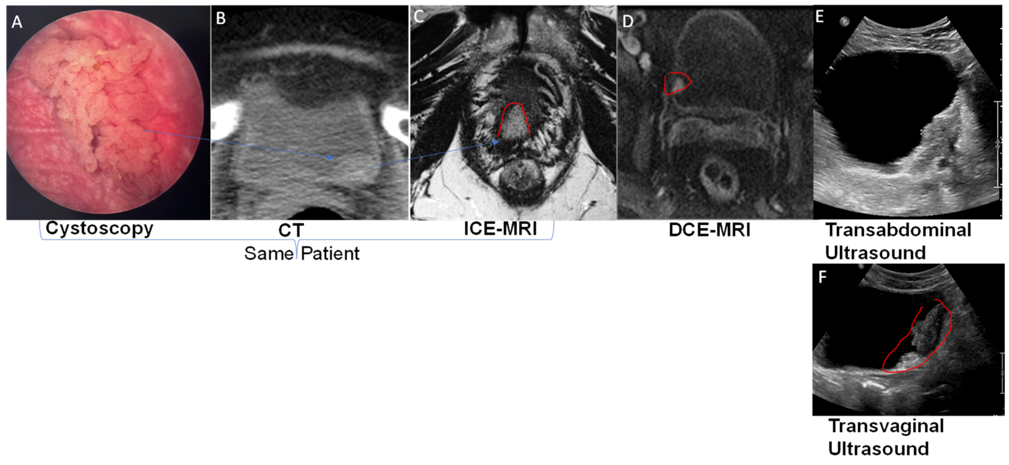
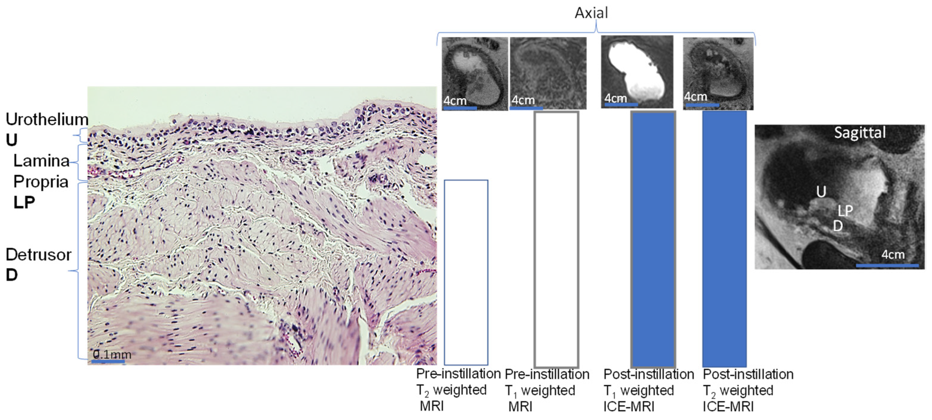
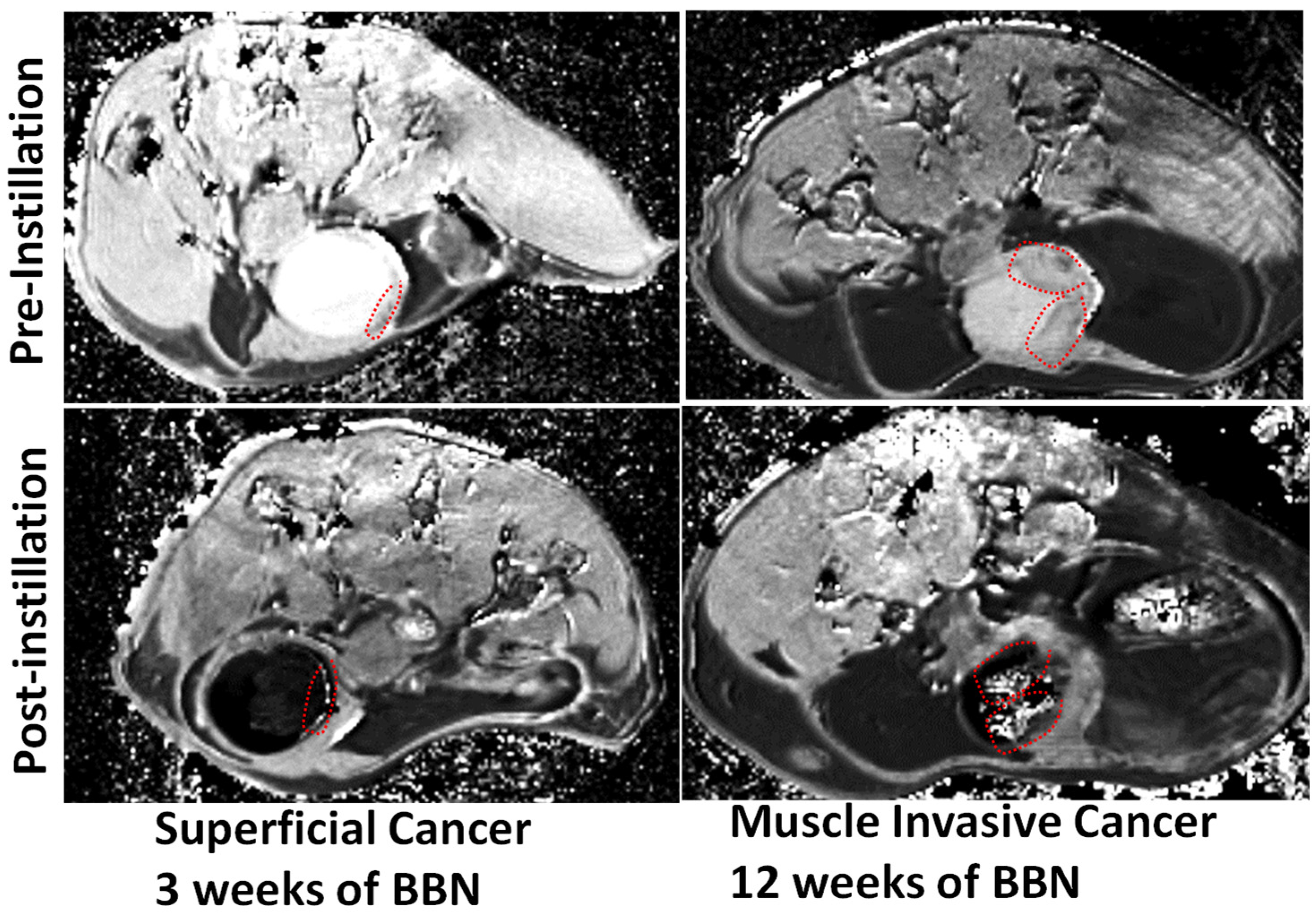
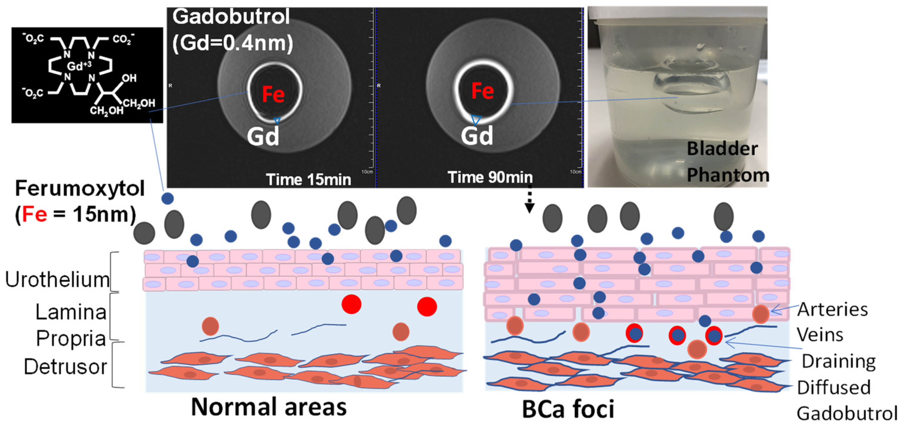
While the poor resolution of soft tissue obtained by widely available imaging options such as abdominal sonography and radiation-based CT leaves them only suitable for measuring the gross tumor volume and bladder wall thickening, dynamic contrast-enhanced magnetic resolution imaging (DCE MRI) is demonstrably superior in resolving muscle invasion. However, major barriers still exist in its adoption. Instead of injection for DCE-MRI, intravesical contrast-enhanced MRI (ICE-MRI) instills Gadolinium chelate (Gadobutrol) together with trace amounts of superparamagnetic agents for measurement of tumor volume, depth, and aggressiveness. ICE-MRI leverages leaky tight junctions to accelerate passive paracellular diffusion of Gadobutrol (604.71 Daltons) by treading the paracellular ingress pathway of fluorescein sodium and of mitomycin (<400 Daltons) into bladder tumor. The soaring cost of diagnosis and care of bladder cancer could be mitigated by reducing the use of expensive operating room resources with a potential non-surgical imaging option for cancer surveillance, thereby reducing over-diagnosis and over-treatment and increasing organ preservation.

bladder cancer staging MRI intravesical

1. Introduction

The cost of the diagnosis and care of bladder cancer is soaring due to several factors: the rise in prevalence, a high recurrence rate of 75–80% [1], and the dependence of the standard of care on expensive operating room resources for BCa staging via transurethral resection of bladder tumor (TURBT). Over 98% of BCas are histologically confirmed to be urothelial cell carcinomas. Urothelial cell carcinomas are histologically stratified into cancers of low and high grade, and further sub-divided into non-muscle-invasive (NMIBC) and muscle-invasive (MIBC) categories depending on the penetration of the urothelial cancer cell into the different layers of the bladder wall (Figure 1). Stage distinction is a critical factor in clinical decision-making, as the treatment goal of low-grade NMIBC with indolent disease history is to reduce the local recurrence and to arrest progression to muscle-invasive disease. Around a third of NMIBCs present with high-grade lesions, and 20–25% of these can progress to muscle invasion (MIBC) within 5 years of TURBT [2]. MIBC is treated with neoadjuvant chemotherapy followed by complete removal of the bladder, whereas more superficial tumors (NIMBC) are amenable to bladder-sparing therapy.
Figure 1. Urothelial carcinoma is staged by its depth of penetration into the three layers of the bladder wall (the inner mucosa, lamina propria and outer muscularis propria). Four bladder tumor stages are recognized: superficial neoplasm, also called non-muscle-invasive bladder cancer (NMIBC- Tis, Ta, and T1), muscle-invasive bladder cancer (MIBC), involving partial wall infiltrating neoplasms (T2), total wall infiltrating neoplasms [T3a (perivesical invasion on histology), T3b (perivesical invasion large enough to be seen on imaging)], and neoplasms involving other pelvic organs (T4).

2. Current Challenges

2.1. Imaging Modalities

Abdominal sonography (Figure 2E,F) [3] and radiation-based imaging modalities such as computed tomography (CT) (Figure 2B) can measure bladder wall thickening secondary to idiopathic, infectious, or non-infectious inflammatory conditions [4], but their poor tissue resolution presents a challenge. Abdominal sonography (Figure 2E) [5][6] has been shown to increase the risk of over-staging NMIBC and under-staging MIBC [7][8], though transvaginal sonography in female bladder cancer patients (Figure 2F) achieves a higher spatial resolution by reducing the physical distance between the ultrasound probe and the diseased area on the bladder wall.
Figure 2. (AC): A cystoscopically confirmed superficial tumor was imaged by CT and then by intravesical contrast-enhanced (ICE)-MRI after the instillation of the Gadolinium-based contrast agent GBCA and (Ferumoxytol) for positive and negative contrast, respectively, creating a clear demarcation of the bladder tumor (red circle) shape, size, and depth of penetration. (DF): Dynamic contrast-enhanced magnetic resonance imaging (DCE-MRI) after intravenous injection of GBCA could image the NMIBC prior to TURBT, whereas the poor soft tissue resolution of CT is only good for estimating gross tumor volume, and the image resolution of transvaginal ultrasound (F) is superior to transabdominal ultrasound (E).
The need for a high radiation dose and an intravenous ionic contrast agent makes CT unsuitable for regular surveillance of BCa [7]. CT is only good for estimating the gross tumor volume, as its poor soft tissue resolution makes it incapable of differentiating between superficial BCa and muscle invasion. Since the available techniques suffer from a ~50% failure rate [9] in detecting deeper muscle invasion, imaging modalities are inadequate in both assessing tumor aggressiveness [10] and in detecting carcinomas in situ (CIS) [11], which adversely impacts [12] the early identification of BCa patients most likely to benefit from chemotherapy or immunotherapy [13]. The superior soft tissue resolution of magnetic resonance imaging (MRI) offers several advantages (Figure 2D), but challenges remain in its use for BCa staging.

2.2. Magnetic Resonance Imaging (MRI)

MRI affords good soft tissue contrast in multiplanar images of visceral organs without the use of ionizing radiation [14], but conventional MRI without contrast enhancement has notable drawbacks [15], as illustrated by unenhanced conventional MRI in Figure 3.
Figure 3. Schematic illustration of mammalian bladder wall anatomy pre and post instillation of contrast mixture for ICE-MRI. Only the muscle layer is visible in pre-contrast T2-weighted MRI (MRI adjacent to histology) compared to the whole bladder wall in T1-weighted MRI acquired in axial orientation. The cystoscopically confirmed muscle-invasive multifocal tumor is more clearly visible in ICE-MRI with all three layers visible in T2-weighted 2D-TSE (TR/TE 4000/100 ms, with a bigger voxel volume of 0.625 × 0.625 × 3 mm3 vs. 0.67 × 0.67 × 1 mm3, and twice the number of signal averages (NSA) than in T1-weighted 3D FLASH (TR/TE 5.24/1.86 ms) at a flip angle (FA) of 20°. Bladder wall segmentation into the urothelium (U), lamina propria (L) and detrusor D is more clearly visible in the magnified view on sagittal orientation with ICE-MRI. Scale bar is there for comparison with axial view.
The MR contrast of cancer foci with respect to normal areas (Figure 2, Figure 3 and Figure 4) depends upon the differences in proton-spin density, magnetic susceptibility, water proton T1 (spin–lattice relaxation time), T2 (spin–spin relaxation time), magnetic susceptibility, molecular diffusion, and perfusion.
Figure 4. Pseudolayering observed in a mouse bladder lumen during multi-slice T1-weighted DCE-MRI, after intravenous injection of Gadobutrol (0.1 mm/kg) manifests the concentration-dependent T1- and T2-shortening effect of Gadobutrol on measured signal intensity (arbitrary units, a.u), with T1 relaxivity dominating at lower concentrations (<1 mM), and T2 relaxivity dominating at higher concentrations (>5 mM).
An MRI image (Figure 2, Figure 3, Figure 4 and Figure 5) is made up of many pixels (two-dimensional (2D) units of image space) corresponding to a three-dimensional (3D) volume called a voxel, defined by a field-of-view (FOV) of 160 × 160 mm2, an acquisition matrix of 256 × 256 and a slice thickness of 3 mm for the multi-slice T2-weighted turbo spin echo image (the leftmost MRI image of Figure 3), with fat suppression in a voxel volume 0.625 × 0.625 × 3 mm3, a 3 mm interslice gap, a repetition time (TR) of 4000 milliseconds (ms), an echo time (TE) of 100 ms, and a number of signal averages (NSA) = 2 for 30 slices acquired in 92 s [16].
Figure 5. Proof of the principle for bladder cancer surveillance and staging by multi-slice, T1-weighted ICE-MRI in axial orientation of 6-week-old B6D2F1 female mice fed carcinogen, N-butyl-N-4-hydroxybutyl nitrosamine (BBN), ad libitum, in drinking water (0.05% w/v) for up to 12 weeks. Tumoritropic infiltration of Gadobutrol accomplishes non-surgical surveillance of small polyps (red dotted circle) at 3 weeks, and stages muscle-invasive cancer (red dotted circle) by 12 weeks without transurethral resection by acquiring ICE-MRI under isoflurane anesthesia. Transurethral instillation of 0.05 mL Gadobutrol (4 mM) and Ferumoxytol (5 mM) for a 30 min period accomplishes voxel-wise T1 mapping at a variable TR of 400–7500 ms, a TE of 6.5 ms, a slice thickness 0.8 mm, an FOV of 28 × 28 mm2, an acquisition matrix 218 × 218, and an NSA = 1.

2.3. DCE-MRI

DCE-MRI following intravenous injection of GBCA visualizes the bladder wall (Figure 2D) in three layers: an inner thin layer of low intensity (mucosa), a middle layer of marked enhancement (lamina propria LP), and a thick outer layer of intermediate intensity (muscularis propria smooth muscle) akin to the segmentation shown in sagittal orientation for ICE-MRI. Upon coming into contact with the paramagnetic, GBCA [17], the T1 water relaxation rate (R1 = 1/T1 relaxation time measured in milliseconds (ms) of a voxel in the tumor increases in linear proportion to the tissue concentration of GBCA in that voxel [18][19]. Tumor-associated angiogenesis delivers a higher concentration of injected GBCA to voxels containing tumor(Figure 2D) compared to normal bladder wall, on the basis of a linear equation [20]: ΔR1 = R1 − R10 = r1[Gd], where R10 and R1 are the pre-contrast (baseline) and post-contrast T1 water relaxation rates (1/T1) of a voxel [18][19], respectively, and r1 is the relaxivity [21] of the T1 relaxation rate constant for GBCA.
As a result, the contrast enhancement in DCE-MRI (middle panel Figure 2) is a function of GBCA concentration delivered to the tumor via arterial perfusion [18][19], but the rapid washout leads to transient enhancement of the tumor (<3 min) [22]. Akin to DCE-MRI images of Figure 2D), post-instillation FLASH images acquired by ICE-MRI at the same FA of 20° could also clearly visualize the tumor (Figure 2C and Figure 3). The imaging parameters remained the same from pre-instillation to post-instillation of the contrast mixture. Contrary to transient (<3 min) enhancement of tumor by DCE-MRI [18][19][23], ICE-MRI enables clear visualization of high-grade urothelial carcinomas (Figure 2 and Figure 5), with a high signal-to-noise ratio for high resolution imaging, without entailing the risk of allergy and heavy metal toxicity with repeated DCE-MRI [24], because there is no injection of intravenous contrast [16].

3. Intravesical Contrast-Enhanced MRI (ICE-MRI)

Given that contrast enhancement of cancer foci in DCE-MRI is a function of GBCA concentration delivered by perfusion, scholarswondered if GBCA delivered by urothelial diffusion to tumor overcomes the above-stated drawbacks of DCE-MRI. This question inspired an intravesical offshoot of DCE-MRI with minimal invasiveness, called ICE-MRI [20][21][25][26]. Since past studies have reported that instillation of GBCA does not offer any advantage in bladder cancer staging over DCE-MRI [27], ICE-MRI is centered on bladder instillation via a transurethral catheter of two FDA-approved agents, Gadobutrol and Ferumoxytol, as a mixture [20][25][26]. Instead of arriving to the tumor by perfusion, ICE-MRI is predicated on the diffusion of instilled Gadobutrol along the downhill concentration gradient from the lumen to reach the highest concentration in the inner layer of the mucosa [28] for positive contrast and negative contrast in the lumen, resulting from the luminal retention of Ferumoxytol (Figure 5 and Figure 6). ICE-MRI ensures uniformity of contrast in lumen by picking a Gadobutrol concentration from Figure 4, which is insensitive to any dilution from fresh urine. Simply stated, the ICE-MRI seeks to repurpose FDA-approved agents for differential contrast enhancement of neoplastic and non-neoplastic lesions on the bladder wall by leveraging the published histological differences between neoplastic [29][30] and non-neoplastic lesions [31], and the structural differences in urothelial permeability [32].
Figure 6. MRI of a bladder phantom. The principle of Stokesian diffusion dictates passive, isotropic, time-dependent diffusion of Gadobutrol (0.4 nm) into >20 times bigger pores of polyacrylamide gel over 90 min (indicated by bigger blue∇ in the right image). The bright ring increasing from 15 to 90 min, which depicts paracellular Gd diffusion, is thrown into sharp relief by the dark cavity mimicking the luminal retention of a larger-sized Ferumoxytol (Fe 15 nm), with its larger magnetic moment decaying the Gadobutrol signal within the lumen.
Figure 2 and Figure 5 illustrate that ICE-MRI [25][26] provides stable positive contrast in rodent and human bladders, and the period of artifact-free visualization can be extended nearly 10-fold compared to DCE-MRI [22]. On the other hand, the reduced bioavailability of Gadobutrol dose 1 mmol (seven times lower than the recommended intravenous dose) [14][25][27] instilled into bladder eliminates the inherent risks of heavy metal toxicity and allergic reaction associated with GBCA injection [33][34]. Preclinical findings of a dark lumen adjacent to a bright bladder wall [20][21][26], generated by ICE-MRI at 7T [26][35], and a 9.4T animal scanner [21] were reproduced in the T2-weighted turbo spin echo images acquired at clinical scanner 3T (Figure 2C and Figure 3).
The graded decline in the signal intensity across bladder wall tissue layers (Figure 3 and Figure 5) manifests the logarithmic decline of diffusing Gadobutrol concentration [20] from the mucosa to deeper tissue layers. The logarithmic decline in diffused Gadobutrol concentration [20] stems from homeostatic venous clearance of any instilled drug reaching mucosa, [36]. Angiogenesis of the bladder tumor [35] augments the venous drainage of diffused Gadobutrol to accentuate the concentration gradient which may accelerate the Fickian diffusion of instilled Gadobutrol. As a result, the intensity of enhancement descends from aggressive cancer lesion > indolent cancer lesion > non-cancerous bladder wall (Figure 5). Cancerous lesions [37][38] on the luminal surface of the bladder are characterized by a disrupted tight junction barrier [39], and tumoritropic infiltration [35] of GBCA generates localized enhancement (Figure 2C).

3.1. Past Attempts of Adding Negative Contrast to Bladder

Since pseudolayering in the bladder lumen with DCE-MRI results from a lack of uniform contrast in the lumen, several group resorted to insufflation air [40] or instillation of Ferumoxytol [41] to ensure uniformity of contrast in the human bladder lumen during DCE-MRI. While Ferumoxytol instillation alone was directly tried in humans, air insufflation was also tried in a mouse bladder together with GBCA instillation [42]. However, both approaches failed to offer any advantage in improving the accuracy of BCa staging. As extensively reported by several groups [27][43], bladder instillation of just GBCA alone without the inclusion of a negative MR contrast (i.e., a hypo-intensity signal) is unable to achieve a clear visualization of bladder wall, which is a prerequisite for BCa staging.

3.2. Principle

The innovation of ICE-MRI capitalizes on the inverse relationship between the diffusion rate and the Stokes–Einstein radius of instilled drugs. Stokesian diffusion dictates the paracellular diffusion of smaller Gadobutrol (with a Stokes–Einstein radius of 0.4 nm and a molecular weight of 604.7 Daltons), whereas the diffusion of larger-sized Ferumoxytol (with a Stokes–Einstein radius of 15 nm and a molecular weight of 731 kiloDaltons) is retarded. Gadobutrol [44] and Ferumoxytol [45][46][47][48] also differ in their magnetic moment for tumorotropic infiltration of Gadobutrol [42][49] to cause tumor enhancement [20][21], while luminal retention of Ferumoxytol darkens the bladder lumen [35] (Figure 5 and Figure 6), as the large magnetic moment of iron [45][46][47][48] induces local magnetic field inhomogeneities to dephase proton spins, causing signal decay within the bladder lumen (Figure 6) [25][26][50].

3.3. Paracellular Path of Diffusion

ICE-MRI is predicated on the perturbed tight junctions [39][51] of bladder tumors relative to the normal areas that are shown to cause tumoritropic infiltration of small molecular weight drugs/dyes: such as mitomycin [36], fluorescein [52], methylene blue [53][54][55]. However, diffusion of high molecular weight radiolabeled probes [56][57][58][59] around tight junctions is slowed in accordance with the principle of Stokesian diffusion. Instilled GBCA is unlikely to enter umbrella cells as the transcellular permeability of umbrella cells is restricted, and GBCA is unable to enter even red blood cells upon injection [60]. The perturbed tight junctions [39][51] of cancer foci are known to compromise the urothelial barrier, which accentuates the passive diffusion of instilled GBCA in both rodent (Figure 5) [35][37][42][49] and human bladder (Figure 2 and Figure 3) [27], analogous to the diffusion of instilled polar dyes in preclinical [61][62][63] and clinical [52][53][54][55] studies. The differential signal enhancement of cancer foci by ICE-MRI replicates the results obtained with other radiation-free approaches [30][52].

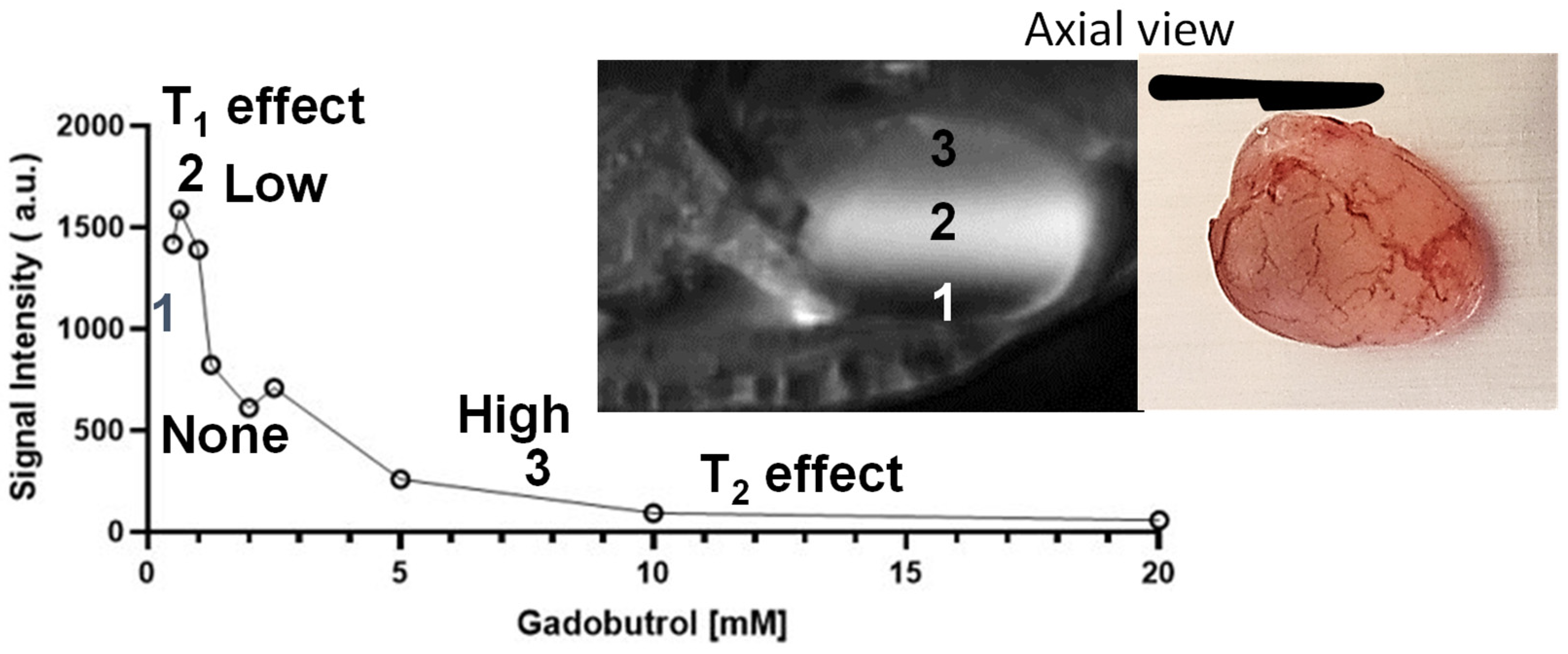
3.4. Effect of Urinary Dilution on Image Contrast

Given that the physical gap of the apico-lateral tight junction [61][62][64] (Figure 2 and Figure 3) can be partly mimicked by the pore size of 12% polyacrylamide gel [65], scholarsrelied on that equivalence to study the paracellular diffusion [20] of Gadobutrol without the confounding influence (Figure 6) of bladder distension and bladder perfusion [66][67]. A spherical bladder-shaped cavity was molded with 12% polyacrylamide gel poured into a plastic container, which was wrapped by a 4-channel flexible receiver coil, for image acquisition using a multi-echo spoiled-gradient echo pulse sequence in 3T scanner (Siemens, BioGraph) Figure 6.

3.5. Clinical Translation of ICE-MRI from 7T to 3T

The ten-fold lower thickness of rodent bladder wall (~0.5 mm) compared to human bladder wall (~5 mm) requires a proportionally higher signal-to-noise ratio of ≥7T for imaging mouse bladder cancer (Figure 5) [35]. The clinical translation of ICE-MRI tackled differences in field strength and pulse sequences from spin echo at a higher field of 7T to gradient echo (FLASH) at 3T for human subjects (Figure 2 and Figure 3) by adjusting the concentration of instilled Gadobutrol and Ferumoxytol. Accordingly, to limit signal dephasing in bladder wall with the use of gradient echo for T1 weighted imaging, scholarslowered the Ferumoxytol concentration [66] from our past study [25] by raising the Gadobutrol concentration [66].

References

  1. Kawada, T.; Yanagisawa, T.; Araki, M.; Pradere, B.; Shariat, S.F. Sequential intravesical gemcitabine and docetaxel therapy in patients with nonmuscle invasive bladder cancer: A systematic review and meta-analysis. Curr. Opin. Urol. 2023, 33, 211–218.
  2. Hugar, L.A.; Yabes, J.G.; Turner, R.M., 2nd; Fam, M.M.; Appleman, L.J.; Davies, B.J.; Jacobs, B.L. Rate and Determinants of Completing Neoadjuvant Chemotherapy in Medicare Beneficiaries With Bladder Cancer: A SEER-Medicare Analysis. Urology 2019, 124, 191–197.
  3. Ge, X.; Lan, Z.K.; Chen, J.; Zhu, S.Y. Effectiveness of contrast-enhanced ultrasound for detecting the staging and grading of bladder cancer: A systematic review and meta-analysis. Med. Ultrason. 2021, 23, 29–35.
  4. Jhang, J.F.; Hsu, Y.H.; Ho, H.C.; Jiang, Y.H.; Lee, C.L.; Yu, W.R.; Kuo, H.C. Possible Association between Bladder Wall Morphological Changes on Computed Tomography and Bladder-Centered Interstitial Cystitis/Bladder Pain Syndrome. Biomedicines 2021, 9, 1306.
  5. Husband, J.E. Staging bladder cancer. Clin. Radiol. 1992, 46, 153–159.
  6. Husband, J.E.; Olliff, J.F.; Williams, M.P.; Heron, C.W.; Cherryman, G.R. Bladder cancer: Staging with CT and MR imaging. Radiology 1989, 173, 435–440.
  7. Setty, B.N.; Holalkere, N.S.; Sahani, D.V.; Uppot, R.N.; Harisinghani, M.; Blake, M.A. State-of-the-art cross-sectional imaging in bladder cancer. Curr. Probl. Diagn. Radiol. 2007, 36, 83–96.
  8. Yaman, O.; Baltaci, S.; Arikan, N.; Yilmaz, E.; Gogus, O. Staging with computed tomography, transrectal ultrasonography and transurethral resection of bladder tumour: Comparison with final pathological stage in invasive bladder carcinoma. Br. J. Urol. 1996, 78, 197–200.
  9. Wang, Y.; Liu, J.; Yang, X.; Liu, Y.; Liu, Y.; Li, Y.; Sun, L.; Yang, X.; Niu, H. Bacillus Calmette-Guerin and anti-PD-L1 combination therapy boosts immune response against bladder cancer. OncoTargets Ther. 2018, 11, 2891–2899.
  10. Roudnicky, F.; Poyet, C.; Buser, L.; Saba, K.; Wild, P.; Otto, V.I.; Detmar, M. Characterization of Tumor Blood Vasculature Expression of Human Invasive Bladder Cancer by Laser Capture Microdissection and Transcriptional Profiling. Am. J. Pathol. 2020, 190, 1960–1970.
  11. Singh, R.; Saleemi, A.; Walsh, K.; Popert, R.; O’Brien, T. Near misses in bladder cancer—An airline safety approach to urology. Ann. R. Coll. Surg. Engl. 2003, 85, 378–381.
  12. Caglic, I.; Panebianco, V.; Vargas, H.A.; Bura, V.; Woo, S.; Pecoraro, M.; Cipollari, S.; Sala, E.; Barrett, T. MRI of Bladder Cancer: Local and Nodal Staging. J. Magn. Reson. Imaging 2020, 52, 649–667.
  13. Zhou, Y.; Abel, G.A.; Hamilton, W.; Singh, H.; Walter, F.M.; Lyratzopoulos, G. Imaging activity possibly signalling missed diagnostic opportunities in bladder and kidney cancer: A longitudinal data-linkage study using primary care electronic health records. Cancer Epidemiol. 2020, 66, 101703.
  14. Rabie, E.; Faeghi, F.; Izadpanahi, M.H.; Dayani, M.A. Role of Dynamic Contrast-Enhanced Magnetic Resonance Imaging in Staging of Bladder Cancer. J. Clin. Diagn. Res. 2016, 10, TC01–TC05.
  15. Wu, L.M.; Chen, X.X.; Xu, J.R.; Zhang, X.F.; Suo, S.T.; Yao, Q.Y.; Fan, Y.; Hu, J. Clinical value of T2-weighted imaging combined with diffusion-weighted imaging in preoperative T staging of urinary bladder cancer: A large-scale, multiobserver prospective study on 3.0-T MRI. Acad. Radiol. 2013, 20, 939–946.
  16. Connell, M.; Dhir, R.; Moon, C.H.; Biatta, S.; Tarin, T.; Maranchie, J.; Tyagi, P. Discrimination of cystitis cystica from bladder cancer by intravesical contrast-enhanced magnetic resonance imaging (ICE-MRI). Can. Urol. Assoc. J. 2022, 16, S158.
  17. Steward, M.C.; Seo, Y.; Rawlings, J.M.; Case, R.M. Water permeability of acinar cell membranes in the isolated perfused rabbit mandibular salivary gland. J. Physiol. 1990, 431, 571–583.
  18. Dickie, B.R.; Banerji, A.; Kershaw, L.E.; McPartlin, A.; Choudhury, A.; West, C.M.; Rose, C.J. Improved accuracy and precision of tracer kinetic parameters by joint fitting to variable flip angle and dynamic contrast enhanced MRI data. Magn. Reson. Med. 2016, 76, 1270–1281.
  19. Kanazawa, Y.; Miyati, T.; Sato, O. Hemodynamic analysis of bladder tumors using T1-dynamic contrast-enhanced fast spin-echo MRI. Eur. J. Radiol. 2012, 81, 1682–1687.
  20. Singh, N.; Zabbarova, I.; Ikeda, Y.; Maranchie, J.; Chermansky, C.; Foley, L.; Hitchens, T.K.; Yoshimura, N.; Kanai, A.; Kaufman, J.; et al. Virtual measurements of paracellular permeability and chronic inflammation via color coded pixel-wise T1 mapping. Am. J. Physiol. Ren. Physiol. 2020, 319, F506–F514.
  21. Saito, T.; Hitchens, T.K.; Foley, L.M.; Singh, N.; Mizoguchi, S.; Kurobe, M.; Gotoh, D.; Ogawa, T.; Minagawa, T.; Ishizuka, O.; et al. Functional and histologic imaging of urinary bladder wall after exposure to psychological stress and protamine sulfate. Sci. Rep. 2021, 11, 19440.
  22. Parikh, N.; Ream, J.M.; Zhang, H.C.; Block, K.T.; Chandarana, H.; Rosenkrantz, A.B. Performance of simultaneous high temporal resolution quantitative perfusion imaging of bladder tumors and conventional multi-phase urography using a novel free-breathing continuously acquired radial compressed-sensing MRI sequence. Magn. Reson. Imaging 2016, 34, 694–698.
  23. Panebianco, V.; Narumi, Y.; Altun, E.; Bochner, B.H.; Efstathiou, J.A.; Hafeez, S.; Huddart, R.; Kennish, S.; Lerner, S.; Montironi, R.; et al. Multiparametric Magnetic Resonance Imaging for Bladder Cancer: Development of VI-RADS (Vesical Imaging-Reporting And Data System). Eur. Urol. 2018, 74, 294–306.
  24. Murata, N.; Gonzalez-Cuyar, L.F.; Murata, K.; Fligner, C.; Dills, R.; Hippe, D.; Maravilla, K.R. Macrocyclic and Other Non-Group 1 Gadolinium Contrast Agents Deposit Low Levels of Gadolinium in Brain and Bone Tissue: Preliminary Results From 9 Patients With Normal Renal Function. Investig. Radiol. 2016, 51, 447–453.
  25. Tyagi, P.; Janicki, J.; Moon, C.H.; Kaufman, J.; Chermansky, C. Novel contrast mixture achieves contrast resolution of human bladder wall suitable for T1 mapping: Applications in interstitial cystitis and beyond. Int. Urol. Nephrol. 2018, 50, 401–409.
  26. Tyagi, P.; Janicki, J.J.; Hitchens, T.K.; Foley, L.M.; Kashyap, M.; Yoshhimura, N.; Kaufman, J. Novel Contrast Mixture Improves Bladder Wall Contrast For Visualizing Bladder Injury. Am. J. Physiol. Ren. Physiol. 2017, 313, F155–F162.
  27. Sparenberg, A.; Hamm, B.; Hammerer, P.; Samberger, V.; Wolf, K.J. . Rofo 1991, 155, 117–122.
  28. Gao, X.; Au, J.L.; Badalament, R.A.; Wientjes, M.G. Bladder tissue uptake of mitomycin C during intravesical therapy is linear with drug concentration in urine. Clin. Cancer Res. 1998, 4, 139–143.
  29. Horikawa, Y.; Sugano, K.; Shigyo, M.; Yamamoto, H.; Nakazono, M.; Fujimoto, H.; Kanai, Y.; Hirohashi, S.; Kakizoe, T.; Habuchi, T.; et al. Hypermethylation of an E-cadherin (CDH1) promoter region in high grade transitional cell carcinoma of the bladder comprising carcinoma in situ. J. Urol. 2003, 169, 1541–1545.
  30. Golijanin, J.; Amin, A.; Moshnikova, A.; Brito, J.M.; Tran, T.Y.; Adochite, R.C.; Andreev, G.O.; Crawford, T.; Engelman, D.M.; Andreev, O.A.; et al. Targeted imaging of urothelium carcinoma in human bladders by an ICG pHLIP peptide ex vivo. Proc. Natl. Acad. Sci. USA 2016, 113, 11829–11834.
  31. Lee, Y.K.; Jhang, J.F.; Jiang, Y.H.; Hsu, Y.H.; Ho, H.C.; Kuo, H.C. Difference in electron microscopic findings among interstitial cystitis/bladder pain syndrome with distinct clinical and cystoscopic characteristics. Sci. Rep. 2021, 11, 17258.
  32. Eldrup, J.; Thorup, J.; Nielsen, S.L.; Hald, T.; Hainau, B. Permeability and ultrastructure of human bladder epithelium. Br. J. Urol. 1983, 55, 488–492.
  33. Ramalho, J.; Ramalho, M.; Jay, M.; Burke, L.M.; Semelka, R.C. Gadolinium toxicity and treatment. Magn. Reson. Imaging 2016, 34, 1394–1398.
  34. Kodzwa, R. ACR Manual on Contrast Media: 2018 Updates. Radiol. Technol. 2019, 91, 97–100.
  35. Ganguly, A.; Foley, L.; Hitchens, T.K.; Maranchie, J.; Ikeda, Y.; Zabbarova, I.; Kanai, A.; Yoshimura, N.; Tyagi, P. Virtual Monitoring of Bladder Cancer Progression In Mice With Intravesical Contrast Enhanced Magnetic Resonance Imaging (ICE-MRI). J. Urol. 2023, 209, e183.
  36. Wientjes, M.G.; Badalament, R.A.; Wang, R.C.; Hassan, F.; Au, J.L. Penetration of mitomycin C in human bladder. Cancer Res. 1993, 53, 3314–3320.
  37. Pan, Y.; Volkmer, J.P.; Mach, K.E.; Rouse, R.V.; Liu, J.J.; Sahoo, D.; Chang, T.C.; Metzner, T.J.; Kang, L.; van de Rijn, M.; et al. Endoscopic molecular imaging of human bladder cancer using a CD47 antibody. Sci. Transl. Med. 2014, 6, 260ra148.
  38. Davis, R.M.; Kiss, B.; Trivedi, D.R.; Metzner, T.J.; Liao, J.C.; Gambhir, S.S. Surface-Enhanced Raman Scattering Nanoparticles for Multiplexed Imaging of Bladder Cancer Tissue Permeability and Molecular Phenotype. ACS Nano 2018, 12, 9669–9679.
  39. Boireau, S.; Buchert, M.; Samuel, M.S.; Pannequin, J.; Ryan, J.L.; Choquet, A.; Chapuis, H.; Rebillard, X.; Avances, C.; Ernst, M.; et al. DNA-methylation-dependent alterations of claudin-4 expression in human bladder carcinoma. Carcinogenesis 2007, 28, 246–258.
  40. Bartolozzi, C.; Caramella, D.; Zampa, V.; Olmastroni, M.; Innocenti, P.; Menchi, I.; Lapini, A.; Amorosi, A. MR imaging with STIR technique and air insufflation for local staging of bladder neoplasms. Acta Radiol. 1992, 33, 577–581.
  41. Beyersdorff, D.; Taupitz, M.; Giessing, M.; Turk, I.; Schnorr, D.; Loening, S.; Hamm, B. . Rofo 2000, 172, 504–508.
  42. Kikuchi, E.; Xu, S.; Ohori, M.; Matei, C.; Lupu, M.; Menendez, S.; Koutcher, J.A.; Bochner, B.H. Detection and quantitative analysis of early stage orthotopic murine bladder tumor using in vivo magnetic resonance imaging. J. Urol. 2003, 170, 1375–1378.
  43. Lee, S.K.; Chang, Y.; Park, N.H.; Kim, Y.H.; Woo, S. Magnetic resonance voiding cystography in the diagnosis of vesicoureteral reflux: Comparative study with voiding cystourethrography. J. Magn. Reson. Imaging 2005, 21, 406–414.
  44. Shen, Y.; Goerner, F.L.; Snyder, C.; Morelli, J.N.; Hao, D.; Hu, D.; Li, X.; Runge, V.M. T1 relaxivities of gadolinium-based magnetic resonance contrast agents in human whole blood at 1.5, 3, and 7 T. Investig. Radiol. 2015, 50, 330–338.
  45. Knobloch, G.; Colgan, T.; Wiens, C.N.; Wang, X.; Schubert, T.; Hernando, D.; Sharma, S.D.; Reeder, S.B. Relaxivity of Ferumoxytol at 1.5 T and 3.0 T. Investig. Radiol. 2018, 53, 257–263.
  46. Finn, J.P.; Nguyen, K.L.; Han, F.; Zhou, Z.; Salusky, I.; Ayad, I.; Hu, P. Cardiovascular MRI with ferumoxytol. Clin. Radiol. 2016, 71, 796–806.
  47. Gupta, T.; Virmani, S.; Neidt, T.M.; Szolc-Kowalska, B.; Sato, K.T.; Ryu, R.K.; Lewandowski, R.J.; Gates, V.L.; Woloschak, G.E.; Salem, R.; et al. MR tracking of iron-labeled glass radioembolization microspheres during transcatheter delivery to rabbit VX2 liver tumors: Feasibility study. Radiology 2008, 249, 845–854.
  48. Mantovani, L.F.; Santos, F.P.S.; Perini, G.F.; Nascimento, C.M.B.; Silva, L.P.; Wroclawski, C.K.; Esposito, B.P.; Ribeiro, M.S.S.; Velloso, E.; Nomura, C.H.; et al. Hepatic and cardiac and iron overload detected by T2* magnetic resonance (MRI) in patients with myelodisplastic syndrome: A cross-sectional study. Leuk. Res. 2019, 76, 53–57.
  49. Chin, J.; Kadhim, S.; Garcia, B.; Kim, Y.S.; Karlik, S. Magnetic resonance imaging for detecting and treatment monitoring of orthotopic murine bladder tumor implants. J. Urol. 1991, 145, 1297–1301.
  50. Tyagi, P.; Moon, C.H.; Janicki, J.; Kaufman, J.; Chancellor, M.; Yoshimura, N.; Chermansky, C. Recent advances in imaging and understanding interstitial cystitis. F1000Research 2018, 7, 1771.
  51. Fellows, G.J. Permeability of normal and diseased human bladder epithelium. Proc. R. Soc. Med. 1972, 65, 299–300.
  52. Sonn, G.A.; Jones, S.N.; Tarin, T.V.; Du, C.B.; Mach, K.E.; Jensen, K.C.; Liao, J.C. Optical biopsy of human bladder neoplasia with in vivo confocal laser endomicroscopy. J. Urol. 2009, 182, 1299–1305.
  53. Fukui, I.; Yokokawa, M.; Mitani, G.; Ohwada, F.; Wakui, M.; Washizuka, M.; Tohma, T.; Igarashi, K.; Yamada, T. In vivo staining test with methylene blue for bladder cancer. J. Urol. 1983, 130, 252–255.
  54. Gill, W.B.; Huffman, J.L.; Lyon, E.S.; Bagley, D.H.; Schoenberg, H.W.; Straus, F.H., 2nd. Selective surface staining of bladder tumors by intravesical methylene blue with enhanced endoscopic identification. Cancer 1984, 53, 2724–2727.
  55. Gill, W.B.; Strauss, F.H. In vivo mapping of bladder cancer (chromocystoscopy for in vivo detection of neoplastic urothelial surfaces). Urology 1984, 23, 63–66.
  56. Malamitsi, J.; Zorzos, J.; Varvarigou, A.D.; Archimandritis, S.; Dassiou, C.; Sivolapenko, G.; Skarlos, D.V.; Serefoglou, A.; Lykourinas, M.; Proukakis, C. Immunotargeting of urothelial cell carcinoma with intravesically administered Tc-99m labeled HMFG1 monoclonal antibody. Cell Biophys. 1994, 24-25, 75–81.
  57. Malamitsi, J.; Zorzos, J.; Varvarigou, A.D.; Archimandritis, S.; Dassiou, C.; Skarlos, D.V.; Dimitriou, P.; Likourinas, M.; Zizi, A.; Proukakis, C. Immunolocalization of transitional cell carcinoma of the bladder with intravesically administered technetium-99m labelled HMFG1 monoclonal antibody. Eur. J. Nucl. Med. 1995, 22, 25–31.
  58. Syrigos, K.N.; Khawaja, M.; Krausz, T.; Williams, G.; Epenetos, A.A. Intravesical administration of radiolabelled tumour-associated monoclonal antibody in bladder cancer. Acta Oncol. 1999, 38, 379–382.
  59. Bamias, A.; Keane, P.; Krausz, T.; Williams, G.; Epenetos, A.A. Intravesical administration of radiolabeled antitumor monoclonal antibody in bladder carcinoma. Cancer Res. 1991, 51, 724–728.
  60. Koenig, S.H.; Spiller, M.; Brown, R.D., 3rd; Wolf, G.L. Relaxation of water protons in the intra- and extracellular regions of blood containing Gd(DTPA). Magn. Reson. Med. 1986, 3, 791–795.
  61. Elsen, S.; Lerut, E.; Van Cleynenbreugel, B.; van der Aa, F.; van Poppel, H.; de Witte, P.A. Biodistribution of Evans blue in an orthotopic AY-27 rat bladder urothelial cell carcinoma model: Implication for the improved diagnosis of non-muscle-invasive bladder cancer (NMIBC) using dye-guided white-light cystoscopy. BJU Int. 2015, 116, 468–477.
  62. Elsen, S.; Lerut, E.; Van Der Aa, F.; Van Cleynenbreugel, B.; Van Poppel, H.; De Witte, P. Evans blue-mediated white-light detection of non-muscle-invasive bladder cancer: A preclinical feasibility and safety study using a rat bladder urothelial cell carcinoma model. Mol. Clin. Oncol. 2016, 5, 678–688.
  63. Eichel, L.; Scheidweiler, K.; Kost, J.; Shojaie, J.; Schwarz, E.; Messing, E.; Wood, R. Assessment of murine bladder permeability with fluorescein: Validation with cyclophosphamide and protamine. Urology 2001, 58, 113–118.
  64. Carattino, M.D.; Prakasam, H.S.; Ruiz, W.G.; Clayton, D.R.; McGuire, M.; Gallo, L.I.; Apodaca, G. Bladder filling and voiding affect umbrella cell tight junction organization and function. Am. J. Physiol. Ren. Physiol. 2013, 305, F1158–F1168.
  65. Holmes, D.L.; Stellwagen, N.C. Estimation of polyacrylamide gel pore size from Ferguson plots of linear DNA fragments. II. Comparison of gels with different crosslinker concentrations, added agarose and added linear polyacrylamide. Electrophoresis 1991, 12, 612–619.
  66. Tyagi, P.; Moon, C.; Singh, N.; Connell, M.; Maranchie, J.; Chermansky, C.; Yoshimura, N.; Kaufman, J. High Resolution 3D T1-Mapping of Pig Bladder Wall by Intravesical Contrast Enhanced MRI At 3T. J. Urol. 2021, 206, e390.
  67. Tyagi, P.; Moon, C.; Singh, N.; Connell, M.; Maranchie, J.; Chermansky, C.; Yoshimura, N.; Kaufman, J. Probing the bladder wall diffusion of instilled gadobutrol by MRI. J. Urol. 2021, 206, e32.
More
Information
Contributors MDPI registered users' name will be linked to their SciProfiles pages. To register with us, please refer to https://encyclopedia.pub/register : , , , , , , , ,
View Times: 334
Revisions: 2 times (View History)
Update Date: 08 Jun 2023
1000/1000
Video Production Service