Submitted Successfully!
To reward your contribution, here is a gift for you: A free trial for our video production service.
Thank you for your contribution! You can also upload a video entry or images related to this topic.
Version Summary Created by Modification Content Size Created at Operation
1 -- 4913 2023-06-06 11:54:37 |
2 format correction -6 word(s) 4907 2023-06-07 05:50:11 |

Video Upload Options

Do you have a full video?

Confirm

Are you sure to Delete?
Cite
If you have any further questions, please contact Encyclopedia Editorial Office.
Sipos, F.; Műzes, G. CAR-Based Therapy for Autoimmune and Immune-Mediated Diseases. Encyclopedia. Available online: https://encyclopedia.pub/entry/45234 (accessed on 27 July 2024).
Sipos F, Műzes G. CAR-Based Therapy for Autoimmune and Immune-Mediated Diseases. Encyclopedia. Available at: https://encyclopedia.pub/entry/45234. Accessed July 27, 2024.
Sipos, Ferenc, Györgyi Műzes. "CAR-Based Therapy for Autoimmune and Immune-Mediated Diseases" Encyclopedia, https://encyclopedia.pub/entry/45234 (accessed July 27, 2024).
Sipos, F., & Műzes, G. (2023, June 06). CAR-Based Therapy for Autoimmune and Immune-Mediated Diseases. In Encyclopedia. https://encyclopedia.pub/entry/45234
Sipos, Ferenc and Györgyi Műzes. "CAR-Based Therapy for Autoimmune and Immune-Mediated Diseases." Encyclopedia. Web. 06 June, 2023.
CAR-Based Therapy for Autoimmune and Immune-Mediated Diseases
Edit

The pervasive application of chimeric antigen receptor (CAR)-based cellular therapies in the treatment of oncological diseases has long been recognized. However, CAR T cells can target and eliminate autoreactive cells in autoimmune and immune-mediated diseases. By doing so, they can contribute to an effective and relatively long-lasting remission. In turn, CAR Treg interventions may have a highly effective and durable immunomodulatory effect via a direct or bystander effect, which may have a positive impact on the course and prognosis of autoimmune diseases. CAR-based cellular techniques have a complex theoretical foundation and are difficult to implement in practice, but they have a remarkable capacity to suppress the destructive functions of the immune system.

chimeric antigen receptor CAR T CAR Treg autoimmune immune-mediated

1. CAR T and CAR Treg Therapies and Clinical Trials in Systemic Lupus Erythematosus

Although CAR T has been used successfully in animal models of SLE, CAR Treg treatments are still uncommon, but both therapies show great promise [1]. Once CD8+ T cells targeting CD19+ B cells transduced with A-MLV retrovirus were transfused into MRL-lpr mice, CAR T cells decreased CD19 gene expression in the spleen, alleviated SLE symptoms, and increased the animals’ survival. Furthermore, CD19-targeted CAR T plasmids were transduced into splenic CD8+ T cells in NZBxNZW F1 mice and then infused into mice with SLE symptoms. The treatment induced CD19+ B-cell hypoplasia, demonstrating the therapy’s effectiveness [2]. CD19-targeted CAR T cell treatment with CD28 or 4-1BB co-stimulatory molecule CAR was also compared to anti-CD19 1D3 monoclonal antibody therapy in MRL-lpr mice. Anti-CD19 CAR T ACT resulted in a more prolonged reduction of B cells in mice than antibody treatment; moreover, the improved therapeutic effectiveness of CAR T cells with 4-1BB was demonstrated. It was also discovered that mice pretreated with mild doses of total body irradiation survived substantially longer [3]. Recently, a 20-year-old female patient with severe (SELENA score: 16), therapy-refractory SLE and type III/A lupus nephritis was treated with anti-CD19 CAR T therapy. After lymphodepletion (also known as conditioning treatment) with fludarabine to prevent immune rejection and to increase CAR T cell expansion and persistence, 1.1 × 10⁶ anti-CD19 CAR T cells were supplied per kilogram of body weight (a CD4+ to CD8+ T cell ratio of 3:1). Five weeks after CAR T cell infusion, the patient’s health improved, the dsDNA autoantibody titer and C3 and C4 complement levels normalized, and proteinuria with nephrotic grade was virtually eradicated. The score for SELENA was 0. In addition, no substantial adverse effects were noted [4]. Afterward, the same group treated an additional four SLE patients with refractory disease. All patients achieved the LLDAS and were able to discontinue all SLE-specific medications, including glucocorticoids, based on preliminary safety and efficacy data. No SLE flare has occurred to date; however, long-term follow-up data are required [5].
In a study [6], five patients (four women and one man) with long-standing, high disease activity and therapy-refractory SLE were enrolled in an anti-CD19 CAR T cell program as a last resort. Autologous T cells from the SLE patients were transduced with a lentiviral anti-CD19 CAR vector, expanded, and then returned to the patients at a dose of 1 × 10⁶ CAR T cells/kg body weight after lymphodepletion with fludarabine and cyclophosphamide. SLE remission according to DORIS criteria occurred in all five patients after 3 months, and the median range of the SLEDAI score was 0 after 3 months. Drug-free remission was maintained beyond 8 months. The B cells that reappeared after a median of 110 days were naïve and were not-class-switched B cells. The CAR T cell treatment was well tolerated, with only mild CRS observed.
In another study, a 41-year-old female patient with stage IV DLBCL and a 20-year history of SLE was treated with compound CAR T (cCAR T) co-expressing anti-BCMA and anti-CD19 bearing the CD137 co-stimulation domain [7]. Prior to the cCAR T infusion, the patient was preconditioned with fludarabine and cyclophosphamide. SLE remained in remission without additional treatment for 20 weeks after treatment, the ANA titer remained negative for 37 weeks, and B cells began to repopulate approximately 28 weeks after treatment (Figure 1).
Figure 1. The compound CAR construction is a two-unit CAR consisting of an entire BCMA-CAR linked to a full CD19-CAR, allowing for the autonomous expression of each of the CAR receptors on the T cell surface. This enables the CAR T cell to target long-lived antibody-producing cells (i.e., CD19+ memory B cells and BCMA+ plasma cells). Red arrows indicate inhibition. The figure was partly created by using www.biorender.com, accessed on 28 May 2023.
According to www.clinicaltrials.com accessed on 11 April 2023, there are currently four clinical trials registered to evaluate CAR T therapy in SLE. The first research study (NCT03030976) is being carried out to assess the safety and efficacy of anti-CD19 CAR T cells in the treatment of SLE patients. Using CD19 as the target and 4-1BB as the co-stimulator, the researchers built a second-generation CAR and adjusted its spatial structure with an appropriate hinge and transmembrane domain sequences. The second study (NCT05030779) also focuses on the B cell-killing effect of cCAR T cells. It aims to investigate the safety and efficacy of CD19/BCMA cCAR T cells in the treatment of refractory SLE. In the third study (NCT05474885) the number and incidence of adverse events after BCMA/CD19 cCAR T cell infusion will be monitored. The research group will evaluate all possible adverse reactions, including the number, incidence, and severity of symptoms such as CRS and neurotoxicity, within 3 months after CAR infusion. The fourth study (NCT05765006) is a Phase I, open-label, single-arm, multicenter trial to assess the safety, tolerability, pharmacokinetics, and pharmacodynamics of anti-CD19 CAR T therapy (Relma-cel) in patients with moderate-to-severe active SLE. The study will use 4 dose levels to assess dose escalation, safety, and tolerability.
Regarding CAR Treg cells, the immunological milieu of an SLE patient provides numerous difficulties [8]. High levels of IL6 and IFNα secreted by DCs impair Treg activity, while IL21 secreted by CD4+ T cells is equally deleterious to Treg survival and function [9]. Strong proinflammatory conditions can transform Treg cells into IL17 (or other inflammatory cytokine)-producing cells.
Although numerous autoantibodies may be expressed in SLE, CAR Treg may be effective in restoring Treg numbers and related inhibitory activities. Infusion of autologous Tregs can activate Tregs in inflamed skin, thereby inhibiting the IFNγ pathway and CD4+ effector cell invasion. It has been demonstrated that low-dose IL2 treatment in sixty individuals with active SLE is successful and restores tolerance [10]. Based on these findings, CAR Treg treatment can restore immunological tolerance and reduce inflammation in skin and renal tissues.

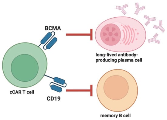
2. Compound CAR T Therapy and Clinical Trials in ANCA-Associated Vasculitis and Autoimmune Hemolytic Anemia

ANCA-associated small vessel vasculitides are the autoimmune inflammatory diseases of the vascular wall in which randomized controlled trials of rituximab and cyclophosphamide have shown the superiority of rituximab in inducing 6 and 24 months of remission of relapsed disease [11][12]. This suggests that repeated rituximab infusions may be needed to maintain disease control and that CAR T therapy may provide a more durable therapeutic option.
AIHA is a cell-specific autoimmune disease characterized by severe anemia due to autoantibodies against red blood cells, which are IgG isotypes in warm AIHA and IgM isotypes in cold AIHA. Treatment with rituximab has beneficial effects in both types, although randomized controlled trials with rituximab have not yet been performed in AIHA [13][14].
The reason for mentioning the two autoimmune diseases together is that there is only one clinical trial involving patients with vasculitis or AIHA (in addition to other diseases such as POEMS syndrome or amyloidosis). In this study (NCT05263817) the safety and effectiveness of CD19/BCMA cCAR T treatment of refractory diseases are assessed. However, based on the results of CAR-based treatment of SLE, it is hypothesized that further promising therapeutic results could be achieved in ANCA-associated vasculitis and AIHA using CAR T cell therapies, but further clinical trials are needed.

3. CAR T and CAR Treg Therapies in Rheumatoid Arthritis

RA is a chronic systemic inflammatory disease marked by persistent symmetric polyarthritis (synovitis) affecting primarily small joints. The synovial membrane is the target of an autoimmune reaction. Significant extra-articular involvement may also occur in organs such as the skin, heart, lungs, and eyes [15]. ACPA have been thoroughly studied in RA and may have a pathogenic function. In mice, ACPA against citrullinated vimentin may increase osteoclast genesis and bone resorption, indicating a pathogenic role for B cells [16][17]. Rituximab has been shown to be effective in patients with active RA, particularly in those with high ACPA levels [18]. Consequently, it is logically hypothesized that the formation of CAAR T cells expressing citrullinated antigens would enable the elimination of anti-citrulline B cells.
In an in vitro study, it was demonstrated that anti-FITC CAR T cells could be precisely redirected and kill hybridoma cells created by immunization with antigenic peptides and autoreactive B cell subsets from RA patients by recognizing appropriate FITC-labeled citrullinated peptide epitopes. Moreover, the cytotoxicity of CAR T cells was dose-dependent and reliant on the presence of peptides [19][20].
Developing particular CAR Tregs that induce tolerance in the synovium of affected joints also appears to be a potential option. CV is a particular antigen discovered only in the extracellular matrix of the inflamed synovial tissue of RA patients [21]. According to unpublished preliminary results, engineered CAR Tregs targeting CV may react with CV expressed in RA synovial fluid. However, further research is required to explore the effect of CAR Tregs in preclinical RA models [22].
Unfortunately, there are currently no ongoing clinical trials for CAR-based therapy for RA.

4. CAR T Therapies and Clinical Trials in Systemic Sclerosis

SSc is a progressive systemic connective tissue disease characterized by autoimmunity, vasculopathy, excessive extracellular matrix deposition and fibrosis, and consequent atrophy of the skin, subcutaneous tissue, muscles, and internal organs (e.g., digestive system, lungs, heart, kidneys, central nervous system) [23]. In SSc, immune cells, including T and B cells, and macrophages, display a variety of immunological abnormalities, as demonstrated by numerous recent investigations [24][25].
The European Scleroderma Study and Research Group published a clinical trial regarding the effect of rituximab on the fibrosis of the skin and lungs [26]. The study showed an improvement in skin fibrosis and a prevention of the progression of pulmonary fibrosis. In an open comparative study, the efficacy of rituximab and oral immunosuppressive therapies (i.e., azathioprine, methotrexate, and mycophenolate mofetil) was studied [27]. Clinical improvement in SSc-associated interstitial lung disease and skin fibrosis was also observed in the group treated with rituximab. In a double-blind, placebo-controlled trial with rituximab, 56 patients with SSc were studied (rituximab vs. placebo). Participants in the rituximab group experienced a significant decrease in the modified Rodnan Skin Score and a progression in the placebo group [28].
Studying the anti-fibrotic effects of CAR T cells found interesting preclinical results. In the mouse model of heart fibrosis, the activity of CAR T cells is directed against the protein that activates fibroblasts. In this model, CAR T CD8+ cells successfully destroyed the heart fibroblast that expressed the xenogeneic antigen [29]. Further examining this approach, during the in vivo ACT of anti-fibrotic CAR T cells, modified messenger RNA was injected into the cells in T cell-targeted lipid nanoparticles in order to increase safety [30].
Despite these promising results in preclinical models, clinical trials in patients SSc are still preliminary. The safety and efficacy of treatment with CD19/BCMA-CAR T were assessed in an open clinical trial (NCT05085444) for therapy-refractory SSc. The primary objective is to investigate dose-restricting toxicity and the incidence of adverse reactions.

5. CAR T, CAAR T and CAR Treg Therapies and Clinical Trials in Immune-Mediated Neurological Disorders

MS is an inflammatory autoimmune demyelinating disease of the CNS in which a large number of patients develop significant neurologic disability, such as weakness, vision loss, and cognitive decline. The spectacular effectiveness of systemic anti-CD20 B cell-depleting monoclonal antibodies in treating relapsing-remitting MS demonstrated that B cells play an essential role in the pathogenesis of the disease [31][32].
Anti-CD19 CAR T cells were studied in an EAE animal model of MS [33]. In this scenario, mice spontaneously acquire EAE accompanied by meningeal B cell aggregates. It was discovered that anti-CD19 CAR T cell therapy decreased the size of B cell aggregates in the meninges but aggravated the clinical illness. This is comparable to what was reported when anti-CD20 B cell depletion caused paradoxical worsening in the same spontaneous EAE or in EAE generated by immunization with MOG peptide (p)35–55 [34][35]. Recent research examined anti-CD19 CAR T cells in a B cell-dependent model of EAE caused by immunization with the extracellular domain of rhMOG protein [36]. Without causing systemic toxicity, this work established the efficacy of anti-CD19 CAR T cells in treating a B cell-dependent model of EAE. Although anti-CD20 monoclonal antibody therapy decreased B cells in both the CNS and the periphery in this mouse model, it is known that anti-CD20 antibodies do not effectively reach the CNS in humans, whereas anti-CD19 CAR T cells do [37][38][39]. Using anti-CD19 CAR T cells, severe and durable B cell depletion has been achieved not only in the periphery but also in the central nervous system, demonstrating that anti-CD19 CAR T cells may hold promise for patients with particular autoimmune disorders [36].
Treg cell therapy has been efficient in the EAE mouse model of MS. In EAE animals, CAR Tregs directed against the antigen MOG exhibited suppressive capability in vitro and could efficiently target several brain areas via intranasal cell delivery. After a second assault with the MOG peptide, the animals exhibited decreased illness symptoms and brain inflammation and remained healthy, indicating that altered Tregs have persistent benefits [40].
Tregs, which express a TCR specific for the MBP, have been constructed as a result of previous developments in leukemia CAR therapy. These TCR-engineered specific Tregs inhibited the proliferation of MBP-reactive T effector cells and ameliorated EAE elicited by MOG [41]. Subsequently, this strategy was expanded by engineering human Tregs to express a functional scFv CAR against MBP or MOG. These scFv CAR-transduced Tregs maintained Foxp3 and Helios levels unique to Treg cells following prolonged in vitro expansion. In addition, these modified CAR Tregs were able to decrease autoimmune pathology in EAE, suggesting that they may be a viable therapy option for MS patients [42].
Conventional ex vivo CAR Treg training necessitates a vast amount of equipment and knowledge, making its implementation for patients with demyelinating disorders challenging. By translating the majority of the ex vivo processes to in vivo, a novel theoretical notion could make CAR Treg therapy viable. The lentivirus HIV, possessing tropism toward CD4+ T cells, is extensively modified and utilized as a vector to introduce particular genes, such as the CAR gene, into cells [43]. Changes include providing the envelope with desired proteins (i.e., pseudotyping), removing its pathogenicity, transmissibility, and replication strength, and equipping the envelope with the appropriate proteins [44]. By using pseudotyping, the lentiviral envelope can be filled with any protein, even designer proteins. The envelope of the lentiviral vector carrying the CAR gene is commonly pseudotyped with vesicular stomatitis virus G protein to ensure broad tropism and, hence, success in currently utilized CAR T cell therapy. Current CAR T cell generation utilizes the ex vivo presentation of lentiviral vectors to T cells; however, viral vectors can be delivered in vivo (e.g., adenoviral vector vaccines against COVID-19). Pseudotyping lentiviral vector envelopes with designed ligands of Treg-specific pairings such as CD25 and CTLA-4 can give Tregs their unique tropism, allowing them to be supplied in vivo as vectors for the CAR gene, leading to the production of host-produced CAR Tregs [45]. The following notion would make Treg therapy viable by shifting the synthesis of CAR Tregs in vivo, leaving only the mass manufacturing of Treg-specific CAR gene lentiviral vectors. According to www.clinicaltrials.gov accessed on 11 April 2023, there are currently no ongoing clinical trials in MS to test the effectiveness of CAR-based therapy.
MG is a chronic autoimmune neuromuscular disease caused by autoantibodies against the AChR, MuSK, or low-density LRP4 expressed in postsynaptic muscle cells [46]. About 80% of patients with MG show anti-AChR antibody positivity, and about 40% of the anti-AChR antibody-negative patients show anti-MuSK antibody positivity. The presence of anti-LRP4 autoantibodies can be detected among patients outside the previous groups [47]. Based on the localization of the affected muscles, ocular and generalized MG can be distinguished. Passive transmission of autoantibodies may be involved in the development of the disease, indicating a primary role for autoantibodies in the pathogenesis of the disease [48].
NMOSD is a rare, progressive demyelinating disease that affects the optic nerves, spinal cord, and less frequently the brain. NMOSD can induce a variety of symptoms, including visual loss, paralysis, persistent hiccups, nausea, and vomiting. Typically, symptoms improve after the initial episode. A blood test for aquaporin-4 IgG antibodies is highly specific for NMOSD and facilitates diagnosis, but some patients with NMOSD may not have detectable antibodies despite exhibiting the disease’s defining characteristics [49].
CIDP is an autoimmune disease that affects peripheral nerves and nerve roots and is characterized by a symmetrical loss of motor and sensory function [42]. Various autoantibodies (including NF155, CNTN1, CASPR1, NF140, and NF186) may be present in the disorder. CIDP is currently treated off-label with glucocorticoids, intravenous immunoglobulin, plasmapheresis, and rituximab [50][51].
IMNM is a subclass of idiopathic inflammatory myopathies, distinguished by elevated blood creatine kinase and necrotic muscle fibers, and associated with autoantibodies against SRP or HMGCR [52][53]. Patients with anti-SRP antibodies respond better to rituximab treatment than those with anti-HMGCR antibodies [53].
Although there have been no experimental model trials to test CAR-based treatments for MG, NMOSD, CIDP, and IMNM, there are three clinical trials investigating this new therapy. In a Phase IIb study (NCT04146051), the safety, tolerability and preliminary efficacy of a repeated dosing schedule of Descartes-08 CAR T cells are evaluated in patients with generalized MG. In a so-called basket trial (NCT04561557) BCMA-CAR T treatment of autoimmune inflammatory neurological diseases (i.e., MG, NMOSD, CIDP, and IMNM) are investigated. The safety, dose-limiting toxicity and efficacy of a novel CAR T cell therapy using CT103A cells are evaluated in patients with relapsed/refractory antibody-mediated idiopathic inflammatory diseases.
A Phase 1 trial (NCT05451212) is investigating the safety and toxicity of different dosing regimens of an investigational cell therapy called MuSK-CAAR T, which can be given to patients with active, anti-MUSK antibody-positive MG. The different dosing regimens of MuSK-CAAR T are being evaluated alone, in combination with cyclophosphamide and in combination with cyclophosphamide and fludarabine.

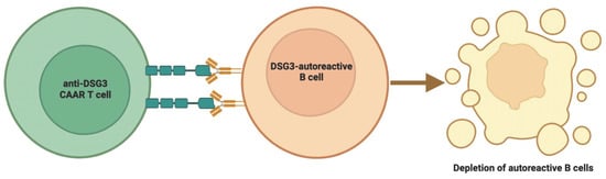
6. CAAR T Therapies and Clinical Trials in Pemphigus Vulgaris

PV is a severe, blistering autoimmune illness that affects the skin and mucous membranes. PV is mediated by Dsg-specific IgG autoantibodies. Autoantibodies bind to keratinocyte adhesion proteins Dsg1 and Dsg3 and impede keratinocyte adhesion, which results in lysis of the spinous layer and blister development. Dsg3 plays an essential role in PV [54].
Based on mice experiments and in vitro results, CAAR T cells made by fusing Dsg3 and CD137-CD3ζ were most effective when they expressed extracellular cadherin domains 1–4 from Dsg3 [55][56][57]. This CAAR T cell directly killed the memory B cells that express anti-Dsg3 sIg and eliminated indirectly the plasma cells that made short-lived, harmful anti-sIg-Dsg3 antibodies. Dsg3-CAAR T cells were not destroyed in the presence of pathogenic IgG, displayed mature specificity, and targeted only pathogenic B cells. In a mouse model, CAAR T cells drastically reduced the amount of anti-Dsg3 IgG-producing B cells without affecting the total number of pathogenic B cells [58]. This showed that CAAR T cells are able to find and kill the harmful anti-Dsg3 B cells in pulmonary vascular disease [46][49]. In mouse models, the T threshold dose of Dsg3-CAAR correlates, suggesting that a prudent fractional starting dose should be employed in future clinical trials [55].
Current standard clinical care for PV with corticosteroids, immunosuppressive medications, and rituximab requires a quite lengthy period to achieve remission, and relapse is common [59][60][61]. Several favorable aspects of CAAR T therapy have been identified, though. In CAAR T-cell experiments, no off-target adverse effects were observed. In addition, anti-Dsg3 B cells are unlikely to cause CRS since they represent a small proportion of total B cells in patients [62][63]. Furthermore, CAAR T cells are “living” agents capable of proliferating and persisting in vivo [55].
Up to a dose of 2.5 × 10⁹ CAAR T cells, preliminary clinical results from the first four cohorts of the DSG3-CAAR T research revealed no dose-limiting adverse effects [64]. At 28 days post-infusion, a dose-dependent rise in DSG3-CAAR T persistence approached the lower range of persistence values (observed in responders receiving anti-CD19 CAR T and lymphodepletion for B-cell leukemia) [65]. This implies that soluble antibodies do not cause CRS and do not destroy CAAR T cells. After DSG3-CAAR T infusion, transient improvements in clinical disease activity and antibody levels with a duration of two months were also seen [64].
One Phase 1 study (NCT04422912) has been conducted to find the maximum tolerated dose and optimal fractionated infusion schedule of the DSG3-CAAR T therapy, that can be given to patients with mucosal-dominant PV who are inadequately managed by standard therapies (Figure 2).
Figure 2. Chimeric autoantibody receptor T cell therapy depletes antigen-specific B cells. The figure was partly created by using www.biorender.com, accessed on 28 May 2023.

7. CAR T and CAR Treg Therapies and Clinical Trials in Dermatomyositis, Adult-Onset Still’s Disease, and Inflammatory Bowel Disease

DM is an idiopathic inflammatory myopathy that affects children and adults and is characterized by unique cutaneous manifestations. This systemic disorder affects the skin and muscles most frequently, but it can also affect the joints, esophagus, lungs, and, less frequently, the heart. The endothelium of the endomysial blood vessels is believed to be the target of an immune response in DM. The pathophysiology of the cutaneous manifestation of DM is poorly known, but it has been hypothesized to be comparable to that of muscle involvement [66].
AOSD is an uncommon inflammatory disorder with an unknown etiology that primarily affects young individuals. It is distinguished by high spiking fevers, arthritis, and an ephemeral, nonpruritic, macular, salmon-colored rash on the trunk and extremities. Organomegaly, lymphadenopathy, serositis, and aseptic meningitis are also possible. Important laboratory results include leukocytosis with a preponderance of neutrophils, negative testing for rheumatoid factor and antinuclear antibodies, as well as elevated serum ferritin and decreased serum glycosylated ferritin [67].
IBD is an idiopathic immune-mediated disease resulting from a dysregulated immune response to the intestinal microbiota. In IBD, the intestinal homeostasis is chronically compromised, and an inflammatory immune response is constitutively active due to disruption of the intestinal epithelial barrier. The two most common forms of IBD are UC, which is restricted to the colonic mucosa, and CD, which can affect any segment of the gastrointestinal tract, is transmural, and involves “skip lesions.” There is a genetic propensity for IBD, and patients with this condition have an increased risk of developing cancer [68].
T cells play an important role in the pathogenesis of all three diseases [69][70][71].
An important area for CAR-based therapies is the potential to treat T cell-mediated autoimmune diseases. To avoid T cell fratricide, anti-CD5/CD7 CAR T therapies are based on the suppression of endogenous CD5 and/or CD7 expression (e.g., by using CRISPR/Cas9-based gene editing or intracellular retention of CD7 by a CD7-binding protein) [72][73]. A high rate and fairly durable remission were achieved in a clinical trial involving subjects with relapsed or refractory T cell acute lymphoblastic leukemia (NCT04689659) with CD7-CAR T, while CD7-negative T cell expansion was observed in the subjects. This suggests selective cytotoxicity and persistence of CD7-CAR T cells [73].
NKG2D-CAR T cells gain selectivity for tumor-expressed stress-induced ligands. However, these stress ligands are also transiently expressed by activated T cells, suggesting that NKG2D-based T cells may undergo self-killing prior to infusion into patients during cell production. To prevent target-driven fratricide and permit the generation of NKG2D-CAR T cells for therapeutic use, two independent techniques were evaluated [74]. The first involved incorporating a phosphoinositol-3-kinase inhibitor into the manufacturing process. A second technique included the use of antibodies to block NKG2D itself. Both procedures had an influence on T cell fratricide, but to varying degrees, with the antibody approach having the most impact on cell production. Accordingly, target-driven CAR T fratricide can be overcome by inhibiting the expression of NKG2D or the action of enzymes in general [74]. CAR T cells engineered using this technique are already being tested in cancer, but trials in autoimmune diseases are still to come.
CD7-negative T cells can also be found in psoriasis, RA, or adult-onset autoimmune enteropathy [75][76][77]. In the latter disease, a decrease in the CD8+CD7− T cell population is associated with clinical improvement [77].
Currently, one basket clinical trial (NCT05239702) is underway to evaluate the safety and efficacy of CD7-CAR T treatment in T cell-dependent autoimmune diseases (i.e., CD, UC, DM, and AOSD). The PREDICT trial (NCT03369353) will investigate the immunology of auto- and alloimmune gastrointestinal disorders, such as IBD, GvHD, and functional gastrointestinal disorders, as well as the immunological manifestations following CAR T and other cellular therapies.
Adoptive transfection of Tregs can prevent and treat autoimmune disorders caused by abnormal Treg cells in animal models [78]. Using a transgenic method, Tregs were made to express a CAR with a TNP-specific antibody-converting region fused to the extracellular and transmembrane domains of the co-stimulatory molecule CD28 and the intracellular domain of the stimulatory receptor chain Fc-γ (TNP-TPCAR Tregs) [79]. All T cells of transgenic mice expressing this receptor were resistant to TNBS-induced colitis. In the inflamed colons of non-transgenic animals receiving tiny numbers of TPCAR Tregs, the cells aggregated and became activated. Due to their antigen specificity, lack of MHC limitation, and independence from co-stimulatory signals, TNP-TPCAR Tregs could heal acute experimental colonic inflammation [80]. TNBS have also been utilized to inhibit colitis in different types of colitis models. Although TPCAR-containing Tregs did not prevent oxazolidone-induced colitis, oxazolidone-induced colitis was healed when a minimal quantity of TNBS was added, showing the bystander effect of TNBS [80].
TNP-TPCAR Tregs may show promise against normal human Tregs. However, the resistance of nTregs to viral vector transduction poses a challenge for the production and clinical application of TNP-TPCAR Tregs [81]. In addition, adoptive transfer of engineered Treg cells can be risky, as the in vivo inflammatory environment can trigger the conversion of Treg cells into antigen-specific pathogenic effector T cells. To address this problem, techniques such as CRISPR can be used for gene editing [82].
Transduction of specific CEA-SCA431 CAR signaling domains fused to CD28-CD3ζ into CD4+CD25+ Treg cells, yields CEA-CAR Tregs [83]. CEA-CAR Tregs are effective at attenuating T cell metastatic colorectal inflammation and inhibiting the development of AOM-DSS-induced colorectal cancer [80]. CEA-CAR Tregs can dwell and aggregate at locations expressing CEA, mostly in the inflammatory colon, with considerably lower amounts in the small intestine and other organs [83]. They are advantageous due to their high antigen specificity, lack of MHC limitation, specific proliferation, independence from co-stimulatory signals, precise targeting, and quick activity. Therefore, CEA-CAR Tregs have significant potential for treating colitis and preventing colitis-associated cancer. However, the longevity of CEA-CAR Tregs is limited (7–9 days) [84]. This could be the result of an immune response to possible epitopes on CAR or luciferase reporter proteins. The relatively short in vivo lifespan of CAR Tregs may be a consequence of activation-induced cell death [83]. Presently, CEA-CAR Tregs cannot be used clinically to treat colitis due to their limited lifespan, and clinical trials have not yet been published.

8. CAR T Therapy in Type 1 Diabetes Mellitus

T1D is a chronic autoimmune condition that destroys pancreatic β-cells that produce insulin [85]. Consequently, insulin deficiency ultimately results in hyperglycemia. Repetitive insulin injections suggest that insulin levels in affected people are not stable and that T1D patients require long-term treatment [85].
To date, T1D CAR treatment has been primarily focused on CAR-T therapy with limited use of CAR Treg. Chimeric MHC molecules augmented with TCR-signaling motifs serve as activation receptors and can steer gene-modified T cells against pathogenic CD8 T cells. In NOD mice, it was shown that CD8+ T cells may be reprogrammed to identify diabetogenic T cells by electroporation of mRNA expressing peptide/β2-microglobulin/CD3ζ. These CAR T cells can target autoreactive cytotoxic T cells in vivo to minimize insulitis and avoid autoimmune diabetes [86]. Similarly, in NOD mice, it was observed that monoclonal antibody-287-targeted CAR T cells retained their specificity and destroyed antigen-presenting cells in vitro. In vivo, they were able to delay the onset of T1D in a well-established preclinical model, although the protection faded with time [87]. In addition, it has been shown that functional CARs against insulin can be created and that the combination of CARs and Foxp3 can generate antigen-specific Tregs from naïve CD4+ effector T cells. The insulin-specific CAR Tregs exhibit the same phenotype and function as their natural counterparts. In NOD mice, insulin-specific CAR Tregs had a lengthy lifespan [88].
Although animal trials seem promising, there are no data on human investigations or ongoing clinical trials of T1D CAR-based therapy.

9. CAR Treg Therapy and Clinical Trials in Graft-Versus-Host Disease

GvHD is a multiorgan complication of ACT that is potentially fatal. It depends on the fact that the host seems non-self to the graft, allowing it to stimulate antigenically using a wide range of immunological processes. GvHD is categorized as acute and chronic [89]. Frequently, the chronic form mimics autoimmune conditions.
GVHD is one of the side effects of CAR-based treatments, however, a recent study has shown in vitro the suppression of IgG antibody production and differentiation of B cells by CD19-CAR Tregs. In the same study, infusion of CD19-CAR Treg in immunodeficient mice reconstituted with human peripheral blood mononuclear cells, suppressed their antibody production and reduced the risk of GvHD [90].

References

  1. Radic, M.; Neeli, I.; Marion, T. Prospects for CAR T cell immunotherapy in autoimmune diseases: Clues from Lupus. Expert Opin. Biol. Ther. 2022, 22, 499–507.
  2. Kansal, R.; Richardson, N.; Neeli, I.; Khawaja, S.; Chamberlain, D.; Ghani, M.; Ghani, Q.-U.; Balazs, L.; Beranova-Giorgianni, S.; Giorgianni, F.; et al. Sustained B cell depletion by CD19-targeted CAR T cells is a highly effective treatment for murine lupus. Sci. Transl. Med. 2019, 11, eaav1648.
  3. Jin, X.; Xu, Q.; Pu, C.; Zhu, K.; Lu, C.; Jiang, Y.; Xiao, L.; Han, Y.; Lu, L. Therapeutic efficacy of anti-CD19 CAR-T cells in a mouse model of systemic lupus erythematosus. Cell. Mol. Immunol. 2021, 18, 1896–1903.
  4. Mougiakakos, D.; Krönke, G.; Völkl, S.; Kretschmann, S.; Aigner, M.; Kharboutli, S.; Böltz, S.; Manger, B.; Mackensen, A.; Schett, G. CD19-Targeted CAR T Cells in Refractory Systemic Lupus Erythematosus. N. Engl. J. Med. 2021, 385, 567–569.
  5. Schett, G.; Boeltz, S.; Müller, F.; Kleyer, A.; Völkl, S.; Aigner, M.; Gary, R.; Kretschmann, S.; Simon, D.; Kharboutli, S.; et al. OP0279 CAR-T cell treatment of refractory systemic lupus erythematosus—Safety and preliminary efficacy data from the first four patients. Ann. Rheum. Dis. 2022, 81, 185.
  6. Mackensen, A.; Müller, F.; Mougiakakos, D.; Böltz, S.; Wilhelm, A.; Aigner, M.; Völkl, S.; Simon, D.; Kleyer, A.; Munoz, L.; et al. Anti-CD19 CAR T cell therapy for refractory systemic lupus erythematosus. Nat. Med. 2022, 28, 2124–2132.
  7. Zhang, W.; Feng, J.; Cinquina, A.; Wang, Q.; Xu, H.; Zhang, Q.; Sun, L.; Chen, Q.; Xu, L.; Pinz, K.; et al. Treatment of Systemic Lupus Erythematosus using BCMA-CD19 Compound CAR. Stem Cell Rev. Rep. 2021, 17, 2120–2123.
  8. Kim, Y.C.; Zhang, A.-H.; Su, Y.; Rieder, S.A.; Rossi, R.J.; Ettinger, R.A.; Pratt, K.P.; Shevach, E.M.; Scott, D.W. Engineered antigen-specific human regulatory T cells: Immunosuppression of FVIII-specific T- and B-cell responses. Blood 2015, 125, 1107–1115.
  9. Guo, H.; Xun, L.; Zhang, R.; Hu, F.; Luan, J.; Lao, K.; Wang, X.; Gou, X. Stability and inhibitory function of Treg cells under inflammatory conditions in vitro. Exp. Ther. Med. 2020, 18, 2443–2450.
  10. He, J.; Zhang, R.; Shao, M.; Zhao, X.; Miao, M.; Chen, J.; Liu, J.; Zhang, X.; Zhang, X.; Jin, Y.; et al. Efficacy and safety of low-dose IL-2 in the treatment of systemic lupus erythematosus: A randomised, double-blind, placebo-controlled trial. Ann. Rheum. Dis. 2019, 79, 141–149.
  11. Stone, J.H.; Merkel, P.A.; Spiera, R.; Seo, P.; Langford, C.A.; Hoffman, G.S.; Kallenberg, C.G.; St Clair, E.W.; Turkiewicz, A.; Tchao, N.K.; et al. Faculty Opinions recommendation of Rituximab versus cyclophosphamide for ANCA-associated vasculitis. N. Engl. J. Med. 2011, 363, 221–232.
  12. Jones, R.B.; Furuta, S.; Tervaert, J.W.; Hauser, T.; Luqmani, R.; Morgan, M.D.; Peh, C.A.; Savage, C.O.; Segelmark, M.; Tesar, V.; et al. Rituximab versus cyclophosphamide in AN-CA-associated renal vasculitis: 2-year results of a randomised trial. Ann. Rheum. Dis. 2015, 74, 1178–1182.
  13. Garvey, B. Rituximab in the treatment of autoimmune haematological disorders. Br. J. Haematol. 2008, 141, 149–169.
  14. Rodrigo, C.; Rajapakse, S.; Gooneratne, L. Rituximab in the treatment of autoimmune haemolytic anaemia. Br. J. Clin. Pharmacol. 2015, 79, 709–719.
  15. Shen, Q.; Du, Y. A comprehensive review of advanced drug delivery systems for the treatment of rheumatoid arthritis. Int. J. Pharm. 2023, 635, 122698.
  16. Harre, U.; Georgess, D.; Bang, H.; Bozec, A.; Axmann, R.; Ossipova, E.; Jakobsson, P.-J.; Baum, W.; Nimmerjahn, F.; Szarka, E.; et al. Induction of osteoclastogenesis and bone loss by human autoantibodies against citrullinated vimentin. J. Clin. Investig. 2012, 122, 1791–1802.
  17. Vossenaar, E.R.; Després, N.; Lapointe, E.; van der Heijden, A.; Lora, M.; Senshu, T.; van Venrooij, W.J.; Ménard, H.A. Rheumatoid arthritis specific anti-Sa antibodies target citrullinated vimentin. Arthritis Res. Ther. 2004, 6, R142–R150.
  18. Zanetakis, E.; Rigby, W.F.; Rubbert-Roth, A.; Peterfy, C.G.; Van Vollenhoven, R.F.; Stohl, W.; Hessey, E.; Chen, A.; Tyrrell, H.; Hackshaw, K.; et al. Inhibition of joint damage and improved clinical outcomes with rituximab plus methotrexate in early active rheumatoid arthritis: The IMAGE trial. Ann. Rheum. Dis. 2011, 70, 39–46.
  19. Zhang, B.; Wang, Y.; Yuan, Y.; Sun, J.; Liu, L.; Huang, D.; Hu, J.; Wang, M.; Li, S.; Song, W.; et al. In vitro elimination of autoreactive B cells from rheumatoid arthritis patients by universal chimeric antigen receptor T cells. Ann. Rheum. Dis. 2021, 80, 176–184.
  20. Li, Y.-J.; Chen, Z. Cell-based therapies for rheumatoid arthritis: Opportunities and challenges. Ther. Adv. Musculoskelet. Dis. 2022, 14, 1759720X221100294.
  21. Van Steendam, K.; Tilleman, K.; De Ceuleneer, M.; De Keyser, F.; Elewaut, D.; Deforce, D. Citrullinated vimentin as an important antigen in immune complexes from synovial fluid of rheumatoid arthritis patients with antibodies against citrullinated proteins. Arthritis Res. Ther. 2010, 12, R132.
  22. Orvain, C.; Boulch, M.; Bousso, P.; Allanore, Y.; Avouac, J. Is There a Place for Chimeric Antigen Receptor-T Cells in the Treatment of Chronic Autoimmune Rheumatic Diseases? Arthritis Rheumatol. 2021, 73, 1954–1965.
  23. Yoshizaki, A. Pathogenic roles of B lymphocytes in systemic sclerosis. Immunol. Lett. 2018, 195, 76–82.
  24. Pillai, S. T and B lymphocytes in fibrosis and systemic sclerosis. Curr. Opin. Rheumatol. 2019, 31, 576–581.
  25. Brown, M.; O’Reilly, S. The immunopathogenesis of fibrosis in systemic sclerosis. Clin. Exp. Immunol. 2019, 195, 310–321.
  26. Jordan, S.; Distler, J.H.; Maurer, B.; Huscher, D.; van Laar, J.M.; Allanore, Y.; Distler, O.; EUSTAR Rituximab study group. Effects and safety of rituximab in systemic sclerosis: An analysis from the European Scleroderma Trial and Research (EUSTAR) group. Ann. Rheum. Dis. 2015, 74, 1188–1194.
  27. Daoussis, D.; Melissaropoulos, K.; Sakellaropoulos, G.; Antonopoulos, I.; Markatseli, T.E.; Simopoulou, T.; Georgiou, P.; Andonopoulos, A.P.; Drosos, A.A.; Sakkas, L.; et al. A multicenter, open-label, comparative study of B-cell depletion therapy with Rituximab for systemic sclerosis-associated interstitial lung disease. Semin. Arthritis Rheum. 2017, 46, 625–631.
  28. Kuzumi, A.; Ebata, S.; Fukasawa, T.; Matsuda, K.M.; Kotani, H.; Yoshizaki-Ogawa, A.; Sato, S.; Yoshizaki, A. Long-term Outcomes After Rituximab Treatment for Patients With Systemic Sclerosis: Follow-up of the DESIRES Trial With a Focus on Serum Immunoglobulin Levels. JAMA Dermatol. 2023, 159, e226340.
  29. Aghajanian, H.; Kimura, T.; Rurik, J.G.; Hancock, A.S.; Leibowitz, M.S.; Li, L.; Scholler, J.; Monslow, J.; Lo, A.; Han, W.; et al. Targeting cardiac fibrosis with engineered T cells. Nature 2019, 573, 430–433.
  30. Rurik, J.G.; Tombácz, I.; Yadegari, A.; Fernández, P.O.M.; Shewale, S.V.; Li, L.; Kimura, T.; Soliman, O.Y.; Papp, T.E.; Tam, Y.K.; et al. CAR T cells produced in vivo to treat cardiac injury. Science 2022, 375, 91–96.
  31. Hauser, S.L.; Bar-Or, A.; Comi, G.; Giovannoni, G.; Hartung, H.P.; Hemmer, B.; Lublin, F.; Montalban, X.; Rammohan, K.W.; Selmaj, K.; et al. Faculty Opinions recommendation of Ocrelizumab versus Interferon Beta-1a in Relapsing Multiple Sclerosis. N. Engl. J. Med. 2017, 376, 221–234.
  32. Montalban, X.; Hauser, S.L.; Kappos, L.; Arnold, D.L.; Bar-Or, A.; Comi, G.; de Seze, J.; Giovannoni, G.; Hartung, H.P.; Hemmer, B.; et al. Faculty Opinions recommendation of Ocrelizumab versus Placebo in Primary Progressive Multiple Sclerosis. N. Engl. J. Med. 2016, 376, 209–220.
  33. Mitsdoerffer, M.; Di Liberto, G.; Dötsch, S.; Sie, C.; Wagner, I.; Pfaller, M.; Kreutzfeldt, M.; Fräßle, S.; Aly, L.; Knier, B.; et al. Formation and immunomodulatory function of meningeal B cell aggregates in progressive CNS autoimmunity. Brain 2021, 144, 1697–1710.
  34. Weber, M.S.; Prod’homme, T.; Patarroyo, J.C.; Molnarfi, N.; Karnezis, T.; Lehmann-Horn, K.; Danilenko, D.M.; Eastham-Anderson, J.; Slavin, A.J.; Linington, C.; et al. B-cell activation influences T-cell polarization and outcome of an-ti-CD20 B-cell depletion in central nervous system autoimmunity. Ann. Neurol. 2010, 68, 369–383.
  35. Häusler, D.; Häusser-Kinzel, S.; Feldmann, L.; Torke, S.; Lepennetier, G.; Bernard, C.C.A.; Zamvil, S.S.; Brück, W.; Lehmann-Horn, K.; Weber, M.S. Functional characterization of reappearing B cells after anti-CD20 treatment of CNS autoimmune disease. Proc. Natl. Acad. Sci. USA 2018, 115, 9773–9778.
  36. Gupta, S.; Simic, M.; Sagan, S.A.; Shepherd, C.; Duecker, J.; Sobel, R.A.; Dandekar, R.; Wu, G.F.; Wu, W.; Pak, J.E.; et al. CAR-T Cell–Mediated B-Cell Depletion in Central Nervous System Autoimmunity. Neurol. Neuroimmunol. Neuroinflamm. 2023, 10, e200080.
  37. Monson, N.L.; Cravens, P.D.; Frohman, E.M.; Hawker, K.; Racke, M.K. Effect of Rituximab on the Peripheral Blood and Cerebrospinal Fluid B Cells in Patients With Primary Progressive Multiple Sclerosis. Arch. Neurol. 2005, 62, 258–264.
  38. Mainero, C.; Louapre, C. Meningeal inflammation in multiple sclerosis: The key to the origin of cortical lesions? Neurology 2015, 85, 12–13.
  39. Abramson, J.S.; McGree, B.; Noyes, S.; Plummer, S.; Wong, C.; Chen, Y.-B.; Palmer, E.; Albertson, T.; Ferry, J.A.; Arrillaga-Romany, I.C. Anti-CD19 CAR T Cells in CNS Diffuse Large-B-Cell Lymphoma. N. Engl. J. Med. 2017, 377, 783–784.
  40. Fransson, M.; Piras, E.; Burman, J.; Nilsson, B.; Essand, M.; Lu, B.; Harris, R.A.; Magnusson, P.U.; Brittebo, E.; Loskog, A.S. CAR/FoxP3-engineered T regulatory cells target the CNS and suppress EAE upon intranasal delivery. J. Neuroinflamm. 2012, 9, 112.
  41. Kim, Y.C.; Zhang, A.H.; Yoon, J.; Culp, W.E.; Lees, J.R.; Wucherpfennig, K.W.; Scott, D.W. Engineered MBP-specific human Tregs ameliorate MOG-induced EAE through IL-2-triggered inhibition of effector T cells. J. Autoimmun. 2018, 92, 77–86.
  42. De Paula Pohl, A.; Schmidt, A.; Zhang, A.H.; Maldonado, T.; Königs, C.; Scott, D.W. Engineered regulatory T cells expressing myelin-specific chimeric antigen receptors suppress EAE progression. Cell Immunol. 2020, 358, 104222.
  43. Feinberg, M.B.; McCune, J.M.; Miedema, F.; Moore, J.P.; Schuitemaker, H. HIV tropism and CD4+ T-cell depletion. Nat. Med. 2002, 8, 537.
  44. Ghaleh, H.E.G.; Bolandian, M.; Dorostkar, R.; Jafari, A.; Pour, M.F. Concise review on optimized methods in production and transduction of lentiviral vectors in order to facilitate immunotherapy and gene therapy. Biomed. Pharmacother. 2020, 128, 110276.
  45. Sedaghat, N.; Etemadifar, M. Inducing chimeric antigen receptor (CAR) regulatory T cells in-vivo: A novel concept for a potential feasible cure of demyelinating diseases. Mult. Scler. Relat. Disord. 2021, 57, 103341.
  46. Gilhus, N.E.; Tzartos, S.; Evoli, A.; Palace, J.; Burns, T.M.; Verschuuren, J.J.G.M. Myasthenia gravis. Nat. Rev. Dis. Prim. 2019, 5, 30.
  47. Lazaridis, K.; Tzartos, S.J. Myasthenia Gravis: Autoantibody Specificities and Their Role in MG Management. Front. Neurol. 2020, 11, 596981.
  48. Behin, A.; Le Panse, R. New Pathways and Therapeutic Targets in Autoimmune Myasthenia Gravis. J. Neuromuscul. Dis. 2018, 5, 265–277.
  49. Bagherieh, S.; Afshari-Safavi, A.; Vaheb, S.; Kiani, M.; Ghaffary, E.M.; Barzegar, M.; Shaygannejad, V.; Zabeti, A.; Mirmosayyeb, O. Worldwide prevalence of neuromyelitis optica spectrum disorder (NMOSD) and neuromyelitis optica (NMO): A systematic review and meta-analysis. Neurol. Sci. 2023, 44, 1905–1915.
  50. Wolbert, J.; Cheng, M.; Zu Horste, G.M.; Su, M.A. Deciphering immune mechanisms in chronic inflammatory demyelinating polyneuropathies. J. Clin. Investig. 2020, 5, e132411.
  51. Bright, R.J.; Wilkinson, J.; Coventry, B.J. Therapeutic options for chronic inflammatory demyelinating polyradiculoneuropathy: A systematic review. BMC Neurol. 2014, 14, 26.
  52. Stuhlmüller, B.; Schneider, U.; González-González, J.B.; Feist, E. Disease specific autoantibodies in idiopathic inflammatory myopathies. Front. Neurol. 2019, 10, 438.
  53. Xiong, A.; Yang, G.; Song, Z.; Xiong, C.; Liu, D.; Shuai, Y.; He, L.; Zhang, L.; Guo, Z.; Shuai, S. Rituximab in the treatment of immune-mediated necrotizing myopathy: A review of case reports and case series. Ther. Adv. Neurol. Disord. 2021, 14, 1756286421998918.
  54. Zhao, L.; Chen, Y.; Wang, M.; Leyang, X. The global incidence rate of pemphigus vulgaris: A systematic review and meta-analysis. Dermatology 2023, 239, 1–9.
  55. Ellebrecht, C.T.; Bhoj, V.G.; Nace, A.; Choi, E.J.; Mao, X.; Cho, M.J.; Di Zenzo, G.; Lanzavecchia, A.; Seykora, J.T.; Cotsarelis, G.; et al. Reengineering chimeric antigen receptor T cells for targeted therapy of autoimmune disease. Science 2016, 353, 179–184.
  56. Parvathaneni, K.; Scott, D.W. Engineered FVIII-expressing cytotoxic T cells target and kill FVIII-specific B cells in vitro and in vivo. Blood Adv. 2018, 2, 2332–2340.
  57. Ohyama, B.; Nishifuji, K.; Chan, P.T.; Kawaguchi, A.; Yamashita, T.; Ishii, N.; Hamada, T.; Dainichi, T.; Koga, H.; Tsuruta, D.; et al. Epitope Spreading Is Rarely Found in Pemphigus Vulgaris by Large-Scale Longitudinal Study Using Desmoglein 2–Based Swapped Molecules. J. Investig. Dermatol. 2012, 132, 1158–1168.
  58. Lee, J.; Lundgren, D.K.; Mao, X.; Manfredo-Vieira, S.; Nunez-Cruz, S.; Williams, E.F.; Assenmacher, C.-A.; Radaelli, E.; Oh, S.; Wang, B.; et al. Antigen-specific B cell depletion for precision therapy of mucosal pemphigus vulgaris. J. Clin. Investig. 2020, 130, 6317–6324.
  59. Schmidt, E.; Spindler, V.; Eming, R.; Amagai, M.; Antonicelli, F.; Baines, J.F.; Belheouane, M.; Bernard, P.; Borradori, L.; Caproni, M.; et al. Meeting Report of the Pathogenesis of Pemphigus and Pemphigoid Meeting in Munich, September 2016. J. Investig. Dermatol. 2017, 137, 1199–1203.
  60. Siddiqi, H.F.; Staser, K.W.; Nambudiri, V.E. Research Techniques Made Simple: CAR T-Cell Therapy. J. Investig. Dermatol. 2018, 138, 2501–2504.e1.
  61. Colliou, N.; Picard, D.; Caillot, F.; Calbo, S.; Le Corre, S.; Lim, A.; Lemercier, B.; Le Mauff, B.; Maho-Vaillant, M.; Jacquot, S.; et al. Long-Term Remissions of Severe Pemphigus After Rituximab Therapy Are Associated with Prolonged Failure of Desmoglein B Cell Response. Sci. Transl. Med. 2013, 5, 175ra30.
  62. Amagai, M. Pemphigus vulgaris and its active disease mouse model. Curr. Dir. Autoimmun. 2008, 10, 167–181.
  63. Nishifuji, K.; Amagai, M.; Kuwana, M.; Iwasaki, T.; Nishikawa, T. Detection of Antigen-Specific B Cells in Patients with Pemphigus Vulgaris by Enzyme-Linked Immunospot Assay: Requirement of T Cell Collaboration for Autoantibody Production. J. Investig. Dermatol. 2000, 114, 88–94.
  64. Basu, S.; Volkov, J.; Nunez, D.; Fouch, M.; Stadanlick, J.; Binder, G.; Chang, D.; Hoffman, K.; Porter, D.; Abedi, M.; et al. Characterization of DSG3-CAART cells prior to & following adoptive transfer in mucosal Pemphigus Vulgaris. Hum. Gene Ther. 2022, 33, A123.
  65. Mueller, K.T.; Maude, S.L.; Porter, D.L.; Frey, N.; Wood, P.; Han, X.; Waldron, E.; Chakraborty, A.; Awasthi, R.; Levine, B.L.; et al. Cellular kinetics of CTL019 in relapsed/refractory B-cell acute lymphoblastic leukemia and chronic lymphocytic leukemia. Blood 2017, 130, 2317–2325.
  66. Tomaras, S.; Feist, E. Myositissyndrome . Inn. Med. 2023, 64, 152–163.
  67. Mavragani, C.P.; Spyridakis, E.G.; Koutsilieris, M. Adult-Onset Still’s Disease: From Pathophysiology to Targeted Therapies. Int. J. Inflamm. 2012, 2012, 879020.
  68. Otte, M.L.; Tamang, R.L.; Papapanagiotou, J.; Ahmad, R.; Dhawan, P.; Singh, A.B. Mucosal healing and inflammatory bowel disease: Therapeutic implications and new targets. World J. Gastroenterol. 2023, 29, 1157–1172.
  69. Gordon, H.; Rodger, B.; Lindsay, J.O.; Stagg, A.J. Recruitment and residence of intestinal T cells—Lessons for therapy in IBD. J. Crohn’s Colitis 2023, 17, jjad027.
  70. Radziszewska, A.; Moulder, Z.; Jury, E.C.; Ciurtin, C. CD8+ T Cell Phenotype and Function in Childhood and Adult-Onset Connective Tissue Disease. Int. J. Mol. Sci. 2022, 23, 11431.
  71. Jung, J.-Y.; Choi, B.; Sayeed, H.; Suh, C.-H.; Kim, Y.W.; Kim, H.-A.; Sohn, S. Characteristic patterns of HLA presentation and T cell differentiation in adult-onset Still’s disease. Int. J. Immunopathol. Pharmacol. 2018, 32, 2058738418791284.
  72. Dai, Z.; Mu, W.; Zhao, Y.; Cheng, J.; Lin, H.; Ouyang, K.; Jia, X.; Liu, J.; Wei, Q.; Wang, M.; et al. T cells expressing CD5/CD7 bispecific chimeric antigen receptors with fully human heavy-chain-only domains mitigate tumor antigen escape. Signal Transduct. Target. Ther. 2022, 7, 85.
  73. Pan, J.; Tan, Y.; Wang, G.; Deng, B.; Ling, Z.; Song, W.; Seery, S.; Zhang, Y.; Peng, S.; Xu, J.; et al. Donor-Derived CD7 Chimeric Antigen Receptor T Cells for T-Cell Acute Lymphoblastic Leukemia: First-in-Human, Phase I Trial. J. Clin. Oncol. 2021, 39, 3340–3351.
  74. Breman, E.; Demoulin, B.; Agaugué, S.; Mauën, S.; Michaux, A.; Springuel, L.; Houssa, J.; Huberty, F.; Jacques-Hespel, C.; Marchand, C.; et al. Overcoming Target Driven Fratricide for T Cell Therapy. Front. Immunol. 2018, 9, 2940.
  75. Moll, M.; Reinhold, U.; Kukel, S.; Abken, H.; Müller, R.; Oltermann, I.; Kreysel, H.W. CD7-negative helper T cells accumulate in inflammatory skin lesions. J. Investig. Dermatol. 1994, 102, 328–332.
  76. Schmidt, D.; Goronzy, J.J.; Weyand, C.M. CD4+ CD7- CD28- T cells are expanded in rheumatoid arthritis and are characterized by autoreactivity. J. Clin. Investig. 1996, 97, 2027–2037.
  77. Bishu, S.; Arsenescu, V.; Lee, E.Y.; Vargas, H.D.; De Villiers, W.J.; Arsenescu, R. Autoimmune enteropathy with a CD8+ CD7-T-cell small bowel intraepithelial lymphocytosis: Case report and literature review. BMC Gastroenterol. 2011, 11, 131.
  78. Bao, L.; Bo, X.-C.; Cao, H.-W.; Qian, C.; Wang, Z.; Li, B. Engineered T cells and their therapeutic applications in autoimmune diseases. Zool. Res. 2022, 43, 150–165.
  79. Elinav, E.; Adam, N.; Waks, T.; Eshhar, Z. Amelioration of Colitis by Genetically Engineered Murine Regulatory T Cells Redirected by Antigen-Specific Chimeric Receptor. Gastroenterology 2009, 136, 1721–1731.
  80. Elinav, E.; Waks, T.; Eshhar, Z. Redirection of Regulatory T Cells With Predetermined Specificity for the Treatment of Experimental Colitis in Mice. Gastroenterology 2008, 134, 2014–2024.
  81. McGovern, J.L.; Wright, G.P.; Stauss, H.J. Engineering Specificity and Function of Therapeutic Regulatory T Cells. Front. Immunol. 2017, 8, 1517.
  82. Freen-van Heeren, J.J. Using CRISPR to enhance T cell effector function for therapeutic applications. Cytokine X 2020, 3, 100049.
  83. Blat, D.; Zigmond, E.; Alteber, Z.; Waks, T.; Eshhar, Z. Suppression of Murine Colitis and its Associated Cancer by Carcinoembryonic Antigen-Specific Regulatory T Cells. Mol. Ther. 2014, 22, 1018–1028.
  84. Zmievskaya, E.; Valiullina, A.; Ganeeva, I.; Petukhov, A.; Rizvanov, A.; Bulatov, E. Application of CAR-T Cell Therapy beyond Oncology: Autoimmune Diseases and Viral Infections. Biomedicines 2021, 9, 59.
  85. Syed, F.Z. Type 1 Diabetes Mellitus. Ann. Intern. Med. 2022, 175, ITC33–ITC48.
  86. Fishman, S.; Lewis, M.D.; Siew, L.K.; De Leenheer, E.; Kakabadse, D.; Davies, J.; Ziv, D.; Margalit, A.; Karin, N.; Gross, G.; et al. Adoptive Transfer of mRNA-Transfected T Cells Redirected against Diabetogenic CD8 T Cells Can Prevent Diabetes. Mol. Ther. 2017, 25, 456–464.
  87. Zhang, L.; Sosinowski, T.; Cox, A.R.; Cepeda, J.R.; Sekhar, N.S.; Hartig, S.M.; Miao, D.; Yu, L.; Pietropaolo, M.; Davidson, H.W. Chimeric antigen receptor (CAR) T cells targeting a pathogenic MHC class II: Peptide complex modulate the progression of autoimmune diabetes. J. Autoimmun. 2019, 96, 50–58.
  88. Tenspolde, M.; Zimmermann, K.; Weber, L.C.; Hapke, M.; Lieber, M.; Dywicki, J.; Frenzel, A.; Hust, M.; Galla, M.; Buitrago-Molina, L.E.; et al. Regulatory T cells engineered with a novel insulin-specific chimeric antigen receptor as a candidate immunotherapy for type 1 diabetes. J. Autoimmun. 2019, 103, 102289.
  89. Sobkowiak-Sobierajska, A.; Lindemans, C.; Sykora, T.; Wachowiak, J.; Dalle, J.H.; Bonig, H.; Gennery, A.; Lawitschka, A. Management of Chronic Graft-vs.-Host Disease in Children and Adolescents With ALL: Present Status and Model for a Personalised Management Plan. Front. Pediatr. 2022, 10, 808103.
  90. Imura, Y.; Ando, M.; Kondo, T.; Ito, M.; Yoshimura, A. CD19-targeted CAR regulatory T cells suppress B cell pathology without GvHD. J. Clin. Investig. 2020, 5, e136185.
More
Information
Subjects: Immunology
Contributors MDPI registered users' name will be linked to their SciProfiles pages. To register with us, please refer to https://encyclopedia.pub/register : ,
View Times: 437
Revisions: 2 times (View History)
Update Date: 07 Jun 2023
1000/1000
Video Production Service