Submitted Successfully!
To reward your contribution, here is a gift for you: A free trial for our video production service.
Thank you for your contribution! You can also upload a video entry or images related to this topic.
Version Summary Created by Modification Content Size Created at Operation
1 -- 2842 2023-05-29 23:51:34 |
2 format correct + 50 word(s) 2892 2023-05-30 05:51:45 |

Video Upload Options

Do you have a full video?

Confirm

Are you sure to Delete?
Cite
If you have any further questions, please contact Encyclopedia Editorial Office.
Hoch, C.C.; Stögbauer, F.; Wollenberg, B. Epithelial–Mesenchymal Transition in Salivary Adenoid Cystic Carcinoma. Encyclopedia. Available online: https://encyclopedia.pub/entry/44984 (accessed on 17 May 2024).
Hoch CC, Stögbauer F, Wollenberg B. Epithelial–Mesenchymal Transition in Salivary Adenoid Cystic Carcinoma. Encyclopedia. Available at: https://encyclopedia.pub/entry/44984. Accessed May 17, 2024.
Hoch, Cosima C., Fabian Stögbauer, Barbara Wollenberg. "Epithelial–Mesenchymal Transition in Salivary Adenoid Cystic Carcinoma" Encyclopedia, https://encyclopedia.pub/entry/44984 (accessed May 17, 2024).
Hoch, C.C., Stögbauer, F., & Wollenberg, B. (2023, May 29). Epithelial–Mesenchymal Transition in Salivary Adenoid Cystic Carcinoma. In Encyclopedia. https://encyclopedia.pub/entry/44984
Hoch, Cosima C., et al. "Epithelial–Mesenchymal Transition in Salivary Adenoid Cystic Carcinoma." Encyclopedia. Web. 29 May, 2023.
Epithelial–Mesenchymal Transition in Salivary Adenoid Cystic Carcinoma
Edit

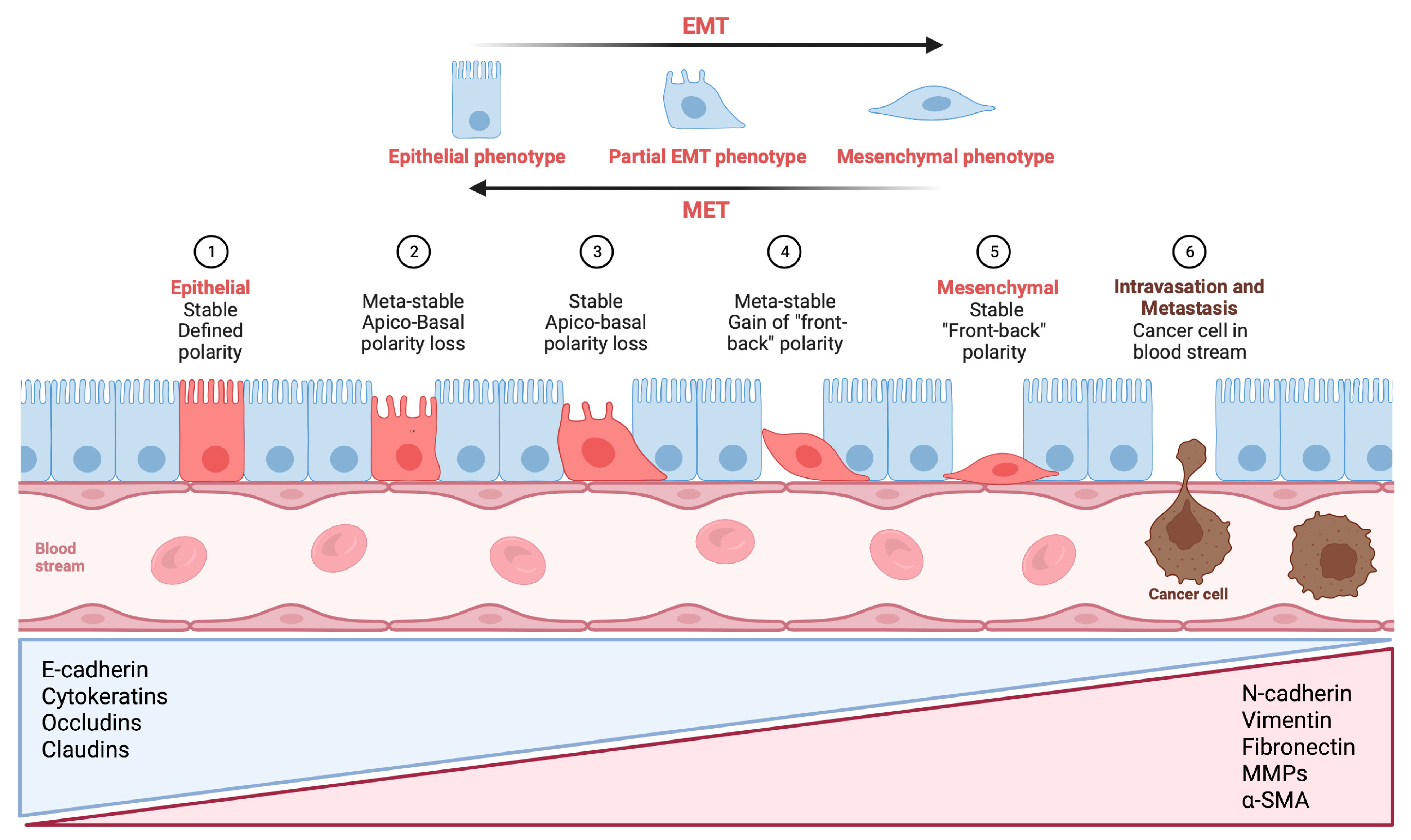
Salivary adenoid cystic carcinoma (SACC) is considered the second most frequent malignant salivary gland neoplasm and exhibits a controversial and poorly understood biological behavior, characterized by slow and indolent growth. SACC typically arises from the submandibular gland and minor salivary glands, while its occurrence in the parotid gland is relatively rare. SACC has an incidence rate of approximately 4.5 cases per million individuals and constitutes 10% of all salivary gland tumors (SGTs). This type of cancer exhibits three distinct histological growth patterns, namely cribriform, tubular, and solid patterns. Among these patterns, the solid pattern represents the most aggressive form of SACC with an increased risk of metastasis, resulting in shorter disease-specific survival.

metastasis biomarkers EMT tumor salivary adenoid cystic carcinoma

1. The Role of Epithelial–Mesenchymal Transition through the Activation of Transcription Factors in Cancer Invasion and Metastasis in Salivary Adenoid Cystic Carcinoma

EMT-TFs serve the purpose of maintaining stem cell characteristics, increasing the tumorigenicity of cells, and connecting to cancer stem cells. Furthermore, EMT-TFs create a phenotype that fosters cell survival by supporting DNA repair, evading immune recognition, resisting treatments, delaying aging, and avoiding cell death, which is advantageous under various stressful conditions. Taken together, the classical EMT functions and the varied, context-dependent, unique, and non-classical functions of EMT-TFs, which are also dynamically modulated by the tumor microenvironment, allow cancer cells to continuously adjust to changing conditions [1].
TWIST is a crucial protein that drives the EMT process by decreasing the expression of E-cadherin and increasing the expression of mesenchymal markers [2]. TWIST consists of two proteins, namely TWIST1 and TWIST2, that exhibit comparable levels of expression in tumors [3]. Studies revealed TWIST to be the most highly expressed factor among EMT-TFs in SACC tumor tissue samples, with the solid variant being the most reactive [4][5]. These results agree with a study performed by Shen et al., which showed significantly higher TWIST expression in SACC compared to pleomorphic adenomas (PAs) and normal parotid gland tissues [6]. Additionally, TWIST expression was documented to be higher in cases with DM than in those without DM [6].
The overexpression of TWIST has been observed to prompt an EMT-like transformation of metastatic cell lines of human SACC, leading to upgraded migration and invasion capabilities [5]. Elevated TWIST expression has also been strongly tied to perineural invasion (PNI), local regional recurrence, and DM of SACC. A high, predominantly nuclear TWIST expression in SACC was recorded when compared to mucoepidermoid carcinoma (MEC), normal glands, and benign tumors [7]. Kerche et al. similarly detected broad nuclear expression of the TWIST protein in SACC, although they did not find any link between TWIST expression and the absence of E-cadherin expression [8]. These findings corroborate previous research conducted on various types of tumors, which demonstrated a possible association between the elevated nuclear expression of TWIST and the increased invasiveness of tumors [9][10].
Recently, siRNA was used to reduce TWIST levels in SACC cells in order to investigate how TWIST is involved in the neurophilic invasion of these cells. Inhibiting TWIST resulted in decreased cancer cell migration, invasion, and PNI ability, as well as a reduction in the expression of genes associated with EMT and Schwann cell markers. When S100A4 was overexpressed using a plasmid, researchers noted a significant growth in the migratory, invasive, and PNI ability of SACC cells, along with a more fibroblast-like appearance and an increase in pseudopodia formation [11].

1.2. SNAIL and SLUG: Key Transcription Factors Driving Epithelial–Mesenchymal Transition and Metastasis in Salivary Adenoid Cystic Carcinoma

According to broad evidence, the SNAIL transcription factor suppresses the expression of E-cadherin, which results in the inhibition of cell–cell adhesion and augmented cancer invasion in different types of cancer [12][13]. SNAIL has a significant role in triggering the process of EMT and its reactivity was shown to be heterogeneous, with a more distinct nuclear pattern in the solid variant and invasive tumor front of SACC [4][14]. Remarkably, high levels of SNAIL expression are linked to PNI, local and regional recurrence, as well as DM of SACC [14]. Zhao et al. pointed out that the increased expression of SNAIL and Integrin-linked kinase (ILK) strongly correlated with the solid pattern, advanced TNM stages, a high risk of recurrences, and DM [15]. Of note, the authors found a correlation between the overexpression of SNAIL and N-cadherin and a neural invasive phenotype of SACC.
Another protein, the small heat-shock protein of 27 kDa (HSP27), has also been connected to cancer progression in SACC. The overexpression of HSP27 in SACC cells encouraged migration and invasion, induced EMT, and upregulated the expression of SNAIL and paired related homeobox protein 1 (PRRX1) [16]. In contrast, HSP27 silencing reduced migration and invasion and contributed to the MET of SACC cells. Additionally, HSP27 downregulated the expression of E-cadherin indirectly via the activation of SNAIL and PRRX1 expressions, while the overexpression of SNAIL or PRRX1 restored migration and invasion in HSP27 knockdown cells. The results of the study align with the clinical outcome of patients with SACC, as HSP27 expression is related to radioresistance and the poor prognosis of SACC patients, as well as with the expression of PRRX1 or SNAIL in SACC tissues.
More recently, Du et al. identified protein kinase D1 (PKD1) as a key regulator of SACC progression [17]. The inhibition of PKD1 resulted in the decreased proliferation, migration, invasion, and EMT of SACC cells. Conversely, the overexpression of kinase-active PKD1 evoked EMT and promoted cell migration in human SACC cells. The downregulation of PKD1 was found to control SNAIL via phosphorylation at Ser-11 on SNAIL protein and the promotion of proteasome-mediated degradation, which ultimately reduced lung metastasis in vivo.
Belulescu et al. reported that SLUG was the second most abundant EMT-TF detected in SACC, with reactivity observed in 78% of cases, mainly in the stroma at the level of cancer-associated fibroblasts (CAFs) and endothelial cells [4]. SLUG expression was noted to be higher at the invasive front and in cases associated with DM and lymph node metastases [4]. Similarly, Tang et al. stated that 72% of studied SACC exhibited reactivity to SLUG, and this correlated significantly with TNM stages, PNI, local regional recurrence, and DM [18]. The mitogen-activated protein kinase (MAPK)–SLUG pathway has been shown to contribute to the metastatic formation of SACC [19]. The high expression of SLUG and the extracellular matrix metalloproteinase inducer (EMMPRIN), along with low E-cadherin expression, were significantly linked with PNI and SACC [20]. Silencing SLUG inhibited the EMT process by reducing EMMPRIN expression and subsequently increasing E-cadherin expression in SACC. Two studies also revealed that elevated levels of BTBD7, a spliced variant of BTBD containing binding sites for significant transcription factors, were significantly linked to SLUG expression in SACC [21][22]. The reduction in BTBD7 considerably impeded the expression of SLUG in SACC cells. Furthermore, Yang et al. found a significant difference in SLUG expression between SACC and normal salivary gland cases, which was significantly linked with TNM stage, tissue typing, and DM.
Liu et al. displayed evidence that the epiregulin (EREG)–epidermal growth factor receptor (EGFR)–SNAIL/SLUG pathway propels EMT and metastatic characteristics in SACC cells, suggesting it could be an effective target for treating metastatic SACC [23]. In accordance with these data, epidermal growth factor (EGF) stimulation resulted in EGRF activation, which stabilized SNAIL expression and provoked EMT [24]. The suppression of SNAIL expression significantly inhibited EGF-induced EMT in these tumors. Jiang et al. further supported the aforementioned findings, as the authors displayed that the EMT process in SACC cell lines was reversed by pretreatment with Nimotuzumab, an anti-EGFR monoclonal antibody [25]. Moreover, high levels of BMI-1, nuclear/cytoplasmic SNAIL and SLUG expression, and low membranous E-cadherin expression were significantly coupled with DM in SACC [26].

1.3. ZEB1/ZEB2: A Key Regulator of Epithelial–Mesenchymal Transition in Salivary Adenoid Cystic Carcinoma

ZEB1/ZEB2 is a critical regulator in various tumor types and is downstream of nuclear factor kappa B (NF-κB) [27][28]. However, the immunohistochemical (IHC) detection of ZEB1 in SACC tumor parenchyma did not depict any positive reaction, while positive staining was examined in the tumor stroma at the level of CAFs and endothelial blood cells in all cases [4]. These results align with a study carried out by Kerche et al., that observed ZEB1 expression on both tumor cells and stromal cells [8]. Moreover, NF-κB directly binds to the promoter region of ZEB1, leading to its transcriptional activation [29]. Insulin-like growth factor-binding protein 2, which is significantly upregulated in SACC, plays a crucial role in the invasion and metastasis of this cancer type by modulating the NF-κB/ZEB1 signaling pathway. Interestingly, the knockdown of cancer cell-derived immunoglobulin G (cancer-IgG) in SACC suppressed EMT by upregulating E-cadherin and downregulating ZEB1/ZEB2, suggesting that cancer-IgG could be a useful prognostic marker for this disease [30]. Additionally, cancer-IgG overexpression was significantly correlated with metastasis, recurrence, and invasion in SACC.

2. Expression of Major Epithelial–Mesenchymal Transition Markers in Salivary Adenoid Cystic Carcinoma

Epithelial cells are transdifferentiated towards a mesenchymal cell phenotype during EMT, as they lose epithelial markers such as E-cadherin or various types of cytokeratin (CK), while gaining mesenchymal markers such as vimentin, fibronectin or N-cadherin [31]. Figure 1 provides a visual illustration that outlines the various phases of EMT.
Figure 1. Visual illustration of a typical epithelial–mesenchymal transition (EMT) program. Epithelial cells exhibit apical–basal polarity and express specific molecules associated with the maintenance of cell polarity and the epithelial state, which are listed in the blue triangular box. As epithelial cells undergo a progressive loss of their distinctive characteristics, they acquire mesenchymal features while retaining certain epithelial traits. In certain situations, they may acquire a complete set of mesenchymal features. Mesenchymal cells display front-to-back polarity, with a highly reorganized cytoskeleton, and they express a distinct set of molecules that promote and preserve the mesenchymal state (listed in the red triangular box). As a result, cells undergoing EMT exhibit increased migration, invasion, and stem-like properties, which can result in treatment resistance, metastasis formation, and recurrences in patients, ultimately affecting their clinical outcomes. Metastatic colonization requires a reversal of EMT, referred to as mesenchymal-to-epithelial transition (MET), which facilitates the expansion of tumor cells, an essential prerequisite for metastatic growth. E-cadherin: epithelial cadherin; N-cadherin: neural cadherin; MMP: matrix metalloproteinase; α-SMA: alpha-smooth muscle actin (Figure created using BioRender, Toronto, ON, Canada).

2.1. Expression of CK in Salivary Adenoid Cystic Carcinoma 

CK is a group of intermediate filaments that offer structural reinforcement to epithelial cells. CK expression is dependent on the tissue or organ and the degree of cell differentiation. Due to the specific cytokeratin patterns associated with different cellular sources, CK can be employed as a diagnostic marker for tumors, particularly epithelial cancers [32][33].
To characterize the CK profile of SACC, several studies have employed IHC examination of CK reactivity. In one study of 32 SACC cases, AE1/AE3 staining was found to be most intense in solid histological subtypes, followed by the tubular and cribriform subtypes [34]. SACC cells demonstrated strong and widespread positivity for AE1/AE3, comparable to the residual parenchyma of the major or minor salivary gland [35]. Furthermore, SACC exhibited positivity for CK7 but negativity for CK20, consistent with the results reported by Lee et al. [36]. Ben Salha et al. observed that AE1/AE3 and CK7 showed widespread and positive reactivity in solid SACC, whereas CK5/6 and CAM5.2 were only reactive in some neoplastic cells [37]. Notably, CK14-positive cells at the invasive front of SACC were reported to stimulate cancer cell invasion and may serve as a potential prognostic marker in these patients [38].

2.2. The Role of E-Cadherin in Salivary Adenoid Cystic Carcinoma: Implications for Tumor Invasion, Metastasis, and Epithelial–Mesenchymal Transition 

E-cadherin is a critical protein that plays a vital role in maintaining the structural integrity of epithelial tissue and is involved in regulating epithelial cell growth, differentiation, and survival. Multiple research projects have proven a reduction in E-cadherin expression in specimens of SACC compared to adjacent normal salivary parenchyma, leading to PNI, regional recurrence, and DM [8][11][15][39][40][41][42]. These studies revealed that regardless of the histological subtype, SACC cells located at the invasive tumor front and near nerve fibers exhibit reduced E-cadherin expression, with the alteration of the subcellular pattern from membranous to predominantly cytoplasmic [34][43]. Interestingly, the co-expression of E-cadherin with the gene cadherin 4 (CDH4), encoding retinal cadherin (R-cadherin), inhibits the growth and metastasis of SACC [44]. Moreover, the overexpression of ILK via the dysregulation of E-cadherin/N-cadherin might trigger EMT and thus facilitate SACC invasion and metastasis. Notably, a reduction in E-cadherin expression has also been explored in the myoepithelial cells of SACC compared to normal gland tissue [8][45], and the absence of E-cadherin expression correlated with PNI, vascular invasion, and DM in SACC patients.
In contrast to previous studies, two groups of researchers have observed E-cadherin expression in most of their SACC samples, suggesting that EMT in SACC could be related to the transformation of ductal epithelial cells into myoepithelial cells [46][47]. However, van der Wal et al. reported a favorable prognosis of SACC patients with diminished E-cadherin expression [48]. Wu et al. conducted a study on the invasion of neoplastic epithelial cells into adjacent tissue in SACC samples and noticed that the cells invaded as multicellular units and maintained their expression of E-cadherin [49]. The study’s IHC results also showed that cancer cells at the invasive front did not exhibit a complete EMT signature. The three-dimensional (3D) spheroid invasion assay revealed that leader cells displayed an elongated phenotype while still retaining E-cadherin expression, representing a partial EMT phenotype. These findings are consistent with previous studies that have noted that leader cells lack a complete EMT signature and cannot be identified through canonical mesenchymal markers [50][51]. EMT is not usually uniformly evident throughout the entire tumor tissue, and partial EMT is more common in cancer than complete EMT [52][53].

2.3. Vimentin: A Key Intermediate Filament in Mesenchymal Cells and Its Role in Salivary Adenoid Cystic Carcinoma Metastasis

Vimentin is a type of intermediate filament that is exclusively present in mesenchymal cells. It is a key structural protein that is widely distributed in different types of cells, including fibroblasts, vascular endothelial cells, smooth muscle cells, cartilage and bone cells, and nerve sheath cells, that form the connective tissue [32][54].
Belulescu et al. reported that the solid variant of SACC exhibited a more prominent EMT process, likely due to the more obvious vimentin expression perceived in these cases [34]. Furthermore, mesenchymal features were more pronounced in neoplastic cells located at the invasive tumor front and those associated with PNI, as demonstrated by Zhang et al., who found enlarged vimentin expression in areas of nerve invasion [11]. In accordance with these results, various authors declared positive vimentin staining in SACC together with the co-expression of keratin in these cells [55][56].

2.4. The Complex Role of Fibronectin in Salivary Adenoid Cystic Carcinoma: Promoting Perineural Invasion, Inhibiting Invasion, and Potential Prognostic Marker

Fibronectin is a type of glycoprotein that creates an extracellular matrix. It is a polypeptide that combines to form a dimer with a molecular weight of approximately 250 kDa. Its primary function is to facilitate the attachment of various cell types such as fibroblasts, hepatocytes, and nerve cells. The cell surface-specific receptor known as Integrin plays a significant role in processes such as cell adhesion, migration, and phagocytosis, particularly in the context of tissue damage [32][57].
Similar to the results regarding vimentin expression, fibronectin seems to act as a key player in the process of SACC invasiveness. In solid subtypes, fibronectin expression was observed to diminish in the outer region of invasive tumor clusters, while strong expression was detected in neoplastic cells that surround and infiltrate the nerve fibers, suggesting its potential dual role in promoting PNI, while potentially inhibiting invasion at the invasive tumor front [34][58][59][60]. In contrast, other studies have found lower fibronectin expression in the solid SACC variant compared to the tubular and cribriform subtypes [61][62]. Interestingly, the authors found a significant association between fibronectin expression, low TNM stages, and non-metastatic SACC cases [61]. Moreover, they postulated a potential inverse correlation between BTBD7 and fibronectin, indicating BTBD7′s potential prognostic role in predicting the invasion and metastasis of SACC.

2.5. N-Cadherin Expression in Salivary Adenoid Cystic Carcinoma: Association with Perineural Invasion and Epithelial–Mesenchymal Transition 

N-cadherin is a transmembrane glycoprotein that belongs to the classic cadherin superfamily and has a molecular weight of 130-kDa. Its expression has been detected in a range of cell types, such as neurons, endothelial cells, and cardiomyocytes [32][63].
Zhao et al. found that N-cadherin was expressed in 68.1% of the investigated SACC cases, with a membranous and/or cytoplasmic reaction pattern, and the highest expression levels were found in cases with PNI [15]. These findings are supported by Zhang et al., who also reported heightened N-cadherin expression in the area of nerve invasion in SACC [11]. In contrast, Belulescu et al. observed N-cadherin reactivity in only 35% of SACC cases, with low intensity and no significant difference between the three histological subtypes [34]. The discrepancy in results may be due to differences in the sample size, technique, or antibodies used for N-cadherin detection. Additionally, N-cadherin expression was found to be negatively aligned with E-cadherin expression and positively associated with the expression of SNAIL and ILK in SACC, implying that ILK overexpression might induce EMT by dysregulating the expression of these cadherins, leading to SACC invasion and metastasis [15]. Jiang et al. reported that thioredoxin 1 (TXN) and thioredoxin reductase 1 were overexpressed in SACC cases with metastasis and were partnered with high N-cadherin expression and low E-cadherin expression, proposing that TXN incites an EMT-like phenotype that promotes SACC migration and invasion [64]. These findings highlight the potential of N-cadherin, SNAIL, ILK, and TXN as prognostic markers for SACC.

References

  1. Puisieux, A.; Brabletz, T.; Caramel, J. Oncogenic roles of EMT-inducing transcription factors. Nat. Cell Biol. 2014, 16, 488–494.
  2. Thiery, J.P.; Acloque, H.; Huang, R.Y.J.; Nieto, M.A. Epithelial-Mesenchymal Transitions in Development and Disease. Cell 2009, 139, 871–890.
  3. Wang, S.M.; Coljee, V.W.; Pignolo, R.J.; Rotenberg, M.O.; Cristofalo, V.J.; Sierra, F. Cloning of the human twist gene: Its expression is retained in adult mesodermally-derived tissues. Gene 1997, 187, 83–92.
  4. Belulescu, I.C.; Mărgăritescu, C.; Dumitrescu, C.I.; Munteanu, M.C.; Dăguci, L.; Mărgăritescu, O.C.; Matei, M. The immunophenotype of epithelial to mesenchymal transition inducing transcription factors in salivary gland adenoid cystic carcinomas. Rom. J. Morphol. Embryol. 2020, 61, 769–782.
  5. Zhou, C.; Liu, J.; Tang, Y.; Zhu, G.; Zheng, M.; Jiang, J.; Yang, J.; Liang, X. Coexpression of hypoxia-inducible factor-2α, TWIST2, and SIP1 may correlate with invasion and metastasis of salivary adenoid cystic carcinoma. J. Oral Pathol. Med. 2012, 41, 424–431.
  6. Shen, M.; Wen, Y.; Hua, C.; Xiao, J. The expression of Twist in salivary adenoid cystic carcinoma and its clinicopathological significance. Chin. -Ger. J. Clin. Oncol. 2010, 9, 187–192.
  7. Pardis, S.; Zare, R.; Jaafari-Ashkavandi, Z.; Ashraf, M.J.; Khademi, B. Twist expression in pleomorphic adenoma, adenoid cystic carcinoma and mucoepidermoid carcinoma of salivary glands. Turk. J. Pathol. 2016, 32, 15–21.
  8. Kerche, L.E.; de Sousa, E.A.; Squarize, C.H.; Oliveira, K.K.; Marchi, F.A.; Bettim, B.B.; Kowalski, L.P.; Soares, F.A.; Lourenço, S.V.; Coutinho-Camillo, C.M. EMT in salivary gland tumors: The expression of microRNAs miR-155 and miR-200c is associated with clinical-pathological parameters. Mol. Biol. Rep. 2022, 49, 2157–2167.
  9. Silva, B.S.D.F.; Silva, F.P.Y.; Pontes, H.A.R.; Junior, D.D.S.P. E-cadherin downregulation and Twist overexpression since early stages of oral carcinogenesis. J. Oral Pathol. Med. 2014, 43, 125–131.
  10. Yuen, H.-F.; Chua, C.-W.; Chan, Y.-P.; Wong, Y.-C.; Wang, X.; Chan, K.-W. Significance of TWIST and E-cadherin expression in the metastatic progression of prostatic cancer. Histopathology 2007, 50, 648–658.
  11. Zhang, M.; Zheng, M.; Dai, L.; Zhang, W.; Fan, H.; Yu, X.; Pang, X.; Liao, P.; Chen, B.; Wang, S.; et al. CXCL12/CXCR4 facilitates perineural invasion via induction of the Twist/S100A4 axis in salivary adenoid cystic carcinoma. J. Cell. Mol. Med. 2021, 25, 7901–7912.
  12. Yokoyama, K.; Kamata, N.; Hayashi, E.; Hoteiya, T.; Ueda, N.; Fujimoto, R.; Nagayama, M. Reverse correlation of E-cadherin and snail expression in oral squamous cell carcinoma cells in vitro. Oral Oncol. 2001, 37, 65–71.
  13. Batlle, E.; Sancho, E.; Francí, C.; Domínguez, D.; Monfar, M.; Baulida, J.; De Herreros, A.G. The transcription factor Snail is a repressor of E-cadherin gene expression in epithelial tumour cells. Nat. Cell Biol. 2000, 2, 84–89.
  14. Jiang, J.; Tang, Y.; Zhu, G.; Zheng, M.; Yang, J.; Liang, X. Correlation between transcription factor Snail1 expression and prognosis in adenoid cystic carcinoma of salivary gland. Oral Surg. Oral Med. Oral Pathol. Oral Radiol. Endodontol. 2010, 110, 764–769.
  15. Zhao, D.; Yang, K.; Tang, X.-F.; Lin, N.-N.; Liu, J.-Y. Expression of integrin-linked kinase in adenoid cystic carcinoma of salivary glands correlates with epithelial–mesenchymal transition markers and tumor progression. Med. Oncol. 2013, 30, 619.
  16. Chen, W.; Ren, X.; Wu, J.; Gao, X.; Cen, X.; Wang, S.; Sheng, S.; Chen, Q.; Tang, Y.; Liang, X. HSP 27 associates with epithelial–mesenchymal transition, stemness and radioresistance of salivary adenoid cystic carcinoma. J. Cell. Mol. Med. 2018, 22, 2283–2298.
  17. Du, Y.; Lv, D.; Cui, B.; Li, X.; Chen, H.; Kang, Y.; Chen, Q.; Feng, Y.; Zhang, P.; Chen, J.; et al. Protein kinase D1 induced epithelial–mesenchymal transition and invasion in salivary adenoid cystic carcinoma via E-cadherin/Snail regulation. Oral Dis. 2022, 28, 1539–1554.
  18. Tang, Y.; Liang, X.; Zhu, G.; Zheng, M.; Yang, J.; Chen, Y. Expression and importance of zinc-finger transcription factor Slug in adenoid cystic carcinoma of salivary gland. J. Oral Pathol. Med. 2010, 39, 775–780.
  19. He, Q.; Zhou, X.; Li, S.; Jin, Y.; Chen, Z.; Chen, D.; Cai, Y.; Liu, Z.; Zhao, T.; Wang, A. MicroRNA-181a suppresses salivary adenoid cystic carcinoma metastasis by targeting MAPK–Snai2 pathway. Biochim. Biophys. Acta (BBA) Gen. Subj. 2013, 1830, 5258–5266.
  20. Wu, B.; Wei, J.; Hu, Z.; Shan, C.; Wang, L.; Zhang, C.; Yang, X.; Yang, X.; Lei, D. Slug silencing inhibited perineural invasion through regulation of EMMPRIN expression in human salivary adenoid cystic carcinoma. Tumor Biol. 2016, 37, 2161–2169.
  21. Yang, L.; Wang, T.; Zhang, J.; Wang, X. BTBD7 silencing inhibited epithelial- mesenchymal transition (EMT) via regulating Slug expression in human salivary adenoid cystic carcinoma. Cancer Biomark. 2017, 20, 461–468.
  22. Yang, L.; Wang, T.; Zhang, J.; Liu, Z.; Wang, X. Expression of BTBD7 in primary salivary adenoid cystic carcinoma and correlation with Slug and prognosis. Cancer Biomark. 2016, 17, 179–185.
  23. Liu, S.; Ye, D.; Xu, D.; Liao, Y.; Zhang, L.; Liu, L.; Yu, W.; Wang, Y.; He, Y.; Hu, J.; et al. Autocrine epiregulin activates EGFR pathway for lung metastasis via EMT in salivary adenoid cystic carcinoma. Oncotarget 2016, 7, 25251–25263.
  24. Wang, Y.; Hu, J.; Wang, Y.; Ye, W.; Zhang, X.; Ju, H.; Xu, D.; Liu, L.; Ye, D.; Zhang, L.; et al. EGFR activation induced Snail-dependent EMT and myc-dependent PD-L1 in human salivary adenoid cystic carcinoma cells. Cell Cycle 2018, 17, 1457–1470.
  25. Jiang, Y.; Ge, X.-Y.; Liu, S.-M.; Zheng, L.; Huang, M.-W.; Shi, Y.; Fu, J.; Zhang, J.-G.; Li, S.-L. Nimotuzumab suppresses epithelial–mesenchymal transition and enhances apoptosis in low-dose UV-C treated salivary adenoid cystic carcinoma cell lines in vitro. Anti-Cancer Drugs 2014, 25, 1052–1060.
  26. Yi, C.; Li, B.-B.; Zhou, C.-X. Bmi-1 expression predicts prognosis in salivary adenoid cystic carcinoma and correlates with epithelial-mesenchymal transition–related factors. Ann. Diagn. Pathol. 2016, 22, 38–44.
  27. Xu, X.M.; Liu, W.; Cao, Z.-H.; Liu, M.-X. Effects of ZEB1 on regulating osteosarcoma cells via NF-κB/iNOS. Eur. Rev. Med. Pharmacol. Sci. 2017, 21, 1184–1190.
  28. Gu, K.; Ming-Ming, L.; Jing, S.; Fang, L.; Jing-Yan, C.; Shi, J.; Yan, Y. Interleukin-17-induced EMT promotes lung cancer cell migration and invasion via NF-κB/ZEB1 signal pathway. Am. J. Cancer Res. 2015, 5, 1169–1179.
  29. Yao, X.; Wang, Y.; Duan, Y.; Zhang, Q.; Li, P.; Jin, R.; Tao, Y.; Zhang, W.; Wang, X.; Jing, C.; et al. IGFBP2 promotes salivary adenoid cystic carcinoma metastasis by activating the NF-κB/ZEB1 signaling pathway. Cancer Lett. 2018, 432, 38–46.
  30. Peng, J.; Wang, H.-C.; Liu, Y.; Jiang, J.-H.; Lv, W.-Q.; Yang, Y.; Li, C.-Y.; Qiu, X.-Y. Involvement of non-B cell-derived immunoglobulin G in the metastasis and prognosis of salivary adenoid cystic carcinoma. Oncol. Lett. 2017, 14, 4491–4498.
  31. Slack, J.M.; Tosh, D. Transdifferentiation and metaplasia—switching cell types. Curr. Opin. Genet. Dev. 2001, 11, 581–586.
  32. Matsumiya-Matsumoto, Y.; Morita, Y.; Uzawa, N. Pleomorphic Adenoma of the Salivary Glands and Epithelial–Mesenchymal Transition. J. Clin. Med. 2022, 11, 4210.
  33. Kuburich, N.A.; Hollander, P.D.; Pietz, J.T.; Mani, S.A. Vimentin and cytokeratin: Good alone, bad together. Semin. Cancer Biol. 2022, 86 Pt 3, 816–826.
  34. Belulescu, I.C.; Mărgăritescu, C.; Dumitrescu, C.I.; Munteanu, M.C.; Mărgăritescu, O.C. Immunophenotypical alterations with impact on the epithelial–mesenchymal transition (EMT) process in salivary gland adenoid cystic carcinomas. Rom. J. Morphol. Embryol. 2020, 61, 175–187.
  35. Meer, S.; Altini, M. CK7+/CK20? immunoexpression profile is typical of salivary gland neoplasia. Histopathology 2007, 51, 26–32.
  36. Lee, J.-H.; Lee, J.H.; Kim, A.; Kim, I.; Chae, Y.-S. Unique expression of MUC3, MUC5AC and cytokeratins in salivary gland carcinomas. Pathol. Int. 2005, 55, 386–390.
  37. Ben Salha, I.; Bhide, S.; Mourtzoukou, D.; Fisher, C.; Thway, K. Solid Variant of Adenoid Cystic Carcinoma: Difficulties in Diagnostic Recognition. Int. J. Surg. Pathol. 2016, 24, 419–424.
  38. Gao, X.-L.; Wu, J.-S.; Cao, M.-X.; Gao, S.-Y.; Cen, X.; Jiang, Y.-P.; Wang, S.-S.; Tang, Y.-J.; Chen, Q.-M.; Liang, X.-H.; et al. Cytokeratin-14 contributes to collective invasion of salivary adenoid cystic carcinoma. PLoS ONE 2017, 12, e0171341.
  39. Ge, M.-H.; Ling, Z.-Q.; Tan, Z.; Chen, C.; Xu, J.-J.; Yu, J.-L. Expression and significance of E-cadherin in adenoid cystic carcinoma of salivary glands. Zhonghua Yi Xue Za Zhi 2012, 92, 106–109.
  40. Lai, F.-Y.; Zhang, Q.; Wu, Q.-L.; Qing, J.; Cao, Y. Expression and significance of E-cadherin in adenoid cystic carcinoma of the salivary glands. Ai Zheng 2007, 26, 1025–1028.
  41. Maruya, S.-I.; Kurotaki, H.; Wada, R.; Saku, T.; Shinkawa, H.; Yagihashi, S. Promoter methylation and protein expression of the E-cadherin gene in the clinicopathologic assessment of adenoid cystic carcinoma. Mod. Pathol. 2004, 17, 637–645.
  42. Prabhu, S.; Kaveri, H.; Rekha, K. Benign; malignant salivary gland tumors: Comparison of immunohistochemical expression of e-cadherin. Oral Oncol. 2009, 45, 594–599.
  43. Zhang, M.; Li, Z.; Wang, H.; Wang, S.; Yu, X.; Wu, J.; Pang, X.; Wu, J.; Yang, X.; Tang, Y.; et al. MIF promotes perineural invasion through EMT in salivary adenoid cystic carcinoma. Mol. Carcinog. 2019, 58, 898–912.
  44. Xie, J.; Feng, Y.; Lin, T.; Huang, X.-Y.; Gan, R.-H.; Zhao, Y.; Su, B.-H.; Ding, L.-C.; She, L.; Chen, J.; et al. CDH4 suppresses the progression of salivary adenoid cystic carcinoma via E-cadherin co-expression. Oncotarget 2016, 7, 82961–82971.
  45. Jia, S.; Wang, W.; Hu, Z.; Shan, C.; Wang, L.; Wu, B.; Yang, Z.; Yang, X.; Lei, D. BDNF mediated TrkB activation contributes to the EMT progression and the poor prognosis in human salivary adenoid cystic carcinoma. Oral Oncol. 2015, 51, 64–70.
  46. Cavalcante, R.B.; Nonaka, C.F.W.; Rabenhorst, S.H.B.; Miguel, M.C.D.C.; Pinto, L.P.; de Souza, L.B. Pleomorphic adenoma and adenoid cystic carcinoma of salivary glands: E-cadherin immunoexpression and analysis of the CDH1 -160C/A polymorphism. Arch. Oral Biol. 2017, 73, 48–54.
  47. Phattarataratip, E.; Kositkittiwanit, N.; Kajornkiatkul, P.; Yeunyong, P.; Ratanapitak, R. P120 catenin expression and its correlation with E-cadherin in salivary gland neoplasms. J. Oral Biol. Craniofacial Res. 2019, 9, 57–62.
  48. Van Der Wal, J.E.; Sgaramella, N.; Spaak, L.N.; Zborayova, K.; Nylander, K. High podoplanin and low E-cadherin levels correlate with better prognosis in adenoid cystic carcinoma. Clin. Exp. Dent. Res. 2019, 5, 350–355.
  49. Wu, J.; Li, Z.; Wang, H.; Yu, X.; Pang, X.; Wu, J.; Wang, S.; Zhang, M.; Yang, X.; Cao, M.; et al. Cathepsin B defines leader cells during the collective invasion of salivary adenoid cystic carcinoma. Int. J. Oncol. 2019, 54, 1233–1244.
  50. Westcott, J.M.; Prechtl, A.M.; Maine, E.A.; Dang, T.; Esparza, M.; Sun, H.; Zhou, Y.; Xie, Y.; Pearson, G.W. An epigenetically distinct breast cancer cell subpopulation promotes collective invasion. J. Clin. Investig. 2015, 125, 1927–1943.
  51. Konen, J.; Summerbell, E.; Dwivedi, B.; Galior, K.; Hou, Y.; Rusnak, L.; Chen, A.; Saltz, J.; Zhou, W.; Boise, L.H.; et al. Image-guided genomics of phenotypically heterogeneous populations reveals vascular signalling during symbiotic collective cancer invasion. Nat. Commun. 2017, 8, 15078.
  52. Krakhmal, N.V.; Zavyalova, M.; Denisov, E.V.; Vtorushin, S.V.; Perelmuter, V. Cancer Invasion: Patterns and Mechanisms. Acta Nat. 2015, 7, 17–28.
  53. Krebs, M.G.; Metcalf, R.L.; Carter, L.; Brady, G.; Blackhall, F.H.; Dive, C. Molecular analysis of circulating tumour cells—Biology and biomarkers. Nat. Rev. Clin. Oncol. 2014, 11, 129–144.
  54. Ostrowska-Podhorodecka, Z.; McCulloch, C.A. Vimentin regulates the assembly and function of matrix adhesions. Wound Repair Regen. 2021, 29, 602–612.
  55. Caselitz, J.; Becker, J.; Seifert, G.; Weber, K.; Osborn, M. Coexpression of keratin and vimentin filaments in adenoid cystic carcinomas of salivary glands. Virchows Arch. 1984, 403, 337–344.
  56. Chomette, G.; Auriol, M.; Vaillant, J.M.; Kasai, T.; Okada, Y.; Mori, M. Heterogeneity and co-expression of intermediate filament proteins in adenoid cystic carcinoma of salivary glands. Pathol. Biol. 1991, 39, 110–116.
  57. Patten, J.; Wang, K. Fibronectin in development and wound healing. Adv. Drug Deliv. Rev. 2021, 170, 353–368.
  58. D’Ardenne, A.J.; Kirkpatrick, P.; Wells, C.A.; Davies, J.D. Laminin and fibronectin in adenoid cystic carcinoma. J. Clin. Pathol. 1986, 39, 138–144.
  59. Dong, F.; Wang, X.; Zhang, P. Study of electron microscopy histochemistry and immunohistochemistry of extracellular matrix in adenoid cystic carcinoma. Hua Xi Kou Qiang Yi Xue Za Zhi 1997, 15, 306–307.
  60. Wegner, A.; Waśniewska, E.; Golusiński, W.; Golusiński, P. Assessment of extracellular matrix proteins (laminin and fibronectin) in adenoid cystic carcinoma of salivary gland using morphometric method. Rep. Pract. Oncol. Radiother. 2007, 12, 339–343.
  61. Liu, Y.; Song, J.; Zhang, J.; Yang, L.; Liu, Z.; Wang, X. BTB/POZ domain-containing protein 7 is inversely associated with fibronectin expression in salivary adenoid cystic carcinoma. Oral Surg. Oral Med. Oral Pathol. Oral Radiol. 2018, 125, 468–477.
  62. Tian, Z.; Wang, Y.; Zhang, W. Immunohistochemical study of basement membrane proteins expression in adenoid cystic carcinoma of salivary glands. Hua Xi Kou Qiang Yi Xue Za Zhi 1999, 17, 236–238,253.
  63. Cao, Z.-Q.; Wang, Z.; Leng, P. Aberrant N-cadherin expression in cancer. Biomed. Pharmacother. 2019, 118, 109320.
  64. Jiang, Y.; Feng, X.; Zheng, L.; Li, S.-L.; Ge, X.-Y.; Zhang, J.-G. Thioredoxin 1 mediates TGF-β-induced epithelial-mesenchymal transition in salivary adenoid cystic carcinoma. Oncotarget 2015, 6, 25506–25519.
More
Information
Contributors MDPI registered users' name will be linked to their SciProfiles pages. To register with us, please refer to https://encyclopedia.pub/register : , ,
View Times: 189
Revisions: 2 times (View History)
Update Date: 30 May 2023
1000/1000