Submitted Successfully!
To reward your contribution, here is a gift for you: A free trial for our video production service.
Thank you for your contribution! You can also upload a video entry or images related to this topic.
Version Summary Created by Modification Content Size Created at Operation
1 -- 2194 2023-05-09 20:32:53 |
2 format change Meta information modification 2194 2023-05-10 03:21:55 |

Video Upload Options

Do you have a full video?

Confirm

Are you sure to Delete?
Cite
If you have any further questions, please contact Encyclopedia Editorial Office.
Bin Wan Hassan, W.M.N.; Mistretta, F.; Molinaro, S.; Russo, R.; Bosco, G.; Gambino, A.; Bergui, M. Spontaneous Intracranial Hypotension. Encyclopedia. Available online: https://encyclopedia.pub/entry/44062 (accessed on 18 August 2024).
Bin Wan Hassan WMN, Mistretta F, Molinaro S, Russo R, Bosco G, Gambino A, et al. Spontaneous Intracranial Hypotension. Encyclopedia. Available at: https://encyclopedia.pub/entry/44062. Accessed August 18, 2024.
Bin Wan Hassan, Wan Muhammad Nazief, Francesco Mistretta, Stefano Molinaro, Riccardo Russo, Giovanni Bosco, Andrea Gambino, Mauro Bergui. "Spontaneous Intracranial Hypotension" Encyclopedia, https://encyclopedia.pub/entry/44062 (accessed August 18, 2024).
Bin Wan Hassan, W.M.N., Mistretta, F., Molinaro, S., Russo, R., Bosco, G., Gambino, A., & Bergui, M. (2023, May 09). Spontaneous Intracranial Hypotension. In Encyclopedia. https://encyclopedia.pub/entry/44062
Bin Wan Hassan, Wan Muhammad Nazief, et al. "Spontaneous Intracranial Hypotension." Encyclopedia. Web. 09 May, 2023.
Spontaneous Intracranial Hypotension
Edit

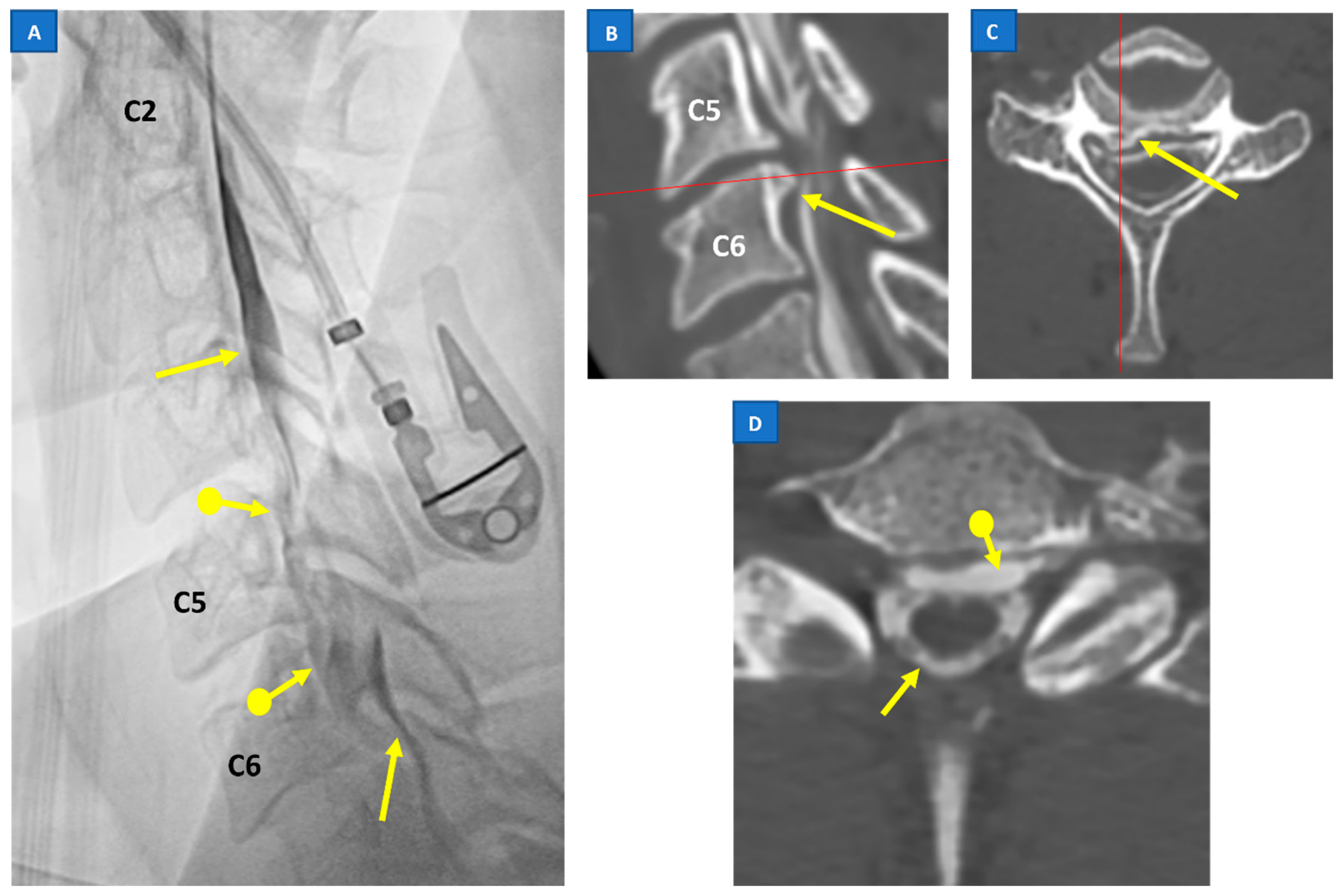
Spontaneous intracranial hypotension (SIH) occurs due to a leakage of the cerebrospinal fluid (CSF) lowering the pressure of subarachnoid space, mostly caused by a dural breach or discogenic microspur. As a result of less support provided by CSF pressure, intracranial structures are stretched downward, leading to a constellation of more or less typical MRI findings, including venous congestion, subdural effusions, brainstem sagging and low-lying cerebellar tonsils. 

spontaneous intracranial hypotension (SIH) myelographic imaging techniques

1. Introduction

The term spontaneous intracranial hypotension (SIH) describes a clinical condition characterized by an incapacitating orthostatic headache syndrome caused by a spontaneous leakage of the cerebrospinal fluid (CSF). More often than not, it is accompanied by stigmata of findings on intracranial magnetic resonance imaging (MRI). In 1939, Schaltenbrand wrote about the incidence of spontaneous CSF hypotension leading to various clinical symptoms, most notably positional headache. He also denoted that the entity itself had been discussed in the French literature twenty years earlier, using several terms such as ‘hypotension of the spinal fluid’ and ‘ventricular collapse’ [1]. The availability of MRI has raised the interest in SIH in the past few decades. While historically perceived to be a rare entity, SIH is now being diagnosed more often, with the incidence currently estimated at 5 per 100,000 annually [2]. However, considering the high occurrence of misdiagnosis—particularly at the initial stage of clinical presentation—the true incidence is likely higher [3]. Although rarely encountered in children, SIH may occur at any age, with the mean age of diagnosis being at around 40 years. Relating this condition, there is female predilection, with a female–male ratio of 2:1 [4][5][6]. Additionally, a higher incidence of SIH is observed in certain connective tissue disorders, particularly the Marfan syndrome and the Ehlers–Danlos syndrome [7][8]. According to the International Classification of Headache Disorders (ICHD) 3rd edition, SIH is diagnosed when there is a spontaneous onset headache, which develops in temporal relation to the occurrence of CSF hypotension (opening CSF pressure < 60 mm H2O); or evidence of CSF leakage (proven through imaging) [9]. However, more recent evidence has challenged the traditional conception of SIH as a condition caused by low CSF pressure.

2. Pathophysiology

The syndrome of SIH, in general, is a consequence of spontaneous CSF leaks along the neuraxis, almost invariably from the thecal sac of the spinal column [10][11][12]. Spontaneous leaks from the skull base are extremely rare [10][13]. Importantly, it has a distinct pathology from postdural puncture headaches or CSF hypovolemic conditions due to postoperative CSF loss or trauma [9].
Although the exact mechanism of spontaneous CSF leaks remain unknown, it is suspected that an underlying weakness of the spinal meninges is an imperative factor [14]. It creates a predisposition for the development of meningeal diverticula or perineural cysts, and subsequently spontaneous dural dehiscence. The presence of calcified degenerative thoracic disc protrusions may cause trivial traumatic events leading to progressive dural tears, which can be either ventral or lateral [7][14][15]. The CSF–venous fistula (CVF) is an important cause of SIH, which pose a diagnostic challenge [16]. Another rare, but interesting cause is the “nude nerve root” phenomenon, which describes the congenital absence of the entire nerve root sleeve [14]. The prevalence of SIH in certain connective-tissue disorders, particularly the Marfan syndrome, the Ehlers–Danlos syndrome (type II), and autosomal dominant polycystic kidney disease (ADPKD), are presumably due to the pre-existing dural fragility [7][8][13].
The old theories of SIH as a condition caused by CSF hypotension have been found to be inaccurate. A series of recent studies on larger samples by Luetmer et al. (2012), Yao et al. (2016), and Kranz et al. (2017), involving 388 patients, proved that only 21% to 55% of SIH patients had a low CSF opening pressure [17][18][19]. Following the data, it has been postulated that the reduction in CSF volume—rather than CSF pressure—is the primary pathogenetic factor [10][13][20]. Kranz et al. further advocated the concept of tissue compliance that determines the physiological relationship between pressure and volume [10]. Compliance with regards to the CSF component is highly dynamic and variable between individuals, as it is influenced by posture (upright vs. recumbent), body habitus, and the extent of the spinal epidural venous plexus [21][22][23]. It has also been demonstrated that the removal of CSF (via lumbar puncture) changes the spinal compliance due to the reduced spinal CSF volume [24]. Considering the fact that the rate of CSF loss is also different between individuals depending on mechanical factors, this may explain why not all SIH patients will have a low CSF opening pressure, despite a common unifying problem of low CSF volume.
In 2019, an international jury of experts awaiting new radiological criteria confirmed the historic radiological definition of CM1 (cerebellar tonsillar position greater than or equal to 5 mm below the level of the foramen magnum) (agreement 83.7%) [25].
Different mechanisms may underlie the pathogenesis of CM1, primarily the underdevelopment of the posterior fossa bony structures. Several studies have shown that many, but not all, patients with CM1 have a small posterior fossa [26].
Another mechanism is represented by the presence of hemodynamic alterations that increase intracranial pressure. According to some authors, the delayed opening of the membrane covering the outlet of the fourth ventricle during fetal development could cause transient obstructive hydrocephalus with a consequent hernia of the hindbrain and cerebellar tonsils [27].
CM1 should be differentiated from cerebellar tonsil herniation secondary to space-occupying lesions (hydrocephalus, arachnoid cysts, brain tumors), and termed ‘acquired tonsillar ectopia’ by an international jury of experts (agreement 95.8%) [25]. Acquired tonsillar ectopia has an identical shape to the shape of the tonsils associated with idiopathic CM1, and may also be secondary to the downward displacement of the central nervous system due to decreased intrathecal pressure (either secondary to the over-drainage of lumboperitoneal shunting or a spontaneous CSF leak) [25][27][28]. In the latter case, careful clinical assessment, ascertaining postural headache, and thorough radiological assessment for intracranial CSF hypotension can help differentiate acquired Chiari malformation from true Chiari I malformation, and guide appropriate treatment (see paragraph 6).
Although these mechanisms that act on the normal cerebellar tonsils, causing their deformation and herniation through the foramen magnum, are well understood, there is no consensus on the pathophysiology of CM1. For example, hydrocephalus has been proposed as both an etiologic cause and a consequence of a Chiari malformation.

3. Classification of SIH

Several classification systems have been proposed as guidance for clinical management, as well as for academic discussion [29][30]. The most widely used classification of SIH was introduced by Schievink et al. in 2016 [29]. This classification is based on the three recognized morphological types of leaks—namely the dural tear, the meningeal diverticulum, and the CSF–venous fistula (CVF)—in combination with the findings of a presence (or absence) of extradural CSF on spinal imaging [31][32] (Table 1).
Table 1. Classification of SIH by Schievink et al. [26].

4. Clinical Presentation of SIH

In a recent meta-analysis of 1694 SIH patients by D’Antona et al., headaches were almost invariably present—being reported in 97% of patients—and in the vast majority, they were orthostatic in nature [6]. This classic headache that characteristically worsens with an upright posture was most pronounced at the occipital region, although frontal and holocephalic headaches were also common. It may be attributed to the traction of the cranial nerves and the pain-sensitive dura mater, caused by the sagging of the brain in the CSF hypovolemia [3][33][34]. Typically, the headache is alleviated within 15–30 min after lying in a recumbent position [5][10]. However, the posture-related component of the headache may also vanish or dampen in a chronic condition [3][35].
Traction, distortion, or compression of some of the cranial nerves, certain structures of the brain, brainstem, mesencephalon, and diencephalon are believed to be the cause of many central nervous system manifestations, as well as the various cranial nerve palsies seen in this disorder [34]. Common associated symptoms include nausea/vomiting, neck pain/stiffness, tinnitus, dizziness, and hearing disturbances. Apart from that, there are a myriad of less frequently reported presentations which include other visual symptoms (photophobia, diplopia, blurred vision, nystagmus, visual loss), back pain, cognitive symptoms (including cognitive impairment, behavioral changes, memory, slow thinking), ear-related symptoms (vertigo, aural fullness, hyperacusis, or unspecified), reduced level of consciousness, and movement disorders (gait disorder, ataxia, dysarthria, tremor, bradykinesia, poor balance) [6].
Other rare, but peculiar symptoms, such as galactorrhea and hyperprolactinemia, have been attributed to the stretching or distortion of the pituitary stalk and/or hypothalamic region [36][37]; meanwhile, the dilatation of the epidural venous plexus or traction and compression of nerve roots are speculated to be the cause of radicular symptoms and incontinence [38].
Other less common, innumerable symptoms have also been described, including dysgeusia, sleepiness, other cranial nerve palsy, fever, fatigue, vocal tics, convulsions, facial spasms/numbness/pain, and dysphagia [6].

5. SIH: Diagnostic Workup and Imaging Strategy

As lumbar puncture is an invasive procedure, in addition to the fact that only about half of the demographic shows a low CSF opening pressure, imaging emerges as the key aspect in the management of SIH. Furthermore, CSF analysis often exhibits considerable variability and is nonspecific for SIH [13].
Imaging plays a pivotal role both in confirming the diagnosis of SIH and localization of the leak point, which allows for the subsequent targeted therapy. A head MRI is the mainstay of initial imaging of SIH, as it is the most sensitive tool in detecting the signs of a CSF leak [6]. Nevertheless, while useful for confirming the diagnosis of SIH, brain MRIs are usually unable to localize the exact site and morphology of the CSF leak. Often, they need to be followed by other myelographic imaging techniques which have the advantage of depicting the direct evidence of CSF leakage (Figure 1), rather than the indirect signs seen with a cranial MRI [15][23].
Figure 1. Prone myelography (A) showing a regular ventral profile of the dural sac (arrows) with anterior extravasation at C5–C6 into the epidural space (arrows with dot). Myelo-CT ((B,C): sagittal and axial planes) shows the presence of a C5–C6 disc herniation, with an osteophyte (bone spur) of the upper somatic margin of C6 (arrows), in the right paramedian site, probable cause of the dural tear (the red line indicates in image (B) the axial plane of image (C) while in image (C) the sagittal plane of image (B)). Myelo-CT (D) also shows the presence of contrast in the epidural space (arrow with dot), anterior to the dural sac (arrow).
Several myelographic imaging techniques have been developed in response to the daunting task of identifying the site of the CSF leakage, which can be anywhere along the neuraxis and with various rates of leakage. These include a spinal MRI, computed tomography myelograms (CTM), dynamic CTM, digital subtraction myelography (DSM), MR myelography, and radionuclide cisternography. Choosing the correct myelographic imaging technique from an array of imaging modalities available today is fundamental for the successful treatment of SIH.
A proposed imaging strategy starts with an MRI of the brain, with a contrast to look for signs of a CSF leakage [15][33][39][40]. Patients showing a high possibility of SIH features should be subjected to a non-directed epidural blood patch (EBP). Those whose head MRI findings were equivocal would benefit from a spinal MRI, which is a non-invasive investigation. A spinal MRI is also indicated in patients who are refractory to non-directed EBP. The findings of a spinal longitudinal extradural collection (SLEC) within the spinal MRI would determine the best myelographic techniques for further delineation of the leaking site. Either DSM or CTM/dynamic CTM performed in certain recommended positions demonstrate a higher yield of CSF leak point detection [40]. If no leak is identified, further exploration with nuclear medicine myelography or MRI myelography (with intrathecal Gadolinium) may reveal an intermittent leak or very slow leaks.

6. Treatment and Prognosis of SIH

Large-scale data and randomized trials are still lacking to guide the standard management of SIH. Current practice of conservative treatment, such as bed rest, high oral fluid intake, caffeine, and an abdominal binder, are often the first-line therapy [41]. Subsequently, non-directed EBP is the treatment of choice if all conservative measures fail [13]. In non-directed EBP, patients’ autologous blood is injected into the epidural space, usually at the lumbar region. This will induce fibrin clot formation at the site of the dural tear, acting as a “patch” to stop the leakage. Symptomatic relief is sometimes immediate, with the majority of patients only requiring one or two sessions of EBP to achieve symptomatic relief [6][41].
For patients with recurrent symptoms after non-directed EBP, further investigations to identify the exact site and type of the CSF leak are necessary. A more targeted approach to the causative pathology can be offered, such as a directed EBP, microsurgical repair of the leak or endovascular treatment for CVF [42][43][44]. The latter is a relatively new treatment of CVF and can be performed by venous embolization of the suspected fistula site: a diagnostic and therapeutic work-up have recently been proposed, showing their feasibility and short-term effectiveness [45].
One complication of a successful CSF leak treatment is the development of rebound high intracranial pressure, which results in reverse orthostatic headaches; the high pressure is generally self-limiting.

References

  1. Schaltenbrand, G. Normal and pathological physiology of the cerebrospinal fluid circulation. Lancet 1953, 261, 805–808.
  2. Schievink, W.I.; Maya, M.; Moser, F.; Tourje, J.; Torbati, S. Frequency of spontaneous intracranial hypotension in the emergency department. J. Headache Pain 2007, 8, 325–328.
  3. Schievink, W.I. Misdiagnosis of spontaneous intracranial hypotension. Arch. Neurol. 2003, 60, 1713–1718.
  4. Schievink, W.I.; Maya, M.M.; Louy, C.; Moser, F.G.; Sloninsky, L. Spontaneous intracranial hypotension in childhood and adolescence. J. Pediatr. 2013, 163, 504–510.e3.
  5. Schievink, W.I.; Meyer, F.B.; Atkinson, J.L.; Mokri, B. Spontaneous spinal cerebrospinal fluid leaks and intracranial hypotension. J. Neurosurg. 1996, 84, 598–605.
  6. D’Antona, L.; Merchan, M.A.J.; Vassiliou, A.; Watkins, L.D.; Davagnanam, I.; Toma, A.K.; Matharu, M.S. Clinical presentation, investigation findings, and treatment outcomes of spontaneous intracranial hypotension syndrome: A systematic review and meta-analysis. JAMA Neurol. 2021, 78, 329–337.
  7. Schievink, W.I. Spontaneous spinal cerebrospinal fluid leaks and intracranial hypotension. JAMA 2006, 295, 2286–2296.
  8. Reinstein, E.; Pariani, M.; Bannykh, S.; Rimoin, D.L.; Schievink, W.I. Connective tissue spectrum abnormalities associated with spontaneous cerebrospinal fluid leaks: A prospective study. Eur. J. Hum. Genet. 2013, 21, 386–390.
  9. Olesen, J.; Dodick, D.; Ducros, A.; Evers, S.; First, M.; Goadsby, P. The International Classification of Headache Disorders, (ICHD-3). Cephalalgia 2018, 38, 1–211.
  10. Kranz, P.G.; Gray, L.; Amrhein, T.J. Spontaneous intracranial hypotension: 10 myths and misperceptions. Headache J. Head Face Pain 2018, 58, 948–959.
  11. Schievink, W.; Maya, M.; Louy, C.; Moser, F.; Tourje, J. Diagnostic criteria for spontaneous spinal CSF leaks and intracranial hypotension. Am. J. Neuroradiol. 2008, 29, 853–856.
  12. Schievink, W.I.; Schwartz, M.S.; Maya, M.M.; Moser, F.G.; Rozen, T.D. Lack of causal association between spontaneous intracranial hypotension and cranial cerebrospinal fluid leaks. J. Neurosurg. 2012, 116, 749–754.
  13. Mokri, B. Spontaneous intracranial hypotension. Contin. Lifelong Learn. Neurol. 2015, 21, 1086–1108.
  14. Schievink, W.I.; Jacques, L. Recurrent spontaneous spinal cerebrospinal fluid leak associated with “nude nerve root” syndrome: Case report. Neurosurgery 2003, 53, 1216–1219.
  15. Kranz, P.G.; Luetmer, P.H.; Diehn, F.E.; Amrhein, T.J.; Tanpitukpongse, T.P.; Gray, L. Myelographic techniques for the detection of spinal CSF leaks in spontaneous intracranial hypotension. Am. J. Roentgenol. 2016, 206, 8–19.
  16. Schievink, W.I.; Moser, F.G.; Maya, M.M. CSF–venous fistula in spontaneous intracranial hypotension. Neurology 2014, 83, 472–473.
  17. Luetmer, P.H.; Schwartz, K.; Eckel, L.; Hunt, C.; Carter, R.; Diehn, F. When should I do dynamic CT myelography? Predicting fast spinal CSF leaks in patients with spontaneous intracranial hypotension. Am. J. Neuroradiol. 2012, 33, 690–694.
  18. Yao, L.-L.; Hu, X.-Y. Factors affecting cerebrospinal fluid opening pressure in patients with spontaneous intracranial hypotension. J. Zhejiang Univ. Sci. B 2017, 18, 577–585.
  19. Kranz, P.G.; Tanpitukpongse, T.P.; Choudhury, K.R.; Amrhein, T.J.; Gray, L. How common is normal cerebrospinal fluid pressure in spontaneous intracranial hypotension? Cephalalgia 2016, 36, 1209–1217.
  20. Mokri, B. Spontaneous cerebrospinal fluid leaks: From intracranial hypotension to cerebrospinal fluid hypovolemia—Evolution of a concept. Mayo Clin. Proc. 1999, 74, 1113–1123.
  21. Alperin, N.; Lee, S.H.; Sivaramakrishnan, A.; Hushek, S.G. Quantifying the effect of posture on intracranial physiology in humans by MRI flow studies. J. Magn. Reson. Imaging 2005, 22, 591–596.
  22. Tain, R.W.; Bagci, A.M.; Lam, B.L.; Sklar, E.M.; Ertl-Wagner, B.; Alperin, N. Determination of cranio-spinal canal compliance distribution by MRI: Methodology and early application in idiopathic intracranial hypertension. J. Magn. Reson. Imaging 2011, 34, 1397–1404.
  23. Medina, J.H.; Abrams, K.; Falcone, S.; Bhatia, R.G. Spinal imaging findings in spontaneous intracranial hypotension. Am. J. Roentgenol. 2010, 195, 459–464.
  24. Alperin, N.; Bagci, A.; Lee, S.; Lam, B. Automated quantitation of spinal CSF volume and measurement of craniospinal CSF redistribution following lumbar withdrawal in idiopathic intracranial hypertension. Am. J. Neuroradiol. 2016, 37, 1957–1963.
  25. Ciaramitaro, P.; Massimi, L.; Bertuccio, A.; Solari, A.; Farinotti, M.; Peretta, P.; Saletti, V.; Chiapparini, L.; Barbanera, A.; Garbossa, D.; et al. Diagnosis and treatment of Chiari malformation and syringomyelia in adults: International consensus document. Neurol. Sci. 2022, 43, 1327–1342.
  26. Stovner, L.J.; Bergan, U.; Nilsen, G.; Sjaastad, O. Posterior cranial fossa dimensions in the Chiari I malformation: Relation to pathogenesis and clinical presentation. Neuroradiology 1993, 35, 113–118.
  27. Buell, T.J.; Heiss, J.D.; Oldfield, E.H. Pathogenesis and Cerebrospinal Fluid Hydrodynamics of the Chiari I Malformation. Neurosurg. Clin. N. Am. 2015, 26, 495–499.
  28. Tachibana, S.; Harada, K.; Abe, T.; Yamada, H.; Yokota, A. Syringomyelia secondary to tonsillar herniation caused by posterior fossa tumors. Surg. Neurol. 1995, 43, 470–475, discussion: 475–477.
  29. Schievink, W.I.; Maya, M.M.; Jean-Pierre, S.; Nuño, M.; Prasad, R.S.; Moser, F.G. A classification system of spontaneous spinal CSF leaks. Neurology 2016, 87, 673–679.
  30. Farb, R.I.; Forghani, R.; Lee, S.; Mikulis, D.; Agid, R. The venous distension sign: A diagnostic sign of intracranial hypotension at MR imaging of the brain. Am. J. Neuroradiol. 2007, 28, 1489–1493.
  31. Mokri, B. The Monro–Kellie hypothesis: Applications in CSF volume depletion. Neurology 2001, 56, 1746–1748.
  32. Cushing, H. The third circulation. In Studies in Intracranial Physiology and Surgery; Oxford University Press: London, UK, 1926; pp. 1–51.
  33. Luetzen, N.; Dovi-Akue, P.; Fung, C.; Beck, J.; Urbach, H. Spontaneous intracranial hypotension: Diagnostic and therapeutic workup. Neuroradiology 2021, 63, 1765–1772.
  34. Mokri, B. Headache associated with abnormalities in intracranial structure or function: Low cerebrospinal-fluid-pressure headache. In Wolff’s Headache and Other Head Pain; Oxford University Press: Oxford, UK, 2007; pp. 513–531.
  35. Mokri, B. Spontaneous CSF leaks: Low CSF volume syndromes. Neurol. Clin. 2014, 32, 397–422.
  36. Yamamoto, M.; Suehiro, T.; Nakata, H.; Nishioka, T.; Itoh, H.; Nakamura, T.; Hashimoto, K. Primary low cerebrospinal fluid pressure syndrome associated with galactorrhea. Intern. Med. 1993, 32, 228–231.
  37. Schievink, W.I.; Nuño, M.; Rozen, T.D.; Maya, M.M.; Mamelak, A.N.; Carmichael, J.; Bonert, V.S. Hyperprolactinemia due to spontaneous intracranial hypotension. J. Neurosurg. 2015, 122, 1020–1025.
  38. Albayram, S.; Wasserman, B.A.; Yousem, D.M.; Wityk, R. Intracranial hypotension as a cause of radiculopathy from cervical epidural venous engorgement: Case report. Am. J. Neuroradiol. 2002, 23, 618–621.
  39. Farb, R.; Nicholson, P.; Peng, P.; Massicotte, E.; Lay, C.; Krings, T. Spontaneous intracranial hypotension: A systematic imaging approach for CSF leak localization and management based on MRI and digital subtraction myelography. Am. J. Neuroradiol. 2019, 40, 745–753.
  40. Dobrocky, T.; Grunder, L.; Breiding, P.S.; Branca, M.; Limacher, A.; Mosimann, P.J.; Mordasini, P.; Zibold, F.; Haeni, L.; Jesse, C.M.; et al. Assessing spinal cerebrospinal fluid leaks in spontaneous intracranial hypotension with a scoring system based on brain magnetic resonance imaging findings. JAMA Neurol. 2019, 76, 580–587.
  41. Schievink, W.I. Spontaneous intracranial hypotension. N. Engl. J. Med. 2021, 385, 2173–2178.
  42. Brinjikji, W.; Savastano, L.; Atkinson, J.; Garza, I.; Farb, R.; Cutsforth-Gregory, J. A novel endovascular therapy for CSF hypotension secondary to CSF-venous fistulas. Am. J. Neuroradiol. 2021, 42, 882–887.
  43. Mamlouk, M.D.; Shen, P.Y.; Sedrak, M.F.; Dillon, W.P. CT-guided fibrin glue occlusion of cerebrospinal fluid–venous fistulas. Radiology 2021, 299, 409–418.
  44. Pagani-Estévez, G.L.; Cutsforth-Gregory, J.K.; Morris, J.M.; Mokri, B.; Piepgras, D.G.; Mauck, W.D.; Eldrige, J.S.; Watson, J.C. Procedural predictors of epidural blood patch efficacy in spontaneous intracranial hypotension. Reg. Anesth. Pain Med. 2019, 44, 212–220.
  45. Bergui, M.; Mistretta, F.; Bosco, G.; Cester, G.; Chioffi, F.; Gambino, A.; Molinaro, S.; Russo, R.; Sorarù, G.; Causin, F. CSF-venous leak responsible for spontaneous intracranial hypotension treated by endovascular venous route: First cases in Italy. Interv. Neuroradiol. 2022.
More
Information
Subjects: Neurosciences
Contributors MDPI registered users' name will be linked to their SciProfiles pages. To register with us, please refer to https://encyclopedia.pub/register : , , , , , ,
View Times: 300
Revisions: 2 times (View History)
Update Date: 10 May 2023
1000/1000
Video Production Service